Embed Size (px)
Citation preview

APPLIED AND ENVIRONMENTAL MICROBIOLOGY,0099-2240/01/$04.0010 DOI: 10.1128/AEM.67.1.22–32.2001
Jan. 2001, p. 22–32 Vol. 67, No. 1
Copyright © 2001, American Society for Microbiology. All Rights Reserved.
Molecular Ecology of Tetracycline Resistance: Developmentand Validation of Primers for Detection of Tetracycline
Resistance Genes Encoding Ribosomal Protection ProteinsR. I. AMINOV,* N. GARRIGUES-JEANJEAN, AND R. I. MACKIE
Department of Animal Sciences, University of Illinois at Urbana-Champaign, Urbana, Illinois 61801
Received 5 July 2000/Accepted 6 October 2000
Phylogenetic analysis of tetracycline resistance genes encoding the ribosomal protection proteins (RPPs)revealed the monophyletic origin of these genes. The most deeply branching class, exemplified by tet and otrA,consisted of genes from the antibiotic-producing organisms Streptomyces rimosus and Streptomyces lividans.With a high degree of confidence, the corresponding genes of the other seven classes (Tet M, Tet S, Tet O, TetW, Tet Q, Tet T, and TetB P) formed phylogenetically distinct separate clusters. Based on this phylogeneticanalysis, a set of PCR primers for detection, retrieval, and sequence analysis of the corresponding genefragments from a variety of bacterial and environmental sources was developed and characterized. A pair ofdegenerate primers targeted all tetracycline resistance genes encoding RPPs except otrA and tet, and sevenother primer pairs were designed to target the specific classes. The primers were used to detect the circulationof these genes in the rumina of cows, in swine feed and feces, and in swine fecal streptococci. Classes Tet O andTet W were found in the intestinal contents of both animals, while Tet M was confined to pigs and Tet Q wasconfined to the rumen. The tet(O) and tet(W) genes circulating in the microbiota of the rumen and thegastrointestinal tract of pigs were identical despite the differences in animal hosts and antibiotic use regimens.Swine fecal streptococci uniformly possessed the tet(O) gene, and 22% of them also carried tet(M). Thispopulation could be considered one of the main reservoirs of these two resistance genes in the pig gastroin-testinal tract. All classes of RPPs except Tet T and TetB P were found in the commercial components of swinefeed. This is the first demonstration of the applicability of molecular ecology techniques to estimation of thegene pool and the flux of antibiotic resistance genes in production animals.
Antibiotic resistance research has been and still is confinedprimarily to the study of cultivable bacterial isolates of mostlyclinical origin. However, the cultivable isolates may representonly a fraction of the actual microbiota (1) where the antibioticresistance genes reside. For example, no bacteria can be grownfrom more than 80% of all clinical samples sent to clinicalmicrobiology laboratories (4), and certainly no antibiotic resis-tance profile can be determined if cultivation fails. Anotherimportant issue with antibiotic resistance is the fact that thewide use of antibiotics not only selects for drug-resistant patho-genic bacteria but also exerts selective pressure on the normalcommensal microbiota. In light of the ubiquitously demon-strated phenomenon of horizontal antibiotic resistance genetransfer in the microbial world, the presence of such reservoirsmay explain the rapid dissemination of antibiotic resistancefrom commensal organisms to the pathogenic microbiota.However, information regarding the antibiotic resistance poolin commensal microbiotas is very scarce, and the data aremostly phenotypical. Therefore, development of genotypingtools for detection and tracking of antibiotic resistance genesin a variety of commensal and pathogenic bacteria, as well as inthe environment, is essential for understanding the ecology ofantibiotic resistance.
One of the attractive models for studying the ecology of
antibiotic resistance could be the genes conferring resistance totetracyclines. Tetracyclines belong to a family of broad-spec-trum antibiotics that includes tetracycline, chlortetracycline,doxycycline, and minocycline. These antibiotics inhibit proteinsynthesis in gram-positive and gram-negative bacteria by pre-venting the binding of aminoacyl-tRNA molecules to the 30Sribosomal subunit (36). Bacterial resistance to tetracycline ismediated mainly by two mechanisms, protection of ribosomesby large cytoplasmic proteins (5, 6, 23, 33, 43) and energy-dependent efflux of tetracycline (18, 33, 36). A third mecha-nism, enzymatic inactivation of tetracycline, is relatively un-common and has been described in only one species (41). Thefirst nomenclature for tetracycline resistance determinants wasproposed in 1989 (17), and a recent update appeared in 1999(19). The ribosomal protection mechanisms identified so farfall into six classes: Tet M, Tet O, TetB P, Tet Q, Tet S, andotrA (43). Almost all representatives of these classes have beensequenced and have been shown to encode proteins with N-terminal amino acid sequence similarity to translation elonga-tion factors EF-Tu and EF-G (6, 22, 35, 43).
Since their introduction in the 1950s, tetracyclines have beenwidely used in human and veterinary medicine, as growth pro-moters in animal industry, and for prophylaxis in plant agri-culture and aquaculture. At present, resistance to tetracyclineshas spread to almost all bacterial genera, and this situationperhaps is the consequence of previous overuse. Among theribosomal protection determinants, Tet M was described orig-inally in streptococci (5, 24) and subsequently in a broad vari-ety of gram-positive and gram-negative bacteria (32). The Tet
* Corresponding author. Mailing address: Department of AnimalSciences, University of Illinois, 1207 W. Gregory Drive, Urbana, IL61801. Phone: (217) 333-8809. Fax: (217) 333-8804. E-mail: [email protected].
22
on June 4, 2018 by guesthttp://aem
.asm.org/
Dow
nloaded from

S determinant was encountered first on a plasmid in the foodpathogen Listeria monocytogenes (7), later in a number of En-terococcus faecalis strains (8), and recently on a plasmid ofLactococcus lactis isolated from raw milk (31). Tet O-relatedsequences were determined first in plasmids from campylobac-teria (40, 42), then in streptococci (16, 45), and recently in arumen bacterium, Butyrivibrio fibrisolvens (2). Finding nearlyidentical tet(Q) sequences in Prevotella ruminicola (a typicalinhabitant of the rumen) and in Bacteroides (a typical inhabit-ant of the human gastrointestinal tract) (29) suggested thatbacteria normally found in the guts of different species canexchange DNA, presumably during transient colonization ofthe animal intestine by human-associated bacteria or viceversa. This scenario is supported by recent work which de-scribed the occurrence of the tet(W) gene in the rumen and inhuman and pig intestinal microbiotas (37). Sequence analysisof this gene from taxonomically divergent rumen and humanbacterial isolates showed that there was no or just one nucle-otide substitution in a 1.25-kb amplified internal fragment.Misincorporation errors during amplification could not beruled out, suggesting that the sequences may actually be iden-tical in ruminal B. fibrisolvens, Selenomonas ruminantium, andMitsuokella multiacidus isolates and in human isolates of Fu-sobacterium prausnitzii and Bifidobacterium longum (37). Thesefindings demand that there be further research targeted atdevelopment of genotyping tools for tracking the movement ofantibiotic resistance genes in the environment.
In this study, we initiated research to examine the molecularecology of antibiotic resistance. As a model, we used tetracy-cline resistance genes encoding the ribosomal protection pro-teins (RPPs). Phylogenetic analysis revealed the monophyleticorigin of these genes, which allowed us to design a set of PCRprimers suitable for detection of RPP genes in general, as wellas different classes. After validation, this set was used to detectthe corresponding genes in the total DNA of swine fecal andrumen samples and in swine feed, as well as in fecal strepto-coccal isolates from pigs. The primers were also used in aPCR-denaturing gradient gel electrophoresis (DGGE) analysisto demonstrate the overall diversity and similarity of RPPgenes in different ecosystems. The methods used in this workcan be applied to study other phylogenetically coherent anti-biotic resistance gene families.
MATERIALS AND METHODS
Phylogenetic analysis and primer design. All currently available nucleotidesequences encoding RPPs, as well as the phylogenetically most closely relatedelongation factors, EF-Gs, were downloaded from the GenBank database (3).These included the sequences of the following RPP genes (the numbers inparentheses are GenBank accession numbers): E. faecalis DS16 tet(M)(M85225), E. faecalis Tn916 tet(M) (X56353), E. faecalis Tn1545 tet(M)(X04388), Neisseria meningitidis tet(M) (X75073), Ureaplasma urealyticum tet(M)(U08812), Gardnerella vaginalis tet(M) (U58986), Neisseria gonorrhoeae 6418tet(M) (L12241), N. gonorrhoeae 2903 tet(M) (L12242), Staphylococcus aureustet(M) (M21136), Streptococcus pneumoniae Tn5251 tet(M) (X90939), L. mono-cytogenes BM4210/pIP811 tet(S) (L09756), L. lactis K214/pK214 tet(S) (X92946),S. pneumoniae tet(O) (Y07780), Streptococcus mutans DL5 tet(O) (M20925),Campylobacter jejuni tet(O) (M18896), B. fibrisolvens tet(W) (AJ222769), Bacte-roides fragilis tet(Q) (Z21523), Prevotella intermedia PDRC-11 tet(Q) (U73497),P. ruminicola tet(Q) (L33696), B. fragilis BF-2 tet(Q) (Y08615), Bacteroides the-taiotaomicron tet(Q) (X58717), Streptococcus pyogenes A498 tet(T) (L42544),Clostridium perfringens CW92 tetB(P) (L20800), Streptomyces lividans 1326 tet(M74049), and Streptomyces rimosus otrA (X53401). Elongation factor EF-G-encoding genes were obtained from Bacillus subtilis (D64127), E. faecalis (re-
trieved from www.tigr.org), Escherichia coli (X00415), Helicobacter pylori(AE001539), Thermus thermophilus (X16278), and Aquifex aeolicus (AE000669).
Sequences were aligned with the multiple-sequence alignment programCLUSTAL W (44). The two-parameter model of Kimura (14) was used forconstruction of neighbor-joining trees (34). The statistical significance of branch-ing was evaluated by bootstrap analysis (11) involving the construction of 1,000trees from resampled data. Sequences within clusters were separately alignedand compared with each other. PCR primers were designed to satisfy specificityand so that they could potentially be used in multiplex PCR with simultaneouscoamplification of the V3 region of 16S ribosomal DNA (rDNA) (26) and inPCR-DGGE analysis (27). The nine sets of primers and the expected ampliconsizes are shown in Table 1. A GC clamp (CGCCCGGGGCGCGCCCCGGGCGGGGCGGGGGCACGGGGGG) was added to the reverse primers for use inDGGE analysis (26). The primers used for amplification of the bacterial V3region of 16S rDNA were the primers described previously (27).
Environmental samples and DNA extraction. Samples of whole rumen con-tents were obtained from eight fistulated steers maintained at the Research Farmof the University of Illinois at Urbana-Champaign. The animals used are treatedwith antibiotics only in case of disease, and no antibiotics are given for prophy-laxis or growth promotion. No tetracyclines had been used for disease treatmentin this group of animals, and the animals were considered free of tetracyclineselective pressure. Fecal samples from six sows were collected at the SwineResearch Farm of the University of Illinois at Urbana-Champaign. In this facil-ity, in addition to therapeutic use, antibiotics are added to feed for prophylacticand growth-promoting purposes. The sows were routinely fed Tylan (ElancoAnimal Health, Indianapolis, Ind.) at a concentration of 40 mg per kg of feed.The antibiotic was switched to chlortetracycline (400 mg per kg of feed) for 2weeks and then back to Tylan. Fecal samples were collected 3 weeks after theswitch back to Tylan. Rumen and fecal samples were frozen at 220°C for futureisolation of total DNA. Samples of pig feed were stored at room temperaturebefore DNA isolation. Total DNA was isolated from the fecal, rumen, and feedsamples by using a Soil DNA Purification Kit (Mo Bio, Solana Beach, Calif.)according to the manufacturer’s protocol.
Organisms, plasmids, and culture techniques. The organisms and plasmidsused in this study for validation and control are listed in Table 2. C. jejuni subsp.jejuni ATCC 43503 was grown at 37°C under microaerophilic conditions onATCC medium 1115. C. perfringens JIR4202 was grown anaerobically at 37°C onBHIB medium (Difco Laboratories, Detroit, Mich.). S. pyogenes CIP105079 wasgrown aerobically on BHIB medium (Difco) at 37°C. E. coli strains were grownon Luria-Bertani medium at 37°C with aeration. Media were solidified whennecessary with 1.8% (wt/vol) agar (Difco). Tetracycline (Sigma Chemical Co., St.Louis, Mo.) was added at a concentration of 10 mg/ml to cultures of C. jejuni, C.perfringens, and S. pyogenes. The cloned tet genes were maintained in E. coli byselection of plasmid antibiotic markers (ampicillin or kanamycin at a concentra-tion of 50 mg/ml or chloramphenicol at a concentration of 20 mg/ml).
For isolation of fecal streptococci, fresh fecal samples from six pigs wereresuspended in phosphate-buffered saline (pH 7.0), and dilutions were plated onMRS agar (Difco) plates. For detection of tetracycline resistance, colonies weretransferred to the same medium with 10 mg of tetracycline per ml. The taxonomicaffiliations of the resulting isolates were confirmed by sequencing 1.4-kb frag-ments of 16S rDNA amplified with bacterial primers (15). DNA similarity ma-trixes were calculated by using the DNADIST program in the PHYLIP package(12). Restriction fragment length polymorphism (RFLP) analysis was accom-plished by AluI restriction digestion of amplified 1.4-kb fragments, followed byelectrophoresis on a 3.0% agarose gel.
PCR and DGGE. A typical PCR mixture (total volume, 20 ml) contained 25pmol of each primer (except for the degenerate universal primers, which wereused at a concentration of 100 pmol per mixture), 13 ExTaq reaction buffer(Takara Shuzo, Orsu, Japan), each deoxynucleoside triphosphate at a concen-tration of 100 mM, and 1.0 U of ExTaq DNA polymerase (Takara Shuzo). A200-ng portion of purified DNA or one-half of the biomass of a 1- to 2-mm-diameter colony was used as a template. PCR amplification (25 cycles) wasperformed with a GeneAmp 2400 PCR system (Perkin-Elmer, Norwalk, Conn.)as follows: initial denaturation at 94°C for 5 min, followed by 25 cycles of 94°Cfor 30 s, 30 s of annealing at the annealing temperatures shown in Table 1, and30 s of extension at 72°C, and a final extension step at 72°C for 7 min. Atouchdown PCR with the degenerate Ribo2 primers (Table 1) was performed asfollows: initial denaturation at 94°C for 5 min; 22 cycles of denaturation at 94°Cfor 30 s, annealing for 30 s with 1°C decrements at temperatures of 72 to 50°C,and extension at 72°C for 30 s; 20 cycles of 94°C for 30 s, 50°C for 30 s, and 72°Cfor 30 s; and final extension at 72°C for 7 min. If unidentified substances in aDNA preparation inhibited the PCR, 1 ml of the reaction mixture was used as atemplate for further amplification. Aliquots (5 ml) were analyzed by electro-
VOL. 67, 2001 PCR DETECTION OF RPP GENES 23
on June 4, 2018 by guesthttp://aem
.asm.org/
Dow
nloaded from

phoresis on a 2.5% (wt/vol) agarose gel (NuSieve; FMC Bioproducts, Rockland,Maine) containing the fluorescent dye GelStar (FMC Bioproducts). Gels withamplicons generated by the Ribo2 primers were stained with ethidium bromide.
For DGGE, polyacrylamide gels with urea-formamide gradients (8% acryl-
amide, 15 to 60% urea-formamide, 0.53 TAE buffer; pH 7.4) were polymerizedon Gel-Bond support sheets (FMC Bioproducts). Electrophoresis was performedat 60°C and 150 V for 2 h and then at 200 V for 1 h by using the D-Gene System(Bio-Rad Laboratories, Richmond, Calif.). After electrophoresis, the gels wererinsed in double-distilled H2O, fixed in a solution containing 10% ethanol and0.5% acetic acid, and silver stained. Gel images were captured and digitized witha Bio-Rad system that included a GS-710 calibrated imaging densitometer con-nected to a G3 Macintosh computer with the Diversity Database fingerprintingsoftware. For cloning and sequencing of DGGE bands, the corresponding am-plicons were excised from the gels and equilibrated in TE buffer at room tem-perature for 30 min, and 1 ml of the buffer with diffused DNA was used forreamplification.
For estimation of the proportion of antibiotic resistance-carrying microbiota,total DNA from the rumen and the standard strain [C. jejuni with tet(O)] weresubjected to multiplex PCR [using amplification conditions for tet(O)] withprimer sets TetO and V3 (targeting the V3 region of 16S rDNA). In thismultiplex PCR, a C. jejuni DNA template was used at various dilutions. The gelimages were digitized, and densitograms were generated with the NIH Imageprogram (http://rsb.info.nih.gov/nih-image). Then the densitogram of the rumenmultiplex PCR was compared to the range of C. jejuni amplification data. In lineshaving the same density of the TetO signal, the V3 signal intensities werecompared for the total rumen DNA (all bacterial sequences amplified) and C.jejuni DNA. From this comparison, the approximate proportion of the suspectedtet(O)-carrying bacteria was calculated.
Cloning and sequencing of PCR amplicons. PCR products were cloned byusing a TA Cloning kit (Invitrogen, Carlsbad, Calif.). White colonies of ampi-cillin-resistant transformants were screened for the presence of tet fragments byPCR by using the same primer set that was used for amplification. DNA se-quence analysis of recombinant plasmids was performed for both strands (prim-ers M13F and M13R) at the University of Illinois Biotechnology Center. On-linesimilarity searching was performed by using the BLAST (Basic Local AlignmentSearch Tool) family of programs in GenBank (21).
RESULTS
Phylogenetic analysis. Phylogenetic analysis was performedwith 25 complete nucleotide sequences encoding RPPs andwith six complete sequences encoding phylogenetically closelyrelated translation elongation factors belonging to family G
TABLE 1. PCR primers targeting the RPP classes
Primer Class targeted Sequence PCR annealing temp (°C) Amplicon size (bp)
Ribo2-FWa All except Otr A GGMCAYRTGGATTTYWTIGC Touchdownb 1,187Ribo2-RV TCIGMIGGIGTRCTIRCIGGRC
TetB/P-FW TetB P AAAACTTATTATATTATAGTG 46 169TetB/P-RV TGGAGTATCAATAATATTCAC
TetM-FW Tet M ACAGAAAGCTTATTATATAAC 55 171TetM-RV TGGCGTGTCTATGATGTTCAC
TetO-FW Tet O ACGGARAGTTTATTGTATACC 60 171TetO-RV TGGCGTATCTATAATGTTGAC
OTR-FW Otr A GGCATYCTGGCCCACGT 66 212OTR-RV CCCGGGGTGTCGTASAGG
TetQ-FW Tet Q AGAATCTGCTGTTTGCCAGTG 63 169TetQ-RV CGGAGTGTCAATGATATTGCA
TetS-FW Tet S GAAAGCTTACTATACAGTAGC 50 169TetS-RV AGGAGTATCTACAATATTTAC
TetT-FW Tet T AAGGTTTATTATATAAAAGTG 46 169TetT-RV AGGTGTATCTATGATATTTAC
TetW-FW Tet W GAGAGCCTGCTATATGCCAGC 64 168TetW-RV GGGCGTATCCACAATGTTAAC
a FW, forward; RV, reverse.b PCR conditions are described in Materials and Methods.
TABLE 2. Characteristics of bacterial strains andplasmids used in this study
Strain orplasmid
Relevantcharacteristics
Reference orsource
StrainsC. jejuni subsp.
jejuniTcr, tet(O) ATCC 43503
S. pyogenesA498
Tcr, tet(T) 9
C. perfringensJIR4202
Tcr, tetA(P) tetB(P) 38
PlasmidspBT-1 pJRD215 carrying a 2.5-kb SstI
fragment containing thetet(Q) gene of TcrEmrDOT
28
pJIR667 pPR328 carrying a 1.1-kb PstI-EcoRI internal fragment ofthe tetB(P) gene
20
pFD310 Carries the tet(M) gene fromS. agalactiae
39
pGEM-tetW pGEM carrying a 2.4-kb PCRproduct with the tet(W) genefrom B. fibrisolvens
2
pGEM-tetO pGEM carrying the tet(O)gene from B. fibrisolvens
2
pVP2 pUC18 carrying a 5.88-kb ClaIfragment of pK214encompassing the tet(S) gene
31
pAT451 pUC18 carrying a 4.5-kb ClaIfragment of pIP811 with thetet(S) gene
7
pCT10 Contains the tet gene from S.lividans 1326
10
24 AMINOV ET AL. APPL. ENVIRON. MICROBIOL.
on June 4, 2018 by guesthttp://aem
.asm.org/
Dow
nloaded from

(EF-G). With 100% bootstrap support, this analysis confirmedthe monophyletic origin of RPP genes and the early branchingfrom the other group of elongation factors, EF-G (Fig. 1). Thenumber of substitutions per base pair was approximately 2.4times higher in the RPP cluster than in the EF-G cluster.Within the RPP supercluster, there are eight clusters corre-sponding to the recently revised classes Tet M, Tet S, Tet O,Tet W, Tet Q, Tet T, TetB P, and otrA. Three of these clusters,Tet W, Tet T, and TetB P, are represented by only a singlesequence at present.
This analysis suggests that there was early branching be-tween the RPP genes of antibiotic-producing strains, tet andotrA, and the other RPP genes circulating in pathogenic andsaprophytic bacteria (Fig. 1). Thus, based on available se-quence data, no evidence of recent horizontal transfer of RPPgenes from antibiotic-producing strains to other bacteria existsat present. However, there is a high level of similarity, as shownby the extremely short branch lengths, among the sequences intaxonomically distantly related bacteria for classes Tet M, TetS, Tet O, and Tet Q (Fig. 1).
Design and validation of PCR primers targeting RPP genes.Evidence of the monophyletic origin of the RPP genes openedthe possibility of designing primers that target all genes in thecluster. However, early branching and further independentdiversification, together with the high G1C contents of the tetand otrA genes, precluded incorporation of these genes intothe alignment analysis. Thus, the design of the universal primerpair was based on the sequences belonging to seven classes ofRPP genes. As mentioned above, the rate of nucleotide sub-stitution in the RPP cluster is higher than that in other elon-gation factors, and therefore, the overall sequence structure isless conserved. Because of this, the design of the universalprimer pair involved a substantial level of degeneracy (Table1). Primer pairs specific for the individual classes, together withthe expected amplicon sizes, are shown in Table 1.
This set of primers was rigorously tested in PCR performedwith DNA and colony biomasses of control strains (Table 3). Inall cases except the tet gene with OTR primers, amplicons ofthe expected size were produced with positive controls. Wesuspect that the failure to amplify tet with OTR primers wasdue to structural instability of this gene on a high-copy-numberplasmid. The gene was also shown to be structurally unstable inits original host, S. lividans (10). As expected, no signal wasproduced with the pJIR667 template when the universal andclass-specific primer sets were used (Table 3). During a previ-ous gene cloning procedure, the upstream region of the genewas deleted (20). The forward primers of the Ribo2 andTetB/P sets target this lost region, and no amplification isexpected because of this. In all other cases, sequence analysisof PCR-generated amplicons from control strains confirmedthe specificity of the primers and the identity of the amplifiedproduct. To simplify the detection procedure, biomasses fromboth colonies and liquid cultures were used for PCR, and theseamplifications were also successful (Table 3).
Detection of RPP genes in the rumen. Total DNA prepara-tions from the rumen samples were subjected to PCR ampli-fication with the universal primer set, the Ribo2 set, followedby analysis with class-specific primers. Although the Ribo2primer set performed well with pure cultures (Table 3), it wasnot sufficiently selective during amplification from total com-
munity DNA. According to the analysis with class-specificprimers, the rumen microbiota appeared to bear the tet(O),tet(Q), and tet(W) genes (data not shown). The coamplified V3region of 16S rDNA (data not shown) allowed a rough esti-mate of the proportion of the rumen bacterial microbiota car-rying the corresponding resistance genes. Comparative analysisof densitograms by using biomass of C. jejuni as a standardsuggested that up to 5% of ruminal bacteria may carry tet(O).The rumen samples were also subjected to PCR-DGGE anal-ysis with the TetO, TetQ, and TetW primer sets, in which GCclamps were attached to the reverse primers. This analysisrevealed the uniformity of the tet(W) and tet(O) genes circu-lating in the rumen microbiota of the cows (Fig. 2, lanes 2through 9, and Fig. 3, lanes 13 through 20). DGGE bands fromtet(W) samples were excised, reamplified, sequenced, andfound to be identical to the tet(W) sequence (2). However, theDGGE band from this control template migrated farther thanour samples (Fig. 2, lane 13), and careful inspection of thenucleotide sequence of the tet(W) control revealed a nucleo-tide substitution (T3C), which may have been incorporatedduring amplification from B. fibrisolvens (2). A DGGE analysiswith the TetO primers produced similar results, with no vari-ations among animals but with two bands (Fig. 3, lanes 13through 20). Sequence analysis revealed that these two bands,which were in the region of tet(O), were actually identical, butfor unknown reasons, the upper band also contained the re-verse DGGE primer misincorporated at the 59 end of theforward primer, thus increasing the melting temperature andcreating artificial heterogeneity. A DGGE analysis with theTetQ primers revealed diversity among tet(Q) in the rumen, aswell as animal-to-animal variation (Fig. 4). There were at leastfive bands, and sequence analysis confirmed the heterogeneityat the sequence level.
Detection of RPP genes in swine feces. Total DNA prepa-rations from swine fecal samples were subjected to PCR am-plification with the universal primer set, the Ribo2 set, and alsowith class-specific primers. Tet M, Tet O, and Tet W determi-nants were detected in the swine intestinal microbiota (datanot shown). Further PCR-DGGE analysis (Fig. 5) and se-quencing demonstrated that swine tet(M) is identical to thecontrol template, tet(M) cloned from Streptococcus agalactiae.Interestingly, the tet(O) and tet(W) genes circulating in the pigherd had the same mobility on DGGE gels as the correspond-ing genes from the rumina of steers (Fig. 2 and 3). Sequenceanalysis of the excised and cloned major DGGE bands con-firmed that these two classes of genes were identical in the twotypes of animals. The difference was that swine fecal samplesproduced an additional minor tet(W) band migrating fartherthan the major band (Fig. 2, lanes 14 through 19). Severalattempts to clone this minor band were unsuccessful, and se-quence information for this band is not available.
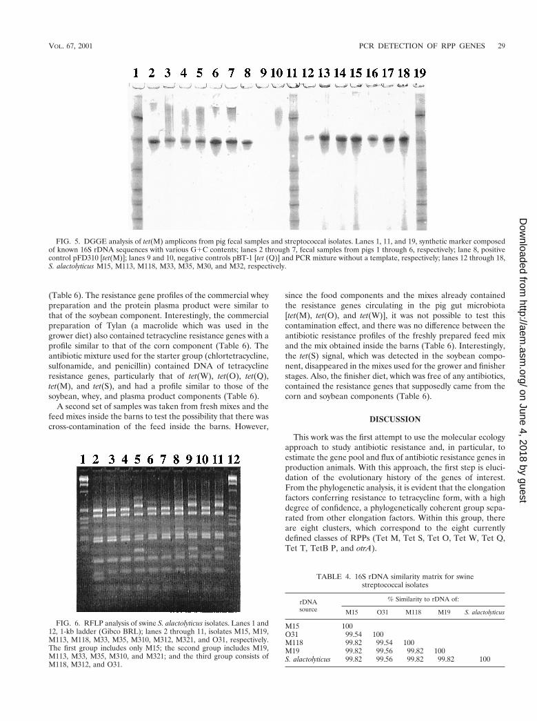
Detection of RPP genes in fecal streptococci from swine.Fecal streptococcal isolates from swine (n 5 150; 25 isolatesfrom each of six animals) were characterized by RFLP and 16SrDNA sequence analyses. As determined by the RFLP analy-sis, these strains could be divided into at least three groups(Fig. 6). Sequence analysis allowed identification (sequencesimilarity, .99%) as strains of Streptococcus alactolyticus (Ta-ble 4). A majority (94.7%) of the isolates were resistant totetracycline at a concentration of 10 mg/ml (Table 5). PCR
VOL. 67, 2001 PCR DETECTION OF RPP GENES 25
on June 4, 2018 by guesthttp://aem
.asm.org/
Dow
nloaded from

FIG. 1. Phylogenetic placement of tetracycline resistance genes encoding RPPs. The sequence of the A. aeolicus fusA gene for translationelongation factor EF-G was used as the outgroup to root the tree. The number at each node is the number of times that that tree configurationoccurred in 1,000 bootstrap trials. The scale bar indicates 0.1 fixed nucleotide substitution per sequence position. The sets of PCR primers (Table1) targeting various classes of RPP genes are shown on the right.
26 AMINOV ET AL. APPL. ENVIRON. MICROBIOL.
on June 4, 2018 by guesthttp://aem
.asm.org/
Dow
nloaded from

analysis with our set of primers revealed that all of the resistantisolates carried the tet(O) gene (Table 5).Approximately 22%of the strains carried tet(M) in addition to tet(O). No othertetracycline resistance determinants conferring ribosomal pro-
tection were detected in these isolates (Table 5). Thus, thetetracycline-resistant swine S. alactolyticus populations werecharacterized by the invariable presence of tet(O), and 22% ofthe strains carried both tet(O) and tet(M).
FIG. 2. DGGE analysis of tet(W) amplicons from steer rumen and pig fecal samples. Lanes 1, 10, and 20, synthetic marker composed of known16S rDNA sequences with various G1C contents; lanes 2 through 9, rumen samples from steers 277, 279, 280, 281, J277, J279, J280, and J281,respectively; lanes 11 and 12, negative controls pBT-1 [tet(Q)] and pJIR667 [DtetB(P)], respectively; lane 13, positive control pGEM-tetW [tet(W)];lanes 14 through 19, fecal samples from pigs 1 through 6, respectively.
TABLE 3. Validation of PCR primers with control templates
TemplateAmplification with PCR primer sets
Ribo2 TetB/P TetM TetO OTR TetQ TetS TetT TetW
Genomic or plasmid DNAC. jejuni subsp. jejuni [tet(O)] 1 2 2 1 2 2 2 2 2S. pyogenes A498 [tet(T)] 1 2 2 2 2 2 2 1 2C. perfringens JIR4202 [tetA(P) tetB(P)] 1 1 2 2 2 2 2 2 2pBT-1 [tet(Q)] 1 2 2 2 2 1 2 2 2pJIR667 [DtetB(P)] 2 2 2 2 2 2 2 2 2pFD310 [tet(M)] 1 2 1 2 2 2 2 2 2pGEM-tetW [tet(W)] 1 2 2 2 2 2 2 2 1pGEM-tetO [tet(O)] 1 2 2 1 2 2 2 2 2pVP2 [tet(S)] 1 2 2 2 2 2 1 2 2pAT451 [tet(S)] 1 2 2 2 2 2 1 2 2pCT10 [tet] 2 2 2 2 2 2 2 2 2
Cell biomassC. jejuni subsp. jejuni [tet(O)] 1 2 2 1 2 2 2 2 2S. pyogenes A498 [tet(T)] 1 2 2 2 2 2 2 1 2C. perfringens JIR4202 [tetA(P) tetB(P)] 1 1 2 2 2 2 2 2 2E. coli(pBT-1) [tet(Q)] 1 2 2 2 2 1 2 2 2E. coli(pJIR667) [DtetB(P)] 2 2 2 2 2 2 2 2 2E. coli(pFD310) [tet(M)] 1 2 1 2 2 2 2 2 2E. coli(pGEM-tetW) [tet(W)] 1 2 2 2 2 2 2 2 1E. coli(pGEM-tetO) [tet(O)] 1 2 2 1 2 2 2 2 2E. coli(pVP2) [tet(S)] 1 2 2 2 2 2 1 2 2E. coli(pAT451) [tet(S)] 1 2 2 2 2 2 1 2 2E. coli(pCT10) [tet] 2 2 2 2 2 2 2 2 2
VOL. 67, 2001 PCR DETECTION OF RPP GENES 27
on June 4, 2018 by guesthttp://aem
.asm.org/
Dow
nloaded from

DGGE and sequence analyses of the amplified tet(M) frag-ments from streptococcal strains demonstrated that these frag-ments were identical (Fig. 5, lanes 12 through 18). Moreover,they were identical to the fragments amplified directly from thetotal DNA, as well as to the control template (Fig. 5). Thecontrol template, tet(M), was originally cloned from S. agalac-tiae, and its 16S rDNA sequence was 97% similar to those ofour streptococcal isolates. Apparently, in our herd S. alacto-lyticus populations could be considered one of the main reser-voirs of the tet(M) gene in the swine intestinal microbiota.
DGGE analysis of streptococcal tet(O) revealed some de-gree of heterogeneity. In particular, the amplicons from two
isolates, O19 and O35, migrated farther through DGGE gelsthan the amplicons from nine other streptococci migrated (theresults for O19 are shown in lane 8 of Fig. 3). Sequence anal-ysis of these two amplicons revealed a single A3G substitution(but the locations were different). However, as with tet(M), themajority of tet(O) amplicons (as exemplified by S. alactolyticusO31 in Fig. 3) had the same melting characteristics as theamplicons amplified from the total swine fecal DNA (Fig. 3).Because of the universal presence of tet(O), S. alactolyticuspopulations could be considered one of the main reservoirs ofthe tet(O) gene in the swine intestinal microbiota. In addition,the TetO-generated DGGE bands from swine streptococcalisolates had the same mobility on DGGE gels as the bandsfrom rumen samples (Fig. 3). The occurrence of tet(O) incultivable rumen bacteria was not studied, and it is not clear inwhich part of the rumen microbiota the gene resides. As in pigsamples, the organisms containing the gene may be the ruminalstreptococci, which have been shown to possess transferabletetracycline resistance (13).
Detection of RPP genes in swine feed. Because of the pres-ence of unidentified inhibitory substances, a second round ofPCR was necessary in the experiments performed with swinefeed, and therefore, the detection limit of this assay was lowerthan that of the assay performed with the fecal and rumensamples. The presence of bacterial DNA in all premix andmixed samples was confirmed by amplification of the V3 regionof bacterial 16S rDNA (Table 6). The presence of RPP genesin these samples was confirmed first with the Ribo2 primer setand then with class-specific primers (Table 6). First, the feedcomponents were sampled before the corresponding diet mixeswere prepared for three different age groups. These groupswere the starters (ages, 3 to 6 weeks), growers (6 weeks to 6months), and finishers (antibiotics were withdrawn beforeslaughtering). The corn component used to prepare the mixesfor all age groups contained tet(W), tet(O), tet(Q), and tet(M),while the soybean component also contained the tet(S) gene
FIG. 3. DGGE analysis of tet(O) amplicons from pig fecal and steer rumen samples. Lanes 1, 12, and 21, synthetic marker composed of known16S rDNA sequences with various G1C contents; lanes 2 through 7, fecal samples from pigs 1 through 6, respectively; lanes 8 and 9, S. alactolyticusO19 and O31, respectively; lanes 10 and 11, negative controls pBT-1 [tet(Q)] and pJIR667 [DtetB(P)], respectively; lanes 13 through 20, rumensamples from steers 277, 279, 280, 281, J277, J279, J280, and J281, respectively.
FIG. 4. DGGE analysis of tet(Q) amplicons from steer rumen sam-ples. Lanes 1 and 11, synthetic marker composed of known 16S rDNAsequences with various G1C contents; lanes 2 through 9, rumen sam-ples from steers 277, 279, 280, 281, J277, J279, J280, and J281, respec-tively; lane 10, positive control pBT-1 [tet(Q)].
28 AMINOV ET AL. APPL. ENVIRON. MICROBIOL.
on June 4, 2018 by guesthttp://aem
.asm.org/
Dow
nloaded from
(Table 6). The resistance gene profiles of the commercial wheypreparation and the protein plasma product were similar tothat of the soybean component. Interestingly, the commercialpreparation of Tylan (a macrolide which was used in thegrower diet) also contained tetracycline resistance genes with aprofile similar to that of the corn component (Table 6). Theantibiotic mixture used for the starter group (chlortetracycline,sulfonamide, and penicillin) contained DNA of tetracyclineresistance genes, particularly that of tet(W), tet(O), tet(Q),tet(M), and tet(S), and had a profile similar to those of thesoybean, whey, and plasma product components (Table 6).
A second set of samples was taken from fresh mixes and thefeed mixes inside the barns to test the possibility that there wascross-contamination of the feed inside the barns. However,
since the food components and the mixes already containedthe resistance genes circulating in the pig gut microbiota[tet(M), tet(O), and tet(W)], it was not possible to test thiscontamination effect, and there was no difference between theantibiotic resistance profiles of the freshly prepared feed mixand the mix obtained inside the barns (Table 6). Interestingly,the tet(S) signal, which was detected in the soybean compo-nent, disappeared in the mixes used for the grower and finisherstages. Also, the finisher diet, which was free of any antibiotics,contained the resistance genes that supposedly came from thecorn and soybean components (Table 6).
DISCUSSION
This work was the first attempt to use the molecular ecologyapproach to study antibiotic resistance and, in particular, toestimate the gene pool and flux of antibiotic resistance genes inproduction animals. With this approach, the first step is eluci-dation of the evolutionary history of the genes of interest.From the phylogenetic analysis, it is evident that the elongationfactors conferring resistance to tetracycline form, with a highdegree of confidence, a phylogenetically coherent group sepa-rated from other elongation factors. Within this group, thereare eight clusters, which correspond to the eight currentlydefined classes of RPPs (Tet M, Tet S, Tet O, Tet W, Tet Q,Tet T, TetB P, and otrA).
FIG. 5. DGGE analysis of tet(M) amplicons from pig fecal samples and streptococcal isolates. Lanes 1, 11, and 19, synthetic marker composedof known 16S rDNA sequences with various G1C contents; lanes 2 through 7, fecal samples from pigs 1 through 6, respectively; lane 8, positivecontrol pFD310 [tet(M)]; lanes 9 and 10, negative controls pBT-1 [tet (Q)] and PCR mixture without a template, respectively; lanes 12 through 18,S. alactolyticus M15, M113, M118, M33, M35, M30, and M32, respectively.
FIG. 6. RFLP analysis of swine S. alactolyticus isolates. Lanes 1 and12, 1-kb ladder (Gibco BRL); lanes 2 through 11, isolates M15, M19,M113, M118, M33, M35, M310, M312, M321, and O31, respectively.The first group includes only M15; the second group includes M19,M113, M33, M35, M310, and M321; and the third group consists ofM118, M312, and O31.
TABLE 4. 16S rDNA similarity matrix for swinestreptococcal isolates
rDNAsource
% Similarity to rDNA of:
M15 O31 M118 M19 S. alactolyticus
M15 100O31 99.54 100M118 99.82 99.54 100M19 99.82 99.56 99.82 100S. alactolyticus 99.82 99.56 99.82 99.82 100
VOL. 67, 2001 PCR DETECTION OF RPP GENES 29
on June 4, 2018 by guesthttp://aem
.asm.org/
Dow
nloaded from

The most deeply branching class, exemplified by tet and otrA,is the class obtained from the antibiotic-producing organismsS. lividans and S. rimosus. Based on the sequence informationavailable, there is no evidence of recent horizontal transfer ofRPP genes from antibiotic-producing strains to commensal orpathogenic microbiotas. Hybridization data indicate, however,that some mycobacteria may actually carry the resistance genesoriginally described in streptomycetes (30). Additional se-quence information concerning the mycobacterial RPP genesis required to decide whether there was a potential horizontaltransfer event from antibiotic-producing strains. Another in-teresting aspect of the two available gene sequences of antibi-otic-producing streptomycetes is that they are quite divergent.The length of the branch between the two genes is actuallycomparable to the length of the branch separating the Tet M,Tet S, and Tet O classes (Fig. 1). If more sequence data fromthis class of genes were available, perhaps definition of at leasttwo new classes would be necessary.
The available sequence data support the scenario that earlybranching and lengthy independent diversification of eight (ormore) clusters of RPPs occurred well before the “antibioticera.” While the functional role of these proteins in antibiotic-producing bacteria is evident (they provide protection againstthe synthesized antibiotics), it is more challenging to explaintheir presence and function in bacteria from other ecologicalniches that have no or limited contact with the soil microbiota(e.g., the gastrointestinal tract). The long evolutionary historyof RPP genes supports the hypothesis that these genes mighthave served some metabolic functions other than providing
antibiotic resistance. Protein synthesis is a vital cell process,and there should be mechanisms that support proper function-ing of the translation machinery and buffer possible undesir-able effects of low-molecular-weight metabolites of the cell.Thus, the alternative elongation factors may have been se-lected in this way in some bacteria and may have assumed arole in protecting ribosomes against tetracyclines only recently.
At the same time, the rapid movement of the tetracycline-resistant elongation factors to taxonomically divergent com-mensal and pathogenic bacteria is a very recent evolutionaryevent on the phylogenetic time scale and can most probably beattributed to horizontal transfers within the clusters in theantibiotic era. Some of the genes are located on plasmids (e.g.,pOZ101, pIP811, or pK214) or conjugative transposons (e.g.,Tn916, Tn5251, or Tn1545), thus facilitating transfer betweenspecies and genus boundaries.
Proof of the monophyletic origin of the RPP genes openedthe possibility of designing primer sets targeting all classes, aswell as class-specific primers. However, it appeared that theearly branching and further independent diversification of theotrA genes, together with a high G1C content, precluded in-corporation of these genes into the alignment. Also, the num-ber of substitutions per base pair appeared to be higher in theRPP genes than in other elongation factors, and therefore, theoverall sequence structure is less conserved. Thus, the overalldesign of the universal primer pair involves a substantial levelof degeneracy and does not include the genes from the anti-biotic-producing streptomycetes. Primers were validated inPCR that included crude bacterial biomass and fecal material,
TABLE 5. Tetracycline resistance phenotypes and genotypes of swine fecal streptococci
Animalno.
No. ofisolates
No. of resistantphenotypes
No. of strainsanalyzed
No. of resistant genotypes
tetB(P) tet(M) tet(Q) tet(O) tet(S) tet(T) tet(W)
1 25 20 (80)a 19 NAb 4 (21.1) 0 19 (100) NA NA NA2 25 25 (100) 18 NA NA NA 18 (100) NA NA NA3 25 23 (92) 22 0 5 (22.7) NA 22 (100) NA NA 04 25 25 (100) NA NA NA NA NA NA NA NA5 25 24 (96) 22 NA NA NA NA NA 0 NA6 25 25 (100) 22 NA NA NA NA 0 NA NATotal 150 142 (94.7) 103 0 9 (21.9) 0 59 (100) 0 0 0
a The values in parentheses are percentages.b NA, not analyzed.
TABLE 6. Detection of RPP genes in swine feed and feed components
SampleDetection with primer sets
V3 region TetB/P TetM TetQ TetO TetS TetT TetW
Corn 1 2 1 1 1 2 2 1Soybean 1 2 1 1 1 1 2 1Whey 1 2 1 1 1 1 2 1Plasma protein 1 2 1 1 1 1 2 1CSP 1 2 1 1 1 1 2 1Tylan 1 2 1 1 1 2 2 1Starter mix (freshly prepared) 1 2 1 1 1 1 2 1Starter mix (inside barn) 1 2 1 1 1 1 2 1Grower mix (freshly prepared) 1 2 1 1 1 2 2 1Grower mix (inside barn) 1 2 1 1 1 2 2 1Finisher mix (freshly prepared) 1 2 1 1 1 2 2 1Finisher mix (inside barn) 1 2 1 1 1 2 2 1
30 AMINOV ET AL. APPL. ENVIRON. MICROBIOL.
on June 4, 2018 by guesthttp://aem
.asm.org/
Dow
nloaded from

which is notorious for the presence of PCR-inhibiting sub-stances. This fact could be useful for rapid screening for thepresence of the RPP genes in bacteria without a DNA isolationstep. The primers also were designed to amplify short se-quences, thus allowing use in PCR-DGGE analysis. Therefore,such an analysis could be performed with total DNA prepara-tions of environmental origin, thus allowing for the first timeaccess to the pool and diversity of RPP genes in a given eco-system.
The primers were used to detect the occurrence of RPPgenes in the rumina of cows, in swine feed and feces, and inswine fecal streptococci. The Tet O and Tet W determinantswere found in the intestinal contents of both types of animals,while Tet M was confined to pigs and Tet Q was confined tothe rumen. Approximate estimates suggest that up to 5% ofthe bacteria in the rumen and swine intestine may carry thetet(O) gene. Another interesting observation is that tet(W) andthe majority of the tet(O) genes circulating in the two differentanimal herds, which had very different antibiotic use regimens,were actually identical. The identity of the tet(W) genes ob-tained from bovine and ovine rumen and human intestinalisolates was demonstrated in a recent study (37). Obviously,this finding could be extended to include yet another animalmodel (pig) and another gene [tet(O)]. The occurrence ofidentical tetracycline resistance genes in different hosts pro-vides additional evidence that there are extensive pools ofantibiotic resistance genes that are actively exchanged at leastbetween domestic animals. However, genetic transfer itself isnot a guarantee that the transferred antibiotic resistance genewill be maintained in another host. The second observationconcerning the persistence of antibiotic resistance in the ap-parent absence of antibiotic selective pressure [cattle that haveno antibiotic in their feed but carry intestinal bacteria withtet(O), tet(Q), and tet(W) and swine feed containing a diversegroup of resistance genes] raises the question of how resistancepersists. The possession of an antibiotic resistance gene by abacterium is certainly advantageous in the presence of thecorresponding antibiotic. In the absence of the antibiotic, how-ever, the cost of carrying of the resistance gene should reducethe bacterial fitness and the resistant phenotype should bereplaced by the sensitive phenotype. However, a recent reex-amination of this topic suggested that bacteria may have beenable to adapt to the burden of resistance with little or no costto their fitness (25). In this scenario, the antibiotic-resistantmicrobiota would successfully compete with the sensitive coun-terpart even in the absence of selection. Such adaptationswould preclude resistant lineages from reverting to sensitivityand make control of antibiotic resistance even more difficult.
The significant outcome of the sequence analysis of tetracy-cline resistance genes in cultivable streptococcal isolates is thatthe nucleotide sequences of tet(M) and the majority of tet(O)genes are identical to those of the corresponding genes ac-quired directly from fecal DNA. This is yet another validationof the in vitro analysis approach and suggests that the pool ofresistance genes, initially discovered in total DNA, could betracked to specific bacterial populations in the gut. In our case,S. alactolyticus could be considered one of the main reservoirsof the tet(M) and tet(O) genes in the swine intestinal micro-biota. Based on RFLP and sequence analyses of 16S rDNA ofS. alactolyticus isolates, this is not a clonal population but is
represented by at least three subpopulations. Therefore, circu-lation of identical tet(M) and tet(O) genes in this geneticallydiverse group of bacteria suggests that there is horizontal ex-change of tetracycline resistance genes rather than coexistenceof several tetracycline-resistant clones.
Compared with the rumen and fecal samples, the compo-nents of the swine feed appeared to be contaminated with amore diverse group of RPPs, and only two classes (Tet T andTetB P) were absent. No attempt to isolate resistant bacteriawas made, but the ubiquitous presence of these genes, togetherwith the bacterial V3 markers, suggests that the feed compo-nents may have been contaminated by bacteria carrying thecorresponding resistance genes. It is not clear whether thesebacteria were dead or viable; regardless, the feed was geneti-cally contaminated. The experiments were designed to detectpossible cross-contamination of the swine feed by on-farm dustand fecal material, but it appeared that the components ofswine feed already carried more diverse markers of tetracy-cline resistance, including that in the swine gut microbiota.This suggests that the actual source of antibiotic resistancegene contamination of swine feed was something else andrequires further independent research. At this time, we hy-pothesize that at least for the corn and soybean componentsthe source may have been manure from farms on which anti-biotics were used, which was applied to the land. Whey, aby-product of cheese manufacturing, may contain a residualbiomass of tetracycline-resistant lactic acid bacteria and pro-pionobacteria. However, we have no information concerningthe source of antibiotic resistance gene contamination in othercomponents of the swine feed, such as the plasma protein andespecially the antibiotic preparations, which are supposedly theproducts of sterile fermentation.
In this study molecular ecology tools were used to study theantibiotic resistance problem, and the results suggest that thisapproach has the potential of uncovering the reservoirs anddetermining the identities of antibiotic resistance genes in avariety of ecosystems. This approach could be easily extendedto other classes of antibiotic resistance genes in order to un-derstand the pathways leading to acquisition of drug resistanceby human- and animal-pathogenic bacteria.
REFERENCES
1. Amann, R. I., W. Ludwig, and K. H. Schleifer. 1995. Phylogenetic identifi-cation and in situ detection of individual microbial cells without cultivation.Microbiol. Rev. 59:143–169.
2. Barbosa, T. M., K. P. Scott, and H. J. Flint. 1999. Evidence for recentintergenic transfer of a new tetracycline resistance gene, tet(W), isolatedfrom Butyrivibrio fibrisolvens, and the occurrence of tet(O), in ruminal bac-teria. Environ. Microbiol. 1:53–64.
3. Benson, D. A., M. S. Boguski, D. J. Lipman, J. Ostell, B. F. Ouellette, B. A.Rapp, and D. L. Wheeler. 1999. GenBank. Nucleic Acids Res. 27:12–17.
4. Bergeron, M. G., and M. Quellette. 1995. Diagnosing bacterial infectiousdiseases in one hour: an essential upcoming revolution. Infection 23:69–72.
5. Burdett, V. 1986. Streptococcal tetracycline resistance mediated at the levelof protein synthesis. J. Bacteriol. 165:564–569.
6. Burdett, V. 1991. Purification and characterization of Tet(M), a protein thatrenders ribosomes resistant to tetracycline. J. Biol. Chem. 266:2872–2877.
7. Charpentier, E., G. Gerbaud, and P. Courvalin. 1993. Characterization of anew class of tetracycline-resistance gene tet(S) in Listeria monocytogenesBM4210. Gene 131:27–34.
8. Charpentier, E., G. Gerbaud, and P. Courvalin. 1994. Presence of the Lis-teria tetracycline resistance gene tet(S) in Enterococcus faecalis. Antimicrob.Agents Chemother. 38:2330–2335.
9. Clermont, D., O. Chesneau, G. De Cespedes, and T. Horaud. 1997. Newtetracycline resistance determinants coding for ribosomal protection instreptococci and nucleotide sequence of tet(T) isolated from Streptococcuspyogenes A498. Antimicrob. Agents Chemother. 41:112–116.
VOL. 67, 2001 PCR DETECTION OF RPP GENES 31
on June 4, 2018 by guesthttp://aem
.asm.org/
Dow
nloaded from
10. Dittrich, W., and H. Schrempf. 1992. The unstable tetracycline resistancegene of Streptomyces lividans 1326 encodes a putative protein with similari-ties to translational elongation factors and Tet(M) and Tet(O) proteins.Antimicrob. Agents Chemother. 36:1119–1124.
11. Felsenstein, J. 1985. Confidence limits on phylogenies: an approach usingthe bootstrap. Evolution 39:783–791.
12. Felsenstein, J. 1993. PHYLIP (Phylogeny Inference Package) version 3.53c.Department of Genetics, University of Washington, Seattle.
13. Jonecova, Z., M. Marekova, and V. Kmei. 1994. Conjugative transfer oftetracycline resistance in rumen streptococcal strains. Folia Microbiol. 39:83–86.
14. Kimura, M. 1980. A simple model for estimating evolutionary rates of basesubstitutions through comparative studies of nucleotide sequences. J. Mol.Evol. 16:111–120.
15. Lane, D. J. 1991. 16S/23S rRNA sequencing, p. 115–175. In E. Stackebrandtand M. Goodfellow (ed.), Nucleic acid techniques in bacterial systematics.John Wiley and Sons, New York, N.Y.
16. LeBlanc, D. J., L. N. Lee, B. M. Titmas, C. J. Smith, and F. C. Tenover. 1998.Nucleotide sequence analysis of tetracycline resistance gene tet(O) fromStreptococcus mutans DL5. J. Bacteriol. 170:3618–3626.
17. Levy, S. B., L. M. McMurry, V. Burdett, P. Courvalin, W. Hillen, M. C.Roberts, and D. E. Taylor. 1989. Nomenclature for tetracycline resistancedeterminants. Antimicrob. Agents Chemother. 33:1373–1374.
18. Levy, S. B. 1992. Active efflux mechanisms for antimicrobial resistance.Antimicrob. Agents Chemother. 36:695–703.
19. Levy, S. B., L. M. McMurry, T. M. Barbosa, V. Burdett, P. Courvalin, W.Hillen, M. C. Roberts, J. I. Rood, and D. E. Taylor. 1999. Nomenclature fornew tetracycline resistance determinants. Antimicrob. Agents Chemother.43:1523–1524.
20. Lyras, D., and J. I. Rood. 1996. Genetic organization and distribution oftetracycline resistance determinants in Clostridium perfringens. Antimicrob.Agents Chemother. 40:2500–2504.
21. Madden, T. L., R. L. Tatusov, and J. Zhang. 1996. Application of networkBLAST server. Methods Enzymol. 266:131–141.
22. Manavathu, E. K., K. Hiratsuka, and D. E. Taylor. 1988. Nucleotide se-quence analysis and expression of a tetracycline resistance gene from Campy-lobacter jejuni. Gene 62:17–26.
23. Manavathu, E. K., C. L. Fernandez, B. S. Cooperman, and D. E. Taylor.1990. Molecular studies on the mechanism of tetracycline resistance medi-ated by Tet(O). Antimicrob. Agents Chemother. 34:71–77.
24. Martin, P., P. Trieu-Cuot, and P. Courvalin. 1986. Nucleotide sequence ofthe Tet M tetracycline resistance determinant of the streptococcal conjuga-tive shuttle transposon Tn1545. Nucleic Acids Res. 14:7047–7058.
25. Morris, A., J. D. Kellner, and D. E. Low. 1998. The superbugs: evolution,dissemination and fitness. Curr. Opin. Microbiol. 1:524–529.
26. Muyzer, G., E. C. de Waal, and A. G. Uitterlinden. 1993. Profiling of complexmicrobial populations by denaturing gradient gel electrophoresis analysis ofpolymerase chain reaction-amplified genes coding for 16S rRNA. Appl.Environ. Microbiol. 59:695–700.
27. Muyzer, G., S. Hottentrager, A. Teske, and C. Wawer. 1996. Denaturinggradient gel electrophoresis of PCR-amplified 16S rDNA—a new molecularapproach to analyze the genetic diversity of mixed microbial communities, p.3.4.4.1–3.4.4.22. In A. D. L. Akkermans, J. D. van Elsas, and F. J. de Bruijn
(ed.), Molecular microbial ecology manual. Kluwer Academic Publishers,Dordrecht, The Netherlands.
28. Nikolich, M. P., N. B. Shoemaker, and A. A. Salyers. 1992. A Bacteroidestetracycline resistance gene represents a new class of ribosome protectiontetracycline resistance. Antimicrob. Agents Chemother. 36:1005–1012.
29. Nikolich, M. P., G. Hong, N. B. Shoemaker, and A. A. Salyers. 1994. Evi-dence for the horizontal transfer of tetQ between bacteria that normallycolonize humans and bacteria that normally colonize livestock. Appl. Envi-ron. Microbiol. 60:3255–3260.
30. Pang, Y., B. A. Brown, V. A. Steingrube, R. J. Wallace, Jr., and M. C.Roberts. 1994. Tetracycline resistance determinants in Mycobacterium andStreptomyces species. Antimicrob. Agents Chemother. 38:1408–1412.
31. Perreten, V., F. Schwarz, L. Cresta, M. Boeglin, G. Dasen, and M. Teuber.1997. Antibiotic resistance spread in food. Nature 389:801–802.
32. Roberts, M. C. 1994. Epidemiology of tetracycline-resistance determinants.Trends Microbiol. 2:353–357.
33. Roberts, M. C. 1996. Tetracycline resistance determinants: mechanisms ofaction, regulation of expression, genetic mobility, and distribution. FEMSMicrobiol. Rev. 19:1–24.
34. Saitou, N., and M. Nei. 1987. The neighbor-joining method: a new methodfor reconstructing phylogenetic trees. Mol. Biol. Evol. 4:406–425.
35. Sanchez-Pescador, R., J. T. Brown, M. C. Roberts, and M. S. Urdea. 1988.Homology of the Tet(M) with translational elongation factors: implicationfor potential modes of tet(M)-conferred tetracycline resistance. Nucleic Ac-ids Res. 16:1218.
36. Schnappinger, D., and W. Hillen. 1996. Tetracyclines: antibiotic action, up-take, and resistance mechanisms. Arch. Microbiol. 165:359–369.
37. Scott, K. P., C. M. Melville, T. M. Barbosa, and H. J. Flint. 2000. Occurrenceof the new tetracycline resistance gene tet(W) in bacteria from the humangut. Antimicrob. Agents Chemother. 44:775–777.
38. Sloan, J., L. M. McMurray, D. Lyras, S. B. Levy, and J. I. Rood. 1994. TheClostridium perfringens Tet P determinant comprises two overlapping genes:tetA(P), which mediates active tetracycline efflux, and tetB(P), which is re-lated to the ribosomal protection family of tetracycline-resistance determi-nants. Mol. Microbiol. 11:403–415.
39. Smith, C. J., M. B. Rogers, and M. L. McKee. 1992. Heterologous geneexpression in Bacteroides fragilis. Plasmid 27:141–154.
40. Sougakoff, W., B. Papadopoulou, P. Nordman, and P. Courvalin. 1987.Nucleotide sequence and distribution of tet(O) gene encoding tetracyclineresistance in Campylobacter coli. FEMS Microbiol. Lett. 44:153–159.
41. Speer, B. S., and A. A. Salyers. 1989. Novel aerobic tetracycline resistancegene that chemically modifies tetracycline. J. Bacteriol. 171:148–153.
42. Taylor, D. E., K. Hiratsuka, H. Ray, and E. K. Manavathu. 1987. Charac-terization and expression of a cloned tetracycline resistance determinantfrom Campylobacter jejuni plasmid pUA466. J. Bacteriol. 169:2984–2989.
43. Taylor, D. E., and A. Chau. 1996. Tetracycline resistance mediated by ribo-somal protection. Antimicrob. Agents Chemother. 40:1–5.
44. Thompson, J. D., D. G. Higgins, and T. J. Gibson. 1994. CLUSTAL W:improving the sensitivity of progressive multiple sequence alignment throughsequence weighting, positions-specific gap penalties and weight matrixchoice. Nucleic Acids Res. 22:4673–4680.
45. Widdowson, C. A., K. P. Klugman, and D. Hanslo. 1996. Identification of thetetracycline resistance gene, tet(O), in Streptococcus pneumoniae. Antimi-crob. Agents Chemother. 40:2891–2893.
32 AMINOV ET AL. APPL. ENVIRON. MICROBIOL.
on June 4, 2018 by guesthttp://aem
.asm.org/
Dow
nloaded from