Embed Size (px)
Citation preview

HEP (2007) 178:49–72© Springer-Verlag Berlin Heidelberg 2007
Tetracycline-Controlled Genetic SwitchesR. Sprengel · M. T. Hasan (�)
Max Planck Institute for Medical Research, Jahnstrasse 29, 69120 Heidelberg, [email protected]
1 Introduction . . . . . . . . . . . . . . . . . . . . . . . . . . . . . . . . . . . . 50
2 Principles of the Tet-Controlled Gene Expression . . . . . . . . . . . . . . . . 502.1 Genetic Elements . . . . . . . . . . . . . . . . . . . . . . . . . . . . . . . . . 502.2 Tet-Controlled Gene Expression in Eukaryotes . . . . . . . . . . . . . . . . . 512.2.1 Tet-Controlled Transactivators tTA and rtTA . . . . . . . . . . . . . . . . . . . 522.2.2 tTA- and rtTA-Dependent Promoters (Ptets) . . . . . . . . . . . . . . . . . . . 532.2.3 Tet and Its Derivatives . . . . . . . . . . . . . . . . . . . . . . . . . . . . . . . 54
3 Dox-Controlled Gene Regulation in Transgenic Mice . . . . . . . . . . . . . . 553.1 tTA and rtTA Minigenes . . . . . . . . . . . . . . . . . . . . . . . . . . . . . . 553.2 tTA- and rtTA-Dependent Responder Genes . . . . . . . . . . . . . . . . . . . 553.3 Setting up a Tet-Controlled Expression System in the Mouse . . . . . . . . . . 563.4 Tet-Controlled Reporter Mouse Lines . . . . . . . . . . . . . . . . . . . . . . 583.4.1 Reporter Genes . . . . . . . . . . . . . . . . . . . . . . . . . . . . . . . . . . 583.4.2 Responder Mouse Lines with Uni-directional
Tet Promoters (Ptet) for Dox-Controlled Expressionof Either the Firefly Luciferase or the β-Galactosidase . . . . . . . . . . . . . . 59
3.4.3 Responder Mice with BidirectionalTet Promoter (Ptet-bi) Regulated Co-expressionof Green Fluorescence Protein and β-Galactosidase . . . . . . . . . . . . . . . 60
3.4.4 Responder Mice with Regulated Expression of Cre Recombinase . . . . . . . . 613.5 tTA- and rtTA-Activator Mouse Lines . . . . . . . . . . . . . . . . . . . . . . . 613.6 Tet Mice from the Jackson Laboratory . . . . . . . . . . . . . . . . . . . . . . 623.7 tTA- and rtTA-Dependent Gene Expression in the Mouse . . . . . . . . . . . . 643.7.1 Transgenic Approaches . . . . . . . . . . . . . . . . . . . . . . . . . . . . . . 643.7.2 Gene Targeting Approaches . . . . . . . . . . . . . . . . . . . . . . . . . . . . 653.7.3 Kinetics of Tet-Controlled, Regulated Gene Expression in the Mouse . . . . . 663.8 Transfer of Tet-Controlled Gene Expression Via Viral Systems . . . . . . . . . 673.9 Closing Remarks . . . . . . . . . . . . . . . . . . . . . . . . . . . . . . . . . . 69
References . . . . . . . . . . . . . . . . . . . . . . . . . . . . . . . . . . . . . . . . 69
Abstract Unlike recombinase-mediated gene manipulations, tetracycline (Tet)-controlledgenetic switches permit reversible control of gene expression in the mouse. Trancriptionalactivation can be induced by activators termed tTA (Tet-Off) or rtTA (Tet-On) in the absenceand presence of Tet, respectively. The Tet-Off and Tet-On systems are complementary, andthe decision to choose one over the other depends on the particular experimental strategy.Both systems were optimized over the years and can now be used to develop mouse models.
Keywords Doxycycline · Luciferase · β-Galactosidase · GFP · Rosa26
50 R. Sprengel · M. T. Hasan
1Introduction
The Tet-controlled inducible gene expression systems allow alteration in indi-vidual gene activities in intact animals, including insects, fly, mice and rats.In mice, the Tet-controlled gene expression continues to provide fundamen-tal insight on various biological processes such as development, diseases andbehaviour.
In recentyears, theadventofmouseembryonic stem(ES)cells, bacterial arti-ficial chromosome (BAC) and recombinase techniques have accomplished withgreat precision functional changes by genetic alterations in selective cell popu-lations. Inducible control of gene expression at specifically chosen time pointswould further facilitate cross-correlation analyses to reliably link changes ingene activity with changes in phenotypes both in cell physiology and animalbehaviour.
Over the past several years, the Tet-controlled gene expression has exploredvarious biological processes in the mouse with impressive detail. Here, we willprovide an outline and an overview of different experimental strategies forgenerating mice with functional Tet-controlled genes.
2Principles of the Tet-Controlled Gene Expression
2.1Genetic Elements
Tet-controlled gene expression systems in eukaryotes are derived from thetransposon Tn10 Tet-resistance operon. Essential features of this prokary-otic Tet-controlled gene expression systems were modified to be operativein eukaryotic cells. In Gram-negative bacteria, Tet is a potent antibiotic thatkills bacteria by blocking protein synthesis (Epe and Woolley 1984). Bacte-ria can achieve resistance to Tet by expressing the TetA resistance protein,a proton-[Tet.Mg]+ antiporter, embedded in the cytoplasmic membrane (Ya-maguchi et al. 1990). Under regular conditions, TetA is not expressed sincethe tetracycline repressor (TetR) blocks TetA expression. In the absence ofTet, TetR dimers bind in the TetR and TetA promoter regions to the oper-ators tetO1 and tetO2, respectively, which physically hinders transcriptionalinitiation at the TetR and TetA promoters, thereby down-regulating expres-sion of these two genes. When intracellular Tet concentrations rise, Tet bindsto TetRs. This leads to a conformational change of the Tet–TetR complex,rendering it incapable of binding tetOs and thus opening access to transcrip-tion initiation. As Tet is effluxed out of cells, TetR regains its ability to bindtetOs and transcription of TetA and TetR genes is down-regulated (Hillen and

Tetracycline-Controlled Genetic Switches 51
Berens 1994). Functional TetR protein binds to tetO sequences as a homod-imer. Each polypeptide is composed of 208 amino acids with 10 α-helices (seeSect. 3.2.1) making up interaction surfaces for TetR dimerization and bind-ing sites for tetO and Tet (Orth et al. 2000). The inducers [Tet, doxycycline(Dox) and anhydrotetracycline (ATc)] bind to TetR with very high affinity:[Tet.Mg] + (Ka ∼ 109 M–1) [Dox.Mg] + (Ka ∼ 1010 M–1) [ATc.Mg]+ (Ka ∼ 1011
M–1), which is about three to five orders of magnitude higher than the affin-ity of these drugs to prokaryotic ribosomes (Takahashi et al. 1986; Ledereret al. 1996). The binding of two molecules of [Tet.Mg]+ to a TetR dimer reducesits affinity for tetO by about nine orders of magnitude (Lederer et al. 1996).Sensitive and reversible control of gene expression is made feasible by TetRbinding to tetO even in the context of competing nonspecific DNA sequencesin the genome. Changes in TetR affinity for tetO when Tet binds to TetRis the prerequisite condition for the control of gene expression in higherorganisms.
2.2Tet-Controlled Gene Expression in Eukaryotes
The well-defined elements of the Tn10 Tet operon have been successfully trans-ferred to eukaryotic cells for controlling gene expression by Tet. As mentionedabove, the Tet operon operates with a simple genetic circuit that requires threeessential components: TetR, tetO and Tets. These three components have beenmodified in various ways to optimize stringent control of gene expression ineukaryotes.
The major breakthrough occurred by the pioneering work of Gossen andBujard (Gossen and Bujard 1992), who succeeded in introducing a geneticswitch for controlling gene expression in potentially all eukaryotes. First, theyconverted TetR from a repressor into a Tet-controlled transcriptional activatorby fusing the herpes simplex virus transcription activator (VP16) to the C-terminus of TetR. The TetR-VP16 hybrid protein, named tTA, binds to tetOsequences via the TetRdomain, while the C-terminal VP16 domain participatesin the recruitment of the RNA polymease II (Pol II) transcriptional initiationcomplex to initiate transcription.
Second, they generated a synthetic tTA-dependent promoter (Ptet) whereseven tetO sequences were linked to a short stretch of sequences containingthe Pol II transcriptional start site derived from the human cytomegalovirus(CMV) immediate early gene IE1 promoter (see Sect. 3.2.2). When tTA bindsto Ptet, tTA initiates transcription at a defined site in the short CMV-promoterfragment (Fig. 1A). Without tTA bound to tetOs, the short CMV-promoter frag-ment is transcriptionally inactive and therefore it is also commonly referredto as a CMV minimal promoter (CMVmin).
In the presence of Tet, tTA is unable to bind to Ptet (Fig. 1B), and tran-scription initiation at Ptet is turned-off (Tet-Off). In this way, tTA allows gene

52 R. Sprengel · M. T. Hasan
Fig. 1 A,B Principle of the Tet-Off-system. A When the constitutively expressed tTA binds toPtet, tTA initiates transcription. B In the presence of Tet, tTA is unable to bind to Ptet andPtet-controlled gene transcription is turned-off (Tet-Off)
expression to be switched on and off in response to Tet (Gossen and Bu-jard 1992).
2.2.1Tet-Controlled Transactivators tTA and rtTA
The original tTA has been functionally improved by eliminating sequencesof VP16 to minimal length for transcriptional activation, targeting it to thenucleus of cells and optimizing codon usage (Baron et al. 1997; Kim 2001;Urlinger et al. 2000). Replacement of VP16 activation domain by three copiesof 12 amino acid minimal activation F-domains, improved its tolerance inmammalian cells at a higher concentration and have graded activation poten-tials in range of 1,000-fold (Baron et al. 1997; Kim 2001). Similarly, addition ofa nuclear localization signal to tTA improved the efficiency of Tet-controlledgene expression (Kim 2001).
A new quality of the Tet-controlled system was achieved by exchangingthe TetR of tTA by a TetR mutant with four point mutations to generate thereverse tTA (rtTA) (Fig. 2). The four point mutations E71K, D95N, L101S andG102D reversed the pharmacology of TetR (Hecht et al. 1993) and now Dox isnecessary for rtTA binding to Ptet (Gossen et al. 1995).
While both the rtTA- and the tTA-inducible systems can be used in themouse (Kistner et al. 1996), there are far more functional studies reportedwith tTA than rtTA (e.g. see Sect. 4.6), which might indicate that Tet-controlledgene expression is more difficult to achieve with rtTA in certain tissues, suchas the brain. The comparison of rtTA and tTA in Hela cell cultures showedthat with both transactivators gene expression was regulated fast and tightly.However, the regulation factors of 105 for tTA and 103 for rtTA shows the
Tetracycline-Controlled Genetic Switches 53
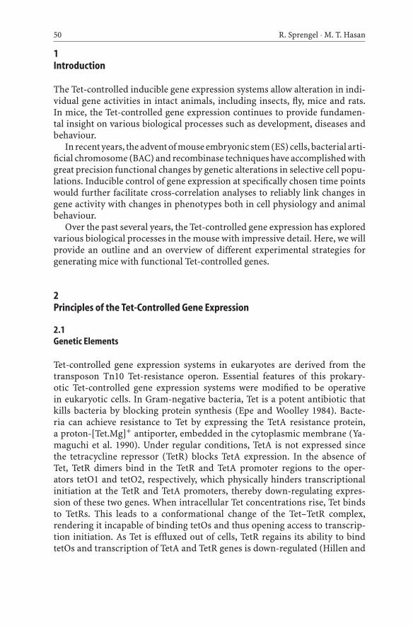
Fig. 2 Schematic drawings for some tTA and rtTA variants, which are currently available.The TetR-core region with 10 α-helices (1–10) contains sites for tetO-binding, Tet-bindingand homodimerization. VP16 or minimal F-domains make up the transactivation domains.The position of the nuclear localization domain (n) is indicated. Amino acid exchanges indifferent rtTAs are indicated by black dots. Codon improved tTA and rtTA variants are inlight grey, original tTA and rtTA are in black. Amino acid positions are numbered startingwith the first amino acid (position +1)
better efficiency of tTA. Similarly, 10 ng/ml of Dox was sufficient to fully inac-tivate tTA-dependent reporter gene transcription in less than 5 min, whereas1 µg/ml of Dox was needed for full gene activation with the rtTA system. Basedon studies in Hela cells, it was estimated that rtTA is approximately 100 timesless sensitive than tTA (Gossen et al. 1995). Novel rtTAs with higher sensitiv-ity towards Dox might overcome these limitations. So far, a genetic screen inyeast has identified two rtTA mutants (rtTA-S2 and rtTA-M2) with reducedbinding to tetO in absence of Dox and increased Dox sensitivity for rtTA-M2(Urlinger et al. 2000).
Currently, several tTA and rtTA variants are available, some of which areindicated in Fig. 2. In our hands, the tTA variant itTA2nls and the rtTA-M2nlsare suitable choices for mice (Urlinger et al. 2000; Hasan et al. 2001; Kim 2001;Krestel et al. 2004). Both itTA and rtTA-M2nls are codon-improved and lackcryptic splice sites.
2.2.2tTA- and rtTA-Dependent Promoters (Ptets)
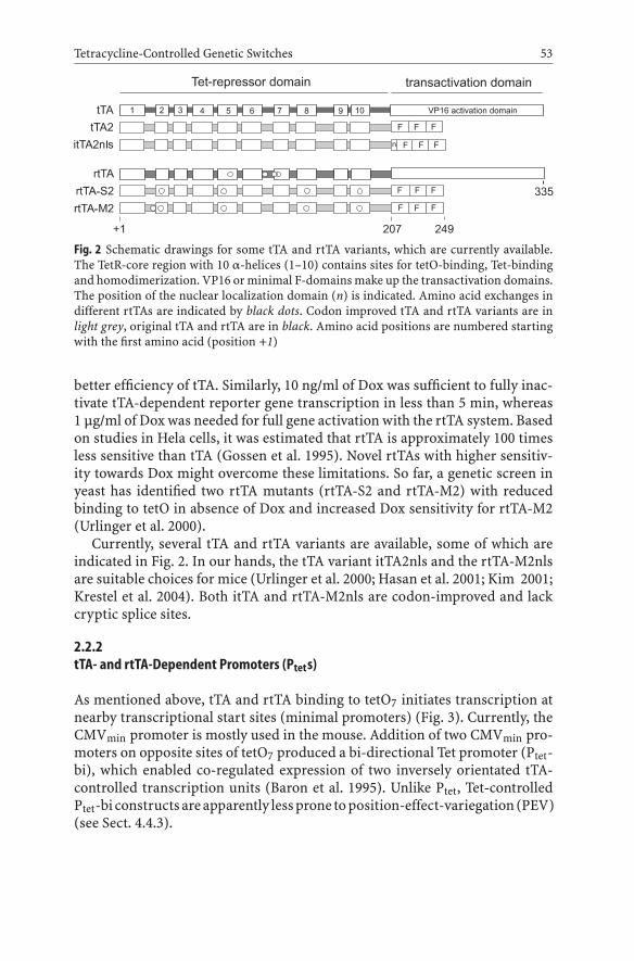
As mentioned above, tTA and rtTA binding to tetO7 initiates transcription atnearby transcriptional start sites (minimal promoters) (Fig. 3). Currently, theCMVmin promoter is mostly used in the mouse. Addition of two CMVmin pro-moters on opposite sites of tetO7 produced a bi-directional Tet promoter (Ptet-bi), which enabled co-regulated expression of two inversely orientated tTA-controlled transcription units (Baron et al. 1995). Unlike Ptet, Tet-controlledPtet-bi constructsareapparently lessprone toposition-effect-variegation(PEV)(see Sect. 4.4.3).
54 R. Sprengel · M. T. Hasan
Fig. 3 Uni- (Ptet) and bi-directional (Ptet-bi) Tet promoters. The different promoter elementsof the two tTA- and rtTA-controlled promoters are depicted. Nucleotide positions relativeto the transcriptional start sites (position +1) are indicated
2.2.3Tet and Its Derivatives
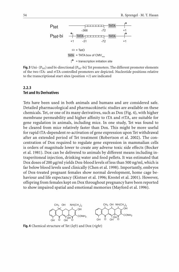
Tets have been used in both animals and humans and are considered safe.Detailed pharmacological and pharmacokinetic studies are available on thesechemicals. Tet, or one of its many derivatives, such as Dox (Fig. 4), with highermembrane permeability and higher affinity to tTA and rtTA, are suitable forgene regulation in animals, including mice. In one study, Tet was found tobe cleared from mice relatively faster than Dox. This might be more usefulfor rapid tTA-dependent re-activation of gene expression upon Tet withdrawalafter an extended period of Tet treatment (Robertson et al. 2002). The con-centration of Dox required to regulate gene expression in mammalian cellsis orders of magnitude lower to create any adverse toxic side effects (Bockeret al. 1981). Dox can be delivered to animals by different means including in-traperitoneal injection, drinking water and food pellets. It was estimated thatDox doses of 200 µg/ml yields Dox-blood levels of less than 500 ng/ml, which isfar below blood levels used clinically (Chen et al. 1998). Importantly, embryosof Dox-treated pregnant females show normal development, home cage be-haviour and life expectancy (Kistner et al. 1996; Krestel et al. 2001). However,offspring from females kept on Dox throughout pregnancy have been reportedto show impaired spatial and emotional memories (Mayford et al. 1996).
Fig. 4 Chemical structure of Tet (left) and Dox (right)

Tetracycline-Controlled Genetic Switches 55
3Dox-Controlled Gene Regulation in Transgenic Mice
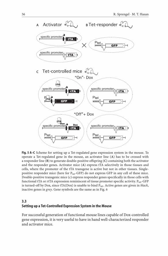
For establishing Dox-controlled gene regulation in the mouse, the geneticcomponents of the Tet system have to be introduced into the mouse genome.The components are first engineered into plasmids, cosmids or BACs andthen transferred into mice by DNA injection into the pronucleus of fertilizedmouse oocytes. Alternatively, genetic components can be inserted at definedchromosomal positions in mouse ES cells, and can be transferred to mice byreconstituting ES cells into early staged embryos (e.g. see the chapter by J.S.Draper and A. Nagy, this volume).
Traditionally, two separate independent mouse lines are generated: a tTA-or rtTA-expressing line (activator) and a line with either the Ptet- or Ptet-bicontrolled gene (responder). Intercrosses between activators and respondersgive rise to double-positive offspring (containing both the activator and theresponder genes) (Fig. 5). These double-positive mice can be tested for tTA- orrtTA-dependent Dox-controlled gene expression.
3.1tTA and rtTA Minigenes
Expression constructs for the tTA minigene are usually composed of a tissuespecific promoter followed by a synthetic 5′-untranslated intron, the tTA orrtTA coding sequences and a polyadenlyation signal (polyA) (Fig. 6). Thechoice of the promoter for a tTA or a rtTA minigene determines when andwhere the transgene is expected to be expressed. To readily achieve cell-typespecificity, larger promoter fragments can be used with the help of cosmids orBACs. In addition, coding sequences for tTA or rtTA can be inserted directlyafter promoter regions by replacing the first coding exon of endogenous genesby gene targeting in ES cells (e.g. see the chapter by J.S. Draper and A. Nagy, thisvolume). However, the latter two experimental approaches have been describedin a few reports only (see Sect. 4.7.2).
3.2tTA- and rtTA-Dependent Responder Genes
A simple responder gene contains Ptet followed by a synthetic 5′-untranslatedintron, the coding sequences of the gene of interest and polyA (Fig. 6). Popularelements of choice are introns derived from SV40, adenovirus or the β-globingene and the polyA signal from SV40, growth hormone or β-globin gene. Notall responder constructs must follow this rule (Fig. 6A).
Responders with two co-regulated genes (Fig. 6B) are generated flankingPtet-bi with two transcription units allowing regulated expression of two genesin a tTA- and rtTA-dependent manner (Baron et al. 1995). In transgenic mice,the Ptet-bi shows faithful and reproducible co-expression and Dox-controlledregulation of both genes in a Ptet-bi module (see Sect. 4.4.3).
56 R. Sprengel · M. T. Hasan
Fig. 5 A–C Scheme for setting up a Tet-regulated gene expression system in the mouse. Tooperate a Tet-regulated gene in the mouse, an activator line (A) has to be crossed witha responder line (B) to generate double-positive offspring (C) containing both the activatorand the responder genes. Activator mice (A) express tTA selectively in those tissues andcells, where the promoter of the tTA transgene is active but not in other tissues. Single-positive responder mice (here for Ptet-GFP) do not express GFP in any cell of these mice.Double-positive transgenic mice (c) express responder genes specifically in those cells withfunctional tTA or rtTA expression reminiscent of tissue promoter specific activity. Ptet-GFPis turned-off by Dox, since tTA(Dox) is unable to bind Ptet. Active genes are given in black,inactive genes in grey. Gene symbols are the same as in Fig. 6
3.3Setting up a Tet-Controlled Expression System in the Mouse
For successful generation of functional mouse lines capable of Dox-controlledgene expression, it is very useful to have in hand well-characterized responderand activator mice.
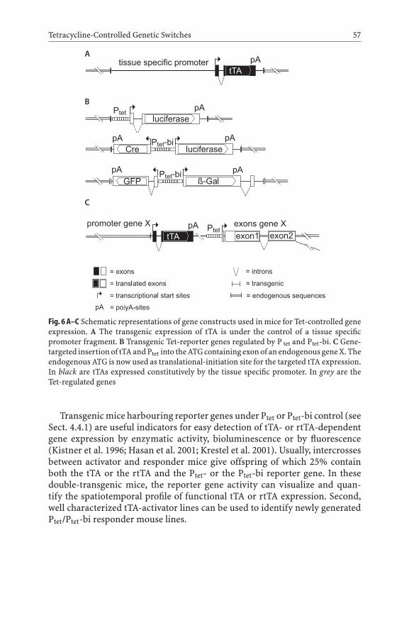
Tetracycline-Controlled Genetic Switches 57
Fig. 6A–C Schematic representations of gene constructs used in mice for Tet-controlled geneexpression. A The transgenic expression of tTA is under the control of a tissue specificpromoter fragment. B Transgenic Tet-reporter genes regulated by P tet and Ptet-bi. C Gene-targeted insertion of tTA and Ptet into the ATG containing exon of an endogenous gene X. Theendogenous ATG is now used as translational-initiation site for the targeted tTA expression.In black are tTAs expressed constitutively by the tissue specific promoter. In grey are theTet-regulated genes
Transgenic mice harbouring reporter genes under Ptet or Ptet-bi control (seeSect. 4.4.1) are useful indicators for easy detection of tTA- or rtTA-dependentgene expression by enzymatic activity, bioluminescence or by fluorescence(Kistner et al. 1996; Hasan et al. 2001; Krestel et al. 2001). Usually, intercrossesbetween activator and responder mice give offspring of which 25% containboth the tTA or the rtTA and the Ptet- or the Ptet-bi reporter gene. In thesedouble-transgenic mice, the reporter gene activity can visualize and quan-tify the spatiotemporal profile of functional tTA or rtTA expression. Second,well characterized tTA-activator lines can be used to identify newly generatedPtet/Ptet-bi responder mouse lines.
58 R. Sprengel · M. T. Hasan
3.4Tet-Controlled Reporter Mouse Lines
3.4.1Reporter Genes
Reporter mice are needed for identifying functional tTA or rtTA mouse lines.Indicated below are four different reporter genes as possible choices.
3.4.1.1Luciferase
Amongst the different reporters, the firefly luciferase gives highest sensitivityand allows exact quantitative analysis of gene activity in vivo. Few molecules ofluciferase can be detected with very low background activity in mouse tissues.With luciferase half-life of 3–4 h in vivo (Leclerc et al. 2000), fast kinetic stud-ies are possible. In mammalian cells, regulation factors of approximately 105
and approximately103 can be achieved for tTA and rtTA, respectively (Kistneret al. 1996). With 20 ng/ml of Dox, tTA-dependent gene expression in Hela cellsis suppressed in less than 5 min and finally leaving merely a few molecules percell. More importantly, upon Dox removal from the culture medium, luciferasegene activity can be monitored within 4 h of Dox removal reaching 20% ofthe steady-state level after 12 h. Techniques are also available for noninvasiveimaging of luciferase expression in the mouse (Hasan et al. 2001). Luciferaseactivity can be easily determined in tissue extracts and tissue explants (Hasanet al. 2004) but so far in these systems has not been demonstrated at a cellularresolution neither by immunohistology nor by optical recording of luciferaseactivity.
3.4.1.2Beta-Galactosidase
Although β-galactosidase (β-gal) is less sensitive than luciferase, it can alsobe quantified in tissue extracts using β-gal enzymatic assays. Now, with β-gal a cellular resolution of enzyme activity can easily be displayed in tissueslices. However, endogenous β-gal hinders detection of very low levels of trans-genically expressed β-gal. Variants of β-gal targeted to the nucleus are highlysensitive for detecting β-gal signal in tissue slices. The detection of β-gal canbe further enhanced in immunohistochemical reactions using anti-β-gal anti-bodies.
3.4.1.3Fluorescent Proteins
Among the different fluorescent proteins (XFPs) which are currently available,the enhanced green fluorescence protein (eGFP) (Chalfie et al. 1994; Cormack
Tetracycline-Controlled Genetic Switches 59
et al. 1996; Zolotukhin et al. 1996) has been used successfully as reporter (Kres-tel et al. 2001). When expression is analysed by live fluorescence, eGFP has byfar the lowest sensitivity since auto-fluorescence of tissue at the wavelength forGFP excitation and emission is quite high. Nevertheless, strong eGFP expres-sioncanbeeasilymonitored formacroscopic andmicroscopic analysis in tissueslices and in the living mouse (Krestel et al. 2001; Hasan et al. 2004). Thus, eGFPreporters can be used in vivo for identifying tTA- and rtTA-expressing mouselines and for visualizing regulated gene expression by noninvasive imagingin living mice. The sensitivity of eGFP detection can be drastically enhancedby the use of anti-eGFPs antibodies. Immunohistochemical reactions permita more detailed high-resolution microscopic analysis in tissue slices and im-munoblots can be used for quantification of protein levels. Apart from eGFP,other GFP variants under the control of Tet inducible systems have been usedin the mouse either by transgenesis (Hasan et al. 2004) or by viral vectors (seeSect. 4.8).
3.4.1.4Cre Recombinase
Similar to the reporters discussed above, Cre immunostains of tissues can beused to detect tTA and rtTA activity at a cellular level. Quantification in im-munoblots from tissue extracts is also achievable. By employing Cre reporters(RosaR26R; see Sect. 4.4.4), tTA- and rtTA-activated Cre expression at ear-lier stages in development can be historically engraved by Cre-induced β-galexpression (See Sect. 4.4.4).
3.4.2Responder Mouse Lines with Uni-directionalTet Promoters (Ptet) for Dox-Controlled Expressionof Either the Firefly Luciferase or the β-Galactosidase
3.4.2.1Tg(tetL)1Bjd/J
A mouse line, previously called L7, where luciferase gene is under the controlof Ptet, has been characterized in detail. Mice of line L7 (Tg(tetL)1Bjd/J; seeSect. 4.6) do not show any background luciferase activity and when activatedby tTA or rtTA, luciferase activity is expressed at high levels and is tightlyregulatable by Dox. In original studies, mice were generated which expressedtransgenic tTA or rtTA under the control of the early human CMV EA1 pro-moter (Tg(tTAhCMV)3Bjd/J and Tg(rtTAhCMV)4Bjd/J; see Sect. 4.6). Bothactivator lines regulated the luciferase reporter of L7 mice to a high degree invarious tissues. When these mice were treated with Dox in the drinking water
60 R. Sprengel · M. T. Hasan
for 1 week, the regulation factor for luciferase activity was up to 105. Similarly,L7 mice crossed with liver-specific tTA-expressing mice (Tg(tTALap)5Bjd/J;see Sect. 4.6) in double-positive mice showed high levels of luciferase activ-ity in the liver, which was tightly regulatable by Dox. By correlating enzy-matic activity to the total number of cells, the authors estimated that thereare approximately 104–105 luciferase molecules per cell in the fully inducedstate and only one molecule per ten cells in the noninduced state (Kistneret al. 1996).
3.4.2.2Tg(tetNZL)2Bjd/J
Alternatively, a responder encoding luciferase inPtet-bimodules canbeused. InTg(tetNZL)2Bjd/J mice (see Sect. 4.6), both luciferase and β-gal with a nuclearlocalization signal are controlled by Ptet-bi. Tg(tetNZL)2Bjd/J mice were testedwith tTA activators of line Tg(tTALap)5Bjd/J (see Sect. 4.6) to demonstrateliver-specific expression of the Tg(tTALap) transgene (Kistner et al. 1996).
3.4.3Responder Mice with BidirectionalTet Promoter (Ptet-bi) Regulated Co-expressionof Green Fluorescence Protein and β-Galactosidase
3.4.3.1Tg(GFPtetO7lacZ)
Transgenic mice harbouring Ptet-bi for dual expression of β-gal and GFP havebeen used to visualize functional tTA. GFP permit the live analysis of tTAexpression down to the cellular level (Krestel et al. 2001). Strong GFP flu-orescence in mouse tissues can instantly identify activator lines with func-tional tTA or rtTA. In cases when no fluorescence signal is detectable, fixedtissues can be stained for β-gal activity by color-based enzyme substrates.Alternatively, immunostaining with anti-GFP and anti-β-gal antibodies dis-play cells and tissues with active tTA. Detailed analyses of GFP/lacZ reportermice [Tg(GFPtetO7lacZ)] with forebrain-specific tTA-expressing mouse line[Tg(Camk2a-tTA)1Mmay/J, see Sect. 4.6] have shown that two different genesunder Ptet-bi can be co-expressed and regulatable by Dox in vivo. Inter-estingly, Ptet-bi is less prone to PEV, probably because the TATA-box ele-ments are shielded by flanking DNA sequences: in this case, β-gal and GFP.[Tg(GFPtetO7lacZ] mice are available from R.S.

Tetracycline-Controlled Genetic Switches 61
3.4.4Responder Mice with Regulated Expression of Cre Recombinase
3.4.4.1LC1
Another reporter mouse employs Ptet-bi controlled Cre and luciferase [lineLC1, (Hasan et al. 2001; Schonig et al. 2002)]. In these mice, Cre and luciferaseexpression occurs only in presence of tTA or rtTA. As mentioned above, Creexpression is historically engraved in the Cre-target gene as soon as Cre isactivated and, therefore, uncovers transient tTA or rtTA activities throughoutmouse development. For this type of analysis, activator and LC1 mice must becrossed with RosaR26R mice, which contain a gene for Cre-activated β-gal inthe Rosa locus (Soriano 1999). In triple-positive mice, ontogenic tTA- or rtTA-induced Cre action is preserved in tissues and detectable by β-gal activity.In addition, immonstains with anti-Cre show tTA or rtTA action at the timewhen the tissue was analysed (Hasan et al. 2001; Schonig et al. 2002; Krestelet al. 2004).
Most importantly, LC1 mice are suitable for Tet-controlled Cre-mediatedgene activations or inactivation, which relies on loxP-modified targeted geneloci (e.g. see the chapter by R. Feil, this volume). Thus, tTA- or rtTA-activatedCre-activity can be restricted to specific cell types in adult mice, thus by-passing phenotypes that might arise when disruption of gene function ei-ther by gene deletion or dominant-negative gene expression occurs duringearly development. In one study, Cre-activated GluR-B(Q) gene was intro-duced in mice along with LC1 and Tg(Camk2a-tTA)1Mmay/J (see Sect. 4.6).Usually, GluR-B(Q) expression leads to early seizure-mediated death in mice(Brusa et al. 1995), but when GluR-B(Q) expression was suppressed duringdevelopment, the function of GluR-B(Q) could be studied in adult mice byinducing GluR-B(Q) expression with tTA-activated Cre expression (Krestelet al. 2004).
3.5tTA- and rtTA-Activator Mouse Lines
With a well characterized tTA-activator mice, both level and pattern of re-sponder gene expression can be analysed. For this analysis, tTA-expressingmice are favored over rtTA activators since tTA function can be analysed with-out Dox treatment. Comparison of single positive responder mice with miceexpressing both the responder and the activator can clearly show whetherresponder gene expression is tTA-dependent and regulatable by Dox. Overseveral years, two activator lines have consistently shown robust and reli-able expression of tTA. The first mouse line, Tg(Camk2a-tTA)1Mmay/J (seeSect. 4.6), expresses functional tTA in principal neurons in the forebrain.
62 R. Sprengel · M. T. Hasan
These mice have been used in numerous studies for controlled gene expres-sion in the brain. The second mouse line, Tg(tTALap)5Bjd/J (see Sect. 4.6),expresses functional tTA in the liver. Both liver and brain tissues can be eas-ily isolated from the mouse and can be analysed for reporter gene expres-sion.
3.6Tet Mice from the Jackson Laboratory
Some of the Tet-lines published and used in research (for complete lists seeSchonig and Bujard 2003 and http://www.tetsystems.com) are available fromthe Jackson laboratories. Indicated below is a brief description of lines from theJackson labs. Details and references can be found on the respective homepage.Currently, the Jackson Laboratory offers over 15 strains. The references anddetails can be found at http://www.jax.org.
tTA expressing mice:
Tg(Ins2-ttTA)2Doi/DoiJ: tTA is expressed in the pancreatic beta cells bythe rat insulin promoter (Ins2, commonly designated RIP).
Tg(Camk2a-tTA)1Mmay/J: tTAisunder thecontrolofa forebrain-specificpromoter derived from a gene encoding for the α-subunit of the calcium/calmodulin-dependent kinase II (α-CaMKII).
Tg(Eno2tTA)5021Nes/J: the rat neuron-specific enolase ( Eno2) promoterwas used for functional tTA expression in the striatum and cerebellum.
Tg(Eno2tTA)5030Nes/J: like line Tg(Eno2tTA)5021Nes/J but the tTA ex-pression pattern is slightly different.
Tg(tTALap)5Bjd/J: the liver-enriched activator protein promoter (PLAP)controls tTA expression in the liver.
Tg(MHCAtTA)6Smbf/J: tTA is under regulatory control of the rat α-myosin heavy chain promoter which directs tTA expression specificallyin cardiac myocytes.
Tg(tTAhCMV)3Bjd/J: the human early cytomegalovirus promoter(PhCMV) was used to express tTA in tissues where PhCMV was knownto be active (e.g. muscle, kidney, thymus, heart, pancreas).
Tg(MMTVtTA)1Mam/J: the MMTV-LTR was used to target tTA expres-sion to the epithelial cells of secretory organs and skin in transgenic mice.

Tetracycline-Controlled Genetic Switches 63
rtTA expressing mice:
Tg(rtTAhCMV)4Bjd/J: the PhCMV promoter was used to drive rtTA ex-pression to the same organs as described for Tg(tTAhCMV)3Bjd/J.
Tg(Nes-rtTA)306Rvs/J: functional rtTA is expressed by the rat nestinpromoter in the neuroepithelium of the developing nervous system. Ex-pression is also observed in some neuron subsets and testes of adult mice.
Tg(Ins2-rtTA)2Doi/DoiJ: the rat insulin promoter (Ins2, commonly des-ignated RIP) was used to express rtTA in the pancreatic beta-cells.
Gt(ROSA)26Sor tm1(rtTA,EGFP)Nagy /J: a genetic module harbouring rtTAand GFP was targeted to the Rosa26 gene locus. Expression of bothrtTA and GFP is achieved by the endogenous Rosa26 promoter aftera Cre recombinase mediated deletion of the loxP -flanked interruptersequence.
Uni-directional rtTA and tTA responsive mice:
Tg(tetL)1Bjd/J: The luciferase gene is regulated by Ptet.
Tg(tetFosb)4468Nes/J: a truncated variant of the FosB transcription fac-tor is expressed by Ptet.
Tg(tetop-lacZ)2Mam/J: the LacZ gene encoding for β-gal is regulated byPtet.
Tg(tetORo1-lacZ)3Conk/J: Ptet-Ro1 and Ptet-lacZ are co-integrated andunder Dox control. Ro1, receptor activated solely by a synthetic ligand.
Tg(tetO-EGFP/FADD)1Doi/DoiJ: a fusion gene between eGFP and theFas-associated death domain (FADD) is controlled by Ptet. The deatheffector domain of FADD is replaced by EGFP.
Tg(TettTALuc)1Dgs/J: tTA and luciferase genes under the control of Ptetwere co-integrated in the mouse genome. Expression of tTA is bothinducible and autoregulatory and luciferase expression was found in allorgans examined (spleen, thymus, lung, liver, kidney, heart, cerebrum,cerebellum, lymph nodes and testes).
Bi-directional rtTA/ tTA responsive mice:
Tg(tetNZL)2Bjd/J: LacZ with a nuclear localization signal and luciferasegenes are under the control of Ptet-bi.
64 R. Sprengel · M. T. Hasan
3.7tTA- and rtTA-Dependent Gene Expression in the Mouse
3.7.1Transgenic Approaches
Detailed analysis of Tet-controlled reporter gene expression in the mouse haverevealed several issues.
First, expression of tTAs and rtTAs as well as responders is influenced bywhere and how the transgene is inserted into the mouse chromosome. An earlyreport employing Dox-controlled gene expression system in the mouse, clearlydemonstrated that two mouse lines [Tg(tTALap)] expressing tTA under controlof a liver-specific promoter induced responder gene expression either exclu-sively in the liveroralso inbrain tissues.Again, tTA-dependentβ-gal expressionwas mosaic in hepatocytes (Kistner et al. 1996). Similar results were also ob-tained in mouse lines with the neuronal specific enolase promotor driving tTA-dependent gene expression in the brain [see Sect. 4.6: Tg(Eno2tTA)5021Nes/Jand Tg(Eno2tTA)5030Nes/J; Chen et al. 1998]. Integration-dependent alter-ation in gene expression pattern was also described for Ptet-responder mouselines (Mayford et al. 1996). Three different independent responder mouselines were crossed with a forebrain-specific mouse line (line B) to generatethree different combinations of double-positive mice. In line B20 line, thePtet-responder [α-CamKII (T-D)] was expressed in forebrain, hippocampus,striatum and amygdala. In line B22, there were moderate levels of α-CamKII(T-D) expression in the hippocampus, subiculum, striatum and amygdala andlittle expression in the neocortex. The expression pattern of line B21 was verymuch restricted to the amygdala. The results show that insertion sites andcopy number of Ptet-controlled transgenes can influence gene expression pat-terns. The integration-dependent alteration in gene expression pattern wasless pronounced in Ptet-bi responder mice Tg(GFPtetO7lacZ). In four differentTg(GFPtetO7lacZ) mouse lines, brain-specific Ptet-bi-controlled gene expres-sion was very similar but levels of GFP and β-gal expression were variableand mosaic (Krestel et al. 2001). Similar results were obtained in transgenicmice harbouring Ptet-bi constructs expressing fluorescent calcium indicatorproteins (Hasan et al. 2004).
Thus, random integration of transgenes in the genome, often as multiplecopies, makes them susceptible to surrounding transcriptional control ele-ments such as enhancer and silencer sequences. The observed variegation ofgene expression levels from cell to cell, known as position-effect-variegation(PEV), can be explained by different degrees of crosstalk between the insertedtransgene and heterochromatin and euchromatin during development.
Responder and activator transgenes engineered into larger DNA fragments,such as BACs, which are less prone to PEV, might allow better recapitula-tion of the endogenous gene activity pattern (Robertson et al. 2002; Heintz
Tetracycline-Controlled Genetic Switches 65
2004). However, more studies are needed before it becomes clear how well theBAC-delivered components would improve Tet-controlled gene expression inmice.
Hence, whenever transgenic responder or activator lines are generated, itis always necessary to screen the expression of tTA, rtTA and activated re-sponder genes in offspring from all founders. Therefore, founders need tobe crossed to activator or reporter mice, and in double-positive offspring theTet-regulated gene expression has to be evaluated. Only those founders withgood expression are selected to establish a mouse line. Using Ptet-bi constructs,we achieved a high success rate and in all our experiments obtained reliableTet-regulated genes (Jerecic et al. 2001; Krestel et al. 2001; Mack et al. 2001;Hasan et al. 2004). Nevertheless, the expression pattern has to be monitoredover several generations since sometimes the transgenes are unstable or si-lenced. Also, by changing the genetic background of the mouse, alterations intransgene expression can occur. In one study, Tet-controlled gene expressionwas described to be highly variable between mice in the CBA/Ca genetic back-ground but it became more uniform, with a higher proportion of expressingcells, in the C57Bl/6 J background (Robertson et al. 2002).
3.7.2Gene Targeting Approaches
To overcome disadvantages of the transgenic systems, the genetic elementsconstituting Tet regulation can be targeted to defined genetic loci by homolo-gous recombination in ES cells for the generation of genetically modified mice.The tTA or rtTA genes can be inserted as single copy at a precise site in thegenome. In this way, instability of transgenes and variability between founderscan be minimized and the analysis of multiple founders is not necessary. Inan elegant study, two alleles of the endothelin-B receptor gene were targetedeither by tTA/rtTA or the Ptet-Ednrb minigene, respectively, and the Ptet-Ednrbgene showed expression patterns similar to the endogenous gene and could beregulated by Dox (Shin et al. 1999).
Dox regulation of an endogenous gene can also be achieved by co-insertingtTA and Ptet in a single construct and targeting it into a defined allele by ho-mologous recombination in ES cells. A good example for this case comes froma study where the tTA and Ptet was inserted by gene targeting into the 5′UTRof the gene for the potassium channel subunit, SK3 (Fig. 6). In the mouse, thetargeted SK3 gene was overexpressed and resulted in a phenotype with an ab-normal respiratory responses to hypoxia and compromised parturition. Bothconditions could be corrected by down-regulating SK3 gene expression withDox (Bond et al. 2000). Furthermore, tTA and rtTA have also been targetedinto the ubiquitously expressed gene locus, Rosa26 (Zambrowicz et al. 1997),permitting Tet-controlled gene expression in most tissues (Belteki et al. 2005;Masui et al. 2005; Yu et al. 2005).
66 R. Sprengel · M. T. Hasan
Thus, targeting of tTA/rtTA to endogenous promoter regions can consis-tently provide mice which should express tTA/rtTA in tissues where the en-dogenous promoter is normally active. Although ectopic rTA/tTA-dependentgene activities have not been described, it remains to be seen whether mosaicexpression is an issue in the gene targeting approach as well.
Another issue with the gene targeting approach is that it cannot predict theexpression level of Tet-controlled genes but fine adjustment of gene expres-sion is possible by keeping animals on specified Dox concentrations (Bondet al. 2000; Bejar et al. 2002).
3.7.3Kinetics of Tet-Controlled, Regulated Gene Expression in the Mouse
3.7.3.1Peripheral Tissue
Tet-controlled mice for luciferase expression [Tg(tetL)1Bjd/J] crossed withmice with rtTA under control of the hCMV-promoter [Tg(rtTAhCMV)4Bjd/J]has alloweddetailedkinetic studies ofDox-induced luciferase expression (Kist-ner et al. 1996; Hasan et al. 2001). Mice treated with Dox in the drinking waterfor various periods of time show activation of luciferase activities after 4 hin most organs and full induction was achieved in 24 h. Mice kept on Dox(20 µg/ml) for 1 week showed partial induction after 24 h in most tissues(tongue, heart, thymus and pancreas) and full gene activities were apparent in1 week. At a lower concentration (0.2 µg/ml), luciferase expression was inducedin the pancreas, but not in kidney. Injecting mice with a single dose of Dox(2 mg) leads to maximum luciferase expression after eight h, which eventuallysubsides to undetectable levels after 48 h (Hasan et al. 2001).
3.7.3.2Brain
In neurons of the brain, which are protected from easy drug access by theblood brain–barrier, Dox control of rtTA-dependent gene expression was alsoachieved, but with slower kinetics. Induction of rtTA-dependent gene expres-sion in cerebellar granular cells was obtained within 3 days when mice weretreated with 2 mg /ml of Dox in drinking water and 6 mg /g of Dox in foodpellets. Dox-controlled regulated gene expression was strongly reduced after7 days of Dox withdrawal and after two more weeks it was decreased downto undetectable levels (Yamamoto et al. 2003). A different study also reportedthat 6 mg /g of Dox in food pellets was sufficient to induce gene expression inthe hippocampus, septum striation and cortical layers within 6 days (Mansuyet al. 1998).
For tTA-dependent gene expression in the brain, lower doses of Dox aresufficient for regulating expression levels.
Tetracycline-Controlled Genetic Switches 67
In neurons of tTA activator mice [Tg(Eno2tTA)5021Nes/J], it has been re-ported that tTA-dependent gene expression was turned-off by 200 µg Dox/mlin drinking water. Similar levels of down-regulation were also observed with25 µg /ml of Dox, and even lower Dox doses substantially, albeit partially,down-regulated tTA-controlled gene expression. This showed that the level ofTet-regulated transgene expression is adjustable in vivo (Chen et al. 1998).
Full reactivation of Dox-suppressed transgenes was reported in earlier stud-ies for adult mice which were kept under 1 mg/ ml of Dox in the drinking waterfor 2–3 weeks (Mayford et al. 1996). Similarly, cycles of reporter gene in-activation (+Dox, 2 mg/ml, 5days) and re-activation (–Dox, 10 days) can beachieved multiple times in single individual mice as monitored by noninvasiveimaging of luciferase activity (Hasan et al. 2001) .
However, it is necessary to treat developing animals early, in utero, withlower Dox doses if rapid and efficient tTA-dependent re-activation is war-ranted at a later time point after Dox withdrawal. Mice kept on high-Dox(2 mg/ml) throughout development showed only partial re-activation of tTA-dependent responder genes after more than 8 weeks of Dox withdrawal (Chenet al. 1998; Mack et al. 2001; Krestel et al. 2004). In contrast, mice treated withlow-Dox (50 µg/ml) showed gene re-activation to more than 50% of maximallevels within 2 weeks of Dox withdrawal. Even more rapid induction was ob-served in mice kept on 25 µg/ml of Dox. Similarly, mice treated with a low-Doxdiet (0.040 mg Dox/g chow) for more than 3 weeks show no expression of theDox-controlled gene. After removing Dox from the food, tTA-induced expres-sion was readily apparent within 2 days and is present at high levels at day 14after switching to a Dox-free diet (Bejar et al. 2002).
3.8Transfer of Tet-Controlled Gene Expression Via Viral Systems
The virus mediated gene transfer (e.g. see the chapter by P. Osten et al., thisvolume) is an attractive alternative to deliver Tet-regulated genes into themouse and other laboratory animals. The tissue selectivity is obtained bylocal virus injection and by promoter elements used in virus constructs. Thetwo components (activator and responder) of the Tet-inducible system can beincorporated into a single or two separate viral vectors.
In a Parkinson mouse disease model, an adenovirus-based tTA system wasused to examine the effects of tyrosine hydroxylase (hTH1) expression onthe dopaminergic nigrostriatal system. The tTA gene was expressed by theubiquitous mouse phosphoglycerate kinase gene promoter (PGK) and hTH1was placed under the control of Ptet (Corti et al. 1999a). A similar vector alsomediates Dox-controlled expression of hTH-1 in brain grafts of human neuralprogenitors (Corti et al. 1999b). In these studies, both components of the Tetsystem (PGK-tTA and Ptet-hTH-1) were placed in the head-to-tail orientation.The complete adenovirus construct (AdPGK.tet.hTH-1) was used to produce
68 R. Sprengel · M. T. Hasan
virus stocks and infected in human neural progenitor cells. High levels ofhTH-1 expression were observed in infected cells and hTH-1 activities werecompletely repressed with Dox. These cells were then transplanted into rodentbrain. Four weeks after transplantation, untreated and Dox-treated animals(1 mg/ml in the drinking water) showed no difference in the size and qualityof transplanted cells. In untreated animals, grafted cells showed intense im-munoreactivity for hTH-1, whereas no detectable reactivity was apparent inthe Dox-treated group. After 1 week of Dox treatment (1 mg/ ml drinking wa-ter) hTH-1 immunoreactivity diminished significantly and was undetectableafter 2 weeks of treatment. Removal of Dox for a period of 11 weeks resulted inthe appearance of hTH-1 expression. Further gene reactivation was observed 4weeks after Dox removal in mice treated with low-Dox (50 µg/ml) in the drink-ing water (Corti et al. 1999b). In another study, two independent adenoviruseswere used to express the tTA or rtTA placed under the control of the hCMVpromoter (abbreviated as AdTet-On or AdTet-Off). The second adenovirusvector contained the EGFP under the control of the minimal Ptet-promoterelement (abbreviated as AdTRE-EGFP). Both viruses (adTet-On or AdTet-Offplus AdTRE-EGFP) were co-infected into the dentate gyrus of hippocampuswith a ratio of 1:20 (Harding et al. 1998). This ratio was chosen in order to avoidany potential nonspecific activation of the transgene in the AdTRE-EGFP con-struct. With the AdTet-Off system, EGFP was visible 3 days after the stereotaxicinjection of virus in rats. After 5 days of Dox treatment, EGFP was significantlyreduced in the hippocampus and was undetectable after 10 days of treatment.When Dox was removed for a period of 3 months, the EGFP signal was fullyvisible. With the AdTet-On system, EGFP was visible by Dox treatment andwas reduced significantly after 7 days of treatment. After 10 days, it was un-detectable. Upon re-administration of Dox, re-induced EGFP expression wasvisible within 3 days and after 10 weeks of Dox treatment, the EGFP signal waspresent with similar intensity. These results show that the adenovirus-basedTet-inducible system is able to mediate long-term regulated gene expression.
Retrovirus and lentivirus systems were used with success to deliver genes inmammalian systems.Retroviral vectors capableof carrying transactivators andtetO driven genes have been engineered (Hwang et al. 1996; Hwang et al. 1997).Cultured dividing cells infected with these viruses show Dox-dependent geneexpression with regulation factor of more than 400-fold. By employing tetO-driven autoregulatory system to drive tTAs with different activation potential,it has been possible to reduce transactivator-related cellular toxicity thereby in-creasing infectivity in hepatocytes both in vitro and in vivo (Kuhnel et al. 2004).The suitability of retroviruses to infect dividing cells make them ideally suitedfor cancer therapy. The possibility of infecting both dividing and nondividingcells became possible by the employment of HIV-based lentiviruses. Lentiviralsystems harbouring tTA and luciferase as reporter have already proven highlysuccessful (Vigna et al. 2002). For example, ex vivo transduction of humanCD34+ hematopoietic cells transplanted into NOD/SCID mice showed mul-
Tetracycline-Controlled Genetic Switches 69
tiple Dox-dependent on/off cycles for up to 20 weeks. In a different study,tTA-dependent expression of ciliary neurotrophic factor (CNTF) under twoseparate lentiviral vectors was tested in a rat model for Huntington disease(Regulier et al. 2002). Rat with high levels of CNTF expression were neuropro-tected from quinolinic acid-induced neuronal damage and showed improvedbehavioral performance compared toanimalswhenCNTFwaseither switched-off by Dox or in control animals not expressing CNTF. More recently, it was alsodemonstrated that single lentiviral vector with the Tet-regulated system ele-ments can achieve Dox-controlled, long-term tissue-specific expression (Vogelet al. 2004).
The recombinant adeno-associated viruses (rAAV) can also provide Tet-controlled gene expression in vitro and in vitro. Two Tet-inducible viral sys-tems were constructed for Dox-controlled regulated GFP expression. In thefirst case, two AAV2 viruses were generated, each carrying activator genesand Ptet-response elements, respectively (McGee Sanftner et al. 2001). In thesecond case, a single AAV2 virus was equipped with both tTA and Ptet-GFP-response genes (Folliot et al. 2003). In both systems, retinal GFP expressionwas monitored in vivo with a noninvasive fluorescence imaging method. GFPexpression was initially observed 1 week after infection. GFP levels were Dox-controlled and multiple on/off cycles of regulated gene expression could beperformed. The Dox-doses were also varied in vivo and showed a correlationto GFP expression levels. Thus, transduction of retinal cells with Tet-induciblerAAV delivered genes allows for tight regulation of gene expression.
3.9Closing Remarks
The temporal and spatial regulated gene expression in various mouse tissuesis opening up unique possibilities to systematically dissect complex biologicalprocesses. Essential components of the Tet-inducible system, transactivatorsand Tets, do not appear to have toxic side effects in mice and Dox can regulatetTA and rtTA-dependent gene expression to a high degree in various tissues.With the employment of improved Tet-regulated modules by BACs and geneknock-in methodologies, it would be feasible to target specific cell-types forTet-controlled gene expression, precisely and reliably to study temporally-controlled changes in physiological conditions in the mouse.
References
Baron U, Freundlieb S, Gossen M, Bujard H (1995) Co-regulation of two gene activities bytetracycline via a bidirectional promoter. Nucleic Acids Res 23:3605–3606
Baron U, Gossen M, Bujard H (1997) Tetracycline-controlled transcription in eukaryotes:novel transactivators with graded transactivation potential. Nucleic Acids Res 25:2723–2729

70 R. Sprengel · M. T. Hasan
Bejar R, Yasuda R, Krugers H, Hood K, Mayford M (2002) Transgenic calmodulin-dependentprotein kinase II activation: dose-dependent effects on synaptic plasticity, learning, andmemory. J Neurosci 22:5719–5726
Belteki G, Haigh J, Kabacs N, Haigh K, Sison K, Costantini F, Whitsett J, Quaggin SE,Nagy A (2005) Conditional and inducible transgene expression in mice through thecombinatorial use of Cre-mediated recombination and tetracycline induction. NucleicAcids Res 33:e51
Bocker R, Estler CJ, Maywald M, Weber D (1981) Comparison of distribution of doxycyclinein mice after oral and intravenous application measured by a high-performance liquidchromatographic method. Arzneimittelforschung 31:2116–2117
Bond CT, Sprengel R, Bissonnette JM, Kaufmann WA, Pribnow D, Neelands T, Storck T,Baetscher M, Jerecic J, Maylie J, Knaus HG, Seeburg PH, Adelman JP (2000) Respirationand parturition affected by conditional overexpression of the Ca(2+)-activated K(+)channel subunit, SK3. Science 289:1942–1946
Brusa R, Zimmermann F, Koh D-S, Feldmeyer D, Gass P, Seeburg PH, Sprengel R (1995)Early-onset epilepsy and postnatal lethality associated with an editing-deficient GluR-Ballele in mice. Science 270:1677–1680
Chalfie M, Tu Y, Euskirchen G, Ward WW, Prasher DC (1994) Green fluorescent protein asa marker for gene expression. Science 263:802–805
Chen J, Kelz MB, Zeng G, Sakai N, Steffen C, Shockett PE, Picciotto MR, Duman RS,Nestler EJ (1998) Transgenic animals with inducible, targeted gene expression in brain.Mol Pharmacol 54:495–503
Cormack BP, Valdivia RH, Falkow S (1996) FACS-optimized mutants of the green fluorescentprotein (GFP). Gene 173:33–38
Corti O, Sabate O, Horellou P, Colin P, Dumas S, Buchet D, Buc-Caron MH, Mallet J (1999a)A single adenovirus vector mediates doxycycline-controlled expression of tyrosine hy-droxylase in brain grafts of human neural progenitors. Nat Biotechnol 17:349–354
Corti O, Sanchez-Capelo A, Colin P, Hanoun N, Hamon M, Mallet J (1999b) Long-termdoxycycline-controlled expression of human tyrosine hydroxylase after direct adeno-virus-mediated gene transfer to a rat model of Parkinson’s disease. Proc Natl Acad SciU S A 96:12120–12125
Epe B, Woolley P (1984) The binding of 6-demethylchlortetracycline to 70S, 50S and 30Sribosomal particles: a quantitative study by fluorescence anisotropy. EMBO J 3:121–126
Folliot S, Briot D, Conrath H, Provost N, Cherel Y, Moullier P, Rolling F (2003) Sustainedtetracycline-regulated transgene expression in vivo in rat retinal ganglion cells usinga single type 2 adeno-associated viral vector. J Gene Med 5:493–501
Gossen M, Bujard H (1992) Tight control of gene expression in mammalian cells bytetracycline-responsive promoters. Proc Natl Acad Sci U S A 89:5547–5551
Gossen M, Freundlieb S, Bender G, Muller G, Hillen W, Bujard H (1995) Transcriptionalactivation by tetracyclines in mammalian cells. Science 268:1766–1769
Harding TC, Geddes BJ, Murphy D, Knight D, Uney JB (1998) Switching transgene expressionin the brain using an adenoviral tetracycline-regulatable system. Nat Biotechnol 16:516
Hasan MT, Schonig K, Berger S, Graewe W, Bujard H (2001) Long-term, noninvasive imagingof regulated gene expression in living mice. Genesis 29:116–122
Hasan MT, Friedrich RW, Euler T, Larkum ME, Giese G, Both M, Duebel J, Waters J, Bujard H,Griesbeck O, Tsien RY, Nagai T, Miyawaki A, Denk W (2004) Functional fluorescent Ca2+indicator proteins in transgenic mice under TET control. PLoS Biol 2:e163
Hecht B, Muller G, Hillen W (1993) Noninducible Tet repressor mutations map from theoperator binding motif to the C terminus. J Bacteriol 175:1206–1210
Heintz N (2004) Gene expression nervous system atlas (GENSAT). Nat Neurosci 7:483

Tetracycline-Controlled Genetic Switches 71
Hillen W, Berens C (1994) Mechanisms underlying expression of Tn10 encoded tetracyclineresistance. Annu Rev Microbiol 48:345–369
Hwang JJ, Scuric Z, Anderson WF (1996) Novel retroviral vector transferring a suicide geneand a selectable marker gene with enhanced gene expression by using a tetracycline-responsive expression system. J Virol 70:8138–8141
Hwang JJ, Li L, Anderson WF (1997) A conditional self-inactivating retrovirus vector thatuses a tetracycline-responsive expression system. J Virol 71:7128–7131
Jerecic J, Schulze CH, Jonas P, Sprengel R, Seeburg PH, Bischofberger J (2001) ImpairedNMDA receptor function in mouse olfactory bulb neurons by tetracycline-sensitive NR1(N598R) expression. Brain Res Mol Brain Res 94:96–104
Kim J (2001) Improvement and establishment of the tTA dependent inducible system in themouse brain. In: Max-Planck-Institut for Medical Research, Molecular Neurobiology.University of Heidelberg, Heidelberg, p. 80
Kistner A, Gossen M, Zimmermann F, Jerecic J, Ullmer C, Lubbert H, Bujard H (1996)Doxycycline-mediated quantitative and tissue-specific control of gene expression intransgenic mice. Proc Natl Acad Sci U S A 93:10933–10938
Krestel HE, Mayford M, Seeburg PH, Sprengel R (2001) A GFP-equipped bidirectionalexpression module well suited for monitoring tetracycline-regulated gene expression inmouse. Nucleic Acids Res 29:e39.
Krestel HE, Shimshek DR, Jensen V, Nevian T, Kim J, Geng Y, Bast T, Depaulis A, Schonig K,Schwenk F, Bujard H, Hvalby O, Sprengel R, Seeburg PH (2004) A genetic switch forepilepsy in adult mice. J Neurosci 24:10568–10578
Kuhnel F, Fritsch C, Krause S, Mundt B, Wirth T, Paul Y, Malek NP, Zender L, Manns MP,KubickaS (2004)Doxycycline regulation in a single retroviral vectorbyanautoregulatoryloop facilitates controlled gene expression in liver cells. Nucleic Acids Res 32:e30
Leclerc GM, Boockfor FR, Faught WJ, Frawley LS (2000) Development of a destabilizedfirefly luciferase enzyme for measurement of gene expression. Biotechniques 29:590–591, 594–596, 598
Lederer T, Kintrup M, Takahashi M, Sum PE, Ellestad GA, Hillen W (1996) Tetracyclineanalogs affecting binding to Tn10-Encoded Tet repressor trigger the same mechanismof induction. Biochemistry 35:7439–7446
Mack V, Burnashev N, Kaiser KM, Rozov A, Jensen V, Hvalby O, Seeburg PH, Sakmann B,Sprengel R (2001) Conditional restoration of hippocampal synaptic potentiation in Glur-A deficient mice. Science 292:2501–2504
Mansuy IM, Winder DG, Moallem TM, Osman M, Mayford M, Hawkins RD, Kandel ER(1998) Inducible and reversible gene expression with the rtTA system for the study ofmemory. Neuron 21:257–265
Masui S, Shimosato D, Toyooka Y, Yagi R, Takahashi K, Niwa H (2005) An efficient systemto establish multiple embryonic stem cell lines carrying an inducible expression unit.Nucleic Acids Res 33:e43
Mayford M, Bach ME, Huang YY, Wang L, Hawkins RD, Kandel ER (1996) Control of memoryformation through regulated expression of a CaMKII transgene. Science 274:1678–1683
McGee Sanftner LH, Rendahl KG, Quiroz D, Coyne M, Ladner M, Manning WC, Flannery JG(2001) Recombinant AAV-mediated delivery of a tet-inducible reporter gene to the ratretina. Mol Ther 3:688–696
Orth P, Schnappinger D, Hillen W, Saenger W, Hinrichs W (2000) Structural basis of generegulation by the tetracycline inducible Tet repressor-operator system. Nat Struct Biol7:215–219

72 R. Sprengel · M. T. Hasan
Regulier E, Pereira de Almeida L, Sommer B, Aebischer P, Deglon N (2002) Dose-dependentneuroprotective effect of ciliary neurotrophic factor delivered via tetracycline-regulatedlentiviral vectors in the quinolinic acid rat model of Huntington’s disease. Hum GeneTher 13:1981–1990
Robertson A, Perea J, Tolmachova T, Thomas PK, Huxley C (2002) Effects of mouse strain,position of integration and tetracycline analogue on the tetracycline conditional systemin transgenic mice. Gene 282:65–74
Schonig K, Bujard H (2003) Generating conditional mouse mutants via tetracycline-con-trolled gene expression. Methods Mol Biol 209:69–104
SchonigK, SchwenkF,RajewskyK,BujardH(2002) Stringentdoxycyclinedependent controlof CRE recombinase in vivo. Nucleic Acids Res 30:e134
Shin MK, Levorse JM, Ingram RS, Tilghman SM (1999) The temporal requirement forendothelin receptor-B signalling during neural crest development. Nature 402:496–501
Soriano P (1999) Generalized lacZ expression with the ROSA26 Cre reporter strain. NatGenet 21:70–71
Takahashi M, Altschmied L, Hillen W (1986) Kinetic and equilibrium characterization of theTet repressor-tetracycline complex by fluorescence measurements. Evidence for divalentmetal ion requirement and energy transfer. J Mol Biol 187:341–348
Urlinger S, Baron U, Thellmann M, Hasan MT, Bujard H, Hillen W (2000) Exploring thesequence space for tetracycline-dependent transcriptional activators: novel mutationsyield expanded range and sensitivity. Proc Natl Acad Sci U S A 97:7963–7968
Vigna E, Cavalieri S, Ailles L, Geuna M, Loew R, Bujard H, Naldini L (2002) Robust andefficient regulation of transgene expression in vivo by improved tetracycline-dependentlentiviral vectors. Mol Ther 5:252–261
Vogel R, Amar L, Thi AD, Saillour P, Mallet J (2004) A single lentivirus vector mediatesdoxycycline-regulated expression of transgenes in the brain. Hum Gene Ther 15:157–165
Yamaguchi A, Udagawa T, Sawai T (1990) Transport of divalent cations with tetracycline asmediated by the transposon Tn10-encoded tetracycline resistance protein. J Biol Chem265:4809–4813
Yamamoto M, Wada N, Kitabatake Y, Watanabe D, Anzai M, Yokoyama M, Teranishi Y,Nakanishi S (2003) Reversible suppression of glutamatergic neurotransmission of cere-bellar granule cells in vivo by genetically manipulated expression of tetanus neurotoxinlight chain. J Neurosci 23:6759–6767
Yu HM, Liu B, Chiu SY, Costantini F, Hsu W (2005) Development of a unique system forspatiotemporal and lineage-specific gene expression in mice. Proc Natl Acad Sci U S A102:8615–8620
Zambrowicz BP, Imamoto A, Fiering S, Herzenberg LA, Kerr WG, Soriano P (1997) Dis-ruption of overlapping transcripts in the ROSA beta geo 26 gene trap strain leadsto widespread expression of beta-galactosidase in mouse embryos and hematopoieticcells. Proc Natl Acad Sci U S A 94:3789–3794
Zolotukhin S, Potter M, Hauswirth WW, Guy J, Muzyczka N (1996) A “humanized” greenfluorescent protein cDNA adapted for high-level expression in mammalian cells. J Virol70:4646–4654