Embed Size (px)
Citation preview
RESEARCH ARTICLE
Molecular recognition and maturation of
SOD1 by its evolutionarily destabilised
cognate chaperone hCCS
Fernanda A. Sala1,2,3☯, Gareth S. A. WrightID1☯, Svetlana V. AntonyukID
1, Richard
C. GarrattID3, S. Samar HasnainID
1*
1 Molecular Biophysics Group, Institute of Integrative Biology, Faculty of Health and Life Sciences, University
of Liverpool, Liverpool, United Kingdom, 2 Instituto de Quımica de São Carlos, Universidade de São Paulo,
São Carlos, Brazil, 3 Instituto de Fısica de São Carlos, Universidade de São Paulo, São Carlos, Brazil
☯ These authors contributed equally to this work.
Abstract
Superoxide dismutase-1 (SOD1) maturation comprises a string of posttranslational modifi-
cations which transform the nascent peptide into a stable and active enzyme. The succes-
sive folding, metal ion binding, and disulphide acquisition steps in this pathway can be
catalysed through a direct interaction with the copper chaperone for SOD1 (CCS). This pro-
cess confers enzymatic activity and reduces access to noncanonical, aggregation-prone
states. Here, we present the functional mechanisms of human copper chaperone for SOD1
(hCCS)–catalysed SOD1 activation based on crystal structures of reaction precursors, inter-
mediates, and products. Molecular recognition of immature SOD1 by hCCS is driven by sev-
eral interface interactions, which provide an extended surface upon which SOD1 folds.
Induced-fit complexation is reliant on the structural plasticity of the immature SOD1 disul-
phide sub-loop, a characteristic which contributes to misfolding and aggregation in neurode-
generative disease. Complexation specifically stabilises the SOD1 disulphide sub-loop,
priming it and the active site for copper transfer, while delaying disulphide formation and
complex dissociation. Critically, a single destabilising amino acid substitution within the
hCCS interface reduces hCCS homodimer affinity, creating a pool of hCCS available to
interact with immature SOD1. hCCS substrate specificity, segregation between solvent and
biological membranes, and interaction transience are direct results of this substitution. In
this way, hCCS-catalysed SOD1 maturation is finessed to minimise copper wastage and
reduce production of potentially toxic SOD1 species.
Author summary
Cellular complexity necessitates an equally complex network of courier proteins to inter-
nalise, sort, and deliver biologically useful metals like copper. These relay systems negoti-
ate a landscape of metal-binding sites through handshake–handoff interactions, but the
mechanisms that impart a necessary transience are often not clear. Superoxide dismutase-
PLOS Biology | https://doi.org/10.1371/journal.pbio.3000141 February 8, 2019 1 / 22
a1111111111
a1111111111
a1111111111
a1111111111
a1111111111
OPEN ACCESS
Citation: Sala FA, Wright GSA, Antonyuk SV,
Garratt RC, Hasnain SS (2019) Molecular
recognition and maturation of SOD1 by its
evolutionarily destabilised cognate chaperone
hCCS. PLoS Biol 17(2): e3000141. https://doi.org/
10.1371/journal.pbio.3000141
Academic Editor: Raquel L. Lieberman, Georgia
Institute of Technology, UNITED STATES
Received: August 14, 2018
Accepted: January 22, 2019
Published: February 8, 2019
Copyright: © 2019 Sala et al. This is an open
access article distributed under the terms of the
Creative Commons Attribution License, which
permits unrestricted use, distribution, and
reproduction in any medium, provided the original
author and source are credited.
Data Availability Statement: Crystallographic data
are deposited in Protein Data Bank (6FOI, 6FN8,
6FOL, 6FON, and 6FP6). Other than this, all
relevant data are within the paper and its
Supporting Information files, which have been
made publicly available online. PDB data sets
would be made publicly available on the next
available date of January 30, 2019.
Funding: This work was funded by the Motor
Neurone Disease Association and the MRC-MRF,
grant numbers Hasnain/Apr15/833-791 and MRF-
1 (SOD1) is one of the most abundant human proteins and is an important part of our
antioxidant, redox signalling and respiratory control mechanisms. If newly synthesised
SOD1 is not correctly processed by the addition of copper, zinc, and an unusual disul-
phide bond, it will remain inactive or can misfold, as is the case in some neurodegenera-
tive diseases. Here, we discover the mechanisms that govern SOD1 maturation and
stabilisation through interaction with the chaperone protein hCCS. Conservation of our
proposed mechanism across eukaryotes indicates it developed very soon after the gene
duplication event that separated SOD1 and CCS coding sequences. SOD1 stability appears
to have been quickly traded at the expense of CCS following the dawn of eukaryotic life, in
order to efficiently produce this important enzyme.
Introduction
Copper binding and disulphide bond formation are strongly discouraged in the eukaryotic
cytoplasm despite widespread use within extracellular spaces and organelles. In the latter case,
the preponderance of reduced glutathione and the thioredoxin system almost completely pre-
vent their existence. In the former case, copper concentration is minimised to prevent adventi-
tious binding or toxic chemistry [1]. However, production of superoxide dismutase-1 (SOD1)
requires sequential copper and intra-subunit disulphide bond acquisition by a zinc-loaded pre-
cursor [2,3]. SOD1 folding, zinc and copper acquisition, and disulphide bond formation can
be catalysed by the copper chaperone for SOD1 (CCS), which forms a transient heterodimeric
complex with SOD1 to facilitate its maturation [4–8].
CCS is thought to retrieve copper primarily from membrane-bound sources, including
direct transfer from copper transporter-1 (Ctr1) or indirectly through Atox1 and glutathione
[9–12]. It then interacts with a pool of pre-existing SOD1 and selects the zinc-metalated, disul-
phide-reduced SOD1 substrate from at least 16 possible other states [5,13]. The molecular rec-
ognition event that dictates CCS specificity is a fulcrum point for the efficient management of
intracellular copper, maintenance of an adequate antioxidant response, and redox signalling
but also helps to avoid accumulation of aggregation-prone immature human SOD1 (hSOD1)
[14,15] (S1A Fig). Indeed, the human copper chaperone for SOD1 (hCCS) activates at least
80% of hSOD1 molecules [16]. The harmful effects of incomplete hSOD1 maturation are
clearly seen in the motor system diseases amyotrophic lateral sclerosis (ALS) and possibly Par-
kinson disease [17,18]. Typically of neurodegenerative disease, reduced stability of hSOD1
potentiates formation of toxic oligomers [19], aggregation [20], and irretrievable sequestration
into distinct cytoplasmic compartments [21,22]. Once posttranslation modification (PTM)
transfer processes are complete, hCCS must disengage from hSOD1. Transience of the interac-
tion is paramount, as hSOD1 must homodimerise for full activity and stability [23], but it is
not clear how that ephemerality has been engineered into the system.
Here, we describe several structures of the hSOD1 activating complex crystallised by inhib-
iting complex dissociation and aggregation through discerning mutagenesis of cysteines
involved in normal and aberrant disulphide bond formation. Combinations of mutants
yielded crystallographic structures of full-length hCCS in two different conformers complexed
with hSOD1; an hCCS domain II truncation complexed with hSOD1; an hCCS domain II
homodimer structure at higher resolution than previously available (1.55 Å); and the hSOD1
disulphide knock-out mutant used to promote complexation (Fig 1, S1B Fig and S1 Table).
These snapshots of reaction precursors, intermediates, and products are important landmarks
on a journey through a transient interaction that has critical importance in the maintenance of
Mechanisms of SOD1 activation by hCCS
PLOS Biology | https://doi.org/10.1371/journal.pbio.3000141 February 8, 2019 2 / 22
060-0002-RG-HASNA, respectively awarded to
SSH, SVA, and GSAW. SSH and RCG would like to
acknowledge the award of a PVE grant (407438/
2013-0) from CNPq, the Brazilian National
Research Council that enabled this collaborative
research, and an award to FAS for a Science
without Border placement. RCG and FAS also thank
the São Paulo Research Foundation (FAPESP) for
grants 2015/00062-1 and 2016/24686-7.
Competing interests: The authors have declared
that no competing interests exist.
Abbreviations: ALS, amyotrophic lateral sclerosis;
ASU, asymmetric unit; CCS, copper chaperone for
SOD1; CHOL, cholesterol; Ctr1, copper transporter-
1; Cu/ZnSOD, copper/zinc superoxide dismutase;
CXC, Cys-Xxx-Cys; DTT, dithiothreitol; GDNT,
Gly51-Asp52-Asn53-Thr54; GDXT, Gly51-Asp52-
Xxx-Thr54; hCCS, human copper chaperone for
SOD1; hSOD1, human superoxide dismutase-1;
NMR, nuclear magnetic resonance; PC, 1,2-
dipalmitoyl-sn-glycero-3-phosphocholine; POPS,
1-palmitoyl-2-oleoyl-sn-glycero-3-phospho-L-
serine; PTM, posttranslation modification; SAXS,
small-angle x-ray scattering; SOD1, superoxide
dismutase-1; Tris-HCl, tris(hydroxymethyl)
aminomethane-HCl; yCCS, yeast copper chaperone
for SOD1.
normal metabolic processes, including regulation of respiration. We find the functionality of
the complex is driven by an evolutionarily fine-tuned affinity gradient. The initial molecular
recognition and complexation event imposes a structure on hSOD1 and facilitates sequential
copper and disulphide PTMs. At the core of this system, a single methyl group resulting from
the conserved substitution of alanine for glycine within the hCCS dimer interface orchestrates
sequential steps in the folding and PTM pathway to produce stable and active hSOD1.
Results and discussion
hCCS is destabilised by an interface methyl group
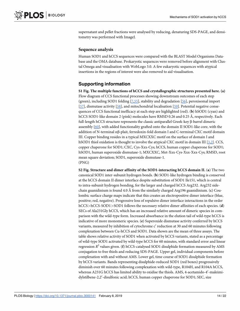
Molecular recognition and affinity are dictated by binding free energy. When homodimeric
hSOD1 and hCCS domain II are physiologically zinc metalated, many of the terms that com-
prise binding free energy are equal due to their sequence and structural similarity. Indeed, ori-
entation of monomers and the presence of four inter-subunit hydrogen bonds found in
homodimeric hSOD1 are maintained by hCCS (S1B Fig, S2A and S2B Fig, S2 and S3 Table).
While fully mature hSOD1 has low nanomolar affinity homodimer affinity [24], zinc-meta-
lated, disulphide-reduced hSOD1 has a dimer dissociation constant of 51 μM [25]. Thus,
within cells, the majority of hCCS’s substrate will be in a monomeric state. It is axiomatic that
hCCS has a higher affinity for this species than for itself, but given mature hCCS forms SOD1-
like homodimers, the question remains, why? The presence of a stabilising Coulombic interac-
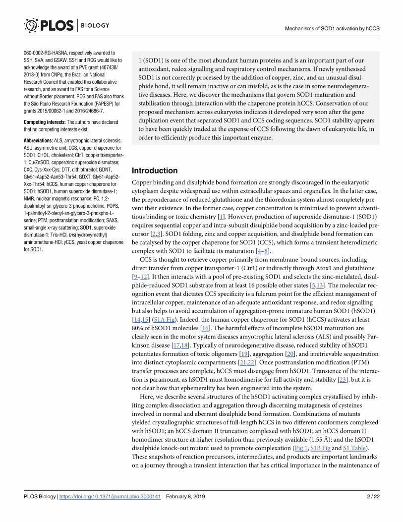
tion between opposing hCCS Arg104 and Asp136 residues (3.55 Å) (Fig 2A), which is not
present in hSOD1, appears counterintuitive in this regard. However, the hCCS dimer interface
is replete with positive charge and therefore not dominated by the hydrophobic effect seen in
hSOD1 and many other protein complexes (Fig 2B).
Most importantly, however, the presence of hCCS Ala231 significantly weakens half of the
homodimer interface hydrogen bonds conserved from SOD1. Specifically, Ala231 side-chain
methyl pushes Arg232 and Gly135 apart through steric repulsion, lengthening the carbonyl-
amine hydrogen bond between them to 3.08 Å from 2.75 Å, in the case of the homologous
SOD1 residues Ile151 and Gly51 (Fig 2C). Without this rearrangement, hCCS Ala231 Cβwould be an energetically unfavourable 2.3 Å from the Gly135 carbonyl oxygen. Introduction
of the hCCS Ala231Gly amino acid substitution to mimic the hSOD1 dimer interface increases
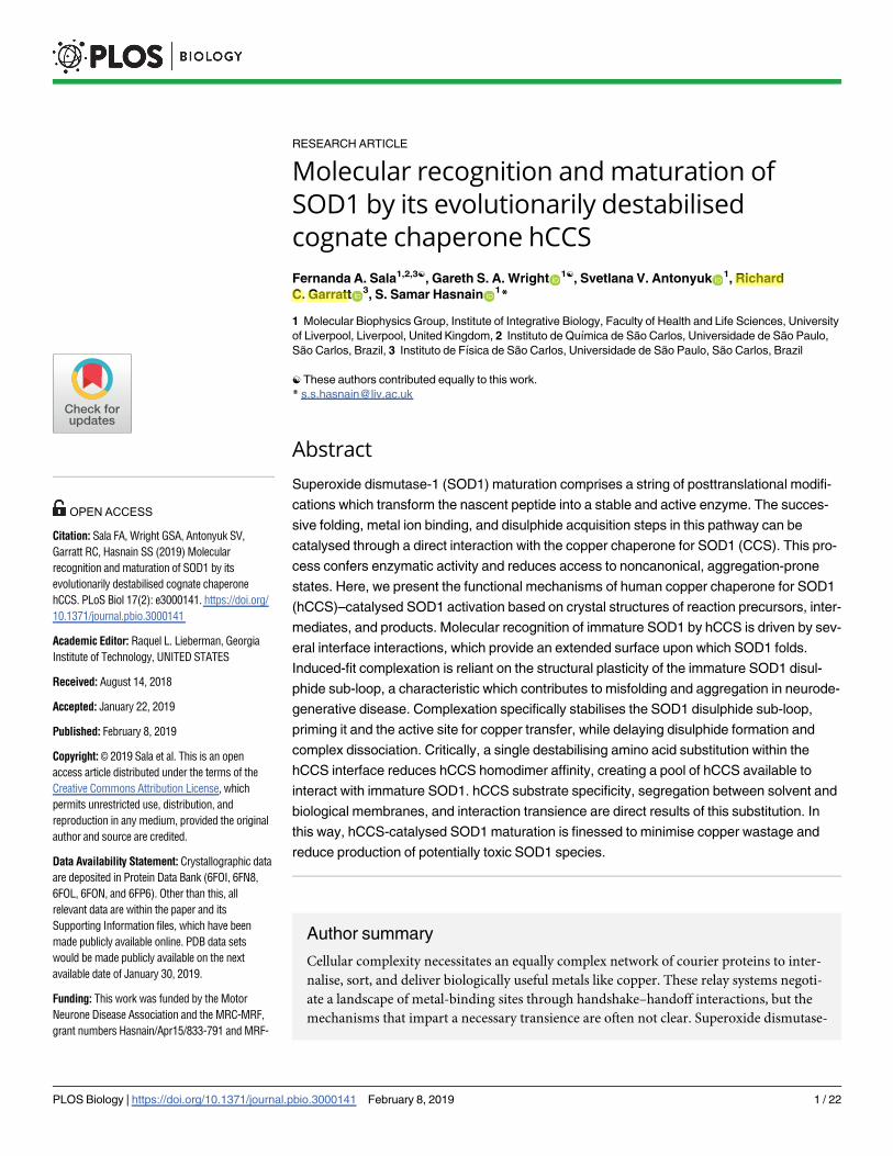
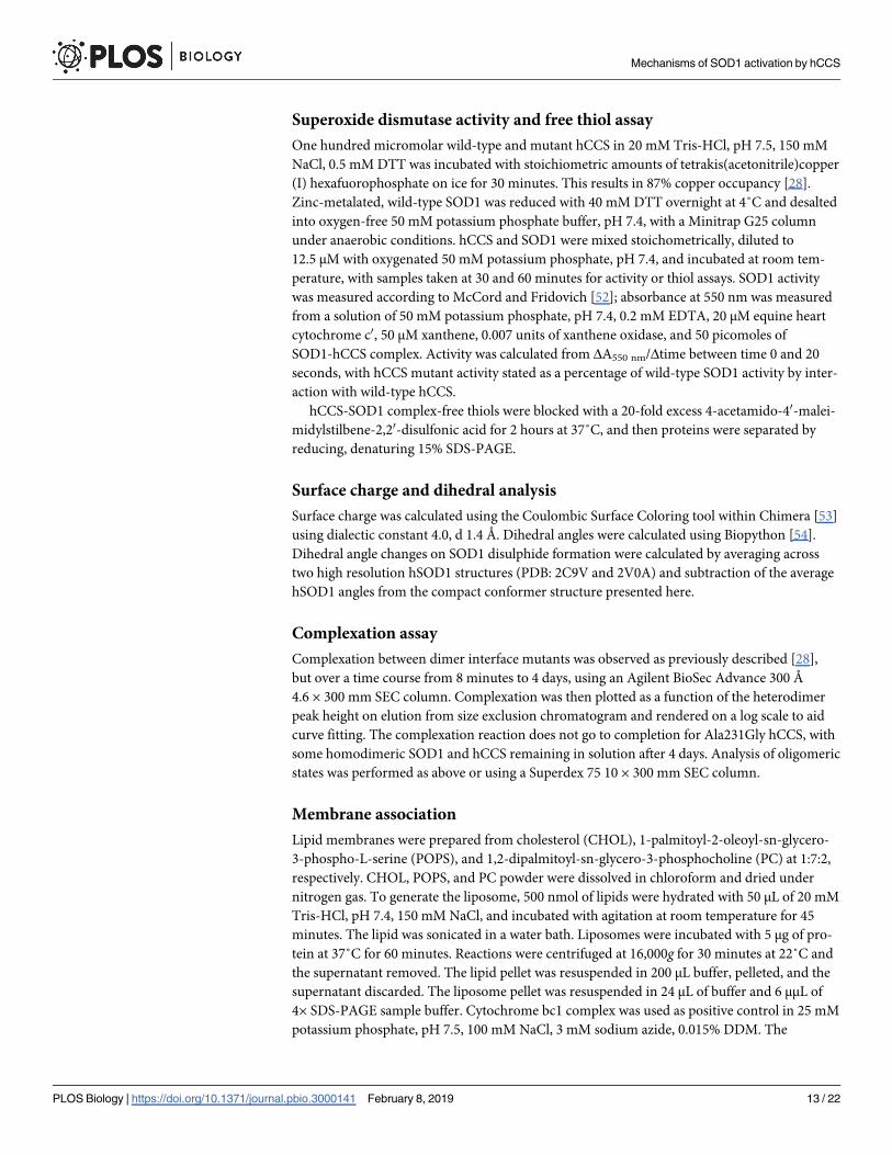
Fig 1. Comparison of human and yeast CCS-SOD1 complex structures. The human CCS-SOD1 complex is
presented in two conformations, elongated (6FON) and compact (6FP6) and is compared with the structure of yeast
CCS-SOD1 (1JK9). Relevant domains and loops are coloured: SOD1 disulphide sub-loop, pink; CCS domain I, green;
CCS C-terminal domain, mauve. For a complete breakdown of structures and mutations presented, please see S1B Fig.
CCS, copper chaperone for SOD1; SOD1, superoxide dismutase-1.
https://doi.org/10.1371/journal.pbio.3000141.g001
Mechanisms of SOD1 activation by hCCS
PLOS Biology | https://doi.org/10.1371/journal.pbio.3000141 February 8, 2019 3 / 22
hCCS dimer affinity (S2D Fig), slows complexation with hSOD1 more than 300-fold (Fig 2D),
impedes hSOD1 activation (S2E Fig), and hSOD1 disulphide formation (S2F Fig). Glycine and
alanine are near ubiquitous at these positions in eukaryotic copper/zinc superoxide dismutases
(Cu/ZnSODs) and their cognate chaperones, respectively (Fig 2E and 2F). Thus, relatively
weak CCS homodimer affinity has been evolutionarily maintained to provide a pool of mono-
meric CCS available to interact with and activate nascent monomeric SOD1 within a physio-
logically relevant timescale. An interesting exception is the nematode CuZnSODs, which have
a deforming alanine in place of hSOD1 Gly150 but are exclusively activated by a CCS-indepen-
dent means [26].
Evolutionarily conserved induced-fit complexation
The SOD1-like, hCCS domain II found within each complex structure has an intact intra-sub-
unit disulphide bond in contrast to the yeast orthologue [27] (S3A Fig). This disulphide does
not dictate complexation (S3B Fig) but does thermally stabilise the hCCS homodimer and the
complex with hSOD1 (S3C and S3D Fig). In contrast, hSOD1 must be in the disulphide-
reduced state to complex with hCCS. We recently predicted hSOD1 disulphide sub-loop
(amino acids His48–His63) movement on complexation with hCCS based on small-angle x-
Fig 2. hCCS dimer interface destabilisation. (A) Electron density map (2Fo-Fc, contoured at 1σ level) showing the
Coulombic interaction between hCCS Arg104 and Asp136/Thr138. (B) Surface charge maps of hSOD1 and hCCS
domain II dimer interface surfaces. (C) The hCCS (pink) dimer interface Arg232-Gly135 hydrogen bond is weakened
by the steric effect of Ala231 side chain. The hCCS Gly135-Asp136 carbonyl rotates to accommodate the methyl group,
and Gly135 is pushed away from Arg232. hSOD1 (cyan) Gly150 maximises hydrogen bond strength between Phe51
and Ile151. (D) Restoring SOD1-like dimer affinity with the Ala231Gly hCCS mutation vastly slows complexation. (E)
Eukaryotic CCS sequence diversity shows Ala231 is very highly conserved despite its detrimental effect on homodimer
affinity. (F) SOD1 Gly150 is equally well conserved, indicating the relative balance of SOD1 and CCS homodimer
affinities is evolutionarily static. AU, absorbance unit; CCS, copper chaperone for SOD1; hCCS, human copper
chaperone for SOD1; hSOD1, human superoxide dismutase-1; SOD1, superoxide dismutase-1; wt, wild-type.
https://doi.org/10.1371/journal.pbio.3000141.g002
Mechanisms of SOD1 activation by hCCS
PLOS Biology | https://doi.org/10.1371/journal.pbio.3000141 February 8, 2019 4 / 22
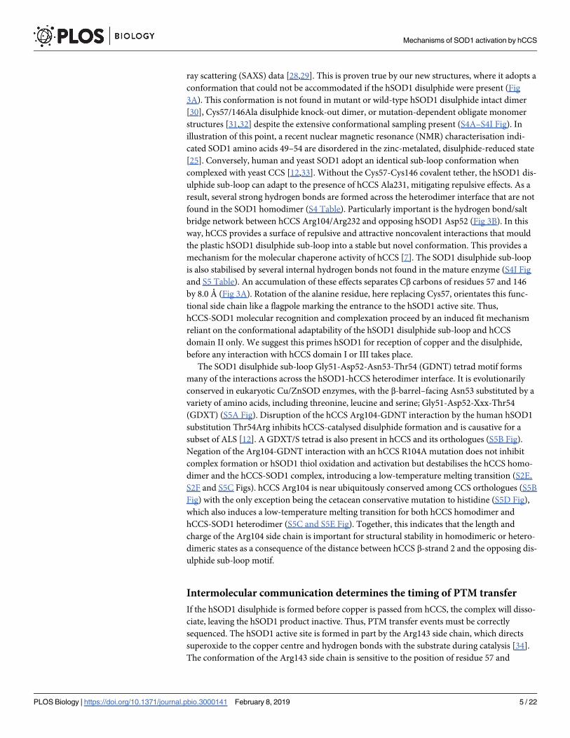
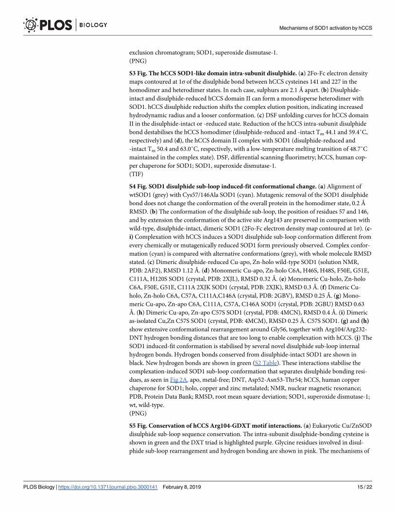
ray scattering (SAXS) data [28,29]. This is proven true by our new structures, where it adopts a
conformation that could not be accommodated if the hSOD1 disulphide were present (Fig
3A). This conformation is not found in mutant or wild-type hSOD1 disulphide intact dimer
[30], Cys57/146Ala disulphide knock-out dimer, or mutation-dependent obligate monomer
structures [31,32] despite the extensive conformational sampling present (S4A–S4I Fig). In
illustration of this point, a recent nuclear magnetic resonance (NMR) characterisation indi-
cated SOD1 amino acids 49–54 are disordered in the zinc-metalated, disulphide-reduced state
[25]. Conversely, human and yeast SOD1 adopt an identical sub-loop conformation when
complexed with yeast CCS [12,33]. Without the Cys57-Cys146 covalent tether, the hSOD1 dis-
ulphide sub-loop can adapt to the presence of hCCS Ala231, mitigating repulsive effects. As a
result, several strong hydrogen bonds are formed across the heterodimer interface that are not
found in the SOD1 homodimer (S4 Table). Particularly important is the hydrogen bond/salt
bridge network between hCCS Arg104/Arg232 and opposing hSOD1 Asp52 (Fig 3B). In this
way, hCCS provides a surface of repulsive and attractive noncovalent interactions that mould
the plastic hSOD1 disulphide sub-loop into a stable but novel conformation. This provides a
mechanism for the molecular chaperone activity of hCCS [7]. The SOD1 disulphide sub-loop
is also stabilised by several internal hydrogen bonds not found in the mature enzyme (S4J Fig
and S5 Table). An accumulation of these effects separates Cβ carbons of residues 57 and 146
by 8.0 Å (Fig 3A). Rotation of the alanine residue, here replacing Cys57, orientates this func-
tional side chain like a flagpole marking the entrance to the hSOD1 active site. Thus,
hCCS-SOD1 molecular recognition and complexation proceed by an induced fit mechanism
reliant on the conformational adaptability of the hSOD1 disulphide sub-loop and hCCS
domain II only. We suggest this primes hSOD1 for reception of copper and the disulphide,
before any interaction with hCCS domain I or III takes place.
The SOD1 disulphide sub-loop Gly51-Asp52-Asn53-Thr54 (GDNT) tetrad motif forms
many of the interactions across the hSOD1-hCCS heterodimer interface. It is evolutionarily
conserved in eukaryotic Cu/ZnSOD enzymes, with the β-barrel–facing Asn53 substituted by a
variety of amino acids, including threonine, leucine and serine; Gly51-Asp52-Xxx-Thr54
(GDXT) (S5A Fig). Disruption of the hCCS Arg104-GDNT interaction by the human hSOD1
substitution Thr54Arg inhibits hCCS-catalysed disulphide formation and is causative for a
subset of ALS [12]. A GDXT/S tetrad is also present in hCCS and its orthologues (S5B Fig).
Negation of the Arg104-GDNT interaction with an hCCS R104A mutation does not inhibit
complex formation or hSOD1 thiol oxidation and activation but destabilises the hCCS homo-
dimer and the hCCS-SOD1 complex, introducing a low-temperature melting transition (S2E,
S2F and S5C Figs). hCCS Arg104 is near ubiquitously conserved among CCS orthologues (S5B
Fig) with the only exception being the cetacean conservative mutation to histidine (S5D Fig),
which also induces a low-temperature melting transition for both hCCS homodimer and
hCCS-SOD1 heterodimer (S5C and S5E Fig). Together, this indicates that the length and
charge of the Arg104 side chain is important for structural stability in homodimeric or hetero-
dimeric states as a consequence of the distance between hCCS β-strand 2 and the opposing dis-
ulphide sub-loop motif.
Intermolecular communication determines the timing of PTM transfer
If the hSOD1 disulphide is formed before copper is passed from hCCS, the complex will disso-
ciate, leaving the hSOD1 product inactive. Thus, PTM transfer events must be correctly
sequenced. The hSOD1 active site is formed in part by the Arg143 side chain, which directs
superoxide to the copper centre and hydrogen bonds with the substrate during catalysis [34].
The conformation of the Arg143 side chain is sensitive to the position of residue 57 and
Mechanisms of SOD1 activation by hCCS
PLOS Biology | https://doi.org/10.1371/journal.pbio.3000141 February 8, 2019 5 / 22
Fig 3. hSOD1 induced-fit complexation. (A) Complexation with hCCS forces the hSOD1 disulphide sub-loop to adopt an open conformation separating the amino
acids involved in disulphide formation. (B) Interactions across the heterodimer interface stabilise the hSOD1 disulphide sub-loop. (C) The hSOD1 Arg143 side chain
interleaved between the β-barrel and disulphide sub-loop, hydrogen bonded with the DNT motif Asn53, and compared with the conformation found in homodimeric
hSOD1 (magenta). (D) hCCS domain III is recruited to the hSOD1 disulphide sub-loop by a restrictive domain I position and hydrogen bonding between hCCS C-
terminal Asn239 and hSOD1 Thr58. This brings the functional hCCS CXC motif into the vicinity of hSOD1 Cys57. 2Fo-Fc electron density maps are contoured at the
1σ level. hSOD1 is shown in cyan, hCCS in pink. CXC, Cys-Xxx-Cys; GDNT, Gly51-Asp52-Asn53-Thr54; hCCS, human copper chaperone for SOD1; hSOD1, human
copper chaperone for SOD1; SOD1, superoxide dismutase-1.
https://doi.org/10.1371/journal.pbio.3000141.g003
Mechanisms of SOD1 activation by hCCS
PLOS Biology | https://doi.org/10.1371/journal.pbio.3000141 February 8, 2019 6 / 22
therefore the propensity of hSOD1 to form a homodimer [31,34] (S4A and S4B Fig). When
hSOD1 complexes with hCCS its copper site is exposed to solvent by a shift in the Arg143 side
chain (Fig 3C). This is due to inability of the Arg143 guanidinium group to hydrogen bond
with Gly61 and Cys57 carbonyls found within the disulphide sub-loop, due to increased dis-
tance (3.0 to 3.5 Å). As a result, it interposes between residues 57 and 146 in the space normally
occupied by the hSOD1 disulphide. In this conformation, the Arg143 guanidinium hydrogen
bonds with the hSOD1 disulphide sub-loop GDNT tetrad Asn53. We propose that this effect is
an integral part of the activation mechanism; communication from hCCS via Arg104 through
the hSOD1 GDNT motif to the Arg143 side chain ensures the amenability of the active site to
receive copper in response to complexation with hCCS. Simultaneously, Arg143 forms a physi-
cal barrier between disulphide bonding residues and occludes the electropositive cavity found
when hSOD1 is complexed with mutant yeast CCS [12]. Arg143 side-chain movement has
been observed in the yeast CCS-SOD1 complex, where it is found hydrogen bonded to the ala-
nine amide of the yeast CCS (yCCS) Cys-Xxx-Cys (CXC) C-terminal motif [33]. Copper trans-
fer or recruitment of the hCCS CXC motif therefore switches the Arg143 side chain removing
a potential block to disulphide formation. This mechanism would prioritise copper transfer
over disulphide formation so that interactions that yield inactive or unstable SOD1 product
are minimised.
From a conformation that facilitates copper acquisition from Ctr1/Ctr2 or Atox1, hCCS
domain I must move to a position that enables transfer to hSOD1. The very high positional
dynamism necessary of the hCCS copper-binding domain is evidenced by intra-lattice confor-
mational flexibility and a 47.4-Å domain movement between conformers (S6A and S6B Fig).
Conformational plasticity is therefore not restricted to the hCCS homodimer state [35] but is
an intrinsic property of the activating complex. The act of copper transfer between hCCS
domain I and hSOD1 is driven by the higher affinity of the hSOD1 tetrahistidine site com-
pared with the domain I bis-cysteine site and facilitated by intermediate chelating side-chain
interactions from the hCCS CXC motif and hSOD1 Cys57 within the disulphide sub-loop
[12,36]. Comparison of different hCCS conformers indicate that the C-terminal tail is also
conformationally dynamic, as has been observed for the yeast orthologue [12,33] and predicted
from SAXS data for hCCS [35]. When hCCS domain I is free to move within the lattice or
inhabits the extended conformer, the position of domain III is partially or entirely unre-
stricted. In the latter case, it forms the interface of a supramolecule comprising four hCCS
monomers and four hSOD1 monomers in crystallo (S6C Fig). By contrast, when domain I is
positioned close to the substrate hSOD1 molecule in the compact conformer, it stabilises
domain III by restricting space and forming a series of conserved interdomain hydrogen
bonds (S6D and S6E Fig). As a result, domain III arches over the hSOD1 disulphide loop and
forms a side-chain hydrogen bond between hCCS Asn239 and hSOD1 Thr58 carbonyl, which
can only exist when the hSOD1 disulphide sub-loop is in the induced fit conformation (Fig 3D
and S6F Fig). Both the conformation and hydrogen bonding are again conserved and provide
the impetus to bring the functionally important hCCS CXC motif into position next to Cys57.
The noncovalently bonded C-terminal conformation existing in the compact structure pre-
sented here is therefore a precursor of the yeast mixed disulphide–bonded structure [33].
Thus, induced-fit SOD1 disulphide sub-loop conformation change upon complexation ulti-
mately recruits the hCCS functional motifs necessary to ensure timely PTM transfer.
A molecular lever affects complex dissociation
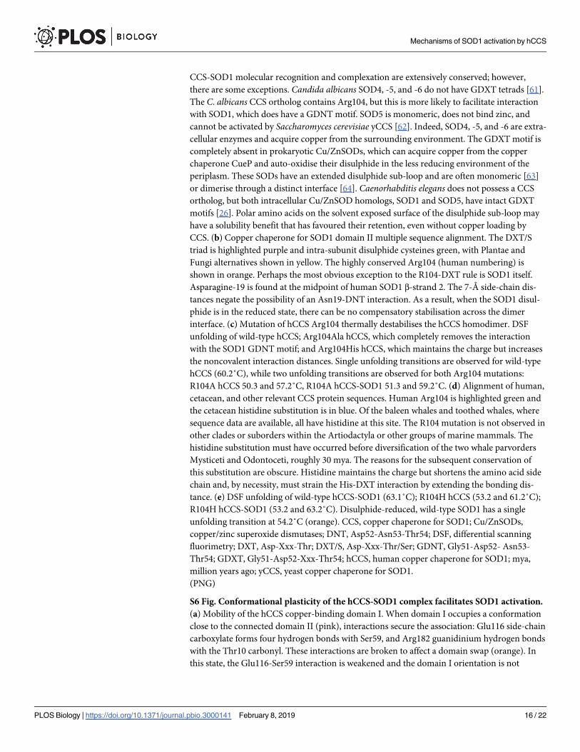
For the Cys57 and 146 sulphydryls to form the disulphide bond, the whole sub-loop pivots on
Gly51, and a Cys57 orientation change is accommodated by Gly56 (Fig 4A). Here, the
Mechanisms of SOD1 activation by hCCS
PLOS Biology | https://doi.org/10.1371/journal.pbio.3000141 February 8, 2019 7 / 22
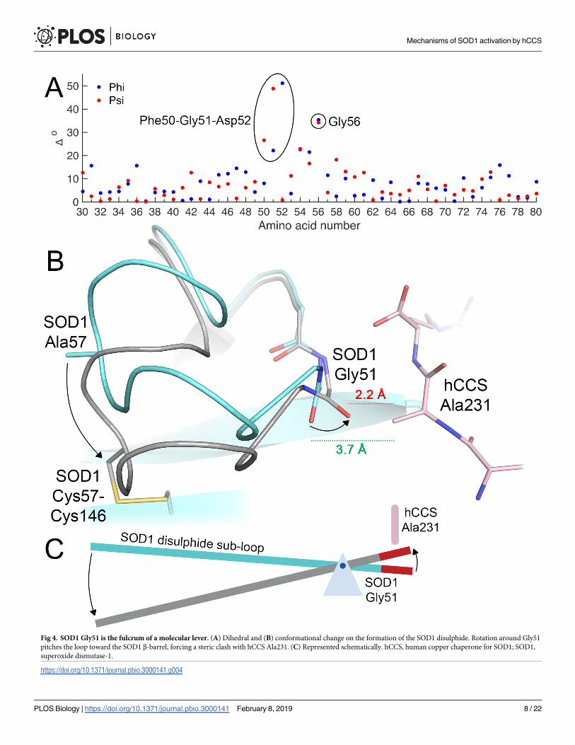
Fig 4. SOD1 Gly51 is the fulcrum of a molecular lever. (A) Dihedral and (B) conformational change on the formation of the SOD1 disulphide. Rotation around Gly51
pitches the loop toward the SOD1 β-barrel, forcing a steric clash with hCCS Ala231. (C) Represented schematically. hCCS, human copper chaperone for SOD1; SOD1,
superoxide dismutase-1.
https://doi.org/10.1371/journal.pbio.3000141.g004
Mechanisms of SOD1 activation by hCCS
PLOS Biology | https://doi.org/10.1371/journal.pbio.3000141 February 8, 2019 8 / 22
disulphide sub-loop operates as a class I lever forcing the Gly51 carbonyl too close to hCCS
Ala231 and deforming the hSOD1 Gly51-hCCS Arg232 interface hydrogen bond (Fig 4B and
4C). hSOD1 then dimerises due to of mitigation of repulsive effects by substitution of Gly150
for hCCS Ala231, formation of the four strong interface hydrogen bonds, breaking of both
Arg104- and Arg232-GDNT heterodimer noncovalent bonding interactions (S6 Table), loss of
electrostatic repulsion as Arg232 is replaced by hSOD1 Ile151, and maximising the stable,
hydrophobic interface surface. Thus, interactions between the GDNT disulphide sub-loop tet-
rad across the heterodimer interface dictate the specificity of hCCS for disulphide-reduced
hSOD1, the timing of copper and disulphide transfer, and complex dissociation. The affinities
that regulate these events are finely balanced as a necessity of the similarity of the proteins
involved and the small disulphide loop conformation change that directs the interaction. Only
3% of the amino acids present dictate complex recognition, while the hCCS Ala231 methyl,
which orchestrates complexation and dissociation, constitutes less than 0.04% of the total mass
of the complex. In addition, while hSOD1 disulphide flexibility is viewed negatively as an
aspect of the pathogenesis of hSOD1-related ALS and now possibly Parkinson disease [17,18],
here we find that Gly51-pivoted sub-loop conformational switching is an absolute necessity
for hCCS-catalysed hSOD1 activation.
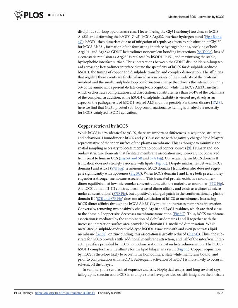
Copper retrieval by hCCS
While hCCS is 27% identical to yCCS, there are important differences in sequence, structure,
and behaviour. Homodimeric hCCS and yCCS associate with negatively charged lipid bilayers
representative of the inner surface of the plasma membrane. This is thought to minimise the
spatial sampling necessary to locate membrane-bound copper sources [9]. Primary and sec-
ondary structure elements that facilitate membrane association are, however, not conserved
from yeast to human CCS (Fig 5A and 5B and S7A Fig). Consequently, an hCCS domain II
truncation does not strongly associate with lipids (Fig 5C). Despite similarities between hCCS
domain I and Atox1 (S7B Fig), a monomeric hCCS domain I truncation also does not segre-
gate significantly with liposomes (Fig 5C). When hCCS domain I and II are both present, they
engender a stronger membrane association. This truncated protein exists in a monomer-
dimer equilibrium at low micromolar concentration, with the majority as monomer (S7C Fig).
An hCCS domain II–III construct has increased dimer affinity and exists as a dimer at micro-
molar concentrations (S7D Fig), but a positively charged patch in the conformationally plastic
domain III (S7E and S7F Fig) does not aid association of hCCS to membranes. Increasing
hCCS dimer affinity through the hCCS Ala231Gly mutation increases membrane interaction.
Conversely, removing two positively charged Arg30 and Lys31 residues, which are sited close
to the domain I copper site, decreases membrane association (Fig 5C). Thus, hCCS membrane
association is mediated by the combination of globular domains I and II together with the
increased interaction surface area provided by domain III–mediated dimerisation. While
metal-free, disulphide-reduced wild-type hSOD1 associates with and even penetrates lipid
membrane [37,38], on zinc binding, this association is greatly reduced (Fig 5C). Thus, the sub-
strate for hCCS provides little additional membrane attraction, and half of the interfacial inter-
acting surface provided by hCCS homodimerisation is lost on heterodimerisation. The hCCS-
hSOD1 complex has little affinity for the lipid bilayer as a result (Fig 5C). Copper acquisition
by hCCS is therefore likely to occur in the homodimeric state while membrane bound, and
prior to complexation with hSOD1. Subsequent activation of hSOD1 is more likely to occur in
solvent, off the bilayer.
In summary, the synthesis of sequence analysis, biophysical assays, and long-awaited crys-
tallographic structures of hCCS in multiple states have provided us with insight on the intricate
Mechanisms of SOD1 activation by hCCS
PLOS Biology | https://doi.org/10.1371/journal.pbio.3000141 February 8, 2019 9 / 22
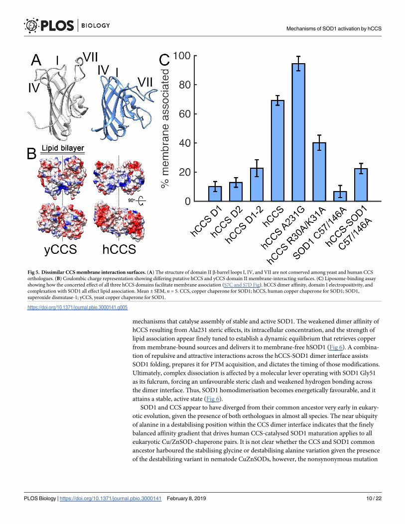
mechanisms that catalyse assembly of stable and active SOD1. The weakened dimer affinity of
hCCS resulting from Ala231 steric effects, its intracellular concentration, and the strength of
lipid association appear finely tuned to establish a dynamic equilibrium that retrieves copper
from membrane-bound sources and delivers it to membrane-free hSOD1 (Fig 6). A combina-
tion of repulsive and attractive interactions across the hCCS-SOD1 dimer interface assists
SOD1 folding, prepares it for PTM acquisition, and dictates the timing of those modifications.
Ultimately, complex dissociation is affected by a molecular lever operating with SOD1 Gly51
as its fulcrum, forcing an unfavourable steric clash and weakened hydrogen bonding across
the dimer interface. Thus, SOD1 homodimerisation becomes energetically favourable, and it
attains a stable, active state (Fig 6).
SOD1 and CCS appear to have diverged from their common ancestor very early in eukary-
otic evolution, given the presence of both orthologues in almost all species. The near ubiquity
of alanine in a destabilising position within the CCS dimer interface indicates that the finely
balanced affinity gradient that drives human CCS-catalysed SOD1 maturation applies to all
eukaryotic Cu/ZnSOD-chaperone pairs. It is not clear whether the CCS and SOD1 common
ancestor harboured the stabilising glycine or destabilising alanine variation given the presence
of the destabilizing variant in nematode CuZnSODs, however, the nonsynonymous mutation
Fig 5. Dissimilar CCS membrane interaction surfaces. (A) The structure of domain II β-barrel loops I, IV, and VII are not conserved among yeast and human CCS
orthologues. (B) Coulombic charge representation showing differing putative hCCS and yCCS domain II membrane-interacting surfaces. (C) Liposome-binding assay
showing how the concerted effect of all three hCCS domains facilitate membrane association (S7C and S7D Fig). hCCS dimer affinity, domain I electropositivity, and
complexation with SOD1 all effect lipid association. Mean ± SEM, n = 5. CCS, copper chaperone for SOD1; hCCS, human copper chaperone for SOD1; SOD1,
superoxide dismutase-1; yCCS, yeast copper chaperone for SOD1.
https://doi.org/10.1371/journal.pbio.3000141.g005
Mechanisms of SOD1 activation by hCCS
PLOS Biology | https://doi.org/10.1371/journal.pbio.3000141 February 8, 2019 10 / 22
leading to this substitution happened very soon after the gene duplication event that separated
SOD1 and CCS coding sequences. Evolution appears to have very quickly traded CCS stability
for SOD1 stability. In great apes, including humans, this effect is particularly pronounced,
with SOD1 having undergone strong positive selection to limit instability and thereby extend
life span [39]. In contrast, a weakened CCS dimer interface does not appear to swamp cellular
proteostasis machinery in the same way that SOD1 dimer interface destabilising mutations do
[20,40], possibly due to reduced relative expression [41] or more efficient degradation. This
adaptation, and the mechanism we propose, may be a critical milestone in the development of
the large, highly compartmentalised forms into which eukaryotic cells have developed.
Materials and methods
Protein expression, purification, and complex formation
Protein expression, purification, and complex formation was performed as previously
described [29,35,42], with the exception of SOD1 C57/146A in the pET3A vector, which was
transformed into Escherichia coli BL21 (DE3), and expression was induced with 0.4 mM of
IPTG with the addition of 0.2 μM ZnCl2 and incubated at 37˚C for 6 hours.
Fig 6. hCCS-catalysed hSOD1 activation. 1. hCCS Ala231 facilitates a dynamic association with membranes while mobilising monomeric hCCS for complexation with
hSOD1. 2. Noncovalent interactions between hCCS domain II and the GDNT motif of the plastic hSOD1 disulphide sub-loop dictate molecular recognition.
Complexation-induced conformational rearrangement prepares hSOD1 to receive copper, while impeding disulphide formation. 3. hSOD1 disulphide formation breaks
GDNT motif interactions, rotates the SOD1 disulphide loop, and weakens heterodimer affinity. 4. hSOD1 forms a stable, active homodimer. hCCS can homodimerise
and relocate back to the cytoplasmic membrane. hCCS, pink; hSOD1, blue. Panels show interactions across dimer interfaces, with disulphide status indicated as SH,
reduced; S-S, intact. Ctr1, copper transporter-1; GDNT, Gly51-Asp52-Asn53-Thr54; hCCS, human copper chaperone for SOD1; hSOD1, human superoxide dismutase-
1.
https://doi.org/10.1371/journal.pbio.3000141.g006
Mechanisms of SOD1 activation by hCCS
PLOS Biology | https://doi.org/10.1371/journal.pbio.3000141 February 8, 2019 11 / 22
Crystallisation
All crystals were grown using the hanging-drop vapour diffusion method at 20˚C from pro-
teins in 20 mM tris(hydroxymethyl)aminomethane-HCl (Tris-HCl), pH 7.4, 150 mM NaCl, 1
mM dithiothreitol (DTT). hSOD1 C57/146A was crystallised from 1.0 μL of protein at 15 mg/
mL, mixed in equal proportions with 0.2 M lithium sulphate; 0.1 M Tris-HCl, pH 8.0; and 24%
w/v PEG 4000. Crystals appeared after 10 days of incubation. hSOD1 C57/146A− hCCS
domain II at 15 mg/mL was crystallised in 25% (w/v) PEG 1500; 0.1 M PCTP buffer, pH 7.0
(sodium propionate, sodium cacodylate, and bis-tris propane in the molar ratios 2:1:2, respec-
tively). Crystals grew after 15 days in space group H32, with four heterodimers in the asym-
metric unit (ASU). Heterocomplexes, hSOD1(C57/146A)—full-length hCCS(C22/25S)
(elongated conformer); hSOD1(C57/146A)—full-length hCCS(C12/22/25/244/246A) (com-
pact conformer); and hCCS D2 were crystallised from nondiffracting seeds prepared from
hSOD1(C57/146A)—full-length hCCS (C22/25S) crystals grown in 0.2 M sodium malonate,
20% (w/v) PEG 3350, frozen in the same solution in liquid nitrogen and stored at −80˚C. For
seed preparation, crystals were crushed and diluted serially: 1:5, 1:25, 1:125, and 1:625 in buffer
dependent on the crystallisation condition with 20% (w/v) PEG 3350. Drop volume ratio con-
sisted of 3 parts protein (1.2 μL):2 parts reservoir solution (0.8 μL):1 part stock (0.4 μL). For
hSOD1(C57/146A)—hCCS(C22/25S) (elongated conformer), the complex was crystallised at
20 mg/mL in 0.1 M MES, pH 6.0, 0.2 M magnesium chloride, 20% (w/v) PEG 6000. Crystals
appeared within 45 days. hSOD1(C57/146A)—full-length hCCS(C12/22/25/244/247A) (com-
pact conformer) at 8 mg/mL was crystallised in 0.1 M PCTP buffer, pH 9.0, 20% (w/v) PEG
3350. Crystals appeared within 25 days. Crystals for hCCS domain II homodimer grew from
1.2 μL of 15 mg/mL full-length hCCS with the addition of 0.2 M sodium chloride, 20% (w/v)
PEG 6000; 0.1 M HEPES, pH 7.0, reservoir solution after 8 months.
Data collection and structure determination
All crystals were transferred into cryoprotective solution consisting of the respective reservoir
solution and 20% glycerol and then flash frozen in liquid nitrogen. Data for all structures
except C57/146A hSOD1 were collected at Soleil on beamline Proxima 1 with 0.97857 Å wave-
length. C57/146A hSOD1 data were collected on Diamond beamline IO3 using 0.97626 Åwavelength. In all cases, a PILATUS 6 M detector was used. Images were integrated with
iMosflm [43] or XDS [44] and scaled with SCALA [45] or AIMLESS [46]. All structures were
solved by molecular replacement using PHASER [47] or MOLREP [48]. hCCS domain II
homodimer and SOD1 C57/146A–hCCS domain II used hCCS and SOD1 structures 1DO5
and 2CV9, respectively, as the search model. Full-length forms were solved using a hSOD1
C57/146A–hCCS domain II heterodimer structure. The structures presented were constructed
with successive rounds of manual model building in COOT [49] and refinement with a combi-
nation of Phenix [50] and Refmac [51]. Structures were validated with PDB validation tool and
deposited in the Protein Data Bank with accession codes 6FOI, 6FN8, 6FOL, 6FON, and 6FP6.
Thermal stability
Thermal stability was assayed by differential scanning fluorometry with a protein concentra-
tion of 10 μM, 20× Sypro Orange, in 20 mM Tris-HCl, 150 mM NaCl, pH 7.4, and with the
addition of 4 mM DDT when necessary. Unfolding was monitored over a temperature gradi-
ent from 25 to 95˚C with 1˚C min−1 ramp rate. Data were normalised and melting transitions
assigned as the peak maximum of the first differential of the unfolding curve.
Mechanisms of SOD1 activation by hCCS
PLOS Biology | https://doi.org/10.1371/journal.pbio.3000141 February 8, 2019 12 / 22
Superoxide dismutase activity and free thiol assay
One hundred micromolar wild-type and mutant hCCS in 20 mM Tris-HCl, pH 7.5, 150 mM
NaCl, 0.5 mM DTT was incubated with stoichiometric amounts of tetrakis(acetonitrile)copper
(I) hexafuorophosphate on ice for 30 minutes. This results in 87% copper occupancy [28].
Zinc-metalated, wild-type SOD1 was reduced with 40 mM DTT overnight at 4˚C and desalted
into oxygen-free 50 mM potassium phosphate buffer, pH 7.4, with a Minitrap G25 column
under anaerobic conditions. hCCS and SOD1 were mixed stoichometrically, diluted to
12.5 μM with oxygenated 50 mM potassium phosphate, pH 7.4, and incubated at room tem-
perature, with samples taken at 30 and 60 minutes for activity or thiol assays. SOD1 activity
was measured according to McCord and Fridovich [52]; absorbance at 550 nm was measured
from a solution of 50 mM potassium phosphate, pH 7.4, 0.2 mM EDTA, 20 μM equine heart
cytochrome c0, 50 μM xanthene, 0.007 units of xanthene oxidase, and 50 picomoles of
SOD1-hCCS complex. Activity was calculated from ΔA550 nm/Δtime between time 0 and 20
seconds, with hCCS mutant activity stated as a percentage of wild-type SOD1 activity by inter-
action with wild-type hCCS.
hCCS-SOD1 complex-free thiols were blocked with a 20-fold excess 4-acetamido-40-malei-
midylstilbene-2,20-disulfonic acid for 2 hours at 37˚C, and then proteins were separated by
reducing, denaturing 15% SDS-PAGE.
Surface charge and dihedral analysis
Surface charge was calculated using the Coulombic Surface Coloring tool within Chimera [53]
using dialectic constant 4.0, d 1.4 Å. Dihedral angles were calculated using Biopython [54].
Dihedral angle changes on SOD1 disulphide formation were calculated by averaging across
two high resolution hSOD1 structures (PDB: 2C9V and 2V0A) and subtraction of the average
hSOD1 angles from the compact conformer structure presented here.
Complexation assay
Complexation between dimer interface mutants was observed as previously described [28],
but over a time course from 8 minutes to 4 days, using an Agilent BioSec Advance 300 Å4.6 × 300 mm SEC column. Complexation was then plotted as a function of the heterodimer
peak height on elution from size exclusion chromatogram and rendered on a log scale to aid
curve fitting. The complexation reaction does not go to completion for Ala231Gly hCCS, with
some homodimeric SOD1 and hCCS remaining in solution after 4 days. Analysis of oligomeric
states was performed as above or using a Superdex 75 10 × 300 mm SEC column.
Membrane association
Lipid membranes were prepared from cholesterol (CHOL), 1-palmitoyl-2-oleoyl-sn-glycero-
3-phospho-L-serine (POPS), and 1,2-dipalmitoyl-sn-glycero-3-phosphocholine (PC) at 1:7:2,
respectively. CHOL, POPS, and PC powder were dissolved in chloroform and dried under
nitrogen gas. To generate the liposome, 500 nmol of lipids were hydrated with 50 μL of 20 mM
Tris-HCl, pH 7.4, 150 mM NaCl, and incubated with agitation at room temperature for 45
minutes. The lipid was sonicated in a water bath. Liposomes were incubated with 5 μg of pro-
tein at 37˚C for 60 minutes. Reactions were centrifuged at 16,000g for 30 minutes at 22˚C and
the supernatant removed. The lipid pellet was resuspended in 200 μL buffer, pelleted, and the
supernatant discarded. The liposome pellet was resuspended in 24 μL of buffer and 6 μμL of
4× SDS-PAGE sample buffer. Cytochrome bc1 complex was used as positive control in 25 mM
potassium phosphate, pH 7.5, 100 mM NaCl, 3 mM sodium azide, 0.015% DDM. The
Mechanisms of SOD1 activation by hCCS
PLOS Biology | https://doi.org/10.1371/journal.pbio.3000141 February 8, 2019 13 / 22
supernatant and pellet fractions were analysed by reducing, denaturing SDS-PAGE, and densi-
tometry was performed with ImageJ.
Sequence analysis
Human SOD1 and hCCS sequences were compared with the BLAST Model Organisms Data-
base and the OMA database. Prokaryotic sequences were removed before alignment with Clus-
tal Omega and visualisation with WebLogo 3.0. A few eukaryotic sequences with atypical
insertions in the regions of interest were also removed to aid visualisation.
Supporting information
S1 Fig. The multiple functions of hCCS and crystallographic structures presented here. (a)
Flow diagram of CCS functional processes showing downstream outcomes of each step
(green), including SOD1 folding [7,55], stability and degradation [56], peroxisomal import
[57], dismutase activity [58], and mitochondrial localisation [59]. Potential negative conse-
quences of CCS functional inefficacy at each step are highlighted (red). (b) hSOD1 (cyan) and
hCCS SOD1-like domain 2 (pink) molecules have RMSD 0.26 and 0.25 Å, respectively. Each
full-length hCCS structure represents the classic antiparallel Greek-key β-barrel dimeric
assembly [60], with added functionality grafted onto the domain II SOD1-like core, with the
addition of N-terminal αβ-plait, ferredoxin-fold domain I and C-terminal CXC motif domain
III. Copper binding resides in a typical MXCXXC motif on the surface of domain I and
hSOD1 thiol oxidation is thought to involve the atypical CXC motif in domain III [3,2]. CCS,
copper chaperone for SOD1; CXC, Cys-Xxx-Cys; hCCS, human copper chaperone for SOD1;
hSOD1, human superoxide dismutase-1; MXCXXC, Met-Xxx-Cys-Xxx-Xxx-Cys; RMSD, root
mean square deviation; SOD1, superoxide dismutase-1.
(PNG)
S2 Fig. Structure and dimer affinity of the SOD1-interacting hCCS domain II. (a) The two
canonical SOD1 inter-subunit hydrogen bonds. (b) SOD1-like hydrogen binding is conserved
at the hCCS domain II dimer interface despite substitution of SOD1 Ile151, which is central
to intra-subunit hydrogen bonding, for the larger and charged hCCS Arg232. Arg232 side-
chain guanidinium is found 4.0 Å from the similarly charged Arg196 guanidinium. (c) Cou-
lombic surface charge maps indicate that this creates an electropositive dimer interface (blue,
positive; red, negative). Progressive loss of repulsive dimer interface interactions in the order
hCCS>hCCS-SOD1>SOD1 follows the necessary relative dimer affinities of each species. (d)
SECs of Ala231Gly hCCS, which has an increased relative amount of dimeric species in com-
parison with the wild-type form. Increased absorbance in the elution tail of wild-type hCCS is
indicative of more monomeric species. (e) Superoxide dismutase activity conferred by hCCS
variants, measured by inhibition of cytochrome c0 reduction at 30 and 60 minutes following
complexation between Cu-hCCS and SOD1. Data shown are the mean of three assays. The
table shows relative activity of SOD1 when activated by hCCS variants, stated as a percentage
of wild-type SOD1 activated by wild-type hCCS for 60 minutes, with standard error and linear
regression R2 values given. (f) hCCS-catalysed SOD1 disulphide formation measured by AMS
conjugation to free thiols and reducing SDS-PAGE. Upper gel, individual components before
complexation with and without AMS. Lower gel, time course of SOD1 disulphide formation
by hCCS variants. Bands representing disulphide-reduced SOD1 (red boxes) progressively
diminish over 60 minutes following complexation with wild-type, R104H, and R104A hCCS,
whereas A231G hCCS has limited ability to oxidise the thiols. AMS, 4-acetamido-40-maleimi-
dylstilbene-2,20-disulfonic acid; hCCS, human copper chaperone for SOD1; SEC, size
Mechanisms of SOD1 activation by hCCS
PLOS Biology | https://doi.org/10.1371/journal.pbio.3000141 February 8, 2019 14 / 22
exclusion chromatogram; SOD1, superoxide dismutase-1.
(PNG)
S3 Fig. The hCCS SOD1-like domain intra-subunit disulphide. (a) 2Fo-Fc electron density
maps contoured at 1σ of the disulphide bond between hCCS cysteines 141 and 227 in the
homodimer and heterodimer states. In each case, sulphurs are 2.1 Å apart. (b) Disulphide-
intact and disulphide-reduced hCCS domain II can form a monodisperse heterodimer with
SOD1. hCCS disulphide reduction shifts the complex elution position, indicating increased
hydrodynamic radius and a looser conformation. (c) DSF unfolding curves for hCCS domain
II in the disulphide-intact or -reduced state. Reduction of the hCCS intra-subunit disulphide
bond destabilises the hCCS homodimer (disulphide-reduced and -intact Tm 44.1 and 59.4˚C,
respectively) and (d), the hCCS domain II complex with SOD1 (disulphide-reduced and
-intact Tm 50.4 and 63.0˚C, respectively, with a low-temperature melting transition of 48.7˚C
maintained in the complex state). DSF, differential scanning fluorimetry; hCCS, human cop-
per chaperone for SOD1; SOD1, superoxide dismutase-1.
(TIF)
S4 Fig. SOD1 disulphide sub-loop induced-fit conformational change. (a) Alignment of
wtSOD1 (grey) with Cys57/146Ala SOD1 (cyan). Mutagenic removal of the SOD1 disulphide
bond does not change the conformation of the overall protein in the homodimer state, 0.2 ÅRMSD. (b) The conformation of the disulphide sub-loop, the position of residues 57 and 146,
and by extension the conformation of the active site Arg143 are preserved in comparison with
wild-type, disulphide-intact, dimeric SOD1 (2Fo-Fc electron density map contoured at 1σ). (c-
i) Complexation with hCCS induces a SOD1 disulphide sub-loop conformation different from
every chemically or mutagenically reduced SOD1 form previously observed. Complex confor-
mation (cyan) is compared with alternative conformations (grey), with whole molecule RMSD
stated. (c) Dimeric disulphide-reduced Cu-apo, Zn-holo wild-type SOD1 (solution NMR,
PDB: 2AF2), RMSD 1.12 Å. (d) Monomeric Cu-apo, Zn-holo C6A, H46S, H48S, F50E, G51E,
C111A, H120S SOD1 (crystal, PDB: 2XJL), RMSD 0.32 Å. (e) Monomeric Cu-holo, Zn-holo
C6A, F50E, G51E, C111A 2XJK SOD1 (crystal, PDB: 2XJK), RMSD 0.3 Å. (f) Dimeric Cu-
holo, Zn-holo C6A, C57A, C111A,C146A (crystal, PDB: 2GBV), RMSD 0.25 Å. (g) Mono-
meric Cu-apo, Zn-apo C6A, C111A, C57A, C146A SOD1 (crystal, PDB: 2GBU) RMSD 0.63
Å. (h) Dimeric Cu-apo, Zn-apo C57S SOD1 (crystal, PDB: 4MCN), RMSD 0.4 Å. (i) Dimeric
as-isolated Cu,Zn C57S SOD1 (crystal, PDB: 4MCM), RMSD 0.25 Å. C57S SOD1. (g) and (h)
show extensive conformational rearrangement around Gly56, together with Arg104/Arg232-
DNT hydrogen bonding distances that are too long to enable complexation with hCCS. (j) The
SOD1 induced-fit conformation is stabilised by several novel disulphide sub-loop internal
hydrogen bonds. Hydrogen bonds conserved from disulphide-intact SOD1 are shown in
black. New hydrogen bonds are shown in green (S2 Table). These interactions stabilise the
complexation-induced SOD1 sub-loop conformation that separates disulphide bonding resi-
dues, as seen in Fig 2A. apo, metal-free; DNT, Asp52-Asn53-Thr54; hCCS, human copper
chaperone for SOD1; holo, copper and zinc metalated; NMR, nuclear magnetic resonance;
PDB, Protein Data Bank; RMSD, root mean square deviation; SOD1, superoxide dismutase-1;
wt, wild-type.
(PNG)
S5 Fig. Conservation of hCCS Arg104-GDXT motif interactions. (a) Eukaryotic Cu/ZnSOD
disulphide sub-loop sequence conservation. The intra-subunit disulphide-bonding cysteine is
shown in green and the DXT triad is highlighted purple. Glycine residues involved in disul-
phide sub-loop rearrangement and hydrogen bonding are shown in pink. The mechanisms of
Mechanisms of SOD1 activation by hCCS
PLOS Biology | https://doi.org/10.1371/journal.pbio.3000141 February 8, 2019 15 / 22
CCS-SOD1 molecular recognition and complexation are extensively conserved; however,
there are some exceptions. Candida albicans SOD4, -5, and -6 do not have GDXT tetrads [61].
The C. albicans CCS ortholog contains Arg104, but this is more likely to facilitate interaction
with SOD1, which does have a GDNT motif. SOD5 is monomeric, does not bind zinc, and
cannot be activated by Saccharomyces cerevisiae yCCS [62]. Indeed, SOD4, -5, and -6 are extra-
cellular enzymes and acquire copper from the surrounding environment. The GDXT motif is
completely absent in prokaryotic Cu/ZnSODs, which can acquire copper from the copper
chaperone CueP and auto-oxidise their disulphide in the less reducing environment of the
periplasm. These SODs have an extended disulphide sub-loop and are often monomeric [63]
or dimerise through a distinct interface [64]. Caenorhabditis elegans does not possess a CCS
ortholog, but both intracellular Cu/ZnSOD homologs, SOD1 and SOD5, have intact GDXT
motifs [26]. Polar amino acids on the solvent exposed surface of the disulphide sub-loop may
have a solubility benefit that has favoured their retention, even without copper loading by
CCS. (b) Copper chaperone for SOD1 domain II multiple sequence alignment. The DXT/S
triad is highlighted purple and intra-subunit disulphide cysteines green, with Plantae and
Fungi alternatives shown in yellow. The highly conserved Arg104 (human numbering) is
shown in orange. Perhaps the most obvious exception to the R104-DXT rule is SOD1 itself.
Asparagine-19 is found at the midpoint of human SOD1 β-strand 2. The 7-Å side-chain dis-
tances negate the possibility of an Asn19-DNT interaction. As a result, when the SOD1 disul-
phide is in the reduced state, there can be no compensatory stabilisation across the dimer
interface. (c) Mutation of hCCS Arg104 thermally destabilises the hCCS homodimer. DSF
unfolding of wild-type hCCS; Arg104Ala hCCS, which completely removes the interaction
with the SOD1 GDNT motif; and Arg104His hCCS, which maintains the charge but increases
the noncovalent interaction distances. Single unfolding transitions are observed for wild-type
hCCS (60.2˚C), while two unfolding transitions are observed for both Arg104 mutations:
R104A hCCS 50.3 and 57.2˚C, R104A hCCS-SOD1 51.3 and 59.2˚C. (d) Alignment of human,
cetacean, and other relevant CCS protein sequences. Human Arg104 is highlighted green and
the cetacean histidine substitution is in blue. Of the baleen whales and toothed whales, where
sequence data are available, all have histidine at this site. The R104 mutation is not observed in
other clades or suborders within the Artiodactyla or other groups of marine mammals. The
histidine substitution must have occurred before diversification of the two whale parvorders
Mysticeti and Odontoceti, roughly 30 mya. The reasons for the subsequent conservation of
this substitution are obscure. Histidine maintains the charge but shortens the amino acid side
chain and, by necessity, must strain the His-DXT interaction by extending the bonding dis-
tance. (e) DSF unfolding of wild-type hCCS-SOD1 (63.1˚C); R104H hCCS (53.2 and 61.2˚C);
R104H hCCS-SOD1 (53.2 and 63.2˚C). Disulphide-reduced, wild-type SOD1 has a single
unfolding transition at 54.2˚C (orange). CCS, copper chaperone for SOD1; Cu/ZnSODs,
copper/zinc superoxide dismutases; DNT, Asp52-Asn53-Thr54; DSF, differential scanning
fluorimetry; DXT, Asp-Xxx-Thr; DXT/S, Asp-Xxx-Thr/Ser; GDNT, Gly51-Asp52- Asn53-
Thr54; GDXT, Gly51-Asp52-Xxx-Thr54; hCCS, human copper chaperone for SOD1; mya,
million years ago; yCCS, yeast copper chaperone for SOD1.
(PNG)
S6 Fig. Conformational plasticity of the hCCS-SOD1 complex facilitates SOD1 activation.
(a) Mobility of the hCCS copper-binding domain I. When domain I occupies a conformation
close to the connected domain II (pink), interactions secure the association: Glu116 side-chain
carboxylate forms four hydrogen bonds with Ser59, and Arg182 guanidinium hydrogen bonds
with the Thr10 carbonyl. These interactions are broken to affect a domain swap (orange). In
this state, the Glu116-Ser59 interaction is weakened and the domain I orientation is not
Mechanisms of SOD1 activation by hCCS
PLOS Biology | https://doi.org/10.1371/journal.pbio.3000141 February 8, 2019 16 / 22
identical. (b) Lattice arrangement within the compact conformer crystal creates clashes (red)
that prevent one hCCS domain I from forming contacts that stabilise it close to domain II. As
a result, electron density for this hCCS monomer begins at residue Asn85, within the domain
I–domain II linker, and ends at Leu236. (c) hCCS domain III amino acids 241–246 and 250–
255 form the antiparallel β-sheet interface of an octameric supramolecular assembly in crys-
tallo. This β-sheet and the position of the C-terminal cysteines within it are conserved in a
yCCS-hSOD1 chimeric structure [28] (PDB: 5U9M), bringing cysteines involved in SOD1 dis-
ulphide formation into close proximity—here, hCCS Cys246, and in yCCS, Cys231-ySOD1
Cys146. Changing the position and spacing of the C-terminal cysteines is severely detrimental
to SOD1 activation and disulphide formation [12]. (d) hCCS domain I can stabilise domain III
in a position interacting with the SOD1 disulphide loop. (e) Hydrogen bonding interactions
within domain III and between domain III and domain I or SOD1. Amino acids 235–241 are
positioned to bring functional residues 244 and 246 close to SOD1 Cys57. This conformation
therefore facilitates formation of the disulphide-linked complex described previously for the
yeast orthologue [33]. In contrast, the yCCS-hSOD1 chimera structure shows domain I signifi-
cantly shifted from its position in comparison with those of nonchimeric heterodimer forms
[12]. This is due to accommodation of tetrahedral zinc coordination by the domain I copper
binding cysteines in two symmetry-related molecules. (f) Formation of the SOD1 disulphide
bond shifts the position of the disulphide sub-loop and breaks hydrogen bonding between
hCCS Asn239 and the SOD1 Thr58 (2.7 to 6.7 Å). Blue, hCCS domain III; cyan, SOD1 disul-
phide loop within the complex; pink, SOD1 disulphide loop conformation after formation of
the SOD1 disulphide (2C9V). hCCS, human copper chaperone for SOD1; hSOD1, human
superoxide dismutase-1; PDB, Protein Data Bank; SOD1, superoxide dismutase-1; yCCS, yeast
copper chaperone for SOD1.
(PNG)
S7 Fig. Structural basis of hCCS membrane association. (a) Sequence alignment of yeast and
human CCS proteins showing the residues known to facilitate yCCS association with mem-
branes (purple) [9]. The charge of these residues are not conserved (with the exception of
Arg172) in the human sequence or the structure of β-barrel loops I, IV, and VII, which are
shortened or conformationally rearranged to form a highly electronegative β-barrel end sur-
face. It is noteworthy that changes to the sequence and position of loops I and VII (Fig 3A) are
a direct result of hCCS domain II zinc binding, a PTM not present in yCCS. There appears to
have been an evolutionary divergence in the structure and behaviour of CCS orthologues from
the Animalia kingdom that bind zinc, have an internal disulphide bond and relatively high
dimer affinity, and associate with membranes through the association of domain I and II, com-
pared with yCCS, which has low dimer affinity, does not bind zinc, and membrane associates
through positively charged residues on the β-barrel surface of the domain II end [9,27,65]. (b)
The copper chaperone protein Atox1 has 28% sequence identity with hCCS domain I, interacts
with Ctr1, and associates with membranes [66]. Sequence alignment of human Atox1 and
hCCS domain I, showing lack of conservation of positively charged residues known to mediate
Atox1–lipid membrane association (purple). However, Arg30 and Lys31 (blue), which are
proximal to the domain I copper binding site, do facilitate membrane association (Fig 5C). (c)
SEC and light scattering–derived molecular weights showing that hCCS domain I–II is mono-
meric at mid–low micromolar concentration. (d) SEC and light scattering–derived molecular
weights showing that hCCS domain II–III is dimeric. Masses are quoted as experimental/pre-
dicted. Each species has a polydispersity index of 1.0. (e) Homodimeric structure of full-length
hCCS, with surface coloured by Coulombic charge showing the positively charged region
Arg225 to Lys267 within domain III. This region evidently does not provide enough surface
Mechanisms of SOD1 activation by hCCS
PLOS Biology | https://doi.org/10.1371/journal.pbio.3000141 February 8, 2019 17 / 22
area to mediate membrane association. Homodimeric hCCS was generated by alignment of
compact hCCS conformers to the hCCS domain II structure and refinement of domain I and
III positions against SAXS data using the method described previously [28,35], which (f) fits
the experimental data with a χ value of 1.51. CCS, copper chaperone for SOD1; Ctr1, copper
transporter-1; hCCS, human copper chaperone for SOD1; PTM, posttranslation modification;
SAXS, small-angle x-ray scattering; SEC, size-exclusion chromatography; yCCS, yeast copper
chaperone for SOD1.
(PNG)
S1 Table. Crystallographic data collection and refinement statistics.
(DOCX)
S2 Table. SOD1 interface hydrogen bonding interactions. SOD1, superoxide dismutase-1.
(DOCX)
S3 Table. hCCS domain II interface noncovalent bonding interactions. hCCS, human cop-
per chaperone for SOD1
(DOCX)
S4 Table. hCCS-SOD1 complex interface noncovalent bonding interactions. hCCS, human
copper chaperone for SOD1; SOD1, superoxide dismutase-1.
(DOCX)
S5 Table. Internal SOD1 disulphide sub-loop hydrogen bonding in the disulphide-intact
homodimeric state in comparison with the conformation present in the heterodimeric
complex with hCCS. hCCS, human copper chaperone for SOD1; SOD1, superoxide dismut-
ase-1.
(DOCX)
S6 Table. The effect of SOD1 disulphide bond formation on hCCS-SOD1 inter-subunit
noncovalent bonding interactions. hCCS, human copper chaperone for SOD1; SOD1, super-
oxide dismutase-1.
(DOCX)
S1 Data. Underlying data for the following Figs 2D, 5C, S2D, S2E, S3, S5C, S5E, S7C, S7D
and S7F.
(XLSX)
Author Contributions
Conceptualization: Svetlana V. Antonyuk, Richard C. Garratt, S. Samar Hasnain.
Formal analysis: Gareth S. A. Wright, Svetlana V. Antonyuk.
Funding acquisition: S. Samar Hasnain.
Investigation: Fernanda A. Sala, Gareth S. A. Wright.
Project administration: S. Samar Hasnain.
Supervision: S. Samar Hasnain.
Validation: Gareth S. A. Wright, Svetlana V. Antonyuk.
Writing – original draft: Fernanda A. Sala, Gareth S. A. Wright, S. Samar Hasnain.
Writing – review & editing: Svetlana V. Antonyuk, Richard C. Garratt, S. Samar Hasnain.
Mechanisms of SOD1 activation by hCCS
PLOS Biology | https://doi.org/10.1371/journal.pbio.3000141 February 8, 2019 18 / 22
References1. Rae TD, Schmidt PJ, Pufahl RA, Culotta VC, O’Halloran TV. Undetectable intracellular free copper: the
requirement of a copper chaperone for superoxide dismutase. Science. 1999; 284: 805–808. PMID:
10221913
2. Furukawa Y, Torres AS, O’Halloran TV. Oxygen-induced maturation of SOD1: a key role for disulfide
formation by the copper chaperone CCS. EMBO J. 2004; 23: 2872–2881. https://doi.org/10.1038/sj.
emboj.7600276 PMID: 15215895
3. Culotta VC, Klomp LW, Strain J, Casareno RL, Krems B, Gitlin JD. The copper chaperone for superox-
ide dismutase. J Biol Chem. 1997; 272: 23469–23472. PMID: 9295278
4. Banci L, Bertini I, Cantini F, Kozyreva T, Massagni C, Palumaa P, et al. Human superoxide dismutase 1
(hSOD1) maturation through interaction with human copper chaperone for SOD1 (hCCS). Proc Natl
Acad Sci U S A. 2012; 109: 13555–13560. https://doi.org/10.1073/pnas.1207493109 PMID: 22869735
5. Schmidt PJ, Kunst C, Culotta VC. Copper activation of superoxide dismutase 1 (SOD1) in vivo. Role for
protein-protein interactions with the copper chaperone for SOD1. J Biol Chem. 2000; 275: 33771–
33776. https://doi.org/10.1074/jbc.M006254200 PMID: 10944535
6. Lamb AL, Torres AS, O’Halloran TV, Rosenzweig AC. Heterodimer formation between superoxide dis-
mutase and its copper chaperone. Biochemistry. 2000; 39: 14720–14727. PMID: 11101286
7. Luchinat E, Barbieri L, Banci L. A molecular chaperone activity of CCS restores the maturation of SOD1
fALS mutants. Sci Rep. 2017; 7: 17433. https://doi.org/10.1038/s41598-017-17815-y PMID: 29234142
8. Boyd SD, Liu L, Bulla L, Winkler DD. Quantifying the Interaction between Copper-Zinc Superoxide Dis-
mutase (Sod1) and its Copper Chaperone (Ccs1). J Proteomics Bioinform. 2018; 11: 1–5. https://doi.
org/10.4172/jpb.1000e36
9. Pope CR, De Feo CJ, Unger VM. Cellular distribution of copper to superoxide dismutase involves scaf-
folding by membranes. Proc Natl Acad Sci U S A. 2013; 110: 20491–20496. https://doi.org/10.1073/
pnas.1309820110 PMID: 24297923
10. Petzoldt S, Kahra D, Kovermann M, Dingeldein APG, Niemiec MS, Åden J, et al. Human cytoplasmic
copper chaperones Atox1 and CCS exchange copper ions in vitro. Biometals Int J Role Met Ions Biol
Biochem Med. 2015; 28: 577–585. https://doi.org/10.1007/s10534-015-9832-1 PMID: 25673218
11. Maryon EB, Molloy SA, Kaplan JH. Cellular glutathione plays a key role in copper uptake mediated by
human copper transporter 1. Am J Physiol-Cell Physiol. 2013; 304: C768–C779. https://doi.org/10.
1152/ajpcell.00417.2012 PMID: 23426973
12. Fetherolf MM, Boyd SD, Taylor AB, Kim HJ, Wohlschlegel JA, Blackburn NJ, et al. Copper-zinc super-
oxide dismutase is activated through a sulfenic acid intermediate at a copper-ion entry site. J Biol
Chem. 2017; 292: 12025–12040. https://doi.org/10.1074/jbc.M117.775981 PMID: 28533431
13. Casareno RL, Waggoner D, Gitlin JD. The copper chaperone CCS directly interacts with copper/zinc
superoxide dismutase. J Biol Chem. 1998; 273: 23625–23628. PMID: 9726962
14. Luchinat E, Barbieri L, Rubino JT, Kozyreva T, Cantini F, Banci L. In-cell NMR reveals potential precur-
sor of toxic species from SOD1 fALS mutants. Nat Commun. 2014; 5: 1–10. https://doi.org/10.1038/
ncomms6502 PMID: 25429517
15. Son M, Puttaparthi K, Kawamata H, Rajendran B, Boyer PJ, Manfredi G, et al. Overexpression of CCS
in G93A-SOD1 mice leads to accelerated neurological deficits with severe mitochondrial pathology.
Proc Natl Acad Sci U S A. 2007; 104: 6072–6077. https://doi.org/10.1073/pnas.0610923104 PMID:
17389365
16. Subramaniam JR, Lyons WE, Liu J, Bartnikas TB, Rothstein J, Price DL, et al. Mutant SOD1 causes
motor neuron disease independent of copper chaperone-mediated copper loading. Nat Neurosci. 2002;
5: 301–307. https://doi.org/10.1038/nn823 PMID: 11889469
17. Karch CM, Prudencio M, Winkler DD, Hart PJ, Borchelt DR. Role of mutant SOD1 disulfide oxidation
and aggregation in the pathogenesis of familial ALS. Proc Natl Acad Sci U S A. 2009; 106: 7774–7779.
https://doi.org/10.1073/pnas.0902505106 PMID: 19416874
18. Trist BG, Davies KM, Cottam V, Genoud S, Ortega R, Roudeau S, et al. Amyotrophic lateral sclerosis-
like superoxide dismutase 1 proteinopathy is associated with neuronal loss in Parkinson’s disease
brain. Acta Neuropathol. 2017; 134: 113–127. https://doi.org/10.1007/s00401-017-1726-6 PMID:
28527045
19. Proctor EA, Fee L, Tao Y, Redler RL, Fay JM, Zhang Y, et al. Nonnative SOD1 trimer is toxic to motor
neurons in a model of amyotrophic lateral sclerosis. Proc Natl Acad Sci. 2016; 113: 614–619. https://
doi.org/10.1073/pnas.1516725113 PMID: 26719414
20. Bruijn LI, Houseweart MK, Kato S, Anderson KL, Anderson SD, Ohama E, et al. Aggregation and motor
neuron toxicity of an ALS-linked SOD1 mutant independent from wild-type SOD1. Science. 1998; 281:
1851–1854. PMID: 9743498
Mechanisms of SOD1 activation by hCCS
PLOS Biology | https://doi.org/10.1371/journal.pbio.3000141 February 8, 2019 19 / 22
21. Weisberg SJ, Lyakhovetsky R, Werdiger A, Gitler AD, Soen Y, Kaganovich D. Compartmentalization of
superoxide dismutase 1 (SOD1G93A) aggregates determines their toxicity. Proc Natl Acad Sci. 2012;
109: 15811–15816. https://doi.org/10.1073/pnas.1205829109 PMID: 22967507
22. Mateju D, Franzmann TM, Patel A, Kopach A, Boczek EE, Maharana S, et al. An aberrant phase transi-
tion of stress granules triggered by misfolded protein and prevented by chaperone function. EMBO J.
2017; 36: 1669–1687. https://doi.org/10.15252/embj.201695957 PMID: 28377462
23. Bertini I, Piccioli M, Viezzoli MS, Chiu CY, Mullenbach GT. A spectroscopic characterization of a mono-
meric analog of copper, zinc superoxide dismutase. Eur Biophys J EBJ. 1994; 23: 167–76. PMID:
7956977
24. McAlary L, Yerbury JJ, Aquilina JA. Glutathionylation potentiates benign superoxide dismutase 1 vari-
ants to the toxic forms associated with amyotrophic lateral sclerosis. Sci Rep. 2013; 3: 3275. https://doi.
org/10.1038/srep03275 PMID: 24253732
25. Culik RM, Sekhar A, Nagesh J, Deol H, Rumfeldt JAO, Meiering EM, et al. Effects of maturation on the
conformational free-energy landscape of SOD1. Proc Natl Acad Sci. 2018; 201721022. https://doi.org/
10.1073/pnas.1721022115 PMID: 29483249
26. Jensen LT, Culotta VC. Activation of CuZn Superoxide Dismutases from Caenorhabditis elegans Does
Not Require the Copper Chaperone CCS. J Biol Chem. 2005; 280: 41373–41379. https://doi.org/10.
1074/jbc.M509142200 PMID: 16234242
27. Lamb AL, Wernimont AK, Pufahl RA, Culotta VC, O’Halloran TV, Rosenzweig AC. Crystal structure of
the copper chaperone for superoxide dismutase. Nat Struct Biol. 1999; 6: 724–729. https://doi.org/10.
1038/11489 PMID: 10426947
28. Wright GSA, Antonyuk SV, Hasnain SS. A faulty interaction between SOD1 and hCCS in neurodegen-
erative disease. Sci Rep. 2016; 6: 27691. https://doi.org/10.1038/srep27691 PMID: 27282955
29. Wright GSA, Lee HC, Schulze-Briese C, Grossmann JG, Strange RW, Hasnain SS. The application of
hybrid pixel detectors for in-house SAXS instrumentation with a view to combined chromatographic
operation. J Synchrotron Radiat. 2013; 20: 383–385. https://doi.org/10.1107/S0909049513001866
PMID: 23412497
30. Strange RW, Antonyuk SV, Hough MA, Doucette PA, Valentine JS, Hasnain SS. Variable metallation of
human superoxide dismutase: atomic resolution crystal structures of Cu-Zn, Zn-Zn and as-isolated
wild-type enzymes. J Mol Biol. 2006; 356: 1152–1162. https://doi.org/10.1016/j.jmb.2005.11.081 PMID:
16406071
31. Banci L, Benedetto M, Bertini I, Del Conte R, Piccioli M, Viezzoli MS. Solution structure of reduced
monomeric Q133M2 copper, zinc superoxide dismutase (SOD). Why is SOD a dimeric enzyme? Bio-
chemistry. 1998; 37: 11780–11791. https://doi.org/10.1021/bi9803473 PMID: 9718300
32. Leinartaite L, Saraboji K, Nordlund A, Logan DT, Oliveberg M. Folding catalysis by transient coordina-
tion of Zn2+ to the Cu ligands of the ALS-associated enzyme Cu/Zn superoxide dismutase 1. J Am
Chem Soc. 2010; 132: 13495–13504. https://doi.org/10.1021/ja1057136 PMID: 20822138
33. Lamb AL, Torres AS, O’Halloran TV, Rosenzweig AC. Heterodimeric structure of superoxide dismutase
in complex with its metallochaperone. Nat Struct Biol. 2001; 8: 751–755. https://doi.org/10.1038/
nsb0901-751 PMID: 11524675
34. Banci L, Bertini I, Cramaro F, Del Conte R, Viezzoli MS. The solution structure of reduced dimeric cop-
per zinc superoxide dismutase. The structural effects of dimerization. Eur J Biochem FEBS. 2002; 269:
1905–1915.
35. Wright GSA, Hasnain SS, Grossmann JG. The structural plasticity of the human copper chaperone for
SOD1: insights from combined size-exclusion chromatographic and solution X-ray scattering studies.
Biochem J. 2011; 439: 39–44. https://doi.org/10.1042/BJ20110948 PMID: 21722094
36. Banci L, Bertini I, Ciofi-Baffoni S, Kozyreva T, Zovo K, Palumaa P. Affinity gradients drive copper to cel-
lular destinations. Nature. 2010; 465: 645–648. https://doi.org/10.1038/nature09018 PMID: 20463663
37. Lim L, Lee X, Song J. Mechanism for transforming cytosolic SOD1 into integral membrane proteins of
organelles by ALS-causing mutations. Biochim Biophys Acta BBA—Biomembr. 2015; 1848: 1–7.
https://doi.org/10.1016/j.bbamem.2014.10.002 PMID: 25306968
38. Chng C-P, Strange RW. Lipid-associated aggregate formation of superoxide dismutase-1 is initiated by
membrane-targeting loops. Proteins Struct Funct Bioinforma. 2014; 82: 3194–3209. https://doi.org/10.
1002/prot.24688 PMID: 25212695
39. Dasmeh P, Kepp KP. Superoxide dismutase 1 is positively selected to minimize protein aggregation in
great apes. Cell Mol Life Sci CMLS. 2017; 74: 3023–3037. https://doi.org/10.1007/s00018-017-2519-8
PMID: 28389720
40. Hough MA, Grossmann JG, Antonyuk SV, Strange RW, Doucette PA, Rodriguez JA, et al. Dimer desta-
bilization in superoxide dismutase may result in disease-causing properties: structures of motor neuron
Mechanisms of SOD1 activation by hCCS
PLOS Biology | https://doi.org/10.1371/journal.pbio.3000141 February 8, 2019 20 / 22
disease mutants. Proc Natl Acad Sci U S A. 2004; 101: 5976–5981. https://doi.org/10.1073/pnas.
0305143101 PMID: 15056757
41. Rothstein JD, Dykes-Hoberg M, Corson LB, Becker M, Cleveland DW, Price DL, et al. The copper chap-
erone CCS is abundant in neurons and astrocytes in human and rodent brain. J Neurochem. 1999; 72:
422–429. PMID: 9886096
42. Wright GSA, Antonyuk SV, Kershaw NM, Strange RW, Samar Hasnain S. Ligand binding and aggrega-
tion of pathogenic SOD1. Nat Commun. 2013; 4: 1758. https://doi.org/10.1038/ncomms2750 PMID:
23612299
43. Battye TGG, Kontogiannis L, Johnson O, Powell HR, Leslie AGW. iMOSFLM: A new graphical interface
for diffraction-image processing with MOSFLM. Acta Crystallogr D Biol Crystallogr. 2011; 67: 271–281.
https://doi.org/10.1107/S0907444910048675 PMID: 21460445
44. Kabsch W. XDS. Acta Crystallogr D Biol Crystallogr. 2010; 66: 125–132. https://doi.org/10.1107/
S0907444909047337 PMID: 20124692
45. The CCP4 suite: programs for protein crystallography. Acta Crystallogr D Biol Crystallogr. 1994; 50:
760–763. https://doi.org/10.1107/S0907444994003112 PMID: 15299374
46. Winn MD, Ballard CC, Cowtan KD, Dodson EJ, Emsley P, Evans PR, et al. Overview of the CCP4 suite
and current developments. Acta Crystallogr D Biol Crystallogr. 2011; 67: 235–242. https://doi.org/10.
1107/S0907444910045749 PMID: 21460441
47. McCoy AJ, Grosse-Kunstleve RW, Adams PD, Winn MD, Storoni LC, Read RJ. Phaser crystallographic
software. J Appl Crystallogr. 2007; 40: 658–674. https://doi.org/10.1107/S0021889807021206 PMID:
19461840
48. Vagin A, Teplyakov A. MOLREP: an Automated Program for Molecular Replacement. J Appl Crystal-
logr. 1997; 30: 1022–1025. https://doi.org/10.1107/S0021889897006766
49. Emsley P, Cowtan K. Coot: model-building tools for molecular graphics. Acta Crystallogr D Biol Crystal-
logr. 2004; 60: 2126–2132. https://doi.org/10.1107/S0907444904019158 PMID: 15572765
50. Adams PD, Afonine PV, Bunkoczi G, Chen VB, Davis IW, Echols N, et al. PHENIX: a comprehensive
Python-based system for macromolecular structure solution. Acta Crystallogr D Biol Crystallogr. 2010;
66: 213–221. https://doi.org/10.1107/S0907444909052925 PMID: 20124702
51. Murshudov GN, Vagin AA, Dodson EJ. Refinement of macromolecular structures by the maximum-like-
lihood method. Acta Crystallogr D Biol Crystallogr. 1997; 53: 240–255. https://doi.org/10.1107/
S0907444996012255 PMID: 15299926
52. McCord JM, Fridovich I. Superoxide dismutase. An enzymic function for erythrocuprein (hemocuprein).
J Biol Chem. 1969; 244: 6049–6055. PMID: 5389100
53. Pettersen EF, Goddard TD, Huang CC, Couch GS, Greenblatt DM, Meng EC, et al. UCSF Chimera—a
visualization system for exploratory research and analysis. J Comput Chem. 2004; 25: 1605–1612.
https://doi.org/10.1002/jcc.20084 PMID: 15264254
54. Cock PJA, Antao T, Chang JT, Chapman BA, Cox CJ, Dalke A, et al. Biopython: freely available Python
tools for computational molecular biology and bioinformatics. Bioinforma Oxf Engl. 2009; 25: 1422–
1423. https://doi.org/10.1093/bioinformatics/btp163 PMID: 19304878
55. Proescher JB, Son M, Elliott JL, Culotta VC. Biological effects of CCS in the absence of SOD1 enzyme
activation: implications for disease in a mouse model for ALS. Hum Mol Genet. 2008; 17: 1728–1737.
https://doi.org/10.1093/hmg/ddn063 PMID: 18337307
56. Kirby K, Jensen LT, Binnington J, Hilliker AJ, Ulloa J, Culotta VC, et al. Instability of superoxide dismut-
ase 1 of Drosophila in mutants deficient for its cognate copper chaperone. J Biol Chem. 2008; 283:
35393–35401. https://doi.org/10.1074/jbc.M807131200 PMID: 18948262
57. Islinger M, Li KW, Seitz J, Volkl A, Luers GH. Hitchhiking of Cu/Zn superoxide dismutase to peroxi-
somes—evidence for a natural piggyback import mechanism in mammals. Traffic Cph Den. 2009; 10:
1711–1721. https://doi.org/10.1111/j.1600-0854.2009.00966.x PMID: 19686298
58. Wong PC, Waggoner D, Subramaniam JR, Tessarollo L, Bartnikas TB, Culotta VC, et al. Copper chap-
erone for superoxide dismutase is essential to activate mammalian Cu/Zn superoxide dismutase. Proc
Natl Acad Sci U S A. 2000; 97: 2886–2891. https://doi.org/10.1073/pnas.040461197 PMID: 10694572
59. Field LS, Furukawa Y, O’Halloran TV, Culotta VC. Factors Controlling the Uptake of Yeast Copper/Zinc
Superoxide Dismutase into Mitochondria. J Biol Chem. 2003; 278: 28052–28059. https://doi.org/10.
1074/jbc.M304296200 PMID: 12748182
60. Richardson J, Thomas KA, Rubin BH, Richardson DC. Crystal structure of bovine Cu,Zn superoxide
dismutase at 3 A resolution: chain tracing and metal ligands. Proc Natl Acad Sci U S A. 1975; 72: 1349–
1353 PMID: 1055410
Mechanisms of SOD1 activation by hCCS
PLOS Biology | https://doi.org/10.1371/journal.pbio.3000141 February 8, 2019 21 / 22
61. Martchenko M, Alarco A-M, Harcus D, Whiteway M. Superoxide Dismutases in Candida albicans: Tran-
scriptional Regulation and Functional Characterization of the Hyphal-induced SOD5 Gene. Mol Biol
Cell. 2004; 15: 456–467. https://doi.org/10.1091/mbc.E03-03-0179 PMID: 14617819
62. Gleason JE, Galaleldeen A, Peterson RL, Taylor AB, Holloway SP, Waninger-Saroni J, et al. Candida
albicans SOD5 represents the prototype of an unprecedented class of Cu-only superoxide dismutases
required for pathogen defense. Proc Natl Acad Sci U S A. 2014; 111: 5866–5871. https://doi.org/10.
1073/pnas.1400137111 PMID: 24711423
63. Battistoni A, Folcarelli S, Gabbianelli R, Capo C, Rotilio G. The Cu,Zn superoxide dismutase from
Escherichia coli retains monomeric structure at high protein concentration. Evidence for altered subunit
interaction in all the bacteriocupreins. Biochem J. 1996; 320 (Pt 3): 713–716.
64. Bourne Y, Redford SM, Steinman HM, Lepock JR, Tainer JA, Getzoff ED. Novel dimeric interface and
electrostatic recognition in bacterial Cu,Zn superoxide dismutase. Proc Natl Acad Sci. 1996; 93:
12774–12779. PMID: 8917495
65. Hall LT, Sanchez RJ, Holloway SP, Zhu H, Stine JE, Lyons TJ, et al. X-ray crystallographic and analyti-
cal ultracentrifugation analyses of truncated and full-length yeast copper chaperones for SOD (LYS7): a
dimer-dimer model of LYS7-SOD association and copper delivery. Biochemistry. 2000; 39: 3611–3623.
PMID: 10736160
66. Flores AG, Unger VM. Atox1 Contains Positive Residues That Mediate Membrane Association and Aid
Subsequent Copper Loading. J Membr Biol. 2013; 246: 903–913. https://doi.org/10.1007/s00232-013-
9592-1 PMID: 24036897
Mechanisms of SOD1 activation by hCCS
PLOS Biology | https://doi.org/10.1371/journal.pbio.3000141 February 8, 2019 22 / 22