Embed Size (px)
Citation preview

Morphological and molecular diversity and phylogenetic
relationships among anuran trypanosomes from the
Amazonia, Atlantic Forest and Pantanal biomes in Brazil
R. C. FERREIRA, M. CAMPANER, L. B. VIOLA, C. S. A. TAKATA, G. F. TAKEDA
and M. M. G. TEIXEIRA*
Department of Parasitology, Institute of Biomedical Sciences, University of Sao Paulo, Sao Paulo, SP, 05508-900, Brazil
(Received 6 March 2007; revised 24 April 2007; accepted 25 April 2007; first published online 19 June 2007)
SUMMARY
We examined for the presence of trypanosomes in blood samples from 259 anurans (47 species from 8 families), themajority
of which were from the Brazilian Amazonia, Atlantic Forest and Pantanal biomes. Trypanosomes were detected by a
combination of microhaematocrit and haemoculture methods in 45% of the anurans, and 87 cultures were obtained: 44
from Hylidae, 22 from Leptodactylidae, 15 from Bufonidae, 5 from Leiuperidae and 1 from an unidentified anuran. High
morphological diversity (11 morphotypes) was observed among blood trypanosomes from anurans of different species
and of the same species as well as among trypanosomes from the same individual. Conversely, morphologically similar
trypanosomes were found in anurans from distinct species and biomes. ITS and SSU rDNA polymorphisms revealed
high diversity among the 82 isolates examined.#Twenty-nine genotypes could be distinguished, themajority distributed in
11 groups. Phylogenetic relationships based on rDNA sequences indicated that isolates frommore phylogenetically related
anurans aremore closely related. Comparison of anuran trypanosomes fromBrazil and other countries revealed several new
species among the isolates examined in this study. Phylogenetic relationships suggest that host restriction, host switching
and overall ecogeographical structure may have played a role in the evolution of the anuran trypanosomes.
Key words: Trypanosoma, Amphibia, Anura, Amazonia, genetic polymorphism, phylogeny, evolution, ribosomal
sequences, morphology.
INTRODUCTION
Amphibians belonging to the ordersAnura (frogs and
toads) and Caudata (salamanders) have long been
known to be infected with trypanosomes. Trypano-
somes in Anura were discovered in 1842 in Europe
in the blood of the frog Rana esculenta and initially
classified as Amoeba rotatoria. A year later, this
species was denominated Trypanosoma rotatorium
by Gruby (1843), who thus created the genus Try-
panosoma. Anuran trypanosomes have been recorded
in all continents, as reviewed by Bardsley and
Harmsen (1973). After their review, new descrip-
tions of trypanosomes in anurans in Canada and the
USA (Werner andWalewski, 1976; Levine and Nye,
1977; Woo and Bogart, 1984; Barta and Desser,
1984), China and Japan (Miyata, 1978; Werner,
1993), Europe (Barta et al. 1989; Zickus, 2002) and
Costa Rica in Central America (Desser, 2001) were
published. No surveys were carried out in South
America, where trypanosomes were only incidentally
mentioned in anurans in Venezuela, Colombia,
Argentina, Peru and Brazil (Bardsley and Harmsen,
1973).
Low parasitaemias make microscopical detection
of trypanosomes difficult in the blood of anurans.
Nevertheless, anuran trypanosomes have tradition-
ally been classified according to the morphology of a
small number of blood trypanosomes, their host and
geographical origin and, sporadically, the results of
cross-infection experiments. Unfortunately, this ap-
proach is not supported by the extreme polymor-
phism of blood trypanosomeswithin the same anuran
species from the same region or by the marked
pleomorphism of life-cycle developmental forms of
these parasites. Moreover, trypanosomes in anurans
from distant geographical regions and distinct host
species can be morphologically indistinguishable
(Bardsley and Harmsen, 1973; Werner and
Walewski, 1976; Reilly and Woo, 1982; Woo and
Bogart, 1984; Martin and Desser 1991a ; Desser,
2001; Martin et al. 2002). Data from natural and
laboratory cross-infections suggested that some
toad trypanosomes evolved through host switching
from frogs to toads. However, these data also re-
vealed a certain degree of host restriction among
* Corresponding author. Fax:+55 11 3818 7417. E-mail :[email protected]# Nucleotide sequence data reported in this paper areavailable in the GenBank database under the Accessionnumbers listed in Table 2.
1623
Parasitology (2007), 134, 1623–1638. f 2007 Cambridge University Press
doi:10.1017/S0031182007003058 Printed in the United Kingdom

anuran trypanosomes, the potential for host-
switching being inversely proportional to the evol-
utionary distance between their hosts (Reilly and
Woo, 1982; Martin and Desser, 1991b ; Martin et al.
1992a, 2002).
Therefore, morphological, behavioural and host-
parasite features render unreliable the identification
of anuran trypanosomes based solely on traditional
taxonomic parameters. However, before this was
recognized, Diamond (1965) referred to 26 species of
anuran trypanosomes in a compilation of the litera-
ture worldwide. In the largest review of amphibian
trypanosomes, Bardsley and Harmsen (1973)
examined 68 species of anuran trypanosomes and
considered most species non-valid. They proposed
guidelines for the taxonomy of these organisms that
included morphology, geographical origin, ver-
tebrate and invertebrate host species, life-cycles and
biochemical features. Miyata (1978) did not take into
account these guidelines and recognized 34 valid
species including 6 new ones based mainly on mor-
phology. All these reviews focused on morphology
and host origin, and no consensus currently exists as
to which species are valid.
Recent phylogenetic studies have revealed that
anuran trypanosomes cluster together with fish try-
panosomes in the ‘Aquatic’ clade, which comprises
species that infect water vertebrates (fishes, turtles
and platypus) and are thought to be transmitted by
leeches, a fact favouring host switching (Stevens et al.
2001; Jakes et al. 2001; Hamilton et al. 2004; Gibson
et al. 2005; Simpson et al. 2006). Anurans may live
all or part of their lives in an aquatic environment
where they may be also preyed upon by leeches
(Martin and Desser, 1991a ; Siddall and Desser,
1992). In addition, anurans are also prey to terrestrial
arthropods such as sand flies and mosquitoes
(Anderson and Ayala, 1968; Ayala, 1970; Desser
et al. 1973, 1975) and to terrestrial leeches (Hamilton
et al. 2005). The interplay of habitats, hosts and
vectors makes anuran trypanosomes a unique model
for evolutionary studies of trypanosomatids
(Simpson et al. 2006).
Although anuran trypanosomes occur worldwide
and have long been cultured, not many cultures are
available, and most studies have included a limited
set of trypanosome species, namely, T. chattoni, T.
fallisi, T. rotatorium and T. ranarum from North
America, T. neveulemairei from Europe and T. mega
from Africa. Biochemical and molecular data for
anuran trypanosomes are limited and include analy-
sis of zymodemes (Martin et al. 1992a, b), ribo-
printing (Clark et al. 1995), karyotyping (Lun and
Desser, 1995) and RAPD patterns (Lun and Desser,
1996). Phylogenetic studies based on SSU rDNA
sequences revealed that all the above species, with
the exception of T. chattoni, which is far from the
other anuran trypanosomes, were very closely related
(Martin et al. 2002). Molecular studies showed that
the traditional taxonomy was insufficient to properly
address the genetic diversity and phylogenetic re-
lationships between anuran trypanosomes (Martin
and Desser, 1990, 1991b ; Clark et al. 1995; Lun and
Desser, 1996; Desser, 2001; Martin et al. 2002).
Since most trypanosome species from Anura were
described on the basis of their morphology and
host origin, their host-parasite and phylogenetic
relationships remain far from understood, and a re-
liable taxonomy of these organisms is still badly
needed. Dealing properly with these questions re-
quires the collection, culturing and comparative
analysis of a large number of trypanosomes from
anuran of distinct species and geographical origins.
In light of this, the aims of the present study were
(a) to estimate the occurrence of trypanosomes in
the blood of anurans from different Brazilian biomes;
(b) to evaluate the morphological diversity of blood
and culture forms; (c) to assess the molecular di-
versity of these trypanosomes by analysing the
polymorphisms of their ITS ribosomal sequences
and (d) to infer phylogenetic relationships between
anuran trypanosomes from Brazil and other
countries by analysis of ITS1 and SSU ribosomal
sequences.
MATERIALS AND METHODS
Capture and identification of anurans
In this paper we report on the presence of trypano-
somes in anurans captured during different seasons
in the period 2000 to 2005 in the following Brazilian
biomes separated by large geographical distances
(the regions in Brazil and the states and cities within
the biomes in which the anurans were collected are
given in parentheses) : Amazonia (AM) (northern
region, Rondonia state, Monte Negro); Atlantic
Forest (AF) (southeast region, Sao Paulo, Sao Paulo,
Guarulhos and Biritiba Mirim); Pantanal (PA),
wetland (central region, Mato Grosso do Sul,
Miranda); and Guapore (GU), a transition region
between the Cerrado and Amazonia (western Brazil,
Mato Grosso, Pontes de Lacerda). Four anuran
specimens from Cerrado (CE), tropical savanna
(southeast region, Sao Paulo, Rio Claro) captured
by Dr Carlos Jared (Butantan Institute, Sao Paulo,
Brazil) were also examined (Fig. 1, Table 1). Anuran
captures were performed according to IBAMA
(The Brazilian Institute for the Environment and
Renewable Natural Resources) recommendations
with the collaboration of Dr Miguel T. Rodrigues
(Department of Zoology, University of Sao Paulo,
Brazil), who also identified and deposited the cap-
tured specimens in the Museum of Zoology of
the University of Sao Paulo. The taxonomy of
anurans was recently revised according to Frost
(2006). The present report is only concerned with the
occurrence of trypanosomes in anurans and their
R. C. Ferreira and others 1624

characterization and was neither planned nor in-
tended to reflect the composition of anuran fauna in
any of the regions studied.
Blood survey, isolation in culture and morphology of
anuran trypanosomes
The anurans captured were bled by heart puncture
using sodium citrate as anticoagulant, and the blood
samples examined for the presence of trypanosomes
using the microhaematocrit (MH) and haemoculture
(HC) methods. In animals captured in Guapore,
part of the anurans from Atlantic Forest, and several
hylids from other biomes, the blood was not ex-
amined by MH because of the very small blood
sample obtained and/or the lack of infield facilities.
Haemocultures were performed by inoculating
0.2–0.5 ml of blood in Vacutainer tubes containing a
biphasic medium consisting of 15% rabbit red blood
cells mixed with 4% Blood Agar Base (DIFCO)
overlaid with liquid LIT medium supplemented
with 10% FBS. Cultures were maintained in this
medium with incubation at 25 xC and expanded for
DNA preparation and cryopreservation in liquid N2.
Some positive HCs could not be propagated despite
attempts with different media and culture con-
ditions. The culture codes and the anuran hosts
and geographical origins are given in Table 2. For
morphological analysis, glass-slide smears of the
blood from anurans and of the cultures were fixed
with methanol and stained with Giemsa.
PCR amplification of ITS1 and SSU rDNA,
restriction analysis, sequencing and data analysis
GenomicDNAwas extracted from cultured trypano-
somes by the classical phenol-chloroform method.
The oligonucleotides employed for PCR amplifi-
cations of whole ITS rDNA (ITS1/5.8S/ITS2),
ITS1 rDNA and the V7–V8 regions of SSU rDNA
have been described before (Maia da Silva et al.
2004; Rodrigues et al. 2006). The PCR-amplified
products of SSU and whole ITS genes were cloned,
and at least 3 clones from each gene and isolate were
sequenced. Length polymorphism of whole ITS
rDNA and ITS1 rDNA and restriction site poly-
morphisms of ITS rDNA digested with Hinf I or
Rsa I enzymes were analysed on 2% agarose gels.
Sequences were aligned using ClustalX and the
alignment obtained was refined manually. There are
no ITS rDNA sequences from anuran trypanosomes
deposited in GenBank. ITS rDNA sequences of
other trypanosome species were not included in the
analysis due to unreliable alignments. Phylogenetic
inferences were assessed by the Parsimony (P) and
Bayesian (B) methods. Analysis was conducted in
PAUP* v4b10 via 100 random-addition sequence
replicates followed by a branch swap (RAS-TBR).
Amazonia
Pampa
PantanalAtlantic Forest
CerradoCaatinga
Fig. 1. Geographical origin of Brazilian isolates of anuran trypanosomes. The cities and states within the biomes in
which the anurans were collected are: Monte Negro (&), Rondonia in Amazonia; Miranda (%), Mato Grosso do
Sul in Pantanal ; Mato Grosso; Pontes de Lacerda (�) in Guapore ; Sao Paulo, Guarulhos and Biritiba Mirim (#)
in Sao Paulo in Atlantic Forest; and Rio Claro (1), Sao Paulo in Cerrado.
Diversity of anuran trypanosomes from Brazil 1625

Table 1. Host and biomes of Brazilian anurans examined in this study
(Trypanosome infection determined by MH and HE methods, isolates obtained in culture, and morphotypes associatedto trypanosomes observed in blood of anurans.)
Host species of originGeographical origin of anuransa
No. of individuals : examined\positivebNo. ofisolatesinculturec
Morpho-typedFamily Genus Species AF AM PA GU CE Total
Bufonidae Chaunus ornatus 2\1 2\1 0granulosus 1\0 1\0 0ictericus 20\2 20\2 2 11marinus 9\3 9\3 2 1, 3, 5, 6schneideri 1\1 20\8 1\0 22\9 6 11
Rhaebo guttatus 5\0 1\1 6\1 1Rhinella margaritifera 12\5 2\0 14\5 4 2, 3
Centrolenidae Hyalino-batrachium
eurygnathum 2\0 2\0 0
Hylidae Aplastodiscus leucopygius 10\3 10\3 2Bokermannohyla circumdata 6\5 6\5 3
hylax 1\0 1\0 0Dendropsophus berthalutzae 3\0 3\0 0
leucophyllatus 1\0 1\0 0microps 4\0 4\0 0nanus 1\0 1\0 0
Hypsiboas albomarginatus 2\2 2\2 1bischoffi 13\6 13\6 6boans 3\3 3\3 6\6 3 9, 10faber 2\2 2\2 2 7, 9geographicus 2\0 1\1 3\1 1prasinus 3\2 3\2 2punctatus 1\1 1\1 1raniceps 4\2 5\5 9\7 5 8, 9, 10
Itapotihyla langsdorffii 1\1 1\1 1Scinax acuminatus 3\2 3\2 2
fuscovarius 2\0 2\0 0hayii 2\2 2\2 2 9nebulosus 1\0 1\0 0perpusillus 1\0 1\0 0ruber 4\1 4\1 1 9
Osteocephalus taurinus 1\1 1\1 1sp 1\1 1\1 1 9
Phyllomedusa hypochondrialis 1\0 1\0 0sp 3\2 1\1 4\3 2 9tomopterna 2\1 2\1 0
Sphaenorhynchus sp 1\0 1\0 0Trachycephalus venulosus 16\12 1\1 17\13 8 8, 10
Brachycephalidae Eleutherodactylus binotatus 2\0 2\0 0fenestratus 2\1 2\1 0guentheri 2\0 2\0 0zeuctotylus 5\1 5\1 0
Hylodidae Hylodes phyllodes 3\0 3\0 0Leptodactylidae Leptodactylus chaquensis 23\22 2\1 25\23 19 2, 3, 4,
8, 9, 10knudseni 1\0 1\0 0labyrinthicus 4\2 4\2 2 3, 8, 10pentadactylus 1\1 1\1 1 3stenodema 1\0 1\0 0
Leiuperidae Engystomops petersi 10\6 10\6 5 3Physalaemus moreirae 1\0 1\0 0
sp 1\0 1\0 0Cycloramphidae Proceratophrys boiei 6\0 6\0 0Microhylidae Ctenophryne geayi 3\1 3\1 0ND ND ND 1\0 9\1 10\1 1Total 21 48 90\27 75\27 68\47 22\14 4/2 259\117 87
a Geographical origin of anurans: AF, Atlantic Forest ; AM, Amazonia; PA, Pantanal; GU, Guapore ; CE, Cerrado.b Number of anuran specimens examined for the presence of trypanosome by a combination of MH and HE methods.c Isolates obtained from the hemocultures of anuran blood samples that propagated in culture and were cryopreserved.d Morphotype: distinct trypanosome forms observed in Giemsa-stained smears of anuran blood samples (Fig. 2).
R. C. Ferreira and others 1626

Gaps were treated as fifth state and branches whose
minimum length was zero were collapsed. Bootstrap
analysis (100 replicates) was done using the same
parameters described for the searches including
only informative characters. Distance matrices were
generated using uncorrected p-distance. Bayesian
analysis was done using MrBayes v3.1.2 (Ronquist
and Huelsenbeck, 2003). Tree searches employed
GTR plus gamma and proportion of invariable sites.
The first 25% of the trees from 100000 generations
were discarded as burn in.
To infer phylogenetic relationships between
anuran trypanosomes from Brazil and those from
other countries, y750 bp of SSU rDNA corre-
sponding to the V7–V8 variable region plus the con-
served flanking region were identified in this study
andalignedwith sequences fromthe following anuran
trypanosomes from GenBank (Accession number):
T. rotatorium (B2-II) (AJ009161); T. neveulemairei
(AF119809); T. mega (AJ223567) ; T. fallisi
(AF119806); T. ranarum (AF119810); and T. chat-
toni (AF119807). T. therezieni (AJ223571) from
Chamaeleo brevicornis, which clustered with anuran
trypanosomes, was also included. The following
sequences of trypanosomes from fish were used as
outgroup for anuran trypanosomes (Hamilton
et al. 2004) : T. boissoni (U39580) ; T. sp. CLAR
(AJ620555) ;T. granulosum (AJ620552) andT. triglae
(U39584). Inaddition,T. sp.K&Afromaquatic leech
(AJ009167) ; T. binneyi from platypus (AJ620565)
and T. chelodinae from aquatic turtle (AF297086),
which clustered with fish trypanosomes, were also
included in the alignment. Alignments used in this
study are available from the authors upon request.
RESULTS
Occurrence of trypanosomes in anurans
Detection of trypanosomes by MH yielded 48 posi-
tive individuals out of 124 examined (39%) whereas
HC yielded 111 positive individuals out of 259 (43%)
(Table 1). Positive haemocultures were obtained
from both MH-positive and MH-negative individ-
uals. The overall prevalence of blood trypanosomes
in anurans assessed by the combination of the two
methods was 45% (117 of 259). Of the 117 positive
animals, 100 belong to 48 nominal species, 7 had only
been identified at the generic level at the time this
study was carried out, and 10 remained unidentified
(Table 1). The prevalence (%), the number of posi-
tive individuals (+), the total number of individuals
examined (N) and the total number of anuran species
recovered (S) in the different biomes were, respect-
ively, 69% (+47, N 68, S 7) in the Pantanal ; 64%
(+14, N 22, S 12) inGuapore ; 36% (+27, N 75, S 17)
in Amazonia; 30% (+27, N 90, S 21) in the Atlantic
Forest; and 50% (+2, N 4, S 1) in Cerrado (Table 1).
The percentage of different anuran species infected
by trypanosomes was highest in the PA (86%), fol-
lowed by Amazonia (65%), Guapore (58%) and the
Atlantic Forest (52%). The occurrence of trypano-
somes in the anuran families examined was 28%
(+21, N 74, S 6) in Bufonidae; 57% (+60, N 106, S
26) in Hylidae; 81% (+26, N 32, S 5) in
Leptodactylidae; 18% (+2, N 11, S 4) in Brachy-
cephalidae; 50% (+6, N 12, S 2) in Leiuperidae; and
33% (+1, N 3, S1) in Microhylidae (Table 1).
Different trypanosome infection indices were
found among anuran of distinct species independent
of their family, namely, 92% for Leptodactylus cha-
quensis (Leptodactylidae) ; 77% for Trachycephalus
venulosus (Hylidae) ; 60% for Engystomops petersi
(Leiuperidae); 46% forHypsiboas bischoffii (Hylidae);
41% for Chaunus schneideri (Bufonidae); 36%
for Rhinella margaritifera (Bufonidae); 30% for
Aplastodiscus leucopygius (Hylidae), and 10% for
Chaunus ictericus (Bufonidae). Only species forwhich
more than 10 individuals were examined are men-
tioned.
Morphology of blood trypanosomes
Microhaematocrit analysis revealed that most anur-
ans had low parasitaemias, with few trypanosomes
found in Giemsa-stained blood smears or even in
smears of buffy-coat layers from MH capillaries.
Microscopy revealed trypanosomes that varied
greatly in size and shape not only between distinct
anuran species but also between individuals of the
same species and even within the same individual.
On the other hand, very similar trypanosomes were
found infecting anurans of distinct species from the
same or different families (Fig. 2).
Comparison of the main morphological features of
Giemsa-stained blood trypanosomes, including
shape, size, kinetoplast position and features of the
nucleus and undulating membrane, revealed at least
11 major morphotypes (M1 to M11) separable in 2
groups (I and II), as shown by the selected photo-
micrographs in Fig. 2. In addition to the major 11
morphotypes, unusual forms represented by a very
small number of individuals and not associatedwith a
particular species were also found in blood samples
(data not shown). Whenever possible, morphotypes
were associated with previously described species of
anuran trypanosomes, but despite careful analysis of
drawings and photomicrographs in the literature,
association of the morphotypes described here with
reported species was very often a particularly difficult
and highly subjective task (Diamond, 1965; Bardsley
and Harmsen, 1973; Miyata, 1978; Reilly and Woo,
1982; Woo and Bogart, 1984; Barta and Desser,
1984; Werner, 1993; Desser, 2001).
A greater number of group I trypanosomes
than group II trypanosomes was detected in blood
samples, indicating a higher parasitaemia for this
type of trypanosome in the anurans examined.
Diversity of anuran trypanosomes from Brazil 1627

Table 2. Host and geographical origin of anuran trypanosome isolates obtained in this study
(Genotyping by polymorphisms of ITS rDNA and morphotypes of blood trypanosomes from their respective anuran of origin.)
Host species
IsolateTryCCa
Geographicaloriginb
Length of ITS rDNAc
GenotypedMorpho-typee
GenBank Accession numbers
Family Species wITS ITS1 ITS1rDNA SSUrDNA
Buf. C. marinus 364 AM 760 & 700 (180) 190 & 250 A1 EF457247-50 EF457295R. margaritifera 367 AM 760 & 700 A1R. margaritifera 399 AM 760 & 700 A1 3
Lep. L. pentadactylus 398 AM 760 & 700 A1 3N.D. 365 AM 760 & 700 A1
Lei. E. petersi 401 AM 760 & 700 A1 3E. petersi 402 AM 760 & 700 A1E. petersi 405 AM 760 & 700 A1 3E. petersi 408 AM 760 & 700 A1 3
Buf. C. marinus 339 AM 780 & 730 (210) 210 & 280 A2 1, 3, 5, 6 EF457244-46 EF457294Hyl. B. circumdata 933 AF 990 290 B
H. bischoffi 932 AF 990 BH. bischoffi 944* AF 990 B
Buf. C. ictericus 858 AF 1020 (287) C1 11 EF457266-69 EF457290C. ictericus 868 AF 1020 C1 11C. schneideri 311 PA 1020 C2C. schneideri 322 PA 1020 (293) C2 EF457263-65 EF457289C. schneideri 325 PA 1020 C2C. schneideri 441 PA 1020 C2 11C. schneideri 598 AF 1020 C2
Hyl. S. hayii 645* AF 1020 C3 9B. circumdata 287* AF 1020 C4H. faber 641 AF 1020 C4 7H. faber 950* AF 1020 C4A. leucopygius 288 AF 1020 C4I. langsdorffii 644 AF 1020 320 C4
Lep. L. chaquensis 324 PA 1020 C5L. chaquensis 439 PA 1020 C5 10L. chaquensis 440 PA 1020 C5 8L. chaquensis 443 PA 1020 C5L. chaquensis 445 PA 1020 C5L. chaquensis 446 PA 1020 C5 8L. chaquensis 447 PA 1020 C5 4, 8, 10L. chaquensis 966 PA 1020 C5
Hyl. H. bischoffi 282* AF 1070 D1H. prasinus 290 AF 1070 D1
Buf. R. margaritifera 346 AM 1070 (262) 280 D2 EF457255-58 EF457292R. margaritifera 362 AM 1080 (266) 280 D3 EF457259-62 EF457293
Lep. L. chaquensis 317 PA 1090 EL. chaquensis 327 PA 1090 EL. chaquensis 436* PA 1090 E 8, 10L. chaquensis 444 PA 1090 (305) 330 E 10 EF457270-73 EF457288
R.C.Ferreira
andoth
ers1628

Hyl. H. boans 616 GU 1100 FOsteocephalus sp 357 AM 1100 270 F 9
Lep L. labyrinthicus 920 CE 1120 350 U1 3Lei. E. petersi 407 AM 1130 G1 3Hyl. S. hayii 660 AF 1130 370 G2
S. ruber 406 AM 1150 310 U2 9Lep. L. labyrinthicus 928 CE 1160 H1 8, 10Hyl. O. taurinus 614 GU 1160 370 H2
T. venulosus 448* PA 1160 H3 8T. venulosus 457 PA 1160 H3H. boans 617 GU 1200 I1 9, 10H. bischoffi 291* AF 1200 440 I2H. albomarginatus 647 AF 1260 J1A. leucopygius 646 AF 1260 (402) J1 EF457278-80 EF457285H. bischoffi 939 AF 1260 J1H. boans 615* GU 1260 J2H. geographicus 612 GU 1260 J2H. punctatus 304 PA 1260 J2H. raniceps 618 GU 1260 J2H. raniceps 613 GU 1260 J2H. raniceps 619 GU 1260 J2H. raniceps 622* GU 1260 J2T. venulosus 305 PA 1260 (401) J2 EF457281-84 EF457286T. venulosus 620* GU 1260 J2T. venulosus 313 PA 1260 J2Phyllomedusa sp 400 AM 1260 J2 9Phyllomedusa sp 358 AM 1260 440 J2S. acuminatus 442 PA 1260 J2S. acuminatus 321 PA 1260 J2T. venulosus 334 PA 1260 J3T. venulosus 465 PA 1260 J3H. raniceps 467 PA 1260 J4 8, 9, 10T. venulosus 315 PA 1260 (385) J4 EF457274-77 EF457287H. bischoffi 653 AF 1380 600 U3H. prasinus 934* AF 1420 & 1070 520 & 330 U4
Lep. L. chaquensis 306 PA 1460 KL. chaquensis 316 PA 1460 (570) 580 K 2, 4, 8 EF457251-54 EF457291L. chaquensis 326 PA 1460 K 3, 8, 10L. chaquensis 449 PA 1460 K 8L. chaquensis 492 PA 1460 K 8
a Cultures of anuran trypanosomes are cryopreserved in the Trypanosomatid Culture Collection of the Department of Parasitology, University of Sao Paulo, Sao Paulo, Brazil.TryCC correspond to number codes of isolates cryopreserved in this collection.b Geographical origin (biomes) of anurans from which cultures were isolated: AM, Amazonia; AF, Atlantic Forest; PA, Pantanal; GU, Guapore ; CE, Cerrado.c Length in base pairs (bp) of PCR-amplified whole ITS (wITS) or ITS1 rDNA sequences determined by separation in agarose gel (approximated length) or by sequencing (inparentheses).d Groups (A–K) and genotypes (indicated by letters and numbers) defined by length and sequencing polymorphism of ITS and SSU rDNA sequences; U, unique genotypes; *Mixedcultures.e Morphotypes (M) of trypanosomes found in blood smears of anurans fromwhich cultures were obtained. Anuran families: Buf, Bufonidae; Lep, Leptodactylidae; Lei, Leiuperidae;Hyl, Hylidae.
Diversity
ofanurantry
panosom
esfrom
Brazil
1629

Group I morphotypes were observed in Bufonidae
(M1, M2, M3, M5 and M6), Leiuperidae (M3) and
Leptodactylidae (M2, M3, M4) whereas group II
morphotypes were found mostly in Hylidae (M7,
M8, M9, M10) and Leptodactylidae (M8, M9,
M10). Interestingly, morphotypes found in Bufoni-
dae were not found in Hylidae and group II
morphotypes were reported in North American
ranids and bufonids (Bardsley and Harmsen, 1973).
Group I comprises elongated trypomastigotes
with pointed ends, which were separated into 6 mor-
photypes (M1–6), of which only 3 were associated
with a previously described species : M1 (T. bocagei-
like that is similar to T. bufophlebotomi), large and
Fig. 2. Photomicrographs selected to illustrate the morphological diversity of trypanosomes found in blood smears
(Giemsa stained) of Brazilian anurans, which are distributed in 2 major groups (I and II) comprising 11 morphotypes
(M1–M11): Group I, elongated trypomastigotes with pointed ends observed in Bufonidae (M1, M2, M3, M5 and M6),
Leiuperidae (M3) and Leptodactylidae (M2, M3, M4), and Group II, leaf-shaped, rounded or elliptical trypanosomes
found mostly in Hylidae (M7, M8, M9, M10) and Leptodactylidae (M8, M9, M10). Morphotypes associated
with previously described species of anuran trypanosomes are: M1 (T. bocagei-like) ; M3 (T. leptodactily-like);
M5 (T. fallisi-like); M7 (T. loricatum-like); M8 (T. rotatorium-like), M10 (T. chattoni-like) ; and M11
(T. tsunezomiytai-like). k, Kinetoplast ; f, flagellum; n, nucleus.
R. C. Ferreira and others 1630

wide forms with central nucleus and kinetoplast,
conspicuous undulant membrane and long free
flagellum; M2, the thinnest trypomastigotes, with
the kinetoplast at the end of a posterior extremity,
long free flagellum and slight undulant membrane;
M3 (T. leptodactily-like), with S-like or roll-shaped
bodies with conspicuous, many-folded undulating
membranes and the kinetoplast at the posterior ex-
tremity; M4, slender trypanosomes with serpentine
bodies and small undulating membranes; M5
(T. fallisi-like), the largest and widest trypomasti-
gotes with many longitudinal striations and large
nucleus at the posterior extremity; and M6, the
smallest forms, with a well-developed undulating
membrane and rounded posterior end (Fig. 2).
Group II is formed by leaf-shaped, rounded or
elliptical trypanosomesdistributed in5morphotypes:
M7 (T. loricatum-like), flagellates with elliptical,
broad and costate bodies, spherical nucleus, and
many-folded undulating membranes without a free
flagellum; M8 (T. rotatorium-like), forms with wide
and large bodies, fusiform nucleus, conspicuous un-
dulating membranes and a short free flagellum; M9
(probably T. rotatorium-like), small dark-staining
forms with a rounded anterior end and a small free
flagellum; M10 (T. chattoni-like), trypanosomes
with largest and irregular rounded bodies with clear
borders, a central small and spherical nucleus with
the kinetoplast appended to it, without undulating
membranes; and M11 (T. tsunezomiytai-like), forms
similar toT. chattoni but with small spherical bodies,
clear and regular borders.
The same anuran species had a maximum of 3
morphotypes (Fig. 2; Table 1). Exceptions were
Leptodactylus chaquensis andChaunus marinus, which
had 6 and 4 morphotypes, respectively, although no
more than 4 morphotypes were observed in the same
animal. Some morphotypes were detected in only 1
anuran species: for example, M1, M5 and M6 in
Chaunus marinus, M7 in Hypsiboas faber and M4 in
Leptodactylus chaquensis (Fig. 2; Table 1).
Isolation in culture of anuran trypanosomes and
morphology
Trypanosomes could be observed after approxi-
mately 10 days in most haemocultures. In recent
cultures, the flagellates multiplied as small round
bodies known as spheromastigotes, which often
clustered into rosettes (Fig. 3I,M,N). After a few
days, the flagellates began to elongate while the ro-
settes disintegrate, releasing free epimastigotes
(Fig. 3I,M). Multiplication occurs by either binary
and/or multiple divisions. Like the blood forms, the
cultured forms of anuran trypanosomes showed high
polymorphisms both among and within cultures.
Although the cultures were not cloned, it was found
by means of molecular analysis that most pleomor-
phic cultures probably consisted of only 1 isolate,
except for a few mixed samples disclosed by ITS
rDNA analysis (Fig. 4). Some cultures shared mor-
phological features while others exhibited unique
and peculiar morphologies (Fig. 3). Epimastigotes
varied in body shape and size, in the length and width
of the body, and in the size and position of the nu-
cleus and kinetoplast, in the development of the
undulating membrane and in the length of the free
flagellum. While the kinetoplast in all the different
blood morphotypes is always very small (Fig. 2), in
the cultured epimastigotes it varies considerably in
size and position (Fig. 3A,B). A few cultures had
very large, slender and pointed epimastigotes, with
the kinetoplast positioned far from the nucleus
(Fig. 3A). Most cultures had smaller epimastigotes
with slender (Fig. 3B,E,K,L), wide (Fig. 3C,D,F)
or short (Fig. 3G,H, J) predominant forms. A small
number of trypomastigote forms, all of which were
distinct from blood trypomastigotes, could be ob-
served in stationary-phase cultures; these included
large and wide forms with a very well-developed
undulant membrane (Fig. 3O,P) and very small
trypomastigote forms (Fig. 3Q,R).
Genetic diversity among anuran trypanosomes
evaluated by ITS rDNA length and restriction
polymorphisms
Analysis of length polymorphism of amplified whole
ITS rDNA or ITS1 rDNA disclosed high diversity
among the 82 trypanosomes examined, which were
isolated from 25 anuran species, but did not allow
phylogenetic relationships to be inferred. Amplified
fragments ranged from y700 to 1460 bp for wITS
(whole ITS) and fromy180 to 600 bp for ITS1 (Fig.
4, Table 2). To evaluate the polymorphism within
the groups, isolates were analysed by restriction
patterns of amplified wITS. Length and restriction
site polymorphisms of the ITS rDNA revealed high
genetic diversity among the 82 isolates examined.
Together, length, restriction and sequence poly-
morphisms permitted most isolates to be distributed
into 11 major groups (A–K). Some isolates showed 2
amplified fragments, suggesting mixed infections
(Fig. 4, Table 2). Polymorphisms of ITS rDNA,
together with sequence polymorphisms of SSU
rDNA sequences, from 11 selected isolates present-
ing different genotypes representative of 6 groups
disclosed high genetic variability, with at least 29
distinguishable genotypes. Most isolates, represent-
ing 25 genotypes distributed in 11 major groups,
each group comprising isolates that shared genotypes
and morphological features in cultures. Four isolates
showed unique and ungrouped genotypes (U1 toU4)
(Fig. 4, Table 2). Some considerations regarding the
distribution of genotypes among the anuran families
deserve to be mentioned. Firstly, there appears to be
a consistent association between trypanosome geno-
type and anuran family, with trypanosome genotypes
Diversity of anuran trypanosomes from Brazil 1631

C1 and C2 found in Bufonidae and genotypes B, C4,
D1, F, H3 and J1–J4 found in Hylidae. There also
appears to be a consistent association between some
anuran species and certain genotypes such as
Leptodactylus chaquensis and genotypes C5, E and K;
Chaunus ictericus and genotype C1; Chaunus sche-
neideri and genotype C2; and Trachycephalus venu-
losus and genotypes J3 and H3. This may suggest the
existence of a certain degree of host-restriction be-
tween anuran species and their trypanosomes.
However, isolates of group A occurred in Bufonidae,
Leptodactylidae and Leuperidae and some anurans
harboured more than 1 distinct genotype, as is the
case of Leiptodactylus chaquensis (3 genotypes),
Hypsiboas bischoffii (5) and Trachycephalus venulosus
(4) (Fig. 4). Thus, these data indicate that if host
restriction does indeed exist, it is not an absolute
axiom for anuran trypanosomes, as some genotypes
are shared by more than 1 species of different genera
and sporadically by species from distinct families of
anurans.
The distribution of the trypanosome genotypes in
the different biomes deserves some considerations.
Some genotypes, in addition to having a host-family
association, were found in only 1 biome. Thus, all
trypanosomes of genotype A (from bufonids, lepto-
daclylids and leiuperids) are from Western
Amazonia; trypanosomes of genotypes B, C4, D1
and J1 (hylids) and C1 (bufonids) are all from the
Atlantic Forest ; trypanosomes of genotypes C5, E
and K (leptodaclylids) and H3 and J3–4 (hylids) are
from the Pantanal. Exceptions were some genotypes
A B C D
E H IF G J
K L M N O P
RQ
Fig. 3. Photomicrographs illustrative of the morphological diversity of culture forms (Giemsa stained) of isolates
from Brazilian anurans. Logarithmic-phase culture epimastigote forms varying in body shape and size, length of free
flagellum and in size and position of kinetoplast, with the following predominant body forms: large and slender (A); of
medium size and slender (B, E, K, L); of medium size and wide (C, D, F); and small (G, H, J). Stationary-phase
cultures showing large and wide (O, P) and very small (Q, R) trypomastigote forms. The following cultures of anuran
isolates are represented: A, 322; B, 858; C, 367; D, 346; E, 339; F, 321; G, 316; H, 407; I, 444; J, 315; K, 291; L,
357; M, 316; N, 287; O, 448; P, 620; Q and R, 321. k, Kinetoplast ; f, flagellum; n, nucleus.
R. C. Ferreira and others 1632

of hylid trypanosomes: J2, from Amazonia, Guapore
and the Pantanal, and F, from Amazonia and
Guapore. It must be remembered, however, that
Amazonia and the Atlantic Forest are biomes se-
parated by a very large geographical distance and
that there is a high level of endemism among
anuran species living in these biomes. In contrast,
the Pantanal is located between these biomes and
shelters anurans from Amazonia and the Atlantic
Forest, and Guapore is a small transition area
between Amazonia and the Pantanal in which anuran
fauna consists of species from both these biomes
(Table 2).
Phylogenetic relationships among Brazilian anuran
trypanosomes using ITS1 rDNA sequences
To infer the phylogenetic relationships among an-
uran trypanosomes ascribed to different genotypes,
we selected 11 genotypes from 8 groups to compare
their ITS1 and SSU rDNA sequences (Table 2).
The high heterogeneity of ITS1 sequences detected
is in accordance with the ITS1 length polymorphism
andwith the polymorphisms on restriction profiles of
wITS rDNA (Fig. 4, Table 2). Analysis of ITS1
aligned sequences disclosed large blocks of deletions
and insertions in addition to regions with numerous
substitutions and few blocks of conserved sequences
(data not shown). Altogether, ITS rDNA sequences
from the 11 isolates shared only 47% ITS1 sequence
similarity, with divergences ranging from y80% to
4.2% among the isolates. Besides significant se-
quence divergence among the isolates, considerable
divergence was also observed among the 3 or 4 cloned
sequences of ITS1 rDNA from the same isolate, with
divergences ranging from 0.0% (isolate 316, 346,
362) up to 9.2% (isolate 339). However, sequences
from the same isolate clustered together despite di-
vergences, indicating that they belong to the same
isolates and are not sequences from mixed cultures.
Dendrograms of ITS1 sequences segregated the
isolates into the following 3 major clusters: An01
(average sequence divergence of y28.3%); An02
(y40.0%); and An03 (y37%). Very similar branch-
ing patterns resulted using the Parsimony (Fig. 5)
and the Bayesian (data not shown) methods.
Cluster An01 comprises 4 flagellates : 3 are tightly
clustered isolates of hylids from the Atlantic Forest
(isolate 646) and Pantanal (305 and 315), while 1 is an
isolate of a leptodactylid from the Pantanal (444).
Cluster An02 grouped 5 isolates: 2 from bufonids
(isolates 322 and 858) from the Pantanal and Atlantic
Forest, respectively, 2 tightly clustered bufonid iso-
lates (346 and 362) from Amazonia, in addition
to more distant isolate 316 of a leptodactylid from
the Pantanal. Cluster An03 consisted of 2 bufonid
A B
C
D
E
D1 D2 E
Fig. 4. Analysis of length and restriction site polymorphism of trypanosome isolates from Brazilian anurans. PCR-
amplified wITS rDNA (A) and ITS1 rDNA (B) sequences from 17 selected isolates plus T. mega to illustrate the high
degree of polymorphism among the anuran trypanosomes. Restriction fragment length polymorphism (RFLP) of PCR-
amplified wITS rDNA (C) digested with the restriction enzymes Hinf I (D) or Rsa I (E). Agarose gels (2%) stained
with ethidium bromide. M, molecular marker (1 kb).
Diversity of anuran trypanosomes from Brazil 1633

isolates (364 and 339) from Amazonia (Fig. 5B).
Although leptodactylid isolates were clustered in
clades An01 andAn02, their sequences were themost
divergent within each clade, suggesting distant re-
lationships of leptodactylid trypanosomes with both
hylid and bufonid trypanosomes.
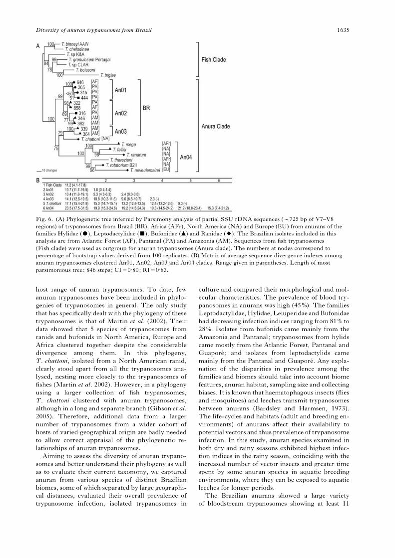
Phylogenetic inferences among anuran trypanosomes
from Brazil and other countries using SSU rDNA
sequences
To infer the phylogenetic positioning of Brazilian
anuran trypanosomes in relation to anuran trypano-
somes from other countries, comparative analysis
was performed using partial SSU rDNA sequences
from the 11 selected isolates and sequences available
on GenBank from trypanosomes of exotic anurans.
All the analyses corroborated the clustering of all the
anuran trypanosomes and indicated the position of
fish trypanosomes as a basal group for the anuran
clade. The branching pattern of the phylogenetic
trees showed very similar topologies in both
Parsimony (Fig. 6A) and Bayesian (data not shown)
analysis. Analysis based on SSU rDNA sequences
confirmed the phylogenetic relationships inferred
by ITS1 sequence analysis, with the isolates also
segregating in the 3 main clades: An01, An02 and
An03. Clades An01 (divergence ranging from 0.4%
to 1.4%), and An02 (0.0% to 3.0% divergence), clus-
tered together (99% bootstrap) and shared y94.8%
similarity (Fig. 6B). All the Brazilian isolates sep-
arated from most species of exotic trypanosomes
(Fig. 6A). T. chattoni was the closest to the Brazilian
isolates despite high divergence (12.39–14.95%) and
was positioned close to clade An03, which consisted
of 2 bufonid isolates from Amazonia (2.3% diver-
gence). Anuran trypanosomes from North America
(T. fallisi, T. ranarum, T. rotatorium), Europe
(T. neveulemairei) and Africa (T. mega) clustered
together (100% bootstrap) in clade An04 despite
significant divergence (15.3%). The divergence
separating Brazilian isolates from the exotic isolates
in clade An04 ranged from 19.2 to 19.9% (Fig. 6B).
Brazilian isolates showed significant sequence
polymorphism in the V7–V8 region of the SSU
rDNA (5.7% average divergence). Because the
V7–V8 SSU rDNA sequences were very conserved
compared with the highly polymorphic ITS1 se-
quences, only 8 of the 11 isolates distinguished by
ITS rDNA polymorphism were separated by sig-
nificant SSU rDNA sequence divergence. Trypano-
somes from the same or from very closely related
anuran species showed identical or very similar SSU
rDNA sequences. This was true for (a) isolates 322
(genotype C2) and 858 (C1) respectively from the
bufonids Chaunus schneideri and Chaunus ictericus,
which shared 100% and y93% SSU and ITS1 se-
quence similarity, respectively; (b) isolates 346 (D2)
and 362 (D3) both from the bufonid Rhinella mar-
garitifera (99.9% and y96% SSU and ITS1 simi-
larity, respectively) and (c) isolates 364 (A1) and 339
(A2) from the bufonid Chaunus marinus (97.7% and
y91% SSU and ITS similarity, respectively).
DISCUSSION
Anuran trypanosomes represent the largest known
assemblage of trypanosomes among vertebrate orders
(Bardsley and Harmsen, 1973). However, their tax-
onomy was largely built on the proven insufficient
criteria of morphology and host origin, with studies
including molecular markers being relatively scarce
(Clark et al. 1995; Martin et al. 1992a, b ; Lun and
Desser, 1995, 1996). As a result, trypanosomes that
probably belong to the same species have been
classified in separate ones, leading to a profusion of
species. Similarly, distinct species may have been
considered as a single species merely because the
trypanosomes come from the same host and/or are
morphologically indistinguishable. The use of un-
reliable taxonomic parameters over a long period
caused considerable confusion and prevented correct
appraisal of the taxonomy, biology, diversity and
A
B
Fig. 5. (A) Unrooted dendrogram based on Parsimony
analysis of 41 cloned ITS1 sequences from 11 isolates (3
to 4 clones from each isolate) of Brazilian anuran
trypanosomes. The numbers at nodes correspond to
percentage of bootstrap values derived from 100
replicates. (B) Matrix of average sequence divergence
among anuran trypanosomes clustered in An01, An02
and An03 clades. Range given in parentheses. Length of
4 most parsimonious trees: 941 steps. Length of strict
consensus: 942 steps; CI=0.87; RI=0.97.
R. C. Ferreira and others 1634
host range of anuran trypanosomes. To date, few
anuran trypanosomes have been included in phylo-
genies of trypanosomes in general. The only study
that has specifically dealt with the phylogeny of these
trypanosomes is that of Martin et al. (2002). Their
data showed that 5 species of trypanosomes from
ranids and bufonids in North America, Europe and
Africa clustered together despite the considerable
divergence among them. In this phylogeny,
T. chattoni, isolated from a North American ranid,
clearly stood apart from all the trypanosomes ana-
lysed, nesting more closely to the trypanosomes of
fishes (Martin et al. 2002). However, in a phylogeny
using a larger collection of fish trypanosomes,
T. chattoni clustered with anuran trypanosomes,
although in a long and separate branch (Gibson et al.
2005). Therefore, additional data from a larger
number of trypanosomes from a wider cohort of
hosts of varied geographical origin are badly needed
to allow correct appraisal of the phylogenetic re-
lationships of anuran trypanosomes.
Aiming to assess the diversity of anuran trypano-
somes and better understand their phylogeny as well
as to evaluate their current taxonomy, we captured
anuran from various species of distinct Brazilian
biomes, some of which separated by large geographi-
cal distances, evaluated their overall prevalence of
trypanosome infection, isolated trypanosomes in
culture and compared their morphological and mol-
ecular characteristics. The prevalence of blood try-
panosomes in anurans was high (45%). The families
Leptodactylidae,Hylidae, Leiuperidae andBufonidae
had decreasing infection indices ranging from 81% to
28%. Isolates from bufonids came mainly from the
Amazonia and Pantanal; trypanosomes from hylids
came mostly from the Atlantic Forest, Pantanal and
Guapore ; and isolates from leptodactylids came
mainly from the Pantanal and Guapore. Any expla-
nation of the disparities in prevalence among the
families and biomes should take into account biome
features, anuran habitat, sampling size and collecting
biases. It is known that haematophagous insects (flies
and mosquitoes) and leeches transmit trypanosomes
between anurans (Bardsley and Harmsen, 1973).
The life-cycles and habitats (adult and breeding en-
vironments) of anurans affect their availability to
potential vectors and thus prevalence of trypanosome
infection. In this study, anuran species examined in
both dry and rainy seasons exhibited highest infec-
tion indices in the rainy season, coinciding with the
increased number of vector insects and greater time
spent by some anuran species in aquatic breeding
environments, where they can be exposed to aquatic
leeches for longer periods.
The Brazilian anurans showed a large variety
of bloodstream trypanosomes showing at least 11
Fig. 6. (A) Phylogenetic tree inferred by Parsimony analysis of partial SSU rDNA sequences (y725 bp of V7–V8
regions) of trypanosomes from Brazil (BR), Africa (AFr), North America (NA) and Europe (EU) from anurans of the
families Hylidae ($), Leptodactylidae (&), Bufonidae (m) and Ranidae (2). The Brazilian isolates included in this
analysis are from Atlantic Forest (AF), Pantanal (PA) and Amazonia (AM). Sequences from fish trypanosomes
(Fish clade) were used as outgroup for anuran trypanosomes (Anura clade). The numbers at nodes correspond to
percentage of bootstrap values derived from 100 replicates. (B) Matrix of average sequence divergence indexes among
anuran trypanosomes clustered An01, An02, An03 and An04 clades. Range given in parentheses. Length of most
parsimonious tree: 846 steps; CI=0.80; RI=0.83.
Diversity of anuran trypanosomes from Brazil 1635

distinct morphotypes, 8 similar to forms of pre-
viously described trypanosome species, but 3 entirely
new. Adoption of the morphological and host-origin
taxonomic criteria would lead to the classification
of some Brazilian trypanosomes as new species.
However, such an approach should be strongly
discouraged because (a) the samemorphotypes could
be found in anurans of different species and from
distant locations, including different continents; (b)
inversely, anurans of the same species and from the
same location could harbour trypanosomes of quite
distinctmorphotypes and (c) cultures of isolates from
anuran blood samples showing identical morpho-
types could have distinct molecular characteristics
or, inversely, blood samples with distinct morpho-
types could generate cultures with identical or very
similar molecular features. The extensive pleo-
morphism of epimastigotes also precludes the use
of the morphology of culture forms for species
discrimination. Thus, data from this study do not
support species identification of anuran trypano-
somes based on morphology, host species and
geographical origin.
To evaluate host, ecological and geographical
diversity, we examined ITS and SSU rDNA se-
quences of Brazilian trypanosomes of anurans from
geographically distant locations corresponding to
distinct biomes. Genetic diversity among 82 isolates
evaluated by polymorphisms of ITS rDNA dis-
tinguished 11 major groups (A–K) comprising 29
genotypes. Phylogenetic analysis using ITS and
SSU rDNA sequences of Brazilian trypanosomes of
anurans and species from North America (T. chat-
toni, T. fallisi, T. ranarum and T. rotatorium), Africa
(T. mega) and Europe (T. neveulemairei) showed that
new isolates from this study and the reference species
clustered together in a clade exclusive of anuran
trypanosomes, providing evidence of the monophyly
of anuran trypanosomes in agreement with previous
studies (Hamilton et al. 2004; Gibson et al. 2005;
Simpson et al. 2006). Phylogenetic relationships of
11 Brazilian isolates inferred using ITS and SSU
rDNA sequences positioned most of them in a major
assemblage formed by 2 clades, An01 and An02.
These clades were separated by a considerable
distance from clade An03, which is composed of 2
Brazilian isolates positioned closer toT. chattoni than
to other Brazilian isolates. Therefore, although still
significantly divergent from all anuran trypano-
somes, in our analysis T. chattoni was positioned
within the clade of anuran trypanosomes, corrob-
orating results from Gibson et al. (2005) in conflict
with its positioning with fish trypanosomes as shown
by Martin et al. (2002). The phylogeny based on
V7–V8 SSU rDNA was totally congruent with
data generated by ITS1 rDNA, in conformity with
our previous analysis using the same approach for
mammalian trypanosomes (Maia da Silva et al. 2004;
Rodrigues et al. 2006; Cortez et al. 2006).
Molecular analysis revealed that the same anuran
species could be infected by distinct trypanosomes
and that the same presumed trypanosome species
could infect distinct anuran species. In addition, our
analyses suggest that closely related trypanosomes
generally come from closely related anuran species.
This is supported by the clustering together of try-
panosomes from anurans of the same host families
and genera but from different biomes and collection
locations, as in the case of trypanosomes from hylids
(Clade An01) or bufonids (Clades An02 and An03).
Comparison of trypanosomes according to their
collection locations might also suggest that there is
some degree of association between genotype and
geographical origin, since isolates from the same
anuran family and genus, but from regions far apart,
were separated in distinct subgroups/genotypes.
Moreover, some genotypes were found to be re-
stricted to certain biomes regardless of their host
family, genus or species. This was the case of group
A, which comprises 10 isolates from 4 anuran species
from 3 families, all of which are from the same
location in Rondonia State in Amazonia. Another
example of a relationship related to geographical
origin is genotype J2, composed of 14 isolates from 6
hylid species living in Pantanal or Guapore, whereas
trypanosomes from hylids of the Atlantic Forest were
clustered in separate groups. This type of association
is supported by previous studies employing iso-
enzyme, RAPD and karyotyping patterns to dem-
onstrate that isolates ofT. rotatorium andT. ranarum
from ranids of the same geographical region had
high similarity whereas isolates from geographical
locations that are far apart exhibited pronounced
genetic polymorphism (Lun and Desser, 1995, 1996;
Desser, 2001). The association between genotype and
geography is expected considering the high degree
of endemism of anuran taxa. For the same reason,
genotypes shared by anurans from Guapore and
Pantanal are also expected. The Brazilian isolates
characterized in this study are from anuran species
whose geographical distribution is restricted to
Brazil or South America. These isolates were
found to be separated from all the trypanosomes
from other countries by large genetic distances, in-
dicating that the culture collection of anuran try-
panosomes obtained in this study contains several
new species.
Finally, in spite of its monophyly, the clade formed
by anuran trypanosomes is undoubtedly a complex
taxon comprising distinct phylogenetic lineages.
Several speciation modes may have played a role
in the evolution of trypanosomes of this clade, in-
cluding co-divergence by host switching and co-
evolution, and sympatric and allopatric speciation
events. The data from this study also suggest a sig-
nificant degree of host restriction and that vector
transmission, which is largely driven by biome
and anuran species-specific ecogeographical features
R. C. Ferreira and others 1636

could be important evolutionary factors for anuran
trypanosomes. Leech transmission is thought to be
a predominant evolutionary factor for anuran try-
panosomes and the entire ‘Aquatic’ clade (Hamilton
et al. 2004, 2005; Simpson et al. 2006). However,
haematophagous insects should be very important
vectors for terrestrial and arboreal anurans, despite
the aquatic breeding environment of these anurans.
We are currently investigating possible trypanosome
vectors for the anurans investigated in this study.
Several analyses of the phylogeography and co-
evolutionary history of diverse host-parasite assem-
blages have been performed. However, few studies
have focused on anuran parasites, one of the most
diverse and complex groups in its parasite com-
munity, and most of the available data relate to the
strictly host-specific Monogenea species (platy-
helminth flatworms) (Sinnappah et al. 2001; Bentz
et al. 2006). Further studies into host-parasite in-
teractions and comparative analysis of the ecology
and phylogeography of anurans and their trypano-
somes would greatly contribute to our understanding
of the evolution of this large, widespread and het-
erogeneous group of trypanosomes. The polymor-
phic DNA markers we have identified in this paper
may facilitate such studies.
We are grateful to several collaborators for their tirelesshelp in the fieldwork. We specially would like to thankArlei Marcili (ICB-USP), Miguel T. Rodrigues (IB-USP), Sandra Favorito (Universidade Bandeirantes, SaoPaulo) and Carlos Jared (Instituto Butantan, Sao Paulo) forinestimable help providing blood samples from anurans.Work in Amazonia was done at the laboratory of theICB5-USP in Monte Negro, Rondonia. We thankFernando Paiva (UFMS) for providing good conditions forfield work in the Pantanal. Many thanks to Professor ErneyP. Camargo for helpful discussions, valuable commentsand constructive criticisms in reviewing our manuscript.This work was funded by the Brazilian agencies ofFAPESP and CNPq. Robson C. Ferreira and Laerte B.Viola are graduate student fellows respectively fromFAPESP and CNPq.
REFERENCES
Anderson, J. R. and Ayala, S. C. (1968). Trypanosome
transmitted by Phlebotomus : first report from
the Americas. Science 161, 1023–1025.
doi: 10.1126/science.161.3845.1023.
Ayala, S. C. (1970). Two new trypanosomes from
California toads and lizards. Journal of Protozoology
17, 370–373.
Bardsley, J. E. and Harmsen, R. (1973). The
trypanosomes of Anura. Advances in Parasitology
11, 1–73.
Barta, J. R., Boulard, Y. and Desser, S. S. (1989).
Blood parasites of Rana esculenta from Corsica:
Comparison of its parasites with those of eastern
North American ranids in the context of host phylogeny.
Transactions of the American Microscopical Society
108, 6–20. doi: 10.2307/3226201.
Barta, J. R. and Desser, S. S. (1984). Blood parasites
of amphibians from Algonquin Park, Ontario.
Journal of Wildlife Diseases 20, 180–189.
Bentz, S., Sinnappaah-Kang, N. D., Lim, L.-HS.,
Lebedev, B., Combes, C. and Verneau, O. (2006).
Historical biogeography of amphibian parasites,
genus Polystoma (Monogenea: Polystomidadee).
Journal of Biogeography 33, 742–749. doi: 10.1111/
j.1365-2699.2005.01402.x.
Clark, C. G., Martin, D. S. and Diamond, L. S. (1995).
Phylogenetic relationships among anuran trypanosomes
as revealed by riboprinting. Journal of Eukaryotic
Microbiology 42, 92–96.
Cortez, A. P., Ventura, R. M., Rodrigues, A. C.,
Batista, J. S., Paiva, F., Anez, N., Machado, R. Z.,
Gibson, W. C. and Teixeira, M. M. G. (2006). The
taxonomic and phylogenetic relationships of
Trypanosoma vivax from South America and Africa.
Parasitology 133, 159–169. doi: 10.1017/
50031182006000254.
Desser, S. S. (2001). The blood parasites of anurans
from Costa Rica with reflections on the taxonomy
of their trypanosomes. Journal of Parasitology
87, 152–160. doi: 10.1645/0022-3395(2001)087[0152 :
TBPOAF]2.0.CO;2.
Desser, S. S., McIver, S. B. and Jez, D. (1975).
Observations on the role of simuliids and culicids
in the transmission of avian and anuran trypanosomes.
International Journal for Parasitology 5, 507–509.
Desser, S. S., McIver, S. B. and Ryckman, A. (1973).
Culex territans as a potential vector of Trypanosoma
rotatorium. I. Development of the flagellate in the
mosquito. Journal of Parasitology 59, 353–358.
Diamond, L. S. (1965). A study of the morphology,
biology and taxonomy of the trypanosomes of anura.
Wildlife Diseases 44, 1–85.
Frost, D. R. (2006). Amphibian Species of the World: an
Online Reference. Version 4 (17 August 2006), http://
research.amnh.org/herpetology/amphibia/index.php.
Gibson, W. C., Lom, J., Peckova, H., Ferris, V. R.
and Hamilton, P. B. (2005). Phylogenetic analysis of
freshwater fish trypanosomes from Europe using
SSUrRNA gene sequences and random amplification
of polymorphic DNA. Parasitology 130, 405–412.
doi: 10.1017/S0031182004006778.
Gruby, M. (1843). Recherches et observations sur
une nouvelle espece d’hematozoaire, Trypanosoma
sanguinis. Comptes Rendus Hebdomadaire des Seances
de l’Academie des Sciences, Paris 55, 1134–1136.
Hamilton, P. B., Stevens, J. R., Gaunt, M. W.,
Gidley, J. and Gibson, W. C. (2004). Trypanosomes
are monophyletic: evidence from genes for
glyceraldehyde phosphate dehydrogenase and small
subunit ribosomal RNA. International Journal for
Parasitology 34, 1393–1404. doi: 10.1016/
j.ijpara.2004.08.011.
Hamilton, P. B., Stevens, J. R., Gidley, J., Holz, P.
and Gibson, W. C. (2005). A new lineage of
trypanosomes from Australian vertebrates and
terrestrial bloodsucking leeches (Haemadipsidae).
International Journal for Parasitology 35, 431–443.
doi: 10.1016/j.ijpara.2004.12.005.
Jakes, K. A., O’Donoghue, P. J. and Adlard, R. D.
(2001). Phylogenetic relationships of Trypanosoma
Diversity of anuran trypanosomes from Brazil 1637

chelodina and Trypanosoma binneyi from Australian
turtles and platypuses inferred from small subunit
rRNAanalyses.Parasitology 123, 483–487. doi: 10.1017/
S0031182001008721.
Levine, N. D. and Nye, R. R. (1977). A survey of blood
and other tissue parasites of leopard frogsRana pipiens in
the United States. Journal of Wildlife Diseases 13, 17–23.
Lun, Z. R. and Desser, S. S. (1995). Karyotype analysis
of anuran trypanosomes by pulsed-field gradient gel
electrophoresis. Journal of Parasitology 81, 1018–1020.
Lun, Z. R. and Desser, S. S. (1996). Analysis of isolates
within species of anuran trypanosomes using random
amplified polymorphic DNA. Parasitology Research
82, 22–27. doi: 10.1007/s004360050062.
Maia da Silva, F., Rodrigues, A. C., Campaner, M.,
Takata, C. S., Brigido, M. C., Junqueira, A. C.,
Coura, J. R., Takeda, G. F., Shaw, J. J. and
Teixeira, M. M. G. (2004). Randomly amplified
polymorphic DNA analysis of Trypanosoma rangeli
and allied species from human, monkeys and
other sylvatic mammals of the Brazilian Amazon
disclosed a new group and a species-specific marker.
Parasitology 128, 283–294. doi: 10.1017/
S0031182003004554.
Martin, D. S. and Desser, S. S. (1990). A light and
electron microscopic study of Trypanosoma fallisi n.
sp. in toads (Bufo americanus) from Algonquin Park,
Ontario. Journal of Protozoology 37, 199–206.
Martin, D. S. and Desser, S. S. (1991a). Development
of Trypanosoma fallisi in the leech, Desserobdella picta,
in toads (Bufo americanus), and in vitro. A light and
electron microscopic study. Parasitology Research 77,
18–26. doi: 10.1007/BF00934379.
Martin, D. S. and Desser, S. S. (1991b). Infectivity of
cultured Trypanosoma fallisi (Kinetoplastida) to various
anuran species and its evolutionary implications.
Journal of Parasitology 77, 498–500.
Martin, D. S., Desser, S. S. and Hong, H. (1992a).
Allozyme comparison of three Trypanosoma species
(Kinetoplastida: Trypanosomatidae) of toads and frogs
by starch-gel electrophoresis. Journal of Parasitology
78, 317–322.
Martin, D. S., Desser, S. S. and Werner, J. K. (1992b).
Allozyme comparison and infectivity of cultured stages
of Trypanosoma fallisi from southern Ontario and a
trypanosome of toads from northern Michigan.
Journal of Parasitology 78, 1083–1086.
Martin, D. S., Wright, A. D., Barta, J. R. and Desser,
S. S. (2002). Phylogenetic position of the giant anuran
trypanosomes Trypanosoma chattoni, Trypanosoma
fallisi, Trypanosoma mega, Trypanosoma neveulemairei,
and Trypanosoma ranarum inferred from 18S rRNA
gene sequences. Journal of Parasitology 88, 566–571.
doi: 10.1645/0022-3395(2002)088[0566:
PPOTGA]2.0.CO;2.
Miyata, A. (1978). Anuran trypanosomes in Kyushu
andRyukyu islands, with descriptions of six new species.
Tropical Medicine 20, 51–80.
Reilly, B. O. and Woo, P. T. K. (1982). The in vivo and
in vitro development of Trypanosoma andersoni Reilly
and Woo, 1982 and Trypanosoma grylli Nigrelli, 1944
(Kinetoplastida). Canadian Journal of Zoology 60,
124–133.
Rodrigues, A. C., Paiva, F., Campaner, M., Stevens,
J. R., Noyes, H. A. and Teixeira, M. M. G.
(2006). Phylogeny of Trypanosoma (Megatrypanum)
theileri and related trypanosomes reveals lineages
of isolates associated with artiodactyl hosts diverging
on SSU and ITS ribosomal sequences. Parasitology
132, 215–224. doi: 10.1017150031182005008929.
Ronquist, F. andHuelsenbeck, J. P. (2003). MrBayes 3:
Bayesian phylogenetic inference under mixed models.
Bioinformatics 19, 1572–1574. doi: 10.1093/
bioinformatics/btg180.
Siddall, M. E. and Desser, S. S. (1992). Alternative leech
vectors of frog and turtle trypanosomes. Journal of
Parasitology 78, 562–563.
Simpson, A. G. B., Stevens, J. R. and Lukes, J. (2006).
The evolution and diversity of kinetoplastid flagellates.
Trends in Parasitology 22, 168–174. doi: 10.1016/
j.pt.2006.02.006.
Sinnappah, N. D., Lim, L.-HS., Rohde, K., Tinsley, R.,
Combes, C. and Verneau, O. (2001). A paedomorphic
parasite associated with a neotenic amphibian host:
phylogenetic evidence suggests a revised systematic
position for Sphyranuridae within anuran and turtle
polystomatoineans. Molecular Phylogenetics and
Evolution 18, 189–201. doi: 10.1006/mpev.2000.0877.
Stevens, J. R., Noyes, H. A., Schofield, C. J.
and Gibson, W. (2001). The molecular evolution
ofTrypanosomatidae.Advances in Parasitology 48, 1–56.
doi: 10.1016/S0065-308X(01)48003-1.
Werner, J. K. (1993). Blood parasites of amphibians
from Sichuan Province, People’s Republic of China.
Journal of Parasitology 79, 356–363.
Werner, J. K. and Walewski, K. (1976). Amphibian
trypanosomes from the McCormick forest, Michigan.
Journal of Parasitology 62, 20–25.
Woo, P. T. and Bogart, J. P. (1984). Trypanosoma spp.
(Protozoa: Kinetoplastida) in Hylidae (Anura) from
eastern North America, with notes on their distribution
and prevalences. Canadian Journal of Zoology 62,
820–824.
Zickus, T. (2002). The first data on the fauna
and distribution of blood parasites of amphibians
in Lithuania. Acta Zoologica Lituanica 12, 197–202.
R. C. Ferreira and others 1638