Embed Size (px)
Citation preview
211
INTRODUCTION
The liver of fishes is a dense organ ventrally locatedin the cranial region of the general cavity. Its size, shape,and volume are adapted to the space available between othervisceral organs. In many teleostei species the liver is dividedinto three lobes. However, no lobulation was recognized insome teleostei (Bruslé & Anadon, 1996). The hepaticparenchyma in fish is made of two cellular plates surroundedby sinusoids. Between two neighboring sinusoids, thehepatocytes are arranged as cords, generally two cells inthickness. The cords extended between central and portalzones (Hinton et al., 1972; Kendall & Hawkins, 1975; Hinton& Pool, 1976 and Bruslé & Anadon).
Previous studies have indicated that in teleost fish,the pancreatic exocrine tissue develops around the portal veinduring ontogenesis. It remains extrahepatic or penetratessomewhat deeply into the liver parenchyma depending onthe species, as Ictalurus punctatus (Kendall & Hawkin andHinton & Pool), Pimelodus maculatus (Marconi Stipp et al.,1980), Micropogon undulatus (Eurell & Haensly, 1982),Serranus cabrilla (Gonzalez et al., 1993). Pancreatic tissuecan be differentiated from hepatic tissue by its acinararrangement. In addition, a thin septa of connective tissueseparates the hepatocytes from the exocrine pancreatic cells(Bruslé & Anadon).
Based on these data, the objective of the present studywas to describe the morphological characteristics of the liverand the intrahepatic exocrine pancreas in Nile tilapia(Oreochromis niloticus). This species is of great interest tofish culture as it means fast growth, in addition to the fact thatits meat is considered of excellent quality.
MATERIAL AND METHOD
This study used 30 samples of adult Nile tilapia,Oreochromis niloticus, cultivated in floating net cages that wereplaced in the Nova Avanhandava reservoir in Buritama, SP,Brazil. The fishes were anaesthetized with methaneasulfonateand the coeloma was opened for liver exposure, which wasthen removed for light microscopy and transmission electronmicroscopy studies. For light microscopy the liver sampleswere fixed in 10% buffered formalin and embedded inhistoresin (Leica, Germany). The histological sections werestained with haematoxylin-eosin and analyzed and documentedphotographically with an Olympus Bx50 microscope (Japan).
For transmission electron microscopy, liver fragmentswere fixed in glutaraldehyde 2,5% in 0,1 M phosphate buffer,
Int. J. Morphol.,23(3):211-216, 2005.
Morphological Study of the Liverin the Teleost Oreochromis niloticus
Estudio Morfológico del Hígado en el Teleósteo Oreochromis niloticus
Vicentini, C. A.; Franceschini-Vicentini, I. B.; Bombonato, M. T. S.; Bertolucci, B.; Lima, S. G. & Santos, A. S.
VICENTINI, C. A.; FRANCESCHINI-VICENTINI, I. B.; BOMBONATO, M. T. S.; BERTOLUCCI, B.; LIMA, S. G. & SANTOS, A. S.Morphological study of the liver in the teleost Orechromis niloticus. Int. J. Morphol., 23(3):211-216, 2005.
SUMMARY: Liver samples of Oreochromis niloticus cultivated in floating net cages were fixed for histological and ultrastructuralstudies with the objective of describing the hepatic parenchymal structure and the intrahepatic exocrine pancreatic tissue. Anatomically, the livershowed only two hepatic lobes. Histological analysis demonstrated that the hepatocytes were spread out as anastomotic cords, arranged in twocellular layers and surrounded by sinusoids. The intrahepatic exocrine pancreatic tissue exhibited an acinar arrangement and was diffused in thehepatic parenchyma. Structural analysis showed that the hepatocytes had a rounded nucleus and a rough endoplasmic reticulum with a paralleldisposition to the nuclear membrane. The exocrine pancreatic cells showed secretion granules at the apical portion and the rough endoplasmicreticulum was concentrically distributed.
KEY WORDS: Liver; Exocrine pancreas; Morphology; Teleost.
Department of Biological Sciences, Faculty of Sciences, UNESP and CAUNESP, Bauru, SP, CEP: 17.033-360, Brazil.This work was supported by grant from the CNPq (Nº 300693/91-5), Brazil.
212
pH 7.2, for 3h, postfixed in 1% osmium tetroxide in phosphatebuffer, washed in the same buffer, dehydrated in a growingacetone series and embedded in Araldite resin (DurcupanACM, Fluka, Sigma-Aldrich , St. Louis , MO, USA). Resinpolymerization was then completed in an oven at 60 ° C, for48 h. Ultrathin sections (60 and 80 nm) were cut andtransported to copper grids, contrasted with uranyl acetate andlead citrate, analyzed and documented photographically witha Philips CEM 100 transmission electron microscope (TEM),at the Electron Microscopy Center of the Institute ofBiosciences of Botucatu, UNESP.
RESULTS
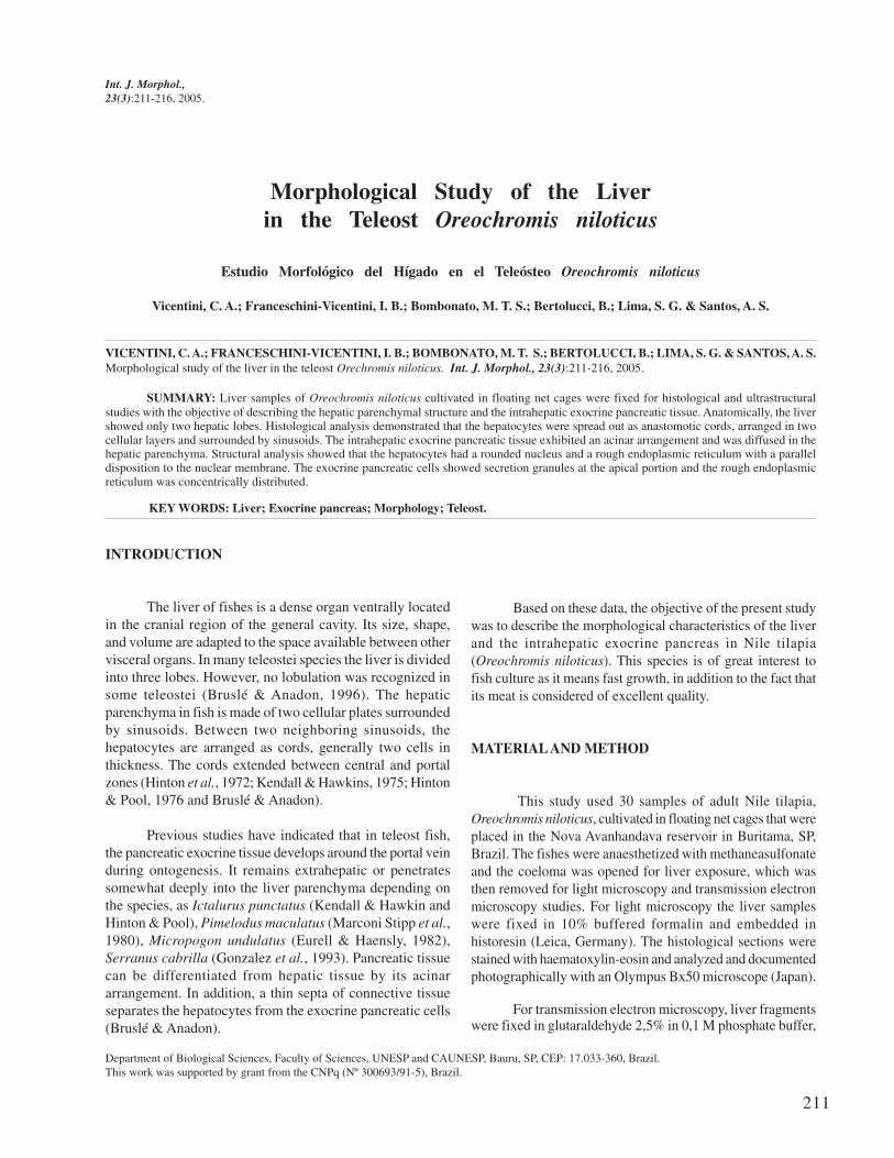
The liver of the Nile tilapia (Oreochromis niloticus) isa large organ and has only two lobes (Fig. 1). The left lobe isbigger and spreads throughout almost the entire corporealcavity. At the visceral face it has the impression of the intestine.The gallbladder is well developed and has a rounded shape.
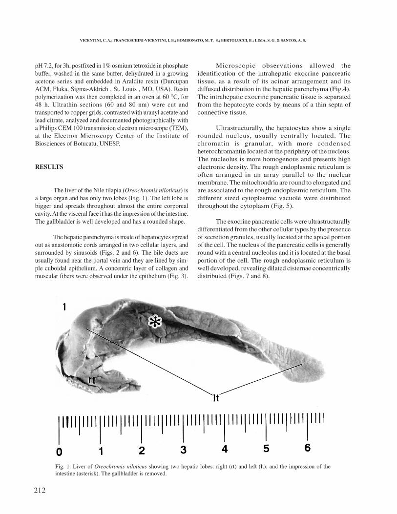
The hepatic parenchyma is made of hepatocytes spreadout as anastomotic cords arranged in two cellular layers, andsurrounded by sinusoids (Figs. 2 and 6). The bile ducts areusually found near the portal vein and they are lined by sim-ple cuboidal epithelium. A concentric layer of collagen andmuscular fibers were observed under the epithelium (Fig. 3).
Microscopic observations allowed theidentification of the intrahepatic exocrine pancreatictissue, as a result of its acinar arrangement and itsdiffused distribution in the hepatic parenchyma (Fig.4).The intrahepatic exocrine pancreatic tissue is separatedfrom the hepatocyte cords by means of a thin septa ofconnective tissue.
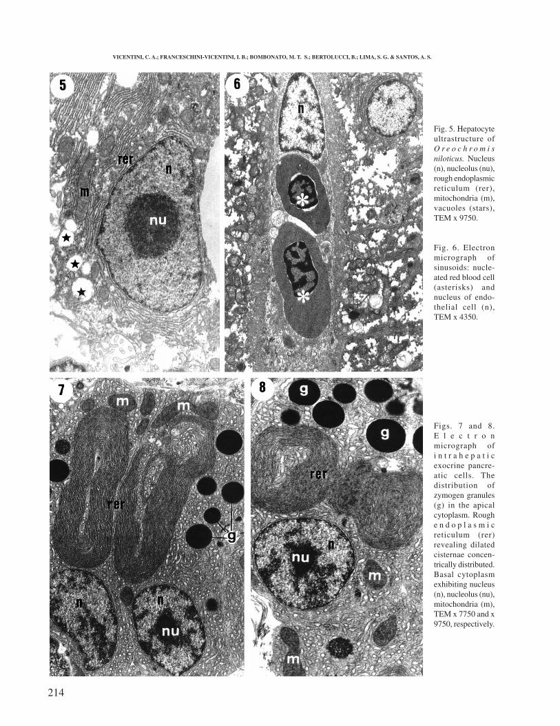
Ultrastructurally, the hepatocytes show a singlerounded nucleus, usually centrally located. Thechromatin is granular, with more condensedheterochromantin located at the periphery of the nucleus.The nucleolus is more homogenous and presents highelectronic density. The rough endoplasmic reticulum isoften arranged in an array parallel to the nuclearmembrane. The mitochondria are round to elongated andare associated to the rough endoplasmic reticulum. Thedifferent sized cytoplasmic vacuole were distributedthroughout the cytoplasm (Fig. 5).
The exocrine pancreatic cells were ultrastructurallydifferentiated from the other cellular types by the presenceof secretion granules, usually located at the apical portionof the cell. The nucleus of the pancreatic cells is generallyround with a central nucleolus and it is located at the basalportion of the cell. The rough endoplasmic reticulum iswell developed, revealing dilated cisternae concentricallydistributed (Figs. 7 and 8).
Fig. 1. Liver of Oreochromis niloticus showing two hepatic lobes: right (rt) and left (lt); and the impression of theintestine (asterisk). The gallbladder is removed.
VICENTINI, C. A.; FRANCESCHINI-VICENTINI, I. B.; BOMBONATO, M. T. S.; BERTOLUCCI, B.; LIMA, S. G. & SANTOS, A. S.
213
Fig. 2. Histology of hepatic parenchyma showing sinusoide arrangement, separated by hepatocyte cords, H/E x 400.Fig. 3. Bile duct showing simple cuboidal epithelium (e) and circular layer of muscular fibers (mc), H/E x 400.Fig. 4. Organization of the intrahepatic exocrine pancreatic tissue around a blood vessel (star). Note the distribution ofzymogen granules (g) in the exocrine cells, H/E x 400.
DISCUSSION
The histological structure of the liver of Oreochromisniloticus showed hepatocytes arrayed in cords, similar towhat is found in many teleosts (Kendall & Hawkins; Hinton& Pool; González et al. and Bruslé & Anadon). The absenceof division into hepatic lobules and the lack of portal triadsare features of Oreochromis niloticus, as evidenced in many
teleosts (Hampton et al., 1985 and González et al.). However,some triads are found in Caranx spp. and Lutjanus bohar(González, 1992).
Observations achieved by optic microscopy alsoevidenced intrahepatic exocrine pancreatic tissue in
Morphological study of the liver in the teleost Orechromis niloticus. Int. J. Morphol., 23(3):211-216, 2005.
214
Fig. 5. Hepatocyteultrastructure ofO r e o c h r o m i sniloticus. Nucleus(n), nucleolus (nu),rough endoplasmicreticulum (rer),mitochondria (m),vacuoles (stars),TEM x 9750.
Fig. 6. Electronmicrograph ofsinusoids: nucle-ated red blood cell(asterisks) andnucleus of endo-thelial cell (n),TEM x 4350.
Figs. 7 and 8.E l e c t r o nmicrograph ofi n t r a h e p a t i cexocrine pancre-atic cells. Thedistribution ofzymogen granules(g) in the apicalcytoplasm. Roughe n d o p l a s m i creticulum (rer)revealing dilatedcisternae concen-trically distributed.Basal cytoplasmexhibiting nucleus(n), nucleolus (nu),mitochondria (m),TEM x 7750 and x9750, respectively.
VICENTINI, C. A.; FRANCESCHINI-VICENTINI, I. B.; BOMBONATO, M. T. S.; BERTOLUCCI, B.; LIMA, S. G. & SANTOS, A. S.
215
Oreochromis niloticus, associated to afferent vases. Yetin some species, the pancreatic tissue was identified asdiffused, surrounding the digestive tract (Beccaria et al.,1992 and Marconi Stipp et al.). The pancreas in Pimelodusmaculatus is compact, enclosed by a thin layer ofconjunctive tissue and is attached to the stomach andintestine wall as small masses of glandular tissue (MarconiStipp et al.).
Ultrastructural characteristics of the liver ofOreochromis niloticus are in accordance with theobservations attained by Kendall & Hawkins; Hinton &Pool and González et al. According to González et al.and Bruslé & Anadon, the hepatocytes in fish are relativelypoor in organelles, suggesting a low synthetic activity forsecretory proteins.
In hepatocytes of various fish, a classical feature isthe high content of glycogen, which fills most of the
cytoplasm. However compared with those of mammals,fish hepatocytes do not metabolize much glycogen (Moonet al.; Hampton et al., 1985 and González et al.).
Exocrine pancreatic cells of Oreochromis niloticusexhibit similar characteristics to those of other teleosts(Kendall & Hawkins; Hinton & Pool; Marconi Stipp etal.; Beccaria et al.).
Sea bass (Beccaria et al.), subjected to long fastingshowed the lumen of excretory ducts narrower and reducedcellular activity demonstrated by the scarcity of zymogengranules. On the other hand, intensively fed fish were seento have increased cellular activity and greater quantity ofzymogen granules. Electrodensed zymogen granules wereabundant in Oreochromis niloticus. They were generallylocated in the apical portion of the cell. The roughendoplasmic reticulum was well developed and showedan organized pattern.
VICENTINI, C. A.; FRANCESCHINI-VICENTINI, I. B.; BOMBONATO, M. T. S.; BERTOLUCCI, B.; LIMA, S. G. & SANTOS, A. S.Estudio morfológico del hígado en el teleósteo Oreochromis niloticus. Int. J. Morphol., 23(3):211-216, 2005.
RESUMEN: Con el objetivo de describir la estructura del parénquima hepático y del páncreas exocrino intrahepático delOreochromis niloticus, fueron fijados para estudios histológicos y ultraestructurales fragmentos de hígado de peces cultivados en jaulasflotantes. Se evidenciaron sólo 2 lóbubos hepáticos. El análisis histológico demostró que los hepatocitos se encontraban organizados enforma de cordones anastomosados, dispuestos en dos capas celulares y cercados por sinusoides. El tejido pancreático exocrino intrahepáticose encontró difuso en el parénquima hepático y se destacó por su organización acinar. El análisis ultraestructural demostró que loshepatocitos presentaban núcleos redondos y el retículo endoplasmático rugoso estaba dispuesto paralelamente a la membrana nuclear.Las células pancreáticas exocrinas presentaban gránulos de secreción localizados en la porción apical y el retículo endoplasmático rugosoestaba organizado de manera concéntrica.
PALABRAS CLAVE: Hígado; Páncreas exocrino; Morfología; Teleósteos.
REFERENCES
Beccaria,C.; Diaz, J. P. & Connes, R. Effects of dietaryconditions on the exocrine pancreas of the sea bass,Dicentrarchus labrax L. (Teleostei). Aquaculture, 101:163-76, 1992.
Bruslé, J. & Anadon, G. G. The Structure and Function ofFish Liver. In: Fish Morphology. Science Publishers,1996. pp 77-93.
Eurell, J. A. & Haensly, W. E. The histology andultrastructure of the liver of Atlantic croackerMicropogon undulatus L. J. Fish Biol., 21:113-25, 1982.
González, G. Contribution à la connaissance des processusciguatérigènes.Thèse de Doctorat (specialitéOceanologie), Université de Perpignan, pp 335, 1992.
González, G.; Crespo, S. & Bruslé, J. Histo-cytologicalstudy of the liver of the cabrilla sea bass, Serranus ca-brilla (Teleostei, Serranidae), an available model formarine fish experimental studies. J. Fish Biol., 43:363-73, 1993.
Hampton, J. A.; McCuskey, P. A.; McCuskey, R. S. & Hinton,D. E. Functional units in rainbow trout (Salmo gairdneri)liver: arrangement and histochemical properties ofhepatocytes. Anat. Rec., 213:166-75, 1985.
Hinton, D. E. & Pool, C. R. Ultrastructure of the liver inchannel catfish Ictalurus punctatus (Rafinesque). J. FishBiol., 8:209-19, 1976.
Hinton, D. E.; Snipes, R. & Kendall, M. W. Morphology
Morphological study of the liver in the teleost Orechromis niloticus. Int. J. Morphol., 23(3):211-216, 2005.
216
and enzyme histochemistry in the liver of largemouthbass (Micropterus salmoides). J. Fish. Res. Bd. Can.,29: 531-4, 1972.
Kendall, M.W. & Hawkins, W. E. Hepatic morphology andacid phosphatase localization in the channel catfish(Ictalurus punctatus). J. Fish. Res. Bd. Can., 32:1459-64, 1975.
Marconi Stipp, A. C.; Ferri, S. & Sesso, A. Fine structuralanalysis of a teleost exocrine pancreas cellularcomponents – A freeze-fracture and transmission electronmicroscopic study. Anat. Anz., 147:60-75, 1980.
Moon, T.W.; Walsh, P.J. & Mommsen, T.P. Fish hepatocytes:a model metabolic system. Can. J. Fish Aquat. Sci., 42:1772-82, 1985.
Correspondence to:Prof. Dr. Carlos Alberto VicentiniFaculdade de Ciências de Bauru – UNESPDepartamento de Ciências BiológicasAv. Luiz Edmundo Carrijo Coube s/nCEP: 17.033-360Bauru – SP,BRASIL
Email: [email protected]
Received: 13-04-2005Accepted: 07-06-2005
VICENTINI, C. A.; FRANCESCHINI-VICENTINI, I. B.; BOMBONATO, M. T. S.; BERTOLUCCI, B.; LIMA, S. G. & SANTOS, A. S.