Embed Size (px)
Citation preview

Wolbachia-Associated Bacterial Protection in theMosquito Aedes aegyptiYixin H. Ye, Megan Woolfit, Edwige Rances, Scott L. O’Neill, Elizabeth A. McGraw*
School of Biological Sciences, Monash University, Clayton, Victoria, Australia
Abstract
Background: Wolbachia infections confer protection for their insect hosts against a range of pathogens including bacteria,viruses, nematodes and the malaria parasite. A single mechanism that might explain this broad-based pathogen protectionis immune priming, in which the presence of the symbiont upregulates the basal immune response, preparing the insect todefend against subsequent pathogen infection. A study that compared natural Wolbachia infections in Drosophilamelanogaster with the mosquito vector Aedes aegypti artificially transinfected with the same strains has suggested thatinnate immune priming may only occur in recent host-Wolbachia associations. This same study also revealed that whileimmune priming may play a role in viral protection it cannot explain the entirety of the effect.
Methodology/Findings: Here we assess whether the level of innate immune priming induced by different Wolbachia strainsin A. aegypti is correlated with the degree of protection conferred against bacterial pathogens. We show that Wolbachiastrains wMel and wMelPop, currently being tested for field release for dengue biocontrol, differ in their protective abilities.The wMelPop strain provides stronger, more broad-based protection than wMel, and this is likely explained by both thehigher induction of immune gene expression and the strain-specific activation of particular genes. We also show thatWolbachia densities themselves decline during pathogen infection, likely as a result of the immune induction.
Conclusions/Significance: This work shows a correlation between innate immune priming and bacterial protectionphenotypes. The ability of the Toll pathway, melanisation and antimicrobial peptides to enhance viral protection or toprovide the basis of malaria protection should be further explored in the context of this two-strain comparison. This workraises the questions of whether Wolbachia may improve the ability of wild mosquitoes to survive pathogen infection or alterthe natural composition of gut flora, and thus have broader consequences for host fitness.
Citation: Ye YH, Woolfit M, Rances E, O’Neill SL, McGraw EA (2013) Wolbachia-Associated Bacterial Protection in the Mosquito Aedes aegypti. PLoS Negl TropDis 7(8): e2362. doi:10.1371/journal.pntd.0002362
Editor: Paulo Filemon Pimenta, Fundacao Oswaldo Cruz, Brazil
Received February 10, 2013; Accepted June 30, 2013; Published August 8, 2013
Copyright: � 2013 Ye et al. This is an open-access article distributed under the terms of the Creative Commons Attribution License, which permits unrestricteduse, distribution, and reproduction in any medium, provided the original author and source are credited.
Funding: This research was supported by a grant from the Foundation for the National Institutes of Health through the Grand Challenges in Global HealthInitiative of the Bill and Melinda Gates Foundation and the National Health and Medical Research Council, Australia. The funders had no role in study design, datacollection and analysis, decision to publish, or preparation of the manuscript.
Competing Interests: The authors have declared that no competing interests exist.
* E-mail: [email protected]
Introduction
Wolbachia pipientis is a maternally inherited intracellular
bacterium that is found in a wide range of arthropod species
and filarial nematodes, with approximately 40% of insect
species infected [1]. Wolbachia spreads rapidly through popula-
tions and to high frequencies by inducing a range of
manipulations of host reproduction that benefit infected
females. In insects, the most common manipulation is cytoplas-
mic incompatibility (CI) [2,3]. Interestingly, some Wolbachia
strains that cannot induce reproductive manipulations still
spread through populations [4]. This would not be predicted
unless there were other positive benefits for Wolbachia-infected
insects. Despite numerous laboratory and semi-field based
experiments examining a range of life history traits, few studies
have identified significant fitness benefits of infection [5,6,7,8].
Most reveal no effect [9] or weak negative effects [10,11,12]. It
is possible, however, that there are benefits to Wolbachia
infections that are only detectable under field conditions or in
circumstances not yet tested in the laboratory.
Recently, Wolbachia was found to either extend the lifespan and/
or increase the survival of Drosophila infected with native viruses,
a trait termed pathogen protection [13,14,15]. Subsequently,
Wolbachia strains native to Drosophila have also been shown to
confer pathogen protection against arboviruses, bacteria, filarial
nematodes and the malaria parasite Plasmodium gallinaceum when
stably transinfected into the mosquito Aedes aegypti [16,17]
[13,17,18,19,20,21,22]. This broad-based pathogen protection
may offer a potential fitness advantage, assisting cytoplasmic
incompatibility in the maintenance and spread of Wolbachia in wild
populations. Understanding the true fitness effects of Wolbachia
infections in mosquitoes is important as these symbiont-infected
mosquitoes are being released into wild populations as part of a
biocontrol strategy for reducing dengue virus transmission to
humans [23].
While the mechanism of pathogen protection is not fully
understood, several recent studies have shed some light on its basis.
It was originally hypothesized that priming of the insect immune
response might provide a single mechanistic explanation for
symbiont-induced protection against viruses, bacteria, nematodes
PLOS Neglected Tropical Diseases | www.plosntds.org 1 August 2013 | Volume 7 | Issue 8 | e2362

and malaria. Under an immune priming model, Wolbachia
infections activate the basal immune response, better preparing
insects against subsequent infection by pathogens. Three different
A. aegypti:Wolbachia strain associations have been created thus far
and in each case infection with the symbiont induces the host
immune response [18,19,20]. The same is true for transient
infections established in the mosquito Anopheles gambiae [22].
Immunity genes upregulated in these mosquitoes include members
of the opsonisation, Toll and melanisation pathways [18,21,24].
Whether the expression of this limited set of insect immunity genes
can confer protection against pathogens other than bacteria [25] is
not clear, although the Toll pathway participates in dengue virus
control [26] and the Imd, Toll, opsonisation and melanisation
pathways assist in Plasmodium control [18,27,28,29,30].
In each case where Wolbachia-associated immunity gene
activation has been reported, the host insects did not have
histories of association with this symbiont. In Drosophila naturally
infected with Wolbachia there is no activation of the immune
response and no bacterial protection [31,32]. There is, however,
weak protection against dengue virus [24] as well as other native
viruses [13,14] indicating that innate immune priming cannot
explain viral protection in this host. Interestingly, in A. aegypti
transinfected with the same Wolbachia strains native to D.
melanogaster, there is both innate immune priming and strong
protection against dengue virus. The comparative study indicates
that innate immune priming alone cannot fully explain pathogen
blocking although it may be contributing to the strength of the
effect in A. aegypti [24,33]. This same study also revealed that
Wolbachia strains differ in their level of immune induction in A.
aegypti [24]. The wMelPop Wolbachia strain, known for causing life
shortening and other fitness effects in its host, is present in more
tissues and grows to higher densities [16,20,34,35] and is
associated with a greater immune response than the wMel strain,
which is present in fewer tissues and grows to much more
moderate densities [17].
While the transcriptional profiles of Wolbachia-infected A. aegypti
predict that they should experience broad protection against
bacterial infection, evidence of bacterial protection in this host
comes from a single study demonstrating the ability of wMelPop to
protect against systemic Erwinia carotorovra infection [21]. Here we
expose A. aegypti stably transinfected with either the wMel or the
wMelPop strain to infection with several bacterial pathogens using
in previous infection studies in D. melanogaster. We characterised the
response to two extracellular bacteria, E. carotovora [36] and the
slow-killing but highly pathogenic Burkholderia cepacia [37], and two
intracellular bacteria, Salmonella typhimurium [37,38] and the slow-
growing Mycobacterium marinum [37]. Following pathogen infection
we then examined mosquito survival and corresponding changes
in Wolbachia and pathogen densities. As a control, we also confirm
that these Wolbachia strains provide no protection against these
same pathogens in D. melanogaster. We studied that the protective
effect of wMelPop and wMel in terms of both survival and delayed
death rates. We examined the association between survivorship
and pathogen load. Our result indicates either a direct effect of
immune priming on the symbiont or an energetic tradeoff, with
sick hosts affecting resources for Wolbachia’s growth and replica-
tion.
Materials and Methods
Ethics statementApproval for blood feeding by human volunteers for mainte-
nance of the mosquito colony was granted by the Monash
University Human Research Ethics Committee (2007001379).
Volunteers provided written informed consent to participate.
Fly and mosquitoThe w1118 fly line infected with wMel (w1118wMel) or wMelPop
(w1118wMelPop) and their respective tetracycline-cured lines
(w1118wMel.tet and w1118wMelPop.tet respectively) were used in
this study [34,39]. PCR using primers specific for the wMel and
wMelPop IS5 repeat was used to confirm the tetracycline-cured
lines to be free of Wolbachia [40]. Flies were reared on standard
yellow corn meal medium at 25uC with 50% relative humidity and
a 12:12 hr light/dark cycle. Around fifty individuals were allowed
to oviposit in bottles with 40 ml of fly food for two days. After
eclosion, adults were transferred to and aged in vials at a density of
,30 individuals per vial.
Mosquito lines used in this study are laboratory lines artificially
infected with wMel (MGYP2) or wMelPop-CLA (PGYP1) and
their tetracycline-cured (PGYP1.tet and MGYP2.tet respectively)
Wolbachia uninfected counterparts [16,17]. Mosquitoes were
reared under standard laboratory conditions (2662uC, 12:12 hr
light/dark cycle, 75% relative humidity). Mosquito larvae were fed
0.1 mg/larvae of TetraMin Tropical Tablets once a day at a
density of 150 larvae per 3 liters of distilled water in trays. Adults
were transferred to cages (measuring 30630630 cm) at emer-
gence at 400 individuals per cage. Adults were supplied with a
basic diet of 10% sucrose solution.
Bacterial cultureE. carotovora strain 15 (Ecc15) and S. typhimurium strain TM11
were cultured in LB medium in a shaker at 37uC [36,37]. B. cepacia
clinical isolate AH1345 was cultured in brain heart infusion broth
(Oxoid, Australia) at 37uC in a shaker [37,38]. M. marinum was
cultured at 29uC in the dark without shaking in Middlebrook 7H9
broth (Difco, Australia) supplemented with OADC [41].
Survival assayFor survival assay, female flies and mosquitoes aged 3–8 days
were used. Insects were anesthetized with CO2 before being
infected by either stabbing with a needle previously dipped into a
Author Summary
Wolbachia is a commonly occurring bacterium or symbiontthat lives inside the cells of insects. Recently, Wolbachiawas artificially introduced into the mosquito vectordengue virus that was naturally Wolbachia-free. Wolbachialimits the growth of a range of pathogens transmitted tohumans, including viruses, bacteria and parasites insidethe mosquito. This ‘‘pathogen protection’’ forms the basisof field trials to determine if releasing Wolbachia into wildmosquito populations could reduce dengue virus inci-dence in humans. The basis of pathogen protection is notfully understood. Previous work suggests that the symbi-ont may activate the basal immune response, preparingthe insect to defend itself against subsequent pathogeninfection. Here we infect mosquitoes harbouring Wolba-chia with a range of bacterial pathogens as a means tounderstand the nature of protection. We show thatdifferent Wolbachia strains vary in their ability to limitpathogen growth and that this correlates with the degreeto which the Wolbachia activates the host immuneresponse. These findings may assist with Wolbachia strainselection for future open field release and raise thequestion whether Wolbachia might provide a fitnessadvantage to mosquitoes in the wild by limiting theirdeath due to bacterial infection.
Wolbachia-Based Bacterial Protection in Mosquitoes
PLOS Neglected Tropical Diseases | www.plosntds.org 2 August 2013 | Volume 7 | Issue 8 | e2362

bacterial culture or injected with 69 nl via an individually
calibrated pulled glass needle attached to a Nanoject II injector
(Drummond Scientific Company, Broomall). PBS mock stabbed
or injected insects were used as control for the infection processes.
For E. carotovora and M. marinum infection, bacterial cultures
were pelleted (OD,20). Flies were infected by injection in the
abdomen and mosquitoes were infected by pricking the thorax
[36,42]. For B. cepacia infection, flies and mosquitoes were infected
by pricking in the thorax from a bacterial culture of OD = 0.1
measured spectrometrically at 600 nm [37]. For S. typhimurium
infection, bacterial culture of OD of 0.1 at 600 nm was injected
into flies and infection in mosquitoes was achieved by pricking in
the thorax [37,38]. Survival data were collected over the entirety
of the insect’s life, however, only the first 200 hours post infection
were used for analysis (when over 90% of death had occurred)
prior to the onset of shortening effects of wMelPop. Survival curves
were analyzed using Kaplan-Meier analysis, and log-rank statistics
(SPSS statistics version 19, SPSS Inc, an IBM Company) were
corrected for false positives using q-value [43].
Bacterial density quantificationWe used qPCR to quantify bacterial density as it is a more
sensitive and specific way to estimate bacterial number than
plating for bacterial growth, especially for bacteria that are difficult
to culture [44]. Specific primers (Table 1) were designed for the
bacterial 16S ribosomal RNA gene of each of the bacterial
pathogens using Primer3 [45]. For Wolbachia previously published
primers for the single copy ankyrin gene WD0550 were employed
[46]. Bacterial gene copy numbers were expressed as a ratio by
normalizing against copy numbers for the host rpS17 [47] gene
(Table 1). To correct for potential differences in body size between
different mosquito lines that would affect host rpS17 copy number,
the change in bacterial density was expressed as the fold increase
of 16S/rpS17 ratio post-infection to the 16S/rpS17 ratio
immediately after infection (zero hour post-infection). Post-
infection mosquitoes were collected at either 8 or 26 hours when
,10% of the individuals had died. Five pairs of females were used
for each bacterial strain.
DNA was extracted from individual mosquitoes using DNeasy
spin columns (QIAGEN, Australia) and qPCR was performed on
LightCycler 480 (Roche Applied Science, Australia) using
PlatinumSYBRGreen (Invitrogen Inc, Carlsbad, CA) according
to manufacturer’s instructions. For each reaction a mastermix of
2 ml RNase-free water, 5 ml of SYBR Supermix and 0.5 ml of each
primer (5 mM) was added to 2 ml of DNA. The cycling protocol
was as follows: 1 cycle Taq activation at 95uC for 2 minutes, 40
cycles of denaturation at 95uC for 5 s, annealing at 60uC for 5 s,
extension at 72uC for 15 s, fluorescence acquisition 78uC, and 1
cycle of melt curve analysis from 68–95uC in 1uC steps. A standard
curve was constructed using serially diluted DNA to calculate the
amplification efficiency of each set of primers. The raw output
data of crossing points (CP) was normalized by taking into
consideration the differences in amplification efficiency of target
and the reference genes using Q-Gene [48]. Scatter plot with
median 6 interquartile range were plotted. Treatment effects were
then examined using Mann-Whitney U tests using Statistica 8.0
(StatSoft, Inc.).
Results
Wolbachia does not protect against bacterial infection inflies
We tested whether w1118wMel and w1118wMelPop fly lines were
protected against either extracellular or intracellular bacterial
infection by comparing their survival to that of their tetracycline-
cured counterparts. The pathogens varied in their virulence as
measured by how quickly they killed flies. Almost all the flies
infected with E. carotovora and S. typhimurium were dead within
24 hours, whereas those infected with B. cepacia and M. marinum
survived for several days. There was no significant difference in
survival, however, between w1118wMel and w1118wMel.tet or
between w1118wMelPop and w1118wMelPop.tet for any of the
pathogens tested (Figure 1A–H, Table S1A).
Wolbachia provides variable protection against bacterialinfection in mosquitoes
We examined mosquitoes infected with wMel (MGYP2) or
wMelPop-CLA (PGYP1) for protection against the four bacterial
strains. After demonstrating no significant difference in survival
between Wolbachia-infected and uninfected mosquitoes when
injected with PBS (Table S1B), direct comparisons were then
made between Wolbachia-infected vs uninfected mosquitoes in the
presence of each of the pathogens. Infection with wMelPop-CLA
provided protection against all four pathogens, but wMel protected
only against E. carotovora and S. typhimurium (Fig. 2, Table S1B). For
these two pathogens, the relative risk ratios (risk of dying for
Wolbachia-uninfected relative to Wolbachia-infected individuals in
the presence of the pathogen) were also greater for PGYP1
compared to MYGYP2 (Fig. 2, A vs E, C vs G) although only
significantly so for S. typhimurium (Table S1B).
The nature of the protection when present also varied. In
response to E. carotovora (Fig. 2A & E), Wolbachia conferred both a
delay in death (lines not parallel) and an increase in survival from
0% to 28% and 0% to 50% for wMel and wMelPop-CLA,
Table 1. Primers used in qPCR determination of bacterial density.
Gene Genbank ID Forward primer (59-39) Reverse primer (39-59)Productsize (bp)
A. aegypti rpS17 AAEL004175-RA CACTCCCAGGTCCGTGGTAT GGACACTTCCGGCACGTAGT 81
E. carotovora 16S AB681950.1 CAGCCACACTGGAACTGAGA GTTAGCCGGTGCTTCTTCTG 204
B. cepacia 16S AB681702.1 ACGCCCTAAACGATGTCAAC AGGATTCCGACCATGTCAAG 202
S. typhimurium16S
EF489442.1 TGGAAACGGTGGCTAATACC CTCAGACCAGCTAGGGATCG 143
M. marinum 16s AB636134.1 TTCATGTCCTGTGGTGGAAA GTGCAATATTCCCCACTGCT 181
W. pipientisWD0550
AY649751.1 CAGGAGTTGCTGTGGGTATATTAG TGCAGGTAATGCAGTAGCGTAAA 74
doi:10.1371/journal.pntd.0002362.t001
Wolbachia-Based Bacterial Protection in Mosquitoes
PLOS Neglected Tropical Diseases | www.plosntds.org 3 August 2013 | Volume 7 | Issue 8 | e2362

Figure 1. Survival curves of Wolbachia-infected (circle) and Wolbachia-uninfected (square) Drosophila infected with pathogenicbacteria (solid) or mock infected with PBS (open). (A&E) E. carotovora, (B&F) B. cepacia, (C&G) S. typhimurium and (D&H) M. marinum. Error barsare SEM calculated from the three replicates. * P-value,0.05, ** P-value,0.01, *** P-value,0.001 denote differences in survival between Wolbachiainfected and uninfected lines by Log-rank statistics (Table S1A).doi:10.1371/journal.pntd.0002362.g001
Wolbachia-Based Bacterial Protection in Mosquitoes
PLOS Neglected Tropical Diseases | www.plosntds.org 4 August 2013 | Volume 7 | Issue 8 | e2362

Wolbachia-Based Bacterial Protection in Mosquitoes
PLOS Neglected Tropical Diseases | www.plosntds.org 5 August 2013 | Volume 7 | Issue 8 | e2362
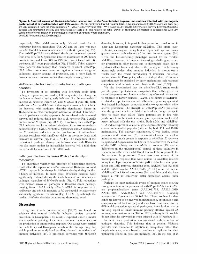
respectively. The wMel strain only delayed death for S.
typhimurium-infected mosquitoes (Fig. 2C) and the same was true
for wMelPop-CLA mosquitoes infected with B. cepacia (Fig. 2F).
The wMelPop-CLA strain delayed death and increased survival
from 0 to 10% for S. typhimurium infected mosquitoes at 200 hours
post-infection and from 38% to 79% for those infected with M.
marinum at 287 hours post-infection (Fig. 2 G&H). Taken together
these patterns demonstrate that, compared to wMel, wMelPop-
CLA offers mosquitoes protection against a broader range of
pathogens, greater strength of protection, and is more likely to
provide increased survival rather than simply delaying death.
Wolbachia infection leads to reductions in pathogendensities
To investigate if co infection with Wolbachia could limit
pathogen replication, we used qPCR to quantify the change in
the bacterial density during early infection. For the extracellular
bacteria E. carotovora (Figure 3A) and B. cepacia (Figure 3B), both
wMel and wMelPop-CLA infected mosquitoes were able to inhibit
the bacteria, with pathogen densities significantly higher in
Wolbachia-uninfected counterparts relative to infected. This differ-
ence in pathogen density appears to be correlated with increased
survival and reduced death rate due to E. carotovora (Fig. 2 A&E)
but less so for B. cepacia (Fig. 2B & F). In contrast, only wMelPop-
CLA infection results in reduced densities of the two intracellular
pathogens (Fig. 3 C&D). For both S. typhimurium and M. marinum, as
for E. carotovora, reduction in the proliferation of intracellular
bacteria correlates with significant delays in mosquito death and
increases in survival (Fig. 2 C–D, G–H). The magnitude of the
reduction in pathogen density due in association with Wolbachia
was also more modest for intracellular bacteria (,3–4 fold) than
for extracellular infections (,30–7000 fold).
Pathogen infection decreases Wolbachia density inmosquitoes
To investigate whether the presence of pathogenic bacteria
could affect the replication and/or survival of Wolbachia, we used
qPCR to quantify the change in Wolbachia density during the first
8 hours of infection. In most cases, Wolbachia densities were
significantly reduced during the early hours of infection with a
pathogen regardless of Wolbachia strain (Fig. 4). Fold reductions
were similar across all pathogen x Wolbachia strain pairings,
ranging from 1.5–2.7. Only wMelPop-CLA in response to S.
typhimurium and wMel in response to M. marinum did not experience
statistically significant reductions (Fig. 4 C & D), although the
median Wolbachia densities demonstrate decreasing trends.
Discussion
In agreement with previous reports [31,32], we found no
evidence that natural Wolbachia infection confers bacterial
protection in Drosophila. This result is expected under a model
where symbiont priming of the innate immune response leads to
the production of antimicrobial peptides. Our assays were carried
out in 3–8 day old Drosophila, which is also the age range for
which previous transcriptional profiling showed no evidence of
immune activation [24]. If protection correlates with Wolbachia
densities, however, it is possible that protection could occur in
older age Drosophila harboring wMelPop. This strain over-
replicates, causing increasing host cell lysis with age and hence
greater contact with effectors of the host immune system [34].
Given the life-shortening phenotype caused by the virulent
wMelPop, however, it becomes increasingly challenging to test
for protection in older insects and to disentangle death due to
symbiont effects from death due to the pathogen. It is becoming
increasingly evident that immune induction in mosquitoes is
results from the recent introduction of Wolbachia. Protection
against virus in Drosophila, which is independent of immune
activation, may be explained by other mechanisms such resources
competition and the natural microbiota of the host [24,33].
We also hypothesized that the wMelPop-CLA strain would
provide greater protection in mosquitoes than wMel, given the
strain’s propensity to colonise a wider array of somatic tissues and
to replicate to higher densities [17,20]. The nature of wMelPop-
CLA-induced protection was indeed broader, operating against all
four bacterial pathogens, compared to the two against which wMel
offered protection. The strength of wMelPop-CLA’s protection
was also greater, conferring higher survival and longer delays in
time to death than wMel. These patterns are in line with
predictions from the innate immune gene expression profiles of A.
aegypti infected with the two strains. Both wMel and wMelPop-
CLA induce expression of a set of genes representing the following
classes: antimicrobial peptides (initiated by both Toll and IMD),
melanisation, Toll pathway constituents, C-type lectins, serine
proteases and Transferrin [24]. In almost all cases, the level of
induction was much greater in response to wMelPop. E. carotovora,
B. cepacia and S. typhimurium are known to be sensitive to the action
of the IMD pathway and the AMPs it produces [49] and so
differences in the transcriptional control of these pathways in
response to wMel versus wMelPop-CLA could be responsible for
the variation in protection. There were also aspects of the
transcriptional response that were unique to wMelPop-infected
mosquitoes. Up-regulation of NF-kappaB Relish-like transcription
factor and IMD pathway signalling gene, AAEL007624 (3.4 fold)
and the AMP, cecropin AAEL015515 (69 fold) occurred only in
wMelPop-CLA infected mosquitoes [24], and this could also have
played a role in conferring better protection against these
pathogens.
Perhaps the most noticeable group of immune genes showing
strong induction in the presence of wMelPop-CLA but not wMel
are prophenoloxidase genes (AAEL011763, AAEL010919,
AAEL014837, AAEL006877 and AAEL011764), exhibiting
upregulation of greater than 30 fold [24]. These prophenoloxidase
genes are known to be involved in melanisation, opsonisation and
encapsulation of bacteria [50] and may have contributed to the
differential protection against all pathogens. Melanisation may be
the only aspect of innate immune priming effective against M.
marinum, as mutations in the Toll or IMD pathway in Drosophila
do not affect its survivorship when infected with M. marinum [41].
In most cases, protection was associated with reduction of
pathogen densities. This indicates that in general Wolbachia
provides true resistance to infection in mosquitoes, rather than
simply tolerance, where bacteria continue to replicate but their
pathogenic effects on hosts are mitigated [51]. In the case of B.
Figure 2. Survival curves of Wolbachia-infected (circle) and Wolbachia-uninfected (square) mosquitoes infected with pathogenicbacteria (solid) or mock infected with PBS (open). (A&E) E. carotovora, (B&F) B. cepacia, (C&G) S. typhimurium and (D&H) M. marinum. Error barsare SEM calculated from the three replicates. * P-value,0.05, ** P-value,0.01, *** P-value,0.001 denote differences in survival between Wolbachiainfected and uninfected lines by Log-rank statistics (Table S1B). The relative risk ratio [EXP(B)] of Wolbachia uninfected to infected lines with 95%confidence intervals shown in parentheses is reported on graphs where significant.doi:10.1371/journal.pntd.0002362.g002
Wolbachia-Based Bacterial Protection in Mosquitoes
PLOS Neglected Tropical Diseases | www.plosntds.org 6 August 2013 | Volume 7 | Issue 8 | e2362

cepacia, both Wolbachia infections reduced pathogen replication, but
wMel did not provide protection and wMelPop-CLA delayed
death only slightly. This pathogen is highly virulent [52], able to
avoid the melanisation response [37] and, like its close relative
Pseudomonas aeruginosa, may require only a few bacteria to kill
insects [53,54].
The wMelPop-CLA strain was better at limiting densities of
intracellular pathogens than wMel. It is not clear if this is because
wMelPop-CLA is triggering greater AMP production [24] that
would operate on intracellular pathogens when they are in the
extracellular environment, or if it differentially induces aspects of
immunity specific to the intracellular environment. Recognition
receptors that operate in the extracellular environment are well-
characterised for insects, including Peptidoglycan binding pro-
teins (PGRPs) and Gram negative binding proteins (GNBP) but
few if any equivalents for the intracellular environment have been
described [55]. The only candidate receptor for the intracellular
space is PGRP-LE given its lack of secretion signal and Toll-
independent activation of autophagy leading to the control of
Listeria monocytogenes infections [56,57]. Expression of the PGRP-
LE homolog (AAEL013112) in A. aegypti, however, was not
upregulated by either wMel or wMelPop-CLA in mosquitoes
[24].
For M. marinum while there is control of pathogen densities, the
magnitude of infection densities remains small relative to the other
pathogens. This may be due to how M. marinum colonises insects,
first establishing itself inside hemocytes with little sign of bacterial
growth before spreading systematically and causing tissue damage
[41].
Lastly, in nearly all cases, Wolbachia densities declined during
pathogen infection. This may be the direct result of innate
immune effectors elicited by the pathogens in addition to those
elicited by Wolbachia. Fold reductions in Wolbachia numbers are in
keeping with those of the intracellular pathogens that would be
exposed to the same aspects of the immune response. A related
study in the mosquito A. albopictus naturally infected with Wolbachia
also reported reductions in symbiont density after co-infection with
the vectored virus Chikungunya [58]. Alternatively, Wolbachia
reductions may spring from indirect effects of innate immune
priming. Mounting an immune response with the production of
AMPs and prophenoloxidases is costly [59]. Infection by
intracellular pathogens also carries with it the added cost of direct
competition for resources within cells. Wolbachia is highly
dependent on its host for nutrition and replication [60] and as
such co-infection with pathogens may cause Wolbachia replication
to slow due to resource limitation. Because change in Wolbachia
numbers was measured over short time periods (8–26 hrs) and
because estimates of Wolbachia’s dividing time are long
(,14 hours) [61], however, our data are more likely to provide
support for control of densities by the direct effect of the immune
response on Wolbachia.
While this study uses the transcription of the inducible immune
response in adult insects to interpret patterns of Wolbachia-
associated bacterial protection, the approach may not capture
other relevant aspects of immunity. At least one study has shown
the ability of Wolbachia infection to affect hemocyte count [62].
This constitutive aspect of immunity, defined early in development
will continue to have real effects on the performance of
phagocytosis in the adult [63]. Also, the recent transinfection of
Wolbachia into new insect hosts has been associated with increases
Figure 3. Median (with interquartile range) fold change inpathogen density variable hours post infection (hpi) inmosquitoes. Five pairs of individuals were used for E. carotovora (A),
B. cepacia (B), S. typhimurium (C) and M. marinum (D). (Mann-Whitney U-test; * P-value,0.05, ** P-value,0.01).doi:10.1371/journal.pntd.0002362.g003
Wolbachia-Based Bacterial Protection in Mosquitoes
PLOS Neglected Tropical Diseases | www.plosntds.org 7 August 2013 | Volume 7 | Issue 8 | e2362

in autophagy [64] and the generation of reactive oxygen species
[65], both of which may not be captured in transcriptional
measures.
Our findings have the following implications for use of
Wolbachia as a biocontrol agent in A. aegypti. Firstly, different
Wolbachia strains may vary substantially in the immune priming
they induce. As the efficacy of Wolbachia is being trialled as a
dengue control agent around the world, one of the main decision
points going forward will be which strain(s) to deploy. A full
understanding of the strain-based differences in pathogen
protection and fitness effects will aid in that decision. Secondly,
bacterial protection may be affecting mosquito fitness in the field.
Recent studies have shown that gut flora can play a role in insect
nutrition [66], behaviour [67] and ability to vector pathogens
[26]. It is possible that innate immune priming may be altering
the composition of the gut microbiome. Priming may also provide
protection against natural infections in the wild and assist with
spread and maintenance of the symbiont. A field-based assess-
ment of the performance of wMel and wMelPop with respect to
native pathogen control is in order although it is difficult to
sample rare and sickly insects in wild populations with systemic
bacterial infections [68].
Lastly, immune priming induced by Wolbachia may also provide
a mechanistic explanation for protection against Plasmodium
gallinaceum, as there appears to be greater evidence of Imd, Toll,
opsonisation and melanisation involvement in control of this
parasite than there is for viruses [18,27,28,29]. Dengue represents
the test case for use of Wolbachia for pathogen protection. Given
that malaria cases outnumber dengue by at least 10-fold [69], the
potential rewards for developing the symbiont for malaria vectors
are great [70]. As the technical challenges around infecting the
host are solved [71], the need to understand the basis of pathogen
blocking becomes immediate [22,72].
ConclusionsOur findings support previous studies indicating that native
Wolbachia infections in D. melanogaster do not confer pathogen
protection against bacteria. In the recently transinfected A. aegypti,
in contrast, we demonstrate pathogen protection that varies by
strain, with wMelPop-CLA exhibiting more effective protection
than wMel against a broader range of bacteria. We also provide
evidence that the expression of innate immunity genes induced by
Wolbachia infection in mosquitoes likely explains these differences
in protection. Future work will need to identify the potential role
for innate immune priming as an enhancer of viral protection,
assess whether bacterial protection is providing benefit for
mosquitoes in the field. These findings may assist with Wolbachia
strain selection for field release.
Supporting Information
Table S1 Adjusted P-Values of log-rank statistics (Mantel-Cox)
comparing the effect of Wolbachia infection or the Wolbachia strain
on survival of bacterial infection in A) flies and B) mosquitoes. * P-
value,0.05, ** P-value,0.01, *** P-value,0.001.
(DOCX)
Figure 4. Median (with interquartile range) relative Wolbachiadensity after infection in mosquitoes. Five pairs of individualswere used for E. carotovora (A), B. cepacia (B), S. typhimurium (C) and M.marinum (D). (Mann-Whitney U-test; * P-value,0.05, ** P-value,0.01).doi:10.1371/journal.pntd.0002362.g004
Wolbachia-Based Bacterial Protection in Mosquitoes
PLOS Neglected Tropical Diseases | www.plosntds.org 8 August 2013 | Volume 7 | Issue 8 | e2362

Acknowledgments
We thank Nichola Kenny for technical assistance and Adam Jenney of
Monash University Department of Medicine/The Alfred Hospital for the
B. cepacia strain.
Author Contributions
Conceived and designed the experiments: YHY SLO EAM. Performed the
experiments: YHY ER. Analyzed the data: YHY MW EAM. Contributed
reagents/materials/analysis tools: MW. Wrote the paper: YHY MW ER
SLO EAM.
References
1. Zug R, Hammerstein P (2012) Still a host of hosts for Wolbachia: analysis of
recent data suggests that 40% of terrestrial arthropod species are infected. PloSOne 7: e38544.
2. O’Neill SL, Hoffmann AA, Werren JH (1997) Influential Passengers: Inherited
Microorganisms amd Arthropod Reproduction. (Oxford Univ Press, Oxford).
3. Werren JH, Baldo L, Clark ME (2008) Wolbachia: master manipulators of
invertebrate biology. Nat Rev Microbiol 6: 741–751.
4. Hoffmann AA, Hercus M, Dagher H (1998) Population dynamics of the
Wolbachia infection causing cytoplasmic incompatibility in Drosophila melanoga-
ster. Genetics 148: 221–231.
5. Calvitti M, Moretti R, Porretta D, Bellini R, Urbanelli S (2009) Effects on male
fitness of removing Wolbachia infections from the mosquito Aedes albopictus. MedVet Entomol 23: 132–140.
6. Friberg U, Miller PM, Stewart AD, Rice WR (2011) Mechanisms promoting thelong-term persistence of a Wolbachia infection in a laboratory-adapted population
of Drosophila melanogaster. PloS One 6: e16448.
7. Weeks AR, Turelli M, Harcombe WR, Reynolds KT, Hoffmann AA (2007)
From parasite to mutualist: rapid evolution of Wolbachia in natural populations of
Drosophila. PLoS Biol 5: e114.
8. Gavotte L, Mercer DR, Stoeckle JJ, Dobson SL (2010) Costs and benefits of
Wolbachia infection in immature Aedes albopictus depend upon sex andcompetition level. J Invertebr Pathol 105: 341–346.
9. Harcombe W, Hoffmann AA (2004) Wolbachia effects in Drosophila melanogaster: in
search of fitness benefits. J Invertebr Pathol 87: 45–50.
10. Brelsfoard CL, Dobson SL (2011) Wolbachia effects on host fitness and the
influence of male aging on cytoplasmic incompatibility in Aedes polynesiensis
(Diptera: Culicidae). J Med Entomol 48: 1008–1015.
11. Rasgon JL (2012) Wolbachia induces male-specific mortality in the mosquito Culex
pipiens (LIN strain). PloS One 7: e30381.
12. Champion de Crespigny FE, Wedell N (2006) Wolbachia infection reduces sperm
competitive ability in an insect. Proc Biol Sci 18.
13. Teixeira L, Ferreira A, Ashburner M (2008) The bacterial symbiont Wolbachia
induces resistance to RNA viral infections in Drosophila melanogaster. PLoS Biol 6:e2.
14. Hedges LM, Brownlie JC, O’Neill SL, Johnson KN (2008) Wolbachia and virusprotection in insects. Science 322: 702–702.
15. Osborne SE, Leong YS, O’Neill SL, Johnson KN (2009) Variation in antiviral
protection mediated by different Wolbachia strains in Drosophila simulans. PLoSPathog 5: e1000656.
16. McMeniman CJ, Lane RV, Cass BN, Fong AW, Sidhu M, et al. (2009) Stableintroduction of a life-shortening Wolbachia infection into the mosquito Aedes
aegypti. Science 323: 141–144.
17. Walker T, Johnson PH, Moreira LA, Iturbe-Ormaetxe I, Frentiu FD, et al.
(2011) The wMel Wolbachia strain blocks dengue and invades caged Aedes aegypti
populations. Nature 476: 450–U101.
18. Kambris Z, Blagborough AM, Pinto SB, Blagrove MS, Godfray HC, et al.
(2010) Wolbachia stimulates immune gene expression and inhibits plasmodiumdevelopment in Anopheles gambiae. PLoS Pathog 6: e1001143.
19. Bian G, Xu Y, Lu P, Xie Y, Xi Z (2010) The endosymbiotic bacterium Wolbachia
induces resistance to dengue virus in Aedes aegypti. PLoS Pathog 6: e1000833.
20. Moreira LA, Iturbe-Ormaetxe I, Jeffery JA, Lu G, Pyke AT, et al. (2009) A
Wolbachia symbiont in Aedes aegypti limits infection with dengue, Chikungunya,and Plasmodium. Cell 139: 1268–1278.
21. Kambris Z, Cook PE, Phuc HK, Sinkins SP (2009) Immune activation by life-shortening Wolbachia and reduced filarial competence in mosquitoes. Science
326: 134–136.
22. Hughes GL, Koga R, Xue P, Fukatsu T, Rasgon JL (2011) Wolbachia infectionsare virulent and inhibit the human malaria parasite Plasmodium falciparum in
Anopheles gambiae. PLoS Pathog 7: e1002043.
23. Hoffmann AA, Montgomery BL, Popovici J, Iturbe-Ormaetxe I, Johnson PH, et
al. (2011) Successful establishment of Wolbachia in Aedes populations to suppressdengue transmission. Nature 476: 454–U107.
24. Rances E, Ye YH, Woolfit M, McGraw EA, O’Neill SL (2012) The relative
importance of innate immune priming in Wolbachia-mediated dengue interfer-ence. PLoS Pathog 8: e1002548.
25. Hoffmann JA, Reichhart JM (2002) Drosophila innate immunity: an evolutionaryperspective. Nat Immunol 3: 121–126.
26. Ramirez JL, Souza-Neto J, Torres Cosme R, Rovira J, Ortiz A, et al. (2012)
Reciprocal tripartite interactions between the Aedes aegypti midgut microbiota,innate immune system and dengue virus influences vector competence. PLoS
Negl Trop D 6: e1561.
27. Pinto SB, Lombardo F, Koutsos AC, Waterhouse RM, McKay K, et al. (2009)
Discovery of Plasmodium modulators by genome-wide analysis of circulatinghemocytes in Anopheles gambiae. Proc Natl Acad Sci U S A 106: 21270–21275.
28. Zou Z, Shin SW, Alvarez KS, Bian G, Kokoza V, et al. (2008) Mosquito
RUNX4 in the immune regulation of PPO gene expression and its effect on
avian malaria parasite infection. Proc Natl Acad Sci U S A 105: 18454–18459.
29. Meister S, Koutsos AC, Christophides GK (2004) The Plasmodium parasite–a
‘new’ challenge for insect innate immunity. Int J Parasitol 34: 1473–1482.
30. Garver LS, Bahia AC, Das S, Souza-Neto JA, Shiao J, et al. (2012) Anopheles
Imd pathway factors and effectors in infection intensity-dependent anti-
Plasmodium action. PLoS Pathog 8: e1002737.
31. Rottschaefer SM, Lazzaro BP (2012) No effect of Wolbachia on resistance to
intracellular infection by pathogenic bacteria in Drosophila melanogaster. PLoS One
7: e40500.
32. Wong ZS, Hedges LM, Brownlie JC, Johnson KN (2011) Wolbachia-mediated
antibacterial protection and immune gene regulation in Drosophila. PLoS One
6: e25430.
33. Xi Z, Ramirez JL, Dimopoulos G (2008) The Aedes aegypti toll pathway controls
dengue virus infection. PLoS Pathog 4: e1000098.
34. Min KT, Benzer S (1997) Wolbachia, normally a symbiont of Drosophila, can be
virulent, causing degeneration and early death. Proc Natl Acad Sci U S A 94:
10792–10796.
35. Turley AP, Moreira LA, O’Neill SL, McGraw EA (2009) Wolbachia infection
reduces blood-feeding success in the dengue fever mosquito, Aedes aegypti. PLoS
Negl Trop D 3: e516.
36. Basset A, Khush RS, Braun A, Gardan L, Boccard F, et al. (2000) The
phytopathogenic bacteria Erwinia carotovora infects Drosophila and activates an
immune response. Proc Natl Acad Sci USA 97: 3376–3381.
37. Schneider DS, Ayres JS, Brandt SM, Costa A, Dionne MS, et al. (2007)
Drosophila eiger mutants are sensitive to extracellular pathogens. PLoS Pathog 3:
e41.
38. Pham LN, Dionne MS, Shirasu-Hiza M, Schneider DS (2007) A specific primed
immune response in Drosophila is dependent on phagocytes. PLoS Pathog 3: e26.
39. Yamada R, Iturbe-Ormaetxe I, Brownlie JC, O’Neill SL (2011) Functional test
of the influence of Wolbachia genes on cytoplasmic incompatibility expression in
Drosophila melanogaster. Insect Mol Biol 20: 75–85.
40. McMeniman CJ, Lane AM, Fong AW, Voronin DA, Iturbe-Ormaetxe I, et al.
(2008) Host adaptation of a Wolbachia strain after long-term serial passage in
mosquito cell lines. Appl Environ Microbiol 74: 6963–6969.
41. Dionne MS, Ghori N, Schneider DS (2003) Drosophila melanogaster is a genetically
tractable model host for Mycobacterium marinum. Infect Immun 71: 3540–3550.
42. Tang H, Kambris Z, Lemaitre B, Hashimoto C (2006) Two proteases defining a
melanization cascade in the immune system of Drosophila. J Biol Chem 281:
28097–28104.
43. Storey JD, Dai JY, Leek JT (2007) The optimal discovery procedure for large-
scale significance testing, with applications to comparative microarray
experiments. Biostatistics 8: 414–432.
44. Postollec F, Falentin H, Pavan S, Combrisson J, Sohier D (2011) Recent
advances in quantitative PCR (qPCR) applications in food microbiology. Food
Microbiol 28: 848–861.
45. Rozen S, Skaletsky HJ, editors (2000) Primer3 on the WWW for general users
and for biologist programmers. Totowa, NJ: Humana Press.
46. Moreira LA, Ye YH, Turner K, Eyles DW, McGraw EA, et al. (2011) The
wMelPop strain of Wolbachia interferes with dopamine levels in Aedes aegypti.
Parasit Vectors 4: 28.
47. Cook PE, Hugo LE, Iturbe-Ormaetxe I, Williams CR, Chenoweth SF, et al.
(2006) The use of transcriptional profiles to predict adult mosquito age under
field conditions. Proc Natl Acad Sci U S A 103: 18060–18065.
48. Simon P (2003) Q-Gene: processing quantitative real-time RT-PCR data.
Bioinformatics 19: 1439–1440.
49. Shirasu-Hiza MM, Schneider DS (2007) Confronting physiology: how do
infected flies die? Cell Microbiol 9: 2775–2783.
50. Cerenius L, Soderhall K (2004) The prophenoloxidase-activating system in
invertebrates. Immunol Rev 198: 116–126.
51. Schneider DS, Ayres JS (2008) Two ways to survive infection: what resistance
and tolerance can teach us about treating infectious diseases. Nat Rev Immunol
8: 889–895.
52. Tegos GP, Haynes MK, Schweizer HP (2012) Dissecting novel virulent
determinants in the Burkholderia cepacia complex. Virulence 3: 234–237.
53. Boman HG (1991) Antibacterial peptides: key components needed in immunity.
Cell 65: 205–207.
54. D’Argenio DA, Gallagher LA, Berg CA, Manoil C (2001) Drosophila as a model
host for Pseudomonas aeruginosa infection. J Bacteriol 183: 1466–1471.
55. Yano T, Kurata S (2011) Intracellular recognition of pathogens and autophagy
as an innate immune host defence. J Biochem 150: 143–149.
Wolbachia-Based Bacterial Protection in Mosquitoes
PLOS Neglected Tropical Diseases | www.plosntds.org 9 August 2013 | Volume 7 | Issue 8 | e2362

56. Kaneko T, Yano T, Aggarwal K, Lim JH, Ueda K, et al. (2006) PGRP-LC and
PGRP-LE have essential yet distinct functions in the Drosophila immune responseto monomeric DAP-type peptidoglycan. Nat Immunol 7: 715–723.
57. Yano T, Mita S, Ohmori H, Oshima Y, Fujimoto Y, et al. (2008) Autophagic
control of listeria through intracellular innate immune recognition in Drosophila.Nat Immunol 9: 908–916.
58. Zouache K, Michelland RJ, Failloux AB, Grundmann GL, Mavingui P (2012)Chikungunya virus impacts the diversity of symbiotic bacteria in mosquito
vector. Mol Ecol 21: 2297–2309.
59. Ye YH, Chenoweth SF, McGraw EA (2009) Effective but costly, evolvedmechanisms of defense against a virulent opportunistic pathogen in Drosophila
melanogaster. PLoS Pathog 5: e1000385.60. Wu M, Sun LV, Vamathevan J, Riegler M, Deboy R, et al. (2004)
Phylogenomics of the reproductive parasite Wolbachia pipientis wMel: astreamlined genome overrun by mobile genetic elements. PLoS Biol 2: E69.
61. Fenollar F, Maurin M, Raoult D (2003) Wolbachia pipientis growth kinetics and
susceptibilities to 13 antibiotics determined by immunofluorescence staining andreal-time PCR. Antimicrob Agents Chemother 47: 1665–1671.
62. Braquart-Varnier C, Lachat M, Herbiniere J, Johnson M, Caubet Y, et al.(2008) Wolbachia mediate variation of host immunocompetence. PLoS One 3:
e3286.
63. Haine ER, Moret Y, Siva-Jothy MT, Rolff J (2008) Antimicrobial Defense andPersistent Infection in Insects. Science 322: 1257–1259.
64. Le Clec’h W, Braquart-Varnier C, Raimond M, Ferdy JB, Bouchon D, et al.(2012) High virulence of Wolbachia after host switching: when autophagy hurts.
PLoS Pathog 8: e1002844.
65. Andrews ES, Crain PR, Fu Y, Howe DK, Dobson SL (2012) Reactive Oxygen
Species Production and Brugia pahangi Survivorship in Aedes polynesiensis with
artificial Wolbachia infection types. PLoS Pathog 8: e1003075.
66. Ridley EV, Wong AC, Westmiller S, Douglas AE (2012) Impact of the resident
microbiota on the nutritional phenotype of Drosophila melanogaster. PLoS One 7:
e36765.
67. Sharon G, Segal D, Ringo JM, Hefetz A, Zilber-Rosenberg I, et al. (2010)
Commensal bacteria play a role in mating preference of Drosophila melanogaster.
Proc Natl Acad Sci U S A 107: 20051–20056.
68. Juneja P, Lazzaro BP (2009) Providencia sneebia sp. nov. and Providencia
burhodogranariea sp. nov., isolated from wild Drosophila melanogaster. Int J Syst
Evol Microbiol 59: 1108–1111.
69. WHO (2004) World Health Report 2004: Changing History. Geneva,
Switzerland: World Health Organization.
70. Rasgon JL, Ren X, Petridis M (2006) Can Anopheles gambiae be infected with
Wolbachia pipientis? Insights from an in vitro system. Appl Environ Microbiol 72:
7718–7722.
71. Bian G, Joshi D, Dong Y, Lu P, Zhou G, et al. (2013) Wolbachia invades Anopheles
stephensi populations and induces refractoriness to Plasmodium infection. Science
340: 748–751.
72. Hughes GL, Vega-Rodriguez J, Xue P, Rasgon JL (2012) Wolbachia strain
wAlbB enhances infection by the rodent malaria parasite Plasmodium berghei in
Anopheles gambiae mosquitoes. Appl Environ Microbiol 78: 1491–1495.
Wolbachia-Based Bacterial Protection in Mosquitoes
PLOS Neglected Tropical Diseases | www.plosntds.org 10 August 2013 | Volume 7 | Issue 8 | e2362