Embed Size (px)
Citation preview
Proc. Nati. Acad. Sci. USAVol. 85, pp. 6232-6236, September 1988Biochemistry
Mouse lysozyme M gene: Isolation, characterization, andexpression studies
(cDNA/macrophage/lysozyme P/tisue specificity/a-lactalbumin)
MICHAEL CROSS, INGE MANGELSDORF, ANGELA WEDEL, AND RAINER RENKAWITZ*
Genzentrum, Max Planck Institut fMr Biochemie, D8033 Martinsried, Federal Republic of Germany
Communicated by M. Lindauer, May 9, 1988 (received for review January 27, 1988)
ABSTRACT We have isolated and characterized bothcDNA and genomic DNA of the mouse lysozyme M gene.Derivation of the amino acid sequence from the nucleotidesequences revealed six positions in the carboxyl terminus thatdiffer from partial sequences previously published. The differ-ential detection of specific mRNAs from the closely relatedlysozymeM and P genes has revealed different but overlappingtissue specificities of expression. The M gene is expressedweakly in myeloblasts, moderately in immature macrophages,and strongly in both mature macrophages and macrophage-rich tissues, while high levels of P transcripts are present onlyin small intestine. Sites of protein accumulation, rather thangene expression, have been identified by comparative quanti-tation of mRNA and enzyme levels.
Since the discovery in 1922 of lysozyme (EC 3.2.1.17) via itsantibacterial effects (1), the enzyme has been adopted as amodel for studies on protein structure and molecular evolution(for review, see ref. 2), while the lysozyme gene has been thesubject of investigations into the determinants of tissue spec-ificity and hormonal control of gene expression (3-6). Com-parison of lysozyme sequences with those of a-lactalbuminhave implied an evolutionary relationship between theseproteins (7), a hypothesis that has been strengthened by thedemonstration of strong similarities within the crystal struc-tures of the proteins (8) and the sequences and arrangementsof their respective genes (9). The involvement of lysozyme inhost defense has prompted numerous studies of the tissuedistribution of lysozyme enzyme activity in animals in bothnormal and pathogenic states. Hence, deviations in lysozymelevels of blood and urine, for example, have been correlatedto particular myeloid and renal abnormalities, and lysozymelevels have subsequently been used to monitor the success oftherapy (10, 11). Although the lysozyme protein can be foundin most tissues, studies at the cellular level have revealed highlevels of lysozyme specifically in phagocytic cells, includingmacrophages, granulocytes, and the Paneth cells of the smallintestine, and in the proximal tubules of kidney (12, 13). Whilethe kidney accumulates at least some of its lysozyme from theblood (14), endogenous lysozyme synthesis has been detectedin mammalian myeloid cells (15). The recent discovery ofhighlevels of intestinal lysozyme in some species ofhouse mice, inwhich the enzyme may have assumed a digestive role, hasrecently stressed the involvement of regulatory mutations inevolution (16, 17). In this case, a regulatory mutation appearsto have affected only one of two closely related lysozymegenes, resulting in a specific overexpression of lysozyme P inthe small intestine and leaving normal expression oflysozymeM in other tissues tested.
Studies on the expression of the chicken lysozyme genehave described particular DNA sequences that demonstrate
a positive or negative effect on gene expression in a macro-phage-specific manner (4-6) and others that bind steroidhormone receptors and mediate hormonal control in oviductcells (3). Our intention is to extend studies of lysozyme generegulation to the mouse, from which the availability of anumber of myeloid cell lines representing different stages ofmyeloid differentiation and the possibility of generatingchimeric or transgenic animals present an extremely versatilesystem. We present here the isolation and characterization ofthe mouse lysozymeM gene.t We have determined the tissuedistribution of both lysozyme protein and mRNA, permittinga distinction to be made between sites ofgene expression andprotein accumulation, and describe a strong dependence oflevels of lysozyme gene expression on the stage of differen-tiation of myeloid cells. Successful resolution of the mRNAsfrom the lysozymeM and P genes has facilitated independentdeterminations of their tissue specificities of expression.
MATERIALS AND METHODSIsolation of Mouse Lysozyme M cDNA and Genomic Clones.
The spleen of a mouse (Mus domesticus) sacrificed 14 daysafter infection with AF1 virus (18) was kindly provided by W.Ostertag (University of Hamburg, Hamburg, F.R.G.). Poly-adenylylated RNA was purified from this tissue by the LiClprecipitation method (19) and subsequent selection on oli-go(dT)-cellulose. A cDNA bank was constructed in AgtlO bythe method ofGubler and Hoffman (20). A screen ofduplicateplaque lifts on GeneScreen membranes (New England Nu-clear) was performed under the manufacturer's recommendedconditions with nick-translated probes from part of a humanlysozyme cDNA, kindly provided by P. Swetly (Ernst Boeh-ringer Institute, Vienna). Hybridization was carried out in 40%oformamide/0.75 M NaCl/75 mM sodium citrate at 50(C.
Fractions of a mouse (M. domesticus; tAE5/1295v) genebank in vector AEMBL3, kindly provided by B. Hermann(European Molecular Biology Laboratory, Heidelberg), werescreened with nick-translated probes from the isolated mouselysozymeM cDNA sequences. Hybridization was performedat 420C in 50%o formamide/0.75 M NaCl/75 mM sodiumcitrate. All standard DNA manipulations were carried outaccording to Maniatis et al. (21) unless otherwise indicated.
Sequencing. Plasmid DNA sequencing by the chain-termination method (22) was performed with a Sequenase kit(United States Biochemical, Cleveland). All reported se-quences were determined independently from both DNAstrands. Sequence analysis was performed with softwarefrom the University ofWisconsin Genetics Computer Group.
Analysis of Mouse Lysozyme mRNA. RNA prepared fromcell lines or M. domesticus BALB/c tissues by the guani-dinium isothiocyanate method was electrophoresed through
*To whom reprint requests should be addressed.tThis sequence is being deposited in the EMBL/GenBank data base(IntelliGenetics, Mountain View, CA, and Eur. Mol. Biol. Lab.,Heidelberg) (accession no. J03846).
6232
The publication costs of this article were defrayed in part by page chargepayment. This article must therefore be hereby marked "advertisement"in accordance with 18 U.S.C. §1734 solely to indicate this fact.
Dow
nloa
ded
by g
uest
on
Apr
il 29
, 202
0
Proc. Natl. Acad. Sci. USA 85 (1988) 6233
1.2% agarose/2.2 M formaldehyde denaturing gels (21), withRNA molecular weight markers (Bethesda Research Labo-ratories). RNA was blotted to GeneScreen membranes,which were subsequently stained with methylene blue toconfirm quantitative transfer (23). Comparative quantitationof lysozyme-specific message in samples of total RNA wasachieved by dot blot titration on GeneScreen membranesunder conditions recommended by the manufacturer. Hy-bridization to nick-translated probes was carried out in 50%oformamide/1 M NaCl at 42°C. Primer extension reactionswere performed by the method of Ghosh et al. (24) with a32-nucleotide primer complementary to part of the mouselysozyme M mRNA. Sequencing reaction mixtures primedwith the same oligonucleotide were used as size markers.Oligonucleotides for both primer extension and sequencingwere synthesized in the laboratory of R. Mertz (Max PlanckInstitut Genzentrum, Martinsried, F.R.G.).RNADNA hybrid analysis by S1 nuclease digestion was
carried out essentially according to Weaver and Weissmann(25). RNARNA hybrids were analyzed by RNase A/RNaseT1 digestion (26).Lysozyme Enzyme Assays. The determination of lysozyme
activity was performed by the lysoplate assay of Ossermanand Lawlor (27) with freshly frozen tissues ground underliquid nitrogen or with freeze-thaw cell lysates. Total proteinconcentrations were determined by using the protein assayreagent and protocol from Bio-Rad.
Cells. The mouse myeloblast cell line Ml (28); immaturemacrophage cell lines WEHI3 and WEHI3B (29); maturemacrophage lines P388D1 (30), J774 (31), HA32, and HA38(AF1 virus-transformed, a gift from W. Ostertag); and thefibroblast cell line Ltk- (32) were grown in Dulbecco'smodified Eagle's medium supplemented with fetal calf serumto 20% (HA32 and HA38) or 10%o (all others). Primarymacrophages were derived from bone marrow by the methodof Stewart (33) and were used after 14 days in culture ontissue culture unprepared Petri dishes.
RESULTS
Mouse LysozymeM cDNA. Screening a fraction of a mousespleen cDNA library with human lysozyme cDNA sequences
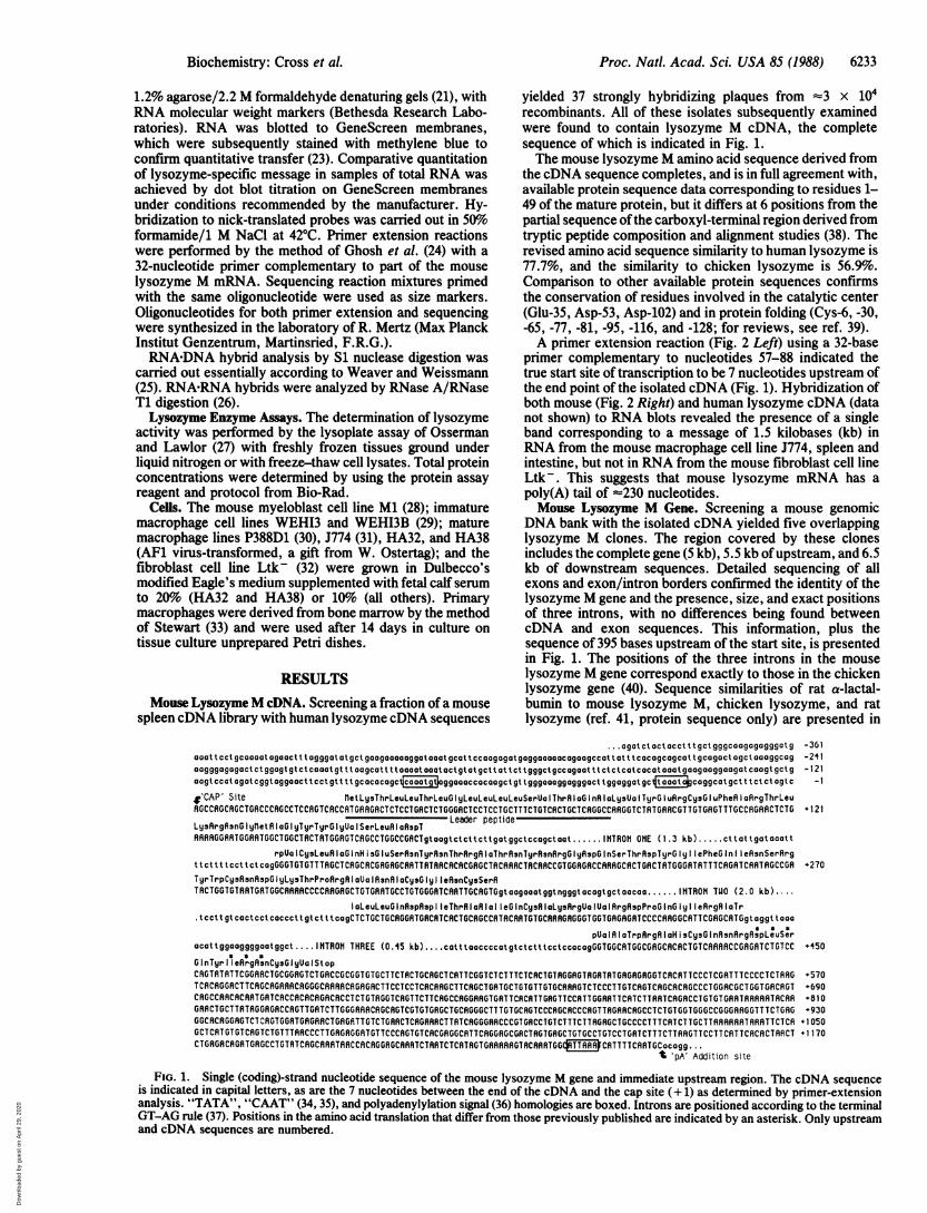
yielded 37 strongly hybridizing plaques from =3 x 10⁴recombinants. All of these isolates subsequently examinedwere found to contain lysozyme M cDNA, the completesequence of which is indicated in Fig. 1.The mouse lysozyme M amino acid sequence derived from
the cDNA sequence completes, and is in full agreement with,available protein sequence data corresponding to residues 1-49 of the mature protein, but it differs at 6 positions from thepartial sequence ofthe carboxyl-terminal region derived fromtryptic peptide composition and alignment studies (38). Therevised amino acid sequence similarity to human lysozyme is77.7%, and the similarity to chicken lysozyme is 56.9%.Comparison to other available protein sequences confirmsthe conservation of residues involved in the catalytic center(Glu-35, Asp-53, Asp-102) and in protein folding (Cys-6, -30,-65, -77, -81, -95, -116, and -128; for reviews, see ref. 39).A primer extension reaction (Fig. 2 Left) using a 32-base
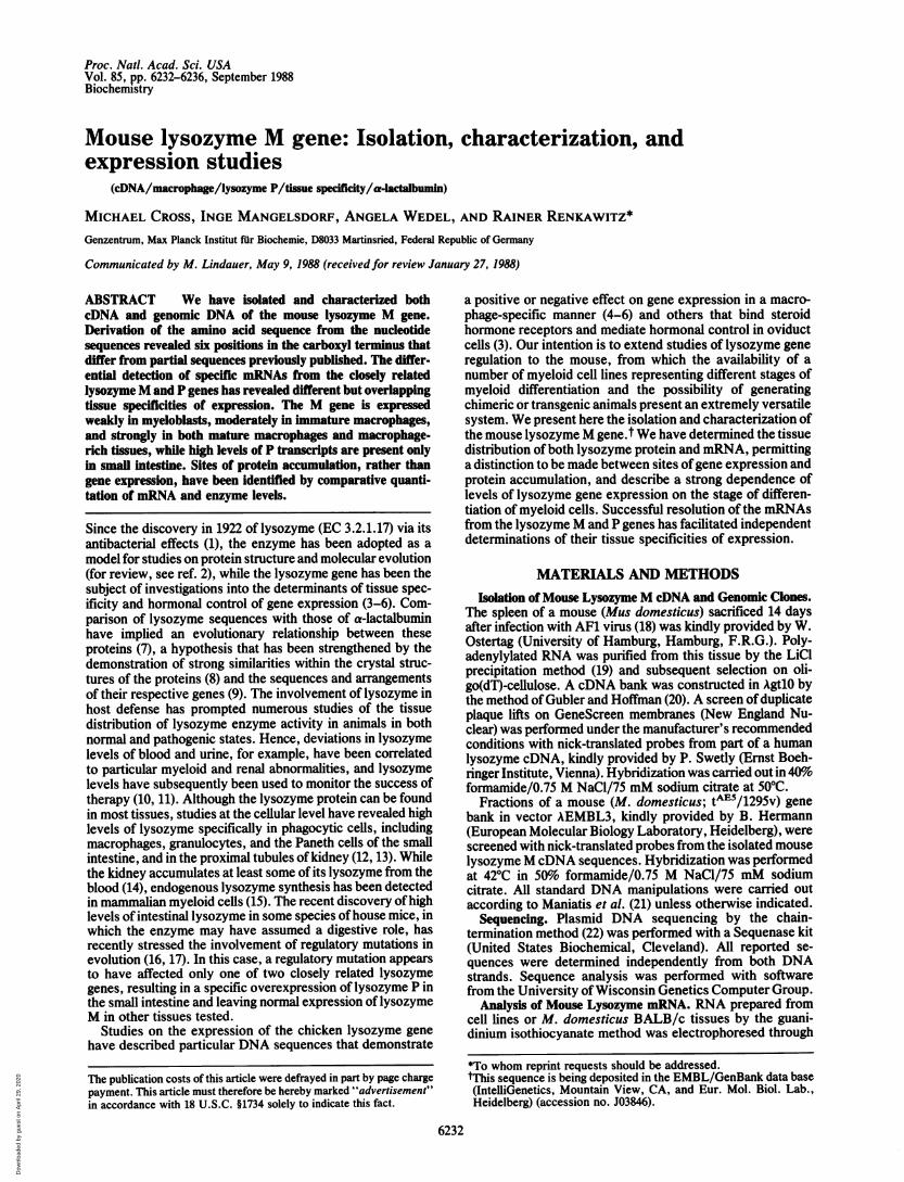
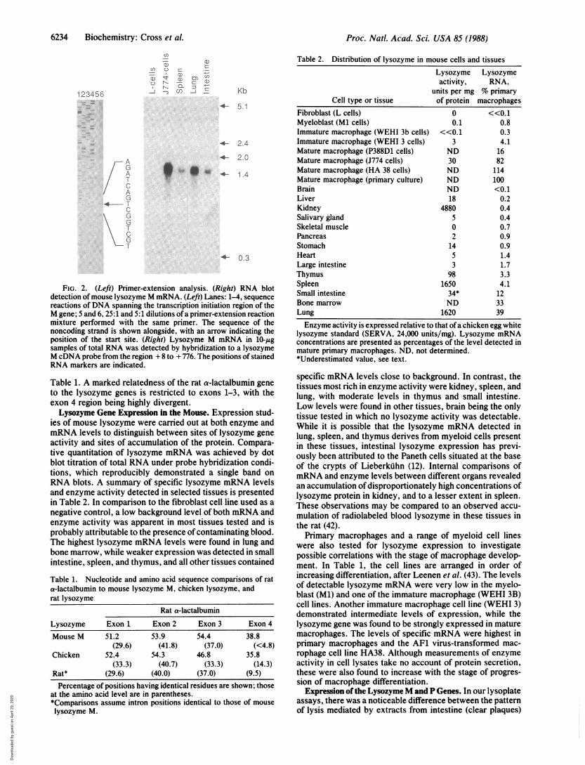
primer complementary to nucleotides 57-88 indicated thetrue start site of transcription to be 7 nucleotides upstream ofthe end point of the isolated cDNA (Fig. 1). Hybridization ofboth mouse (Fig. 2 Right) and human lysozyme cDNA (datanot shown) to RNA blots revealed the presence of a singleband corresponding to a message of 1.5 kilobases (kb) inRNA from the mouse macrophage cell line J774, spleen andintestine, but not in RNA from the mouse fibroblast cell lineLtk-. This suggests that mouse lysozyme mRNA has apoly(A) tail of %'-230 nucleotides.Mouse Lysozyme M Gene. Screening a mouse genomic
DNA bank with the isolated cDNA yielded five overlappinglysozyme M clones. The region covered by these clonesincludes the complete gene (5 kb), 5.5 kb ofupstream, and 6.5kb of downstream sequences. Detailed sequencing of allexons and exon/intron borders confirmed the identity of thelysozyme M gene and the presence, size, and exact positionsof three introns, with no differences being found betweencDNA and exon sequences. This information, plus thesequence of 395 bases upstream of the start site, is presentedin Fig. 1. The positions of the three introns in the mouselysozyme M gene correspond exactly to those in the chickenlysozyme gene (40). Sequence similarities of rat a-lactal-bumin to mouse lysozyme M, chicken lysozyme, and ratlysozyme (ref. 41, protein sequence only) are presented in
... agatctactacctttgctgggcoagagagggotg -361aaat tcct gcaaaat agaact t tagggat at gct gaagaaaaaggaootatgcat tccaagagat gaggaaaaaocagaagccat tat t tccacgcogcat t gcgact agct aaaggcag -2ilGagggogagact ctggagt gt ctcaaat gt t t Gagcot t t toaaat aaat actgt at gct t at t ct tgggct gccagaat t t ct ct cat cocatoaat gaagaaggoagat caogt gct g -121gagtccat gatcggtoggaact t Cct gt t t t gcacacagctiaagtoggaaaccacaagct gt tgggaaaggagggact tggaggat gcftaa0atagcoggcot gct t tct ct ogt c -1
,v'CAP' Site n1etLy3ThrLeuLeuThrLeuGlIyLeuLeuLeuLeuSerUa IThrR IaG InR Ly3UalITyrG IuflrgCy3G IuPheR argThrLeuRGCCRGCRGCTGRCCCRGCCTCCRGTCRCCRTGRRGRCTCTCCTGRCTCTGGGRCTCCTCCTGCTTTCTGTCRCTGCTCRGGCCRRGGTCTRTGRRCGTTGTGRGT TTGCCRGRRCTCTG + 121
Ly3ArqAsnG Iyllet R GIylyrT rGlIyUalISerLeuR I aRpT Leader peptide-
rpUalICy3LeuR IaGlInH isG IuSerRsnTyrRsnThr~rgR IaThrflsnTyrRsnfirgGlIyAspG InSerThrRspTyrGlIy IePheG nIlIeAsnSerflrgttcttttccttctcoyvvGGGTGTGTTTRGCTnCAGCnACGRGRGCRATTATAACACRCGRGnunnTRnXuut TtcRAGGGTGGAGRRGGRAAGTCAcTGAacTRTGGGATRTTTRCAGATRGGAATTCGCCGRGC -241TyrTrpCysRsnRspGlyLysThrProRrgAlaUalRsnRlaCysGIy lIeRsnCysSerRTACTGGTGTRRTGRTGGCARARCCCCRAGRGCTGTGRRTGCCTGTGGGRTCRATTGCRGTGgt aagaoat ggt ngggt acagtgct aacaa ...... TO TUO (2.0 kb)....
IoLeuLeuGInRspRspl IeThrRIaRIaI IeGInCys9IaLys9rgUalJaIArgRspProGInGIy IIeRrgRIaTr.tcct t gt cactcct caccct t gt ct ttcagCTCTGCTGCRGGRTGRCATCRCTGCRGCCATACRATGTGCRARGRGGGTGGTGRGRGRTCCCCRRGGCRTTCGRGCRTGgt aggt t ooo
pUa IA aTrpRrgA aH sCysG nRsnArgApLuSracattggaaggggaatggct.. I NTRON THREE (0.45 kb) .... cattt accccatgtctctttcctccocagGGTGGCATGGCGRGCACRCTGTCARAARCCGAGRTCTGTCC +450G nTyr 11 eRrgRsnCysG yUa 1St opCRGTRTRTTCGGRRCTGCGGAGTCTGRCCGCGGTGTGCTTCTACTGCRGCTCRTTCGGTCTCTTTCTCRCTGTRGGRGTRGRTRTGAGAGRGGTCRCRT TCCCTCGRTTTCCCCTCTR'G+570T CRCRGGRCT TCRGCRGRAACRGGGCRRRRCAGAGRCTTCCTCCTCRCRAGCTTCRGCTGRTGCTGTGTT GTGCRRRGTCTCCCT TGTCRGTCAGCRCRGCC CTGGACGCTGGTGACRGT +690CRGCCRRCRCRRTGR TCRCCRCACAGACRCCT CTGTRGGTCRGT TCTTCRGCCAGGRRGTGRT TCRCRTT GRGTTCCR TT GGRATTCRTCTT RRTCRGRCCT GTGT GRRTRRRRRTRCRR +810GAR CTGCTTATRGGAGRCCRGTTGR TCTTGGGRRRCRGCAGTCGTGTGRGCTGCRGGGCTT TGTGCRGTCCCRGCRCCCRGTTRGRRCRGCCTCTGT GGTGGGCCGGGRRGGTT TCTGRG + 930GGCRCRGGRGTCI wRGTGGRTGRGAACTGRGRTTGTCTGRRCTCRGRRRCTTRTCRGGGRRCCCGT GRCCTGTCTTTCTTAGRGCTGCCCCTTTCRT CT TGCTTRRAARTRRATTCT CA +1 OSOGCTCAT GT GTCRGTCTGTTTRRCCCTTGAG GGRTGTTCCCRGTGTCACGRGGCRTTCRGGRGCGA CTRGTGRGCTGTGCCTGTCCTGATCTTTCTTRRGTTCCTT CAT TCACACTAACT + 1170CTGRG CA GATGAGCCTGTATCRGCRRflTA CCRCAGGRGCRRRTCTRATCTCATRGTGARRR RGTRCAARTGG*TTRR~lCRTTTTCA RTGCcacgg ..
't 'pA' Addition site
FIG. 1. Single (coding)-strand nucleotide sequence of the mouse Iysozyme M gene and immediate upstream region. The cDNA sequenceis indicated in capital letters, as are the 7 nucleotides between the end of the cDNA and the cap site (+ 1) as determined by primer-extensionanalysis. "TATA", "CAAT" (34, 35), and polyadenylylation signal (36) homologies are boxed. Introns are positioned according to the terminalGT-AG rule (37). Positions in the amino acid translation that differ from those previously published are indicated by an asterisk. Only upstreamand cDNA sequences are numbered.
Biochemistry: Cross et al.
If gv
Dow
nloa
ded
by g
uest
on
Apr
il 29
, 202
0
Proc. Natl. Acad. Sci. USA 85 (1988)
123456
A
G
A
T
C
A
G
4 T
C
G
GT
C
G
T
u)
a) 0r
--4 --)
0-
0)(
Kb
5.1
2.4
4- 2.0
4- 1.4
+- 0.3
FIG. 2. (Left) Primer-extension analysis. (Right) RNA blotdetection ofmouse lysozyme M mRNA. (Left) Lanes: 1-4, sequencereactions of DNA spanning the transcription initiation region of theM gene; 5 and 6, 25:1 and 5:1 dilutions of a primer-extension reactionmixture performed with the same primer. The sequence of thenoncoding strand is shown alongside, with an arrow indicating theposition of the start site. (Right) Lysozyme M mRNA in 10-pgsamples of total RNA was detected by hybridization to a lysozymeM cDNA probe from the region + 8 to + 776. The positions of stainedRNA markers are indicated.
Table 1. A marked relatedness of the rat a-lactalbumin geneto the lysozyme genes is restricted to exons 1-3, with theexon 4 region being highly divergent.Lysozyme Gene Expression in the Mouse. Expression stud-
ies of mouse lysozyme were carried out at both enzyme andmRNA levels to distinguish between sites of lysozyme geneactivity and sites of accumulation of the protein. Compara-tive quantitation of lysozyme mRNA was achieved by dotblot titration of total RNA under probe hybridization condi-tions, which reproducibly demonstrated a single band onRNA blots. A summary of specific lysozyme mRNA levelsand enzyme activity detected in selected tissues is presentedin Table 2. In comparison to the fibroblast cell line used as anegative control, a low background level of both mRNA andenzyme activity was apparent in most tissues tested and isprobably attributable to the presence of contaminating blood.The highest lysozyme mRNA levels were found in lung andbone marrow, while weaker expression was detected in smallintestine, spleen, and thymus, and all other tissues contained
Table 1. Nucleotide and amino acid sequence comparisons of rata-lactalbumin to mouse lysozyme M, chicken lysozyme, andrat lysozyme
Rat a-lactalbumin
Lysozyme Exon 1 Exon 2 Exon 3 Exon 4
Mouse M 51.2 53.9 54.4 38.8(29.6) (41.8) (37.0) (<4.8)
Chicken 52.4 54.3 46.8 35.8(33.3) (40.7) (33.3) (14.3)
Rat* (29.6) (40.0) (37.0) (9.5)Percentage of positions having identical residues are shown; those
at the amino acid level are in parentheses.*Comparisons assume intron positions identical to those of mouse
lysozyme M.
Table 2. Distribution of lysozyme in mouse cells and tissuesLysozyme Lysozymeactivity, RNA,
units per mg % primaryCell type or tissue of protein macrophages
Fibroblast (L cells) 0 <<0.1Myeloblast (Ml cells) 0.1 0.8Immature macrophage (WEHI 3b cells) <<0.1 0.3Immature macrophage (WEHI 3 cells) 3 4.1Mature macrophage (P388D1 cells) ND 16Mature macrophage (J774 cells) 30 82Mature macrophage (HA 38 cells) ND 114Mature macrophage (primary culture) ND 100Brain ND <0.1Liver 18 0.2Kidney 4880 0.4Salivary gland 5 0.4Skeletal muscle 0 0.7Pancreas 2 0.9Stomach 14 0.9Heart 5 1.4Large intestine 3 1.7Thymus 98 3.3Spleen 1650 4.1Small intestine 34* 12Bone marrow ND 33Lung 1620 39Enzyme activity is expressed relative to that ofa chicken egg white
lysozyme standard (SERVA, 24,000 units/mg). Lysozyme mRNAconcentrations are presented as percentages of the level detected inmature primary macrophages. ND, not determined.*Underestimated value, see text.
specific mRNA levels close to background. In contrast, thetissues most rich in enzyme activity were kidney, spleen, andlung, with moderate levels in thymus and small intestine.Low levels were found in other tissues, brain being the onlytissue tested in which no lysozyme activity was detectable.While it is possible that the lysozyme mRNA detected inlung, spleen, and thymus derives from myeloid cells presentin these tissues, intestinal lysozyme expression has previ-ously been attributed to the Paneth cells situated at the baseof the crypts of Lieberkuhn (12). Internal comparisons ofmRNA and enzyme levels between different organs revealedan accumulation of disproportionately high concentrations oflysozyme protein in kidney, and to a lesser extent in spleen.These observations may be compared to an observed accu-mulation of radiolabeled blood lysozyme in these tissues inthe rat (42).Primary macrophages and a range of myeloid cell lines
were also tested for lysozyme expression to investigatepossible correlations with the stage of macrophage develop-ment. In Table 1, the cell lines are arranged in order ofincreasing differentiation, after Leenen et al. (43). The levelsof detectable lysozyme mRNA were very low in the myelo-blast (Ml) and one of the immature macrophage (WEHI 3B)cell lines. Another immature macrophage cell line (WEHI 3)demonstrated intermediate levels of expression, while thelysozyme gene was found to be strongly expressed in maturemacrophages. The levels of specific mRNA were highest inprimary macrophages and the AF1 virus-transformed mac-rophage cell line HA38. Although measurements of enzymeactivity in cell lysates take no account of protein secretion,these were also found to increase with the stage of progres-sion of macrophage differentiation.
Expression of the Lysozyme M and P Genes. In our lysoplateassays, there was a noticeable difference between the patternof lysis mediated by extracts from intestine (clear plaques)
6234 Biochemistry: Cross et al.
t94.-- a0 4'
Dow
nloa
ded
by g
uest
on
Apr
il 29
, 202
0
Proc. Natl. Acad. Sci. USA 85 (1988) 6235
and those from other tissues and standard (turbid plaques;data not shown). We have also established that extracts fromintestine have a characteristically low activity in lysoplateassays compared to liquid-phase spectrophotometric assays(results not shown). The values of lysozyme activity fromsmall intestine are therefore underestimated and cannot becompared to the other values (Table 2). These observationsare consistent with reports that lysozyme in the small intes-tine of mouse derives from a structural gene (the lysozyme Pgene), separate from that encoding M lysozyme, and that theproteins differ in amino acid sequence at 6 of 49 positions nearthe amino terminus (16). The corresponding mRNAs provedto be inseparable on denaturing agarose gels (see Fig. 2 Left)and to be indistinguishable by hybridization to probes fromeither translated or untranslated regions of the M gene (datanot shown). To examine further the expression of the twogenes, we attempted to establish conditions allowing differ-ential detection of the two types of transcripts to carry outindependent determinations of their tissue distributions.An S1 nuclease analysis of total RNA hybridized to a
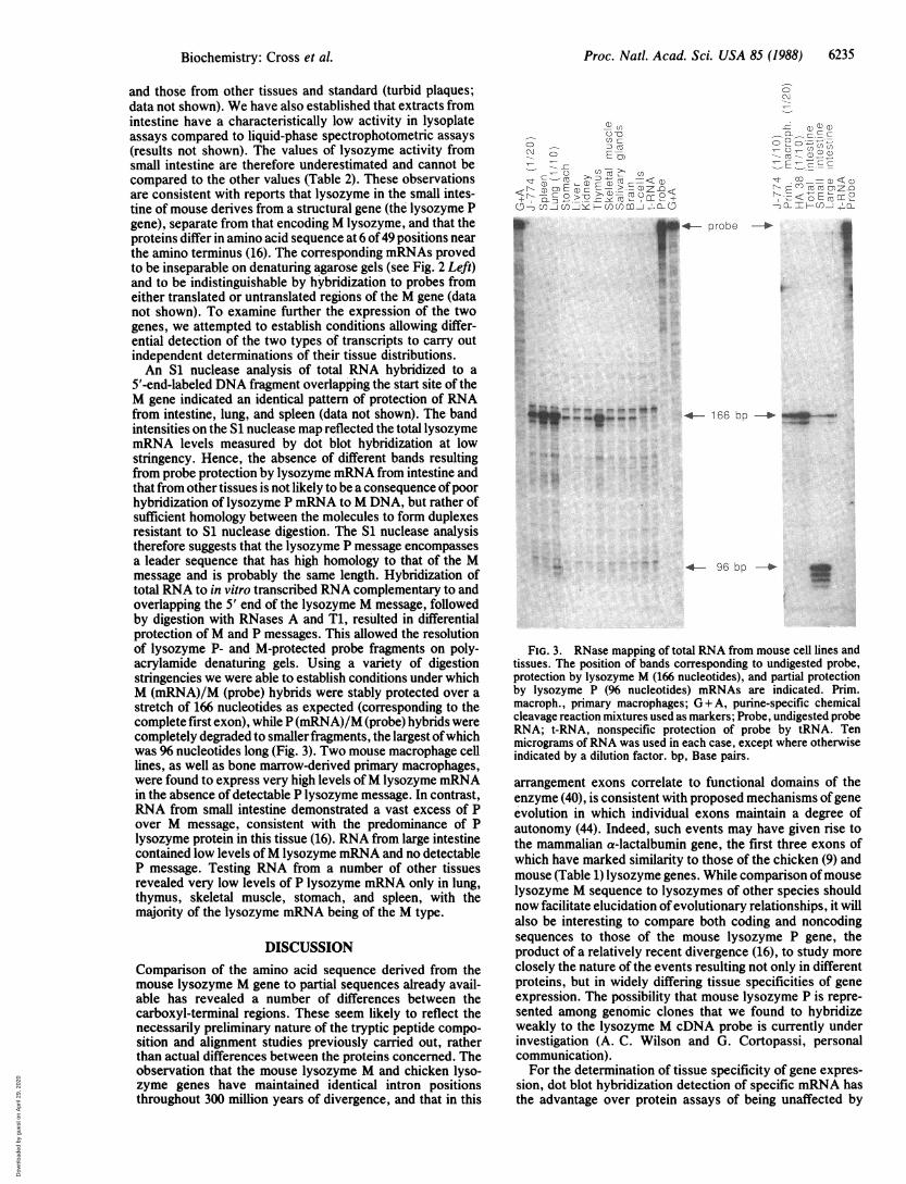
5'-end-labeled DNA fragment overlapping the start site of theM gene indicated an identical pattern of protection of RNAfrom intestine, lung, and spleen (data not shown). The bandintensities on the S1 nuclease map reflected the total lysozymemRNA levels measured by dot blot hybridization at lowstringency. Hence, the absence of different bands resultingfrom probe protection by lysozyme mRNA from intestine andthat from other tissues is not likely to be a consequence of poorhybridization of lysozyme P mRNA to M DNA, but rather ofsufficient homology between the molecules to form duplexesresistant to S1 nuclease digestion. The S1 nuclease analysistherefore suggests that the lysozyme P message encompassesa leader sequence that has high homology to that of the Mmessage and is probably the same length. Hybridization oftotal RNA to in vitro transcribed RNA complementary to andoverlapping the 5' end of the lysozyme M message, followedby digestion with RNases A and T1, resulted in differentialprotection of M and P messages. This allowed the resolutionof lysozyme P- and M-protected probe fragments on poly-acrylamide denaturing gels. Using a variety of digestionstringencies we were able to establish conditions under whichM (mRNA)/M (probe) hybrids were stably protected over astretch of 166 nucleotides as expected (corresponding to thecomplete first exon), while P (mRNA)/M (probe) hybrids werecompletely degraded to smaller fragments, the largest of whichwas 96 nucleotides long (Fig. 3). Two mouse macrophage celllines, as well as bone marrow-derived primary macrophages,were found to express very high levels of M lysozyme mRNAin the absence of detectable P lysozyme message. In contrast,RNA from small intestine demonstrated a vast excess of Pover M message, consistent with the predominance of Plysozyme protein in this tissue (16). RNA from large intestinecontained low levels ofM lysozyme mRNA and no detectableP message. Testing RNA from a number of other tissuesrevealed very low levels of P lysozyme mRNA only in lung,thymus, skeletal muscle, stomach, and spleen, with themajority of the lysozyme mRNA being of the M type.
DISCUSSIONComparison of the amino acid sequence derived from themouse lysozyme M gene to partial sequences already avail-able has revealed a number of differences between thecarboxyl-terminal regions. These seem likely to reflect thenecessarily preliminary nature of the tryptic peptide compo-sition and alignment studies previously carried out, ratherthan actual differences between the proteins concerned. Theobservation that the mouse lysozyme M and chicken lyso-zyme genes have maintained identical intron positionsthroughout 300 million years of divergence, and that in this
cj
CC
C)C
O n co v cpUcf)<4a)Ca0E 2O <)EIzn<
rr 0 \-F- <-)l J) :l
probe
:.:
BJLA-- - -"e--V
I
4- 166 bp-. . .
t..
tour,
96 bp _ 3
FIG. 3. RNase mapping of total RNA from mouse cell lines andtissues. The position of bands corresponding to undigested probe,protection by lysozyme M (166 nucleotides), and partial protectionby lysozyme P (96 nucleotides) mRNAs are indicated. Prim.macroph., primary macrophages; G + A, purine-specific chemicalcleavage reaction mixtures used as markers; Probe, undigested probeRNA; t-RNA, nonspecific protection of probe by tRNA. Tenmicrograms of RNA was used in each case, except where otherwiseindicated by a dilution factor. bp, Base pairs.
arrangement exons correlate to functional domains of theenzyme (40), is consistent with proposed mechanisms of geneevolution in which individual exons maintain a degree ofautonomy (44). Indeed, such events may have given rise tothe mammalian a-lactalbumin gene, the first three exons ofwhich have marked similarity to those of the chicken (9) andmouse (Table 1) lysozyme genes. While comparison ofmouselysozyme M sequence to lysozymes of other species shouldnow facilitate elucidation ofevolutionary relationships, it willalso be interesting to compare both coding and noncodingsequences to those of the mouse lysozyme P gene, theproduct of a relatively recent divergence (16), to study moreclosely the nature of the events resulting not only in differentproteins, but in widely differing tissue specificities of geneexpression. The possibility that mouse lysozyme P is repre-sented among genomic clones that we found to hybridizeweakly to the lysozyme M cDNA probe is currently underinvestigation (A. C. Wilson and G. Cortopassi, personalcommunication).For the determination of tissue specificity of gene expres-
sion, dot blot hybridization detection of specific mRNA hasthe advantage over protein assays of being unaffected by
Biochemistry: Cross et A
Dow
nloa
ded
by g
uest
on
Apr
il 29
, 202
0
Proc. Natl. Acad. Sci. USA 85 (1988)
rates of protein secretion and accumulation, although possi-ble effects of variations in mRNA stability in different tissuescannot be excluded. The high levels of lysozyme expressionrevealed in macrophages by this technique may account forthe strong signals detected in some other tissues, especiallylung, which has a high content of active macrophages as abarrier to infection (45), spleen, in which macrophages arepresent and involved in antigen presentation (46), thymus, inwhich macrophage/thymocyte interactions occur during T-cell maturation (47), and bone marrow, which is rich in allcells of the myeloid lineage. Intestinal lysQzyme P, whichmay play a digestive role, is probably expressed solely by thePaneth cells (12). Despite the detection of low levels oflysozyme P in some other tissues, expression of P in primarymacrophages or macrophage cell lines was not detectable.While it is feasible that differences in the mRNA/enzyme
ratios between tissues reflect variable degrees of posttran-scriptional control, these ratios will also be affected bydifferences in the rates of secretion and accumulation ofenzyme in different tissues. Hence, an accumulation of bloodlysozyme is most likely to explain the extremely high con-centration of enzyme in kidney, which contains only back-ground amounts of specific mRNA. Markedly high levels oflysozyme in the spleen may also be a result ofblood lysozymeaccumulation, although the spleen itself contains moderateamounts of lysozyme mRNA of probable macrophage origin.A distinction between lysozyme synthesis and uptake, anddetermination of the sites of synthesis within tissues, shouldbe possible at the cellular level by in situ hybridization ofspecific lysozyme nucleic acid probes and the parallel detec-tion of protein by immunological techniques.Our studies of lysozyme expression in macrophages and
myeloid cell lines indicate a progressive and strong activationof the gene throughout macrophage differentiation. Thetransfection of DNA into cell lines representing differentstages of myeloid development should now enable us toexamine in detail elements controlling the establishment oftissue-specific gene expression in macrophages.
We are grateful for the excellent technical assistance of DagmarWolf; to Drs. Christopher Franklin and Marc Muller for advice oncDNA cloning and RNase mapping; to Drs. P. Swetly, B. Hermann,R. Mertz, P. Leenen, and W. Ostertag for material; to Marc Mullerfor critical reading; and to Frau Renate Lauermann for typing themanuscript. This work was supported by the Deutsche Forschungs-gemeinschaft (SFB 324).
1. Fleming, A. (1922) Proc. R. Soc. London Ser. B 93, 306-317.2. Jolles, P. & Jolles, J. (1984) Mol. Cell. Biochem. 63, 165-189.3. Renkawitz, R., Schutz, G., von derAhe, D. & Beato, M. (1984)
Cell 37, 503-510.4. Theisen, M., Stief, A. & Sippel, A. E. (1986) EMBO J. 5, 719-
724.5. Steiner, C., Muller, M., Baniahmad, A. & Renkawitz, R. (1987)
Nucleic Acids Res. 15, 4163-4178.6. Baniahmad, A., Muller, M., Steiner, C. & Renkawitz, R. (1987)
EMBO J. 6, 2297-2303.7. Brew, K. & Hill, R. L. (1975) Rev. Physiol. Biochem. Phar-
macol. 72, 105-157.8. Phillips, D. C., Acharya, K. R., Handoll, H. H. G. & Stuart,
D. I. (1987) Biochem. Soc. Trans. 15, 737-744.9. Qasba, P. K. & Sabaya, S. K. (1984) Nature (London) 308,
377-380.10. Finch, S. C., Castro, O., Lippman, M. E., Donadio, J. A. &
Perillie, P. E. (1974) in Lysozyme, eds. Osserman, E. F.,Canfleld, R. E. & Beychok, S. (Academic, New York), pp.335-345.
11. Perillie, P. E. & Finch, S. C. (1974) in Lysozyme, eds. Osser-
man, E. F., Canfield, R. E. & Beychok, S. (Academic, NewYork), pp. 359-372.
12. Klockars, M. & Osserman, E. F. (1974) J. Histochem. Cyto-chem. 22, 139-146.
13. Klockars, M. & Reitamo, S. (1975) J. Histochem. Cytochem.23, 932-940.
14. Perri, G. C., Faulk, M., Shapiro, E. & Money, W. L. (1964)Proc. Soc. Exp. Biol. Med. 115, 189-192.
15. McClelland, D. B. L. & van Furth, R. (1975) Immunology 28,1099-1114.
16. Hammer, M. F., Schilling, J. W., Prager, E. M. & Wilson,A. C. (1987) J. Mol. Evol. 24, 272-279.
17. Hammer, M. F. & Wilson, A. C. (1987) Genetics 115, 521-533.18. Franz, T., Ldhler, J., Fusco, A., Pragnell, I., Nobis, P., Padua,
R. & Ostertag, W. (1985) Nature (LondQn) 315, 149-151.19. Auffrey, C. & Rougeon, F. (1980) Eur. J. Biochem. 107, 303-
314.20. Gubler, U. & Hoffman, B. J. (1983) Gene 25, 263-269.21. Maniatis, T., Fritsch, E. F. & Sambrook, J. (1982) Molecular
Cloning:A Laboratory Manual (Cold Spring Harbor Lab., ColdSpring Harbor, NY).
22. Chen, E. J. & Seeburg, P. H. (1985) DNA 4, 165-170.23. Khandjian, E. W. (1986) Mol. Biol. Rep. 11, 105-115.24. Ghosh, P. K., Reddy, V. B., Piatak, M., Lebowitz, P. &
Weissman, S. M. (1980) Methods Enzymol. 65, 580-595.25. Weaver, R. F. & Weissmann, C. (1979) Nucleic Acids Res. 7,
1175-1193.26. Melton, D. A., Krieg, P. A., Rebagliati, M. R., Maniatis, T.,
Zinn, K. & Green, M. R. (1984) Nucleic Acids Res. 12, 7035-7056.
27. Osserman, E. F. & Lawlor, D. P. (1966) J. Exp. Med. 124, 921-952.
28. Ichikawa, Y. (1969) J. Cell. Physiol. 74, 223-234.29. Warner, N. L., Moore, M. A. S. & Metcalf, D. (1969) J. Natl.
Cancer Inst. 43, 963-977.30. Koren, H. S., Handwerger, B. S. & Wunderlich, J. R. (1975) J.
Immunol. HJ4, 894-897.31. Ralph, P., Pritchard, J. & Cohn, M. (1975) J. Immunol. 114,
898-905.32. Kit, S., Dubbs, D. R., Piekarski, L. J. & Hsu, T. C. (1963) Exp.
Cell Res. 31, 297-312.33. Stewart, C. C. (1981) in Methods for Studying Mononuclear
Phagocytes, eds. Adams, D. O., Edelson, P. J. & Koren, H. S.(Aicademic, New York), pp. 5-20.
34. Breathnach, R. & Chambon, P. (1981) Annu. Rev. Biochem. 50,349-383.
35. Benoist, C., O'Hare, K., Breathnach, R. & Chambon, P. (1980)Nucleic Acids Res. 8, 127-142.
36. Proudfoot, N. J. & Brownlee, G. G. (1976) Nature (London)263, 211-214.
37. Breathnach, R., Benoist, C., O'Hare, K., Gannon, F. &Chambon, P. (1978) Proc. NatI. Acad. Sci. USA 75,4853-4857.
38. Riblet, R. J. (1974) in Lysozyme, eds. Osserman, E. F., Cran-field, R. E. & Beychok; S. (Academic, New York), pp. 89-93.
39. Osserman, E. F., Cranfield, R. E. & Beychok, S., eds. (1974)Lysozyme (Academic, New York).
40. Jung, A., Sippel, A. E., Grez, M. & Shutz, G. (1980) Proc.Natl. Acad. Sci. USA 77, 5759-5763.
41. White, T. J., Mross, G. A., Osserman, E. F. & Wilson, A. C.(1977) Biochemistry 16, 1430-1436.
42. Hansen, N. E., Karle, H. & Anderson, V. (1974) in Lysozyme,eds. Osserman, E. F., Cranfield, R. E. & Beychok, S. (Aca-demic, New York), pp. 307-319.
43. Leenen, P. J. M., Jansen, A. M. A. C. & van Ewijk, W. (1986)Differentiation 32, 157-164.
44. Gilbert, W. (1978) Nature (London) 271, 501.45. Green, G. M. (1970) Am. Rev. Respir. Dis. 102, 691-703.46. Bohnsack, J. F. & Brown, E. J. (1986) Annu. Rev. Med. 37, 49-
59.47. Kyewski, B. A., Rouse, R. V. & Kaplan, H. S. (1982) Proc.
Natl. Acad. Sci. USA 79, 5646-5650.
6236 Biochemistry: Cross et aL
Dow
nloa
ded
by g
uest
on
Apr
il 29
, 202
0