Embed Size (px)
Citation preview
TitleMouse sperm undergo GPI-anchored protein release associatedwith lipid raft reorganization and acrosome reaction to acquirefertility.
Author(s) Watanabe, Hitomi; Kondoh, Gen
Citation Journal of cell science (2011), 124(15): 2573-2581
Issue Date 2011-08-01
URL http://hdl.handle.net/2433/145976
Right © The Company of Biologists Ltd 2011.
Type Journal Article
Textversion publisher
Kyoto University
Research Article 2573
IntroductionThe membranes of mammalian sperm undergo variousmodifications after leaving the testis in male and femalereproductive tracts to acquire full competency to fertilize ova(Sullivan et al., 2005; Yanagimachi, 2009; Ikawa et al., 2010). Oneof the most important steps in this physiological change iscapacitation, which occurs in the female reproductive tract andmight be affected by humoral factors and the interaction betweenthe sperm and the oviductal epithelium (Smith and Yanagimachi,1990). During the capacitation process, it is reported that the spermouter membrane undergoes cholesterol depletion, followed bydownstream signaling processes such as protein tyrosinephosphorylation (Visconti et al., 1995; Visconti et al., 1999; Travisand Kopf, 2002; Bailey, 2010). Because cholesterol is a majorcomponent of lipid rafts (Jacobson et al., 2007; Simons and Toomre,2000), its efflux might also change membrane fluidity and/orinduce rearrangement of lipid rafts (Davis, 1981; Nixon et al.,2008), although other molecular mechanisms involved incapacitation remain to be characterized. Several biologicalprocesses seem to be associated with the lipid microdomain (Mishraand Joshi, 2007). Numerous sperm surface proteins are componentsof this structure and some are directly involved in sperm–zona-pellucida (ZP) binding (Nixon et al., 2008); however, the biologicalimplications of this structure in fertilization have yet to bedetermined. Another important step is the acrosome reaction(Wassarman and Litscher, 2001; Clark and Dell, 2006; Wassarmanand Litscher, 2008; Suarez, 2008). The acrosome is a Golgi-derivedorganelle that overlies the sperm nucleus in the apical region of thehead. It consists of a continuous membrane, called the inner and
outer acrosome membrane. Upon the acrosome reaction, the outeracrosomal membrane and the plasma membrane of sperm fuse,forming multiple hybrid membrane vesicles, following exposureof inner acrosomal membrane and release of acrosomal ingredients.ZP binding of the sperm is a trigger of the acrosome reaction invivo, and this process can be experimentally prompted by treatingsperm with a calcium ionophore to facilitate Ca2+ influx (Costelloet al., 2009). Sperm that have completed the acrosome reaction canexclusively penetrate the ZP and fuse with the egg plasmamembrane.
It is well known that glycosylphosphatidylinositol (GPI)-anchored proteins (GPI-APs) are a major component of lipid rafts(Varma and Mayor, 1998). To monitor the fates of GPI-APs, aswell as lipid rafts, in vivo, we previously developed GPI-anchoredenhanced green fluorescent protein (EGFP–GPI) and examined thelocalization and release of GPI-APs in various tissues (Kondoh etal., 1999). In further studies, we and others showed that the releaseof GPI-APs from the sperm membrane is crucial for male fertility,particularly for the sperm–egg binding step (Kondoh et al., 2005;Ueda et al., 2007).
Izumo1 is a transmembrane protein that is involved in sperm–egg fusion (Inoue et al., 2005), which is also located in lipid rafts(Nixon et al., 2008) and is an indicator of the acrosome reaction(Fujiwara et al., 2010). First, we discovered a linkage betweenGPI-AP release and ganglioside GM1 relocation. GM1 is acharacteristic component and reliable biomarker of lipid rafts.Other studies showed that it dynamically moves with methyl--cyclodextrin (M--CD) treatment in the mouse sperm membrane(Buttke et al., 2006; Selvaraj et al., 2007). Here, we found that M-
SummaryMammalian sperm undergo several maturation steps after leaving the testis to become competent for fertilization. Important changesoccur in sperm within the female reproductive tract, although the molecular mechanisms underlying these processes remain unclear.To investigate sperm membrane remodeling upon sperm maturation, we developed transgenic mouse lines carryingglycosylphosphatidylinositol (GPI)-anchored enhanced green fluorescent protein (EGFP–GPI) and traced the fate of this fluorescentprotein during the fertility-acquiring process in sperm in vitro and in vivo. When the GFP-labeled sperm were treated with compoundsfor promoting the acrosome reaction, EGFP–GPI was released from the sperm surface crosslinked with characteristic relocation of alipid raft marker ganglioside GM1. Sperm ejaculated into the uterus strongly expressed EGFP–GPI in the head region, whereas a partof the oviductal sperm lost fluorescence in a manner that was dependent on the presence of angiotensin-converting enzyme (ACE).Moreover, sperm on the zona pellucida of eggs in the oviduct were all found to have low levels of GFP. These results suggest thatsperm undergoing GPI-anchored protein release associated with reorganization of lipid rafts and the acrosome reaction acquirefertilization potential.
Key words: Sperm, Acrosome reaction, GM1, GPI-anchored protein, Lipid raft, Angiotensin-converting enzyme (ACE)
Accepted 28 March 2011Journal of Cell Science 124, 2573-2581© 2011. Published by The Company of Biologists Ltddoi:10.1242/jcs.086967
Mouse sperm undergo GPI-anchored protein releaseassociated with lipid raft reorganization and acrosomereaction to acquire fertilityHitomi Watanabe and Gen Kondoh*Laboratory of Animal Experiments for Regeneration, Institute for Frontier Medical Sciences, Kyoto University and CREST Program, Japan Scienceand Technology Society, 53 Syogoin-Kawahara-cho, Kyoto 606-8507, Japan*Author for correspondence ([email protected])
Jour
nal o
f Cel
l Sci
ence
-CD induces both GPI-AP release and GM1 relocation. Thepharmacological effect of M--CD is to capture cholesterol, thusit is believed that its main functions in the sperm fertility acquisitionprocess are to facilitate cholesterol efflux and to induce capacitation(Takeo et al., 2008). By monitoring relocation of Izumo1, we alsofound that M--CD can induce the acrosome reaction.
Addition of bovine serum albumin (BSA) alone could induceneither GPI-AP release nor GM relocation, but could do sofollowing treatment with the calcium ionophore A23187. We thusconclude that lipid raft movement occurs upon the acrosomereaction, rather than upon capacitation. Our observations heresuggest a molecular process by which sperm acquire fertility.
ResultsProduction of EGFP(K268Q)-GPI transgenic miceWe previously developed an EGFP–GPI reporter protein to followthe fate of GPI in vivo (Kondoh et al., 1999). However, the originalconstruct encoded a lysine residue adjacent to the GPI attachmentsite that seemed to be a target for serine proteases. To reduce thepossibility of nonspecific cleavage by proteases, we replaced thelysine at residue 268 with glutamine (supplementary material Fig.S1A) and designated the protein EGFP(K268Q)-GPI. Expressionof this construct in cultured cells revealed GFP on the cell surfaceand on the Golgi complex (supplementary material Fig. S1B).Treatment with the phosphatidylinositol-specific phospholipase C(PI-PLC) virtually abrogated the cell-surface signal, confirmingthat the protein is GPI anchored.
We next developed transgenic mouse lines carrying theEGFP(K268Q)-GPI sequence to examine the fate of GPI-anchoredproteins in vivo. Three transgenic founders were obtained by injectingthe transgene into one-cell embryos of the C57BL/6 geneticbackground, with two founders showing protein expression in thesperm head and cytoplasmic droplet (supplementary material Fig.S1C). Transgenic line 2 showed relatively high expression and thuswas selected for further analyses. Male mice of this line showednormal fertility rate (mean no. of litters ± s.d.: 6.83±2.1, n6), whichwas comparable to that of C57BL/6 non-transgenic controls (6–7newborns per litter). The transgene was inherited by their descendantsin accordance with the expected Mendelian frequencies.
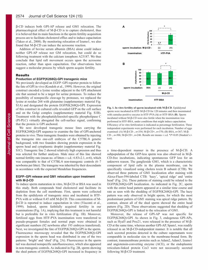
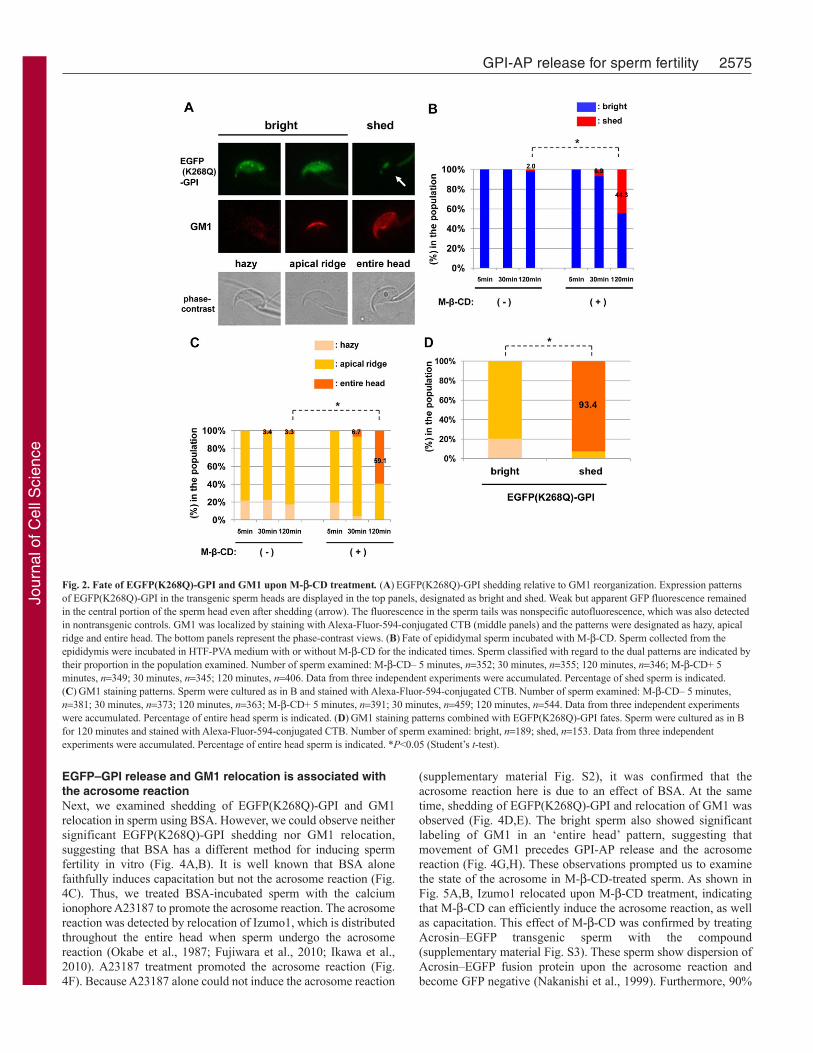
EGFP–GPI release and GM1 relocation upon treatmentwith M--CDTo induce sperm maturation in vitro, M--CD or BSA was used inthis study. Both compounds bind cholesterol and facilitate itsdepletion from the cell membrane. First, sperm were collectedfrom the epididymis of transgenic mice and incubated in HTF-PVA with or without 0.45 mM M--CD. This concentration of M--CD is reported to induce capacitation in vitro (Visconti et al.,1999). Indeed, sperm faithfully acquired fertility in ourexaminations (Fig. 1A), implying that this treatment is not harmfulbut is preferable for in vitro fertilization (Fig. 1B). Moreover,fertilized eggs from HTF-PVA insemination were transferred topseudo-pregnant females and normal pups were obtained insignificant numbers (82 newborns after 193 embryos transferred).Next, we investigated the fate of EGFP(K268Q)-GPI in the sperm.Fluorescence microscopy revealed that the EGFP(K268Q)-GPIexpression in the sperm head was distributed in one of the twopatterns: ‘bright’ and ‘shed’ (Fig. 2A). Fluorescence in the spermtail was deemed nonspecific autofluorescence, which also appearedin non-transgenic controls. As indicated in Fig. 2B, sperm showingthe shed pattern of EGFP(K268Q)-GPI increased in frequency in
a time-dependent manner in the presence of M--CD. Asubpopulation of the GFP-less sperm was also observed in M--CD-free incubations, indicating spontaneous GFP loss for anunknown reason. The ganglioside GM1, which is a characteristiccomponent of lipid rafts in the plasma membrane, can bespecifically visualized using cholera toxin B subunit (CTB). Weobserved three patterns of GM1 localization after staining withAlexa-Fluor-594-labeled CTB: ‘hazy’, ‘apical ridge’ and ‘entirehead’ (Fig. 2A). These patterns of staining could be related to theEGFP(K268Q)-GPI localization. As indicated in Fig. 2C, spermwith the entire head pattern appeared at a similar time course andrate as seen with the shedding of EGFP(K268Q)-GPI. The hazypattern was only observed in bright sperm, for which the mostpredominant pattern of GM1 staining was apical ridge pattern. Bycontrast, almost all of the shed sperm showed the entire headpattern (Fig. 2D). These observations suggested that shedding ofEGFP(K268Q)-GPI is linked to the relocation of GM1.
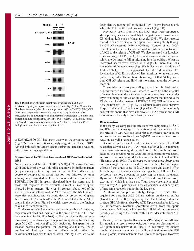
Moreover, the release of GPI-AP was not specific forEGFP(K268Q)-GPI. As shown in Fig. 3, endogenous GPI-APs,such as Hyal5 and Prss21, were released in the presence of M--CD at the same time, whereas another GPI-AP, Spam1, was readilyreleased in an M--CD-independent manner. It is notable that allsuch secreted proteins detected in the culture supernatants werecomparable in molecular size to the membrane-bound form. Bycontrast, transmembrane proteins such as Adam2, Adam3, Izumo1and angiotensin-converting enzyme (ACE), or the endoplasmicreticulum-linked protein Ces3 were not necessarily secretedfollowing M--CD treatment.
2574 Journal of Cell Science 124 (15)
Fig. 1. In vitro fertility of sperm incubated with M--CD. Epididymalsperm were incubated in HTF-M--CD for 120 minutes and then inseminatedwith cumulus-positive oocytes in HTF-PVA (A) or HTF-BSA (B). Spermincubated without M--CD were also fertile when the insemination wasperformed in HTF-BSA, under conditions that might induce capacitation.Efficiency of in vitro fertilization is indicated as percentage fertilization. Threeindependent experiments were performed for each incubation. Number of eggsexamined: (A) M--CD–, n354; M--CD+, n370; (B) BSA, n367; M--CD–, n306; M--CD+, n246. Results are means ± s.d. *P<0.05 (Student’s t-test).
Jour
nal o
f Cel
l Sci
ence
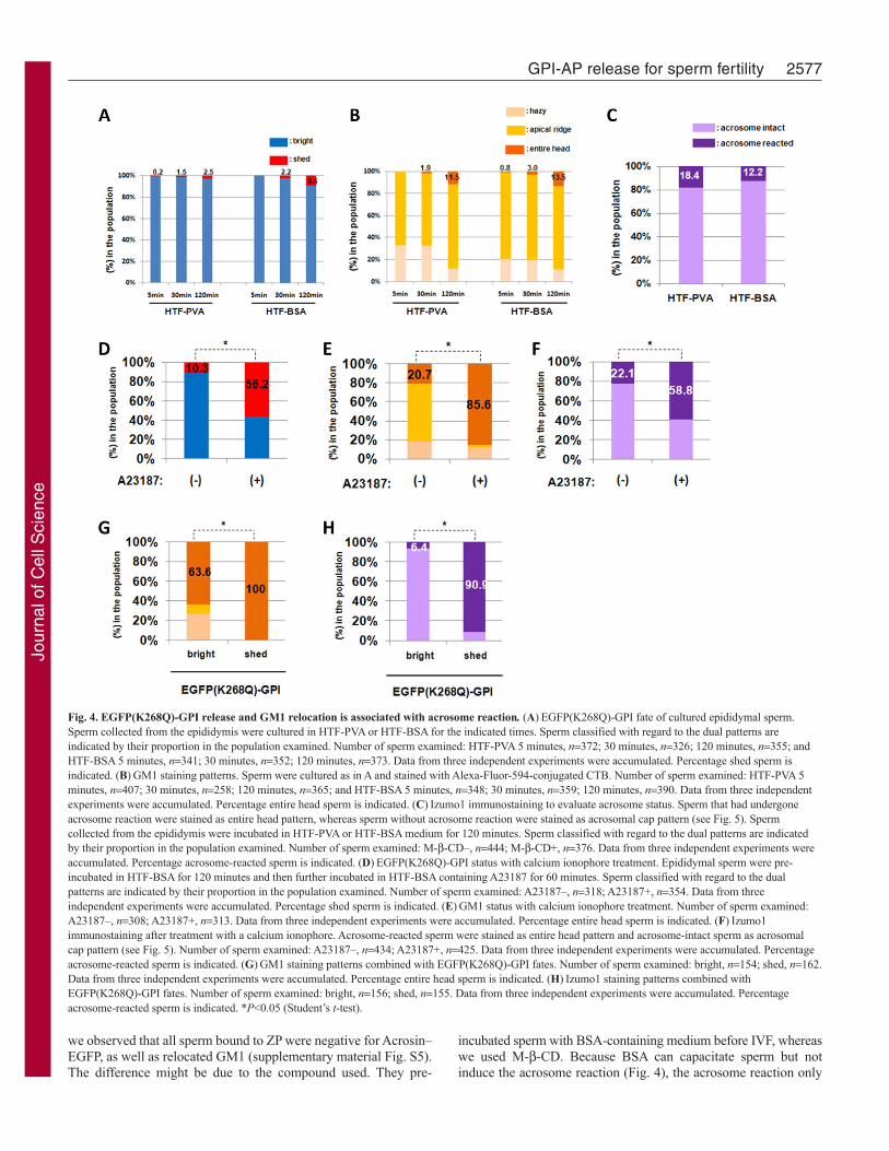
EGFP–GPI release and GM1 relocation is associated withthe acrosome reactionNext, we examined shedding of EGFP(K268Q)-GPI and GM1relocation in sperm using BSA. However, we could observe neithersignificant EGFP(K268Q)-GPI shedding nor GM1 relocation,suggesting that BSA has a different method for inducing spermfertility in vitro (Fig. 4A,B). It is well known that BSA alonefaithfully induces capacitation but not the acrosome reaction (Fig.4C). Thus, we treated BSA-incubated sperm with the calciumionophore A23187 to promote the acrosome reaction. The acrosomereaction was detected by relocation of Izumo1, which is distributedthroughout the entire head when sperm undergo the acrosomereaction (Okabe et al., 1987; Fujiwara et al., 2010; Ikawa et al.,2010). A23187 treatment promoted the acrosome reaction (Fig.4F). Because A23187 alone could not induce the acrosome reaction
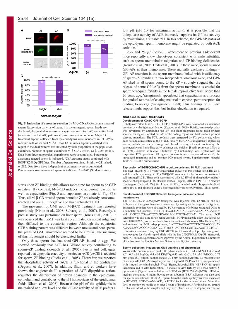
(supplementary material Fig. S2), it was confirmed that theacrosome reaction here is due to an effect of BSA. At the sametime, shedding of EGFP(K268Q)-GPI and relocation of GM1 wasobserved (Fig. 4D,E). The bright sperm also showed significantlabeling of GM1 in an ‘entire head’ pattern, suggesting thatmovement of GM1 precedes GPI-AP release and the acrosomereaction (Fig. 4G,H). These observations prompted us to examinethe state of the acrosome in M--CD-treated sperm. As shown inFig. 5A,B, Izumo1 relocated upon M--CD treatment, indicatingthat M--CD can efficiently induce the acrosome reaction, as wellas capacitation. This effect of M--CD was confirmed by treatingAcrosin–EGFP transgenic sperm with the compound(supplementary material Fig. S3). These sperm show dispersion ofAcrosin–EGFP fusion protein upon the acrosome reaction andbecome GFP negative (Nakanishi et al., 1999). Furthermore, 90%
2575GPI-AP release for sperm fertility
Fig. 2. Fate of EGFP(K268Q)-GPI and GM1 upon M--CD treatment. (A)EGFP(K268Q)-GPI shedding relative to GM1 reorganization. Expression patternsof EGFP(K268Q)-GPI in the transgenic sperm heads are displayed in the top panels, designated as bright and shed. Weak but apparent GFP fluorescence remainedin the central portion of the sperm head even after shedding (arrow). The fluorescence in the sperm tails was nonspecific autofluorescence, which was also detectedin nontransgenic controls. GM1 was localized by staining with Alexa-Fluor-594-conjugated CTB (middle panels) and the patterns were designated as hazy, apicalridge and entire head. The bottom panels represent the phase-contrast views. (B)Fate of epididymal sperm incubated with M--CD. Sperm collected from theepididymis were incubated in HTF-PVA medium with or without M--CD for the indicated times. Sperm classified with regard to the dual patterns are indicated bytheir proportion in the population examined. Number of sperm examined: M--CD– 5 minutes, n352; 30 minutes, n355; 120 minutes, n346; M--CD+ 5minutes, n349; 30 minutes, n345; 120 minutes, n406. Data from three independent experiments were accumulated. Percentage of shed sperm is indicated.(C)GM1 staining patterns. Sperm were cultured as in B and stained with Alexa-Fluor-594-conjugated CTB. Number of sperm examined: M--CD– 5 minutes,n381; 30 minutes, n373; 120 minutes, n363; M--CD+ 5 minutes, n391; 30 minutes, n459; 120 minutes, n544. Data from three independent experimentswere accumulated. Percentage of entire head sperm is indicated. (D)GM1 staining patterns combined with EGFP(K268Q)-GPI fates. Sperm were cultured as in Bfor 120 minutes and stained with Alexa-Fluor-594-conjugated CTB. Number of sperm examined: bright, n189; shed, n153. Data from three independentexperiments were accumulated. Percentage of entire head sperm is indicated. *P<0.05 (Student’s t-test).
Jour
nal o
f Cel
l Sci
ence
of EGFP(K268Q)-GPI shed sperm underwent the acrosome reaction(Fig. 5C). These observations strongly suggest that release of GPI–AP and lipid raft movement occur during the acrosome reaction,rather than during capacitation.
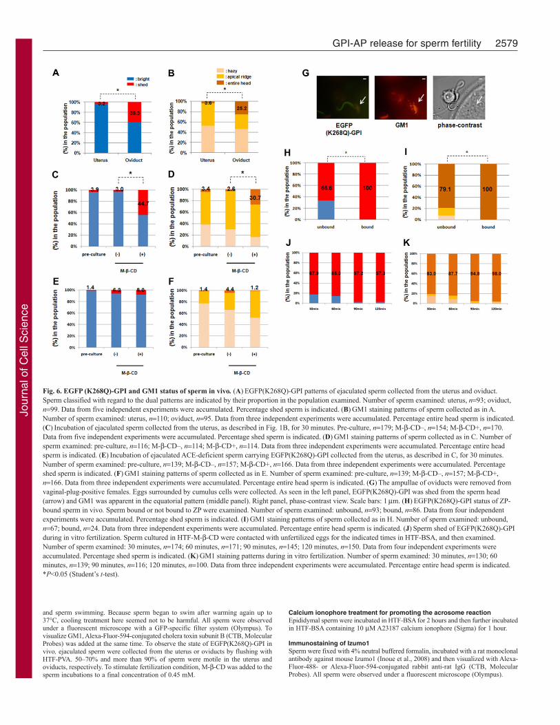
Sperm bound to ZP have low levels of GFP and relocatedGM1We next examined the fate of EGFP(K268Q)-GPI in vivo. BecauseGM1 and Izumo1 always colocalize and move in similar manners(supplementary material Fig. S4), the fate of lipid rafts and thedegree of completed acrosome reaction was followed by GM1staining in in vivo studies. First, we observed the expression ofEGFP(K268Q)-GPI in sperm ejaculated into the uterus and inthose that migrated to the oviducts. Almost all uterine spermshowed a bright pattern (Fig. 6A). By contrast, about 40% of thesperm in the oviducts showed the ‘shed’ pattern. The GM1 patternswere also examined, and we found that the proportion of spermlabeled over the ‘entire head’ with GM1 correlated with the ‘shed’sperm in the oviduct (Fig. 6B), which corresponds to the findingsof our in vitro experiments.
To determine whether uterine sperm are capable of shedding,they were collected and incubated in the presence of M--CD, andthen examined for EGFP(K268Q)-GPI expression by fluorescencemicroscopy. The uterine sperm acquired competency for sheddingupon M--CD treatment (Fig. 6C), indicating that sperm at thislocation possess the potential for shedding and that the limitednumber of shed sperm in the oviducts might reflect theenvironmental capacity to induce sperm fertility. Here, we found
again that the number of ‘entire head’ GM1 sperm increased onlywhen the EGFP–GPI shedding was induced (Fig. 6D).
Previously, sperm from Ace-knockout mice were reported toshow phenotypes such as inability to migrate into the oviduct andZP-binding deficiencies (Hagaman et al., 1998). We also reportedthat ACE can contribute to form sperm–ZP binding ability throughits GPI-AP releasing activity (GPIase) (Kondoh et al., 2005).Therefore, in the present study, we tried to confirm the contributionof ACE to the release of GPI-AP. We also prepared Ace-knockoutmice carrying EGFP(K268Q)-GPI and examined uterine sperm,which are destined to fail in migrating into the oviduct. When therecovered sperm were treated with M--CD, more than 90%retained a bright appearance (Fig. 6E), indicating that shedding ofEGFP(K268Q)-GPI is suppressed by ACE deficiency. Thelocalization of GM1 also showed less transition to the entire headpattern (Fig. 6F). These observations suggest that ACE governsboth GPI-AP release and lipid raft movement upon the acrosomereaction.
To examine our theory regarding the location for fertilization,eggs surrounded by cumulus cells were collected from the ampullaeof mated female oviducts to assess sperm penetrating the cumuluslayer or binding to the ZP of the egg. All sperm that bound to theZP showed the shed pattern of EGFP(K268Q)-GPI and the entirehead pattern for GM1 (Fig. 6G–I). Similar results were observedin sperm with in vitro fertilization (Fig. 6J,K). These results stronglysuggest that sperm that have undergone GPI-AP release and GM1relocation exclusively acquire fertility in vivo.
DiscussionIn this study, we compared the effects of two compounds, M--CDand BSA, for inducing sperm maturation in vitro and revealed thatthe release of GPI-APs and lipid raft movement occur upon theacrosome reaction. We found that M--CD can induce the acrosomereaction, as well as capacitation.
Ace-knockout sperm collected from the uterus showed less GM1relocation, as well as low GPI-AP release, after M--CD treatment.These observations suggest that ACE is involved in the acrosomereaction. In a previous report, ACE-knockout sperm showed normalacrosome reactions induced by treatment with BSA and A23187(Hagaman et al., 1998). The discrepancy between these observationsand ours might be due to a difference in the pharmacologicaleffects of the drugs used. M--CD facilitates cholesterol effluxfrom the sperm membrane and causes capacitation followed by theacrosome reaction, affecting the early step of sperm maturation.By contrast, A23187 facilitates Ca2+ influx and completes acrosomeexocytosis, which is a late step. Thus, these observations mightexplain why ACE participates in the capacitation and/or early stepof acrosome reaction, but not in the late step.
As shown in our previous study, disruption of lipid rafts isrequired for detecting ACE GPIase activity on the cell surface(Kondoh et al., 2005), suggesting that the lipid raft structureprotects GPI-APs from release by ACE. Upon capacitation followedby the acrosome reaction, cholesterol efflux from the spermmembrane is facilitated, which results in lipid raft movement andpossibly loosening of the structure; thus GPI-APs suffer from ACEattack.
Recently, it was reported that sperm–ZP binding is not sufficientfor inducing acrosome exocytosis, but it requires cleavage of theZP2 protein (Baibakov et al., 2007). In this study, the authorsmonitored the acrosome reaction by dispersion of an Acrosin–GFPfusion protein, which took more than 2 hours to complete. However,
2576 Journal of Cell Science 124 (15)
Fig. 3. Distribution of sperm membrane proteins upon M--CDtreatment. Epididymal sperm were incubated as in Fig. 2B for 120 minutes.Membrane fractions (M) and culture supernatants (Cs) of EGFP(K268Q)-GPIsperm were subjected to immunoblotting using 10g of protein, whichrepresented 1/6 of the total protein in membrane fractions and 1/36 of the totalprotein in culture supernatants. GPI-APs: EGFP(K268Q)-GPI, Hyal5, Prss21and Spam1. Transmembrane proteins: Adam2, Adam3, Izumo1 and tACE.Endoplasmic reticulum-associated protein: Ces3.
Jour
nal o
f Cel
l Sci
ence
we observed that all sperm bound to ZP were negative for Acrosin–EGFP, as well as relocated GM1 (supplementary material Fig. S5).The difference might be due to the compound used. They pre-
incubated sperm with BSA-containing medium before IVF, whereaswe used M--CD. Because BSA can capacitate sperm but notinduce the acrosome reaction (Fig. 4), the acrosome reaction only
2577GPI-AP release for sperm fertility
Fig. 4. EGFP(K268Q)-GPI release and GM1 relocation is associated with acrosome reaction. (A)EGFP(K268Q)-GPI fate of cultured epididymal sperm.Sperm collected from the epididymis were cultured in HTF-PVA or HTF-BSA for the indicated times. Sperm classified with regard to the dual patterns areindicated by their proportion in the population examined. Number of sperm examined: HTF-PVA 5 minutes, n372; 30 minutes, n326; 120 minutes, n355; andHTF-BSA 5 minutes, n341; 30 minutes, n352; 120 minutes, n373. Data from three independent experiments were accumulated. Percentage shed sperm isindicated. (B)GM1 staining patterns. Sperm were cultured as in A and stained with Alexa-Fluor-594-conjugated CTB. Number of sperm examined: HTF-PVA 5minutes, n407; 30 minutes, n258; 120 minutes, n365; and HTF-BSA 5 minutes, n348; 30 minutes, n359; 120 minutes, n390. Data from three independentexperiments were accumulated. Percentage entire head sperm is indicated. (C)Izumo1 immunostaining to evaluate acrosome status. Sperm that had undergoneacrosome reaction were stained as entire head pattern, whereas sperm without acrosome reaction were stained as acrosomal cap pattern (see Fig. 5). Spermcollected from the epididymis were incubated in HTF-PVA or HTF-BSA medium for 120 minutes. Sperm classified with regard to the dual patterns are indicatedby their proportion in the population examined. Number of sperm examined: M--CD–, n444; M--CD+, n376. Data from three independent experiments wereaccumulated. Percentage acrosome-reacted sperm is indicated. (D)EGFP(K268Q)-GPI status with calcium ionophore treatment. Epididymal sperm were pre-incubated in HTF-BSA for 120 minutes and then further incubated in HTF-BSA containing A23187 for 60 minutes. Sperm classified with regard to the dualpatterns are indicated by their proportion in the population examined. Number of sperm examined: A23187–, n318; A23187+, n354. Data from threeindependent experiments were accumulated. Percentage shed sperm is indicated. (E)GM1 status with calcium ionophore treatment. Number of sperm examined:A23187–, n308; A23187+, n313. Data from three independent experiments were accumulated. Percentage entire head sperm is indicated. (F)Izumo1immunostaining after treatment with a calcium ionophore. Acrosome-reacted sperm were stained as entire head pattern and acrosome-intact sperm as acrosomalcap pattern (see Fig. 5). Number of sperm examined: A23187–, n434; A23187+, n425. Data from three independent experiments were accumulated. Percentageacrosome-reacted sperm is indicated. (G)GM1 staining patterns combined with EGFP(K268Q)-GPI fates. Number of sperm examined: bright, n154; shed, n162.Data from three independent experiments were accumulated. Percentage entire head sperm is indicated. (H)Izumo1 staining patterns combined withEGFP(K268Q)-GPI fates. Number of sperm examined: bright, n156; shed, n155. Data from three independent experiments were accumulated. Percentageacrosome-reacted sperm is indicated. *P<0.05 (Student’s t-test).
Jour
nal o
f Cel
l Sci
ence
starts upon ZP binding; this allows more time for sperm to be GFPnegative. By contrast, M--CD induces the acrosome reaction aswell as capacitation (Fig. 5 and supplementary material Fig. S3).Thus, all M--CD-treated sperm bound to ZP are already acrosome-reacted and are GFP negative and have relocated GM1.
The movement of GM1 upon M--CD treatment was reportedpreviously (Nixon et al., 2008; Selvaraj et al., 2007). Recently, aprecise study was performed on boar sperm (Jones et al., 2010). Itwas observed that GM1 was first accumulated on apical ridge andthen diffused to the equatorial region. Although the equatorialCTB staining pattern was different between mouse and boar sperm,the paths of GM1 movement seemed to be similar. The meaningof this movement should be elucidated further.
Only those sperm that had shed GPI-APs bound to eggs. Weshowed previously that ACE has GPIase activity contributing tosperm–ZP binding (Kondoh et al., 2005). Fuchs and colleaguesreported that dipeptidase activity of testicular ACE (tACE) is requiredfor sperm–ZP binding (Fuchs et al., 2005). Thereafter, we reportedthat dipeptidase activity of tACE is functional in the epididymis(Deguchi et al., 2007). In addition, Shum and co-workers haveshown that angiotensin II, a product of ACE dipeptidase action,regulates the distribution of proton channels in the epididymalepithelium and contributes to the pH homeostasis of the epididymalfluids (Shum et al., 2008). Because the pH of the epididymis ismaintained at a low level and the GPIase activity of ACE prefers a
low pH (pH 6.5 for maximum activity), it is possible that thedideptidase activity of ACE indirectly supports its GPIase activityby maintaining a suitable pH. In this scheme, the GPI-AP status ofthe epididymal sperm membrane might be regulated by both ACEactivities.
Ace- and Pgap1 (post-GPI attachment to proteins 1)-knockoutmice reportedly show phenotypes consistent with male infertility,such as sperm uterotubular migration and ZP-binding deficiencies(Kondoh et al., 2005; Ueda et al., 2007). In these mice, sperm retainedGPI-APs in their membranes. These mutually exclusive findings –GPI-AP retention in the sperm membrane linked with insufficiencyof sperm–ZP binding in two independent knockout mice, and GPI-AP shed in all sperm bound to the ZP – strongly suggest that therelease of some GPI-APs from the sperm membrane is crucial forsperm to acquire fertility in the female reproductive tract. More than20 years ago, Yanagimachi speculated that capacitation is a processfor gradual removal of coating material to expose sperm receptors forbinding to an egg (Yanagimachi, 1988). Our findings on GPI-APrelease might support this, but further elucidation is required.
Materials and MethodsDevelopment of K268Q-GPI–EGFPK268Q-converted EGFP–GPI (EGFP(K268Q)-GPI) was developed as describedpreviously with slight modification (Kondoh et al., 2009). Briefly, a mutated productwas developed by amplifying the left and right fragments using fixed primersspecific for regions located outside of the coding region and back-to-back primerscarrying mutations. The PCR products were generated by Pfx polymerase, whichdoes not add a nucleotide at the 5� end. Finally, the CAAG ubiquitously expressingvector, which carries a strong and broad driving element containing thecytomegalovirus immediate-early enhancer and chicken -actin promoter (Niwa etal., 1991), cleaved with EcoRI followed by blunt-ending, was ligated with therespective PCR products. All ligated constructs were sequenced to verify theintroduced mutations and to exclude PCR-related errors. Supplementary materialTable S1 lists the primers used.
Expression of EGFP(K268Q)-GPI in culture cells and PI-PLC treatmentThe EGFP(K268Q)-GPI vector constructed above was transfected into CHO cells,and then cells expressing EGFP(K268Q)-GPI were selected by fluorescence-activatedcell sorting (FACS). These cells were treated with 1.0 IU/ml of phosphatidylinositol-specific phospholipase C (Molecular Probes, Eugene, OR) in OPTI-CHO medium(Invitrogen, Carlsbad, CA) for 1 hour at 37°C, washed with phosphate-bufferedsaline (PBS) and observed under a fluorescent microscope (Olympus, Tokyo, Japan).
Development of EGFP(K268Q)-GPI transgenic mice and animalmaintenanceThe CAAG-EGFP K268QGPI transgene was injected into C57BL/6J one-cellembryos and transgenic lines were maintained by mating on the isogenic background.Transgenic founders were obtained by PCR screening of siblings using tail DNA asa template and primers, 5�-TTCTTCAAGGACGACGGCAACTACAAGACC-3�and 5�-GTCACGAACTCCAGCAGGACCATGTGATCG-3�. The same PCRscreening was also used for selecting Acrosin–EGFP transgenic mice. Ace-knockoutmice (JR#002679) were purchased from the Jackson Laboratory (Bar Harbor, ME)and screened by PCR using primers, 5�-CTTGACGAGTTCTTCTGAGG-3�, 5�-AGAAAAGCACGGAGGTATCC-3� and 5�-ACTGCCCGGTCCAGGTTCTG-3�.
Ace-knockout mice carrying EGFP(K268Q)-GPI were developed by mating miceheterozygous for Ace-disrupted allele with the line 2 EGFP(K268Q)-GPI transgenicmice. All animal experiments were approved by the Animal Experiment Committeesof the Institute for Frontier Medical Sciences and Kyoto University.
Sperm collection, incubation, GM1 staining and observationWe used the human tubular fluid (HTF)-base medium (101.61 mM NaCl, 4.69 mMKCl, 0.2 mM MgSO4, 0.4 mM KH2PO4, 6.42 mM CaCl2, 25 mM NaHCO3, 2.77mM glucose, 3.4 g/ml sodium lactate, 0.34 mM sodium pyruvate, 0.2 mM penicillinG sodium salt, 0.03 mM streptomycin and 0.4 l of 0.5% Phenol Red) supplementedwith 1 mg/ml polyvinyl alcohol (PVA) (Sigma, St Louis, MO) (HTF-PVA) for spermincubation and in vitro fertilization. To induce in vitro fertility, 0.45 mM methyl--cyclodextrin (Sigma) was added to the HTF-PVA (HTF-PVA-M--CD). HTF-basemedium containing 4 mg/ml bovine serum albumin (BSA) (Sigma) was also usedfor sperm incubation (HTF-BSA). Sperm from the cauda epididymis were incubatedin HTF-PVA, HTF-PVA-M--CD, or HTF-BSA for the indicated times. More than90% of sperm were motile even after 2 hours of incubation. After incubation, 10 mMEDTA was added to the samples and they were placed on ice to stop further reaction
2578 Journal of Cell Science 124 (15)
Fig. 5. Induction of acrosome reaction by M--CD. (A)Acrosome status ofsperm. Expression patterns of Izumo1 in the transgenic sperm heads aredisplayed, designated as acrosomal cap (acrosome intact, AI) and entire head(acrosome reacted, AR) patterns. (B)Acrosome reaction upon M--CDtreatment. Sperm collected from the epididymis were incubated in HTF-PVAmedium with or without M--CD for 120 minutes. Sperm classified withregard to the dual patterns are indicated by their proportion in the populationexamined. Number of sperm examined: M--CD–, n364; M--CD+, n461.Data from three independent experiments were accumulated. Percentageacrosome-reacted sperm is indicated. (C)Acrosome status combined withEGFP(K268Q)-GPI fates. Number of sperm examined: bright, n211; shed,n212. Data from three independent experiments were accumulated.Percentage acrosome-reacted sperm is indicated. *P<0.05 (Student’s t-test).
Jour
nal o
f Cel
l Sci
ence
and sperm swimming. Because sperm began to swim after warming again up to37°C, cooling treatment here seemed not to be harmful. All sperm were observedunder a fluorescent microscope with a GFP-specific filter system (Olympus). Tovisualize GM1, Alexa-Fluor-594-conjugated cholera toxin subunit B (CTB, MolecularProbes) was added at the same time. To observe the state of EGFP(K268Q)-GPI invivo, ejaculated sperm were collected from the uterus or oviducts by flushing withHTF-PVA. 50–70% and more than 90% of sperm were motile in the uterus andoviducts, respectively. To stimulate fertilization condition, M--CD was added to thesperm incubations to a final concentration of 0.45 mM.
Calcium ionophore treatment for promoting the acrosome reactionEpididymal sperm were incubated in HTF-BSA for 2 hours and then further incubatedin HTF-BSA containing 10 M A23187 calcium ionophore (Sigma) for 1 hour.
Immunostaining of Izumo1Sperm were fixed with 4% neutral buffered formalin, incubated with a rat monoclonalantibody against mouse Izumo1 (Inoue et al., 2008) and then visualized with Alexa-Fluor-488- or Alexa-Fluor-594-conjugated rabbit anti-rat IgG (CTB, MolecularProbes). All sperm were observed under a fluorescent microscope (Olympus).
2579GPI-AP release for sperm fertility
Fig. 6. EGFP (K268Q)-GPI and GM1 status of sperm in vivo. (A)EGFP(K268Q)-GPI patterns of ejaculated sperm collected from the uterus and oviduct.Sperm classified with regard to the dual patterns are indicated by their proportion in the population examined. Number of sperm examined: uterus, n93; oviduct,n99. Data from five independent experiments were accumulated. Percentage shed sperm is indicated. (B)GM1 staining patterns of sperm collected as in A.Number of sperm examined: uterus, n110; oviduct, n95. Data from three independent experiments were accumulated. Percentage entire head sperm is indicated.(C)Incubation of ejaculated sperm collected from the uterus, as described in Fig. 1B, for 30 minutes. Pre-culture, n179; M--CD–, n154; M--CD+, n170.Data from five independent experiments were accumulated. Percentage shed sperm is indicated. (D)GM1 staining patterns of sperm collected as in C. Number ofsperm examined: pre-culture, n116; M--CD–, n114; M--CD+, n114. Data from three independent experiments were accumulated. Percentage entire headsperm is indicated. (E)Incubation of ejaculated ACE-deficient sperm carrying EGFP(K268Q)-GPI collected from the uterus, as described in C, for 30 minutes.Number of sperm examined: pre-culture, n139; M--CD–, n157; M--CD+, n166. Data from three independent experiments were accumulated. Percentageshed sperm is indicated. (F)GM1 staining patterns of sperm collected as in E. Number of sperm examined: pre-culture, n139; M--CD–, n157; M--CD+,n166. Data from three independent experiments were accumulated. Percentage entire head sperm is indicated. (G)The ampullae of oviducts were removed fromvaginal-plug-positive females. Eggs surrounded by cumulus cells were collected. As seen in the left panel, EGFP(K268Q)-GPI was shed from the sperm head(arrow) and GM1 was apparent in the equatorial pattern (middle panel). Right panel, phase-contrast view. Scale bars: 1m. (H)EGFP(K268Q)-GPI status of ZP-bound sperm in vivo. Sperm bound or not bound to ZP were examined. Number of sperm examined: unbound, n93; bound, n86. Data from four independentexperiments were accumulated. Percentage shed sperm is indicated. (I)GM1 staining patterns of sperm collected as in H. Number of sperm examined: unbound,n67; bound, n24. Data from three independent experiments were accumulated. Percentage entire head sperm is indicated. (J)Sperm shed of EGFP(K268Q)-GPIduring in vitro fertilization. Sperm cultured in HTF-M--CD were contacted with unfertilized eggs for the indicated times in HTF-BSA, and then examined.Number of sperm examined: 30 minutes, n174; 60 minutes, n171; 90 minutes, n145; 120 minutes, n150. Data from four independent experiments wereaccumulated. Percentage shed sperm is indicated. (K)GM1 staining patterns during in vitro fertilization. Number of sperm examined: 30 minutes, n130; 60minutes, n139; 90 minutes, n116; 120 minutes, n100. Data from three independent experiments were accumulated. Percentage entire head sperm is indicated.*P<0.05 (Student’s t-test).
Jour
nal o
f Cel
l Sci
ence
In vitro fertilizationAdult C57BL/6J females (more than 10 weeks old) were superovulated by injectingthem with 6.7 IU of pregnant mare serum gonadotropin (Teikoku Zoki, Tokyo,Japan) followed 48 hours later with 6.7 IU of human chorionic gonadotropin(Teikoku Zoki). Ovulated eggs surrounded by a cumulus mass were collected fromthe oviducts 16 hours after the second injection. Eggs with cumuli were incubatedin 300 l of HTF-PVA or HTF-BSA medium, and overlaid with mineral oil. Spermfrom the cauda epididymis were preincubated in 200 l of HTF-PVA, HTF-PVA-M--CD, or HTF-BSA for 2 hours and then added to the egg drop at a final concentrationof ~1.0�105 sperm/ml. Eggs were washed with modified Whitten’s medium (mWM;109.51 mM NaCl, 4.78 mM KCl, 1.19 mM MgSO4, 1.19 mM KH2PO4, 22.62 mMNaHCO3, 5.55 mM glucose, 1.49 mM calcium lactate, 0.23 mM sodium pyruvate,19.1 g/ml EDTA, 10 M -mercaptoethanol, 0.2 mM penicillin G sodium salt, 0.03mM streptomycin, 3 mg/ml BSA and 0.2 l of 0.5% phenol red) after 7 hours contactwith the sperm, and then incubated in fresh mWM for another 16 hours. To quantifyfertilization, the total numbers of unfertilized eggs and two-cell embryos and numbersof two-cell embryos in a population were determined to generate the percentagefertilization value: [(%) fertilization(number of two-cell embryos/total number ofunfertilized eggs and two-cell embryos) �100]. Values are mean ± s.d.
Sperm-egg binding assay in vivo and in vitroAdult CD-1 females (more than 10 weeks old) in estrus were mated withEGFP(K268Q)-GPI transgenic males. Twenty hours after the start of mating, ampullaeof oviduct were removed from the vaginal-plug-positive females and eggs surroundedby cumulus cells were collected in HTF-PVA supplemented with 10 mM EDTA. Forevaluating in vitro sperm–egg binding, sperm cultured in HTF-M--CD for 2 hourswere inseminated with unfertilized eggs for the indicated times in HTF-BSA. Spermbound and unbound to the zona pellucida of eggs were stained with Alexa-Fluor-594-conjugated CTB and observed under a fluorescent microscope.
ImmunoblottingMembrane fractions of incubated sperm were prepared using the ProteoExtractTM
Native Membrane Protein Extraction kit (EMD Biosciences, La Jolla, CA). After thehomogenates and culture supernatants were centrifuged at 25,000 g, the supernatantswere collected and assayed for protein content. Next, 10 g of protein per samplewas subjected to SDS-PAGE and then electrophoretically transferred onto anitrocellulose membrane. The membranes were probed with rabbit polyclonalantibodies against GFP (MBL, Nagoya, Japan), Hyal5 (Kim et al., 2005), Prss21(Honda et al., 2002), Spam1 (Baba et al., 2002) and Ces3 (generated for this studyagainst peptide, CARNGNPNSEGLPS), or mouse monoclonal antibodies againstAdam2, Adam3 (Chemicon International, Temecula, CA) and tACE (Yamaguchi etal., 2006), or a rat monoclonal antibody against mouse Izumo1 (Inoue et al., 2008).Antibody binding was detected and visualized using the ECL Plus system (GEHealthcare Biosciences, Uppsala, Sweden). The blot transfer efficiency was checkedby staining with Coomassie Brilliant Blue (Sigma) after immunoblotting. The densityof each band was measured by a densitometer and the degree of protein release wascalculated as the releasing index in supplementary material Table S2.
Statistical analysisAll data for statistical analyses are displayed in supplementary material Table S3.Differences between two groups were analyzed by Student’s t-test using MicrosoftExcel software. Statistical significance was defined as *P<0.05.
We thank Masaru Okabe (Osaka University, Japan) for providinganti-ACE and anti-Izumo1 antibodies, Acrosin-EGFP transgenic miceand helpful discussions, Tadashi Baba (University of Tsukuba, Japan)for anti-Hyal5, anti-Prss21 and anti-Spam1 antibodies and T. Kinoshitafor helpful discussions. This work was supported by grants from theMinistry of Education, Science, Sports and Culture of Japan and theFujiwara Foundation.
Supplementary material available online athttp://jcs.biologists.org/cgi/content/full/124/15/2573/DC1
ReferencesBaba, D., Kashiwabara, S., Honda, A., Yamagata, K., Wu, Q., Ikawa, M., Okabe, M.
and Baba, T. (2002). Mouse sperm lacking cell surface hyaluronidase PH-20 can passthrough the layer of cumulus cells and fertilize the egg. J. Biol. Chem. 277, 30310-30314.
Baibakov, B., Gauthier, L., Talbot, P., Rankin, T. L. and Dean, J. (2007). Spermbinding to the zona pellucid is not sufficient to induce acrosome exocytosis. Development134, 933-943.
Bailey, J. L. (2010). Factors regulating sperm capacitation. Syst. Biol. Reprod. Med. 56,334-348.
Buttke, D. E., Nelson, J. L., Schlegel, P. N., Hunnicutt, G. R. and Travis, A. J.(2006).Visualization of GM1 with cholera toxin B in live epididymal versus ejaculatedbull, mouse, and human spermatozoa. Biol. Reprod. 74, 889-895.
Clark, G. F. and Dell, A. (2006). Molecular models for murine sperm-egg binding. J.Biol. Chem. 281, 13853-13856.
Costello, S., Michelangeli, F., Nash, K., Lefievre, L., Morris, J., Machado-Oliveria,G., Barratt, C., Kirkman-Brown, J. and Publicover, S. (2009). Ca2+-stores in sperm:their identities and functions. Reproduction 138, 425-437.
Davis, B. K. (1981). Timing of fertilization in mammals: sperm cholesterol/phospholipidratio as a determinant of the capacitation interval. Proc. Natl. Acad. Sci. USA 78, 7560-7564.
Deguchi, E., Tani, T., Watanabe, H., Yamada, S. and Kondoh, G. (2007). Dipeptidase-inactivated tACE action in vivo: selective inhibition of sperm-zona pellucida bindingin the mouse. Biol. Reprod. 77, 794-802.
Fuchs, S., Frenzel, L., Hubert, C., Lyng, R., Muller, L., Michaud, A., Xiao, H. D.,Adams, J. W., Capecchi, M. R., Corvol, P. et al. (2005). Male fertility is dependenton dipeptidase activity of testis ACE. Nat. Med. 11, 1140-1142.
Fujiwara, Y., Murakami, M., Inoue, N., Satouh, Y., Kaseda, K., Ikawa, M. andOkabe, M. (2010). Sperm equatorial segment protein 1, SPESP1, is required fullyfertile sperm in mouse. J. Cell Sci. 123, 1531-1536.
Hagaman, J. R., Moyer, J. S., Bachman, E. S., Sibony, M., Magyar, P. L., Welch, J.E., Smithies, O., Krege, J. H. and O’Brien, D. A. (1998). Angiotensin-convertingenzyme and male fertility. Proc. Natl. Acad. Sci. USA 95, 2552-2557.
Honda, A., Yamagata, K., Sugiura, S., Watanabe, K. and Baba, T. (2002). A mouseserine protease TESP5 is selectively included into lipid rafts of sperm membranepresumably as a glycosylphosphatidylinositol-anchored protein. J. Biol. Chem. 277,16976-16984.
Ikawa, M., Inoue, N., Benham, A. M. and Okabe, M. (2010). Fertilization: a sperm’sjourney to and interaction with the oocyte. J. Clin. Invest. 120, 984-994.
Inoue, N., Ikawa, M., Isotani, A. and Okabe, M. (2005). The immunoglobulin superfamilyprotein Izumo is required for sperm to fuse with eggs. Nature 434, 234-238.
Inoue, N., Ikawa, M. and Okabe, M. (2008). Putative sperm fusion protein IZUMO andthe role of N-glycosylation. Biochem. Biophys. Res. Commun. 377, 910-914.
Jacobson, K., Mouritsen, O. G. and Anderson, R. G. W. (2007). Lipid rafts: at acrossroad between cell biology and physics. Nat. Cell Biol. 9, 7-14.
Jones, R., Howes, E., Dunne, P. D., James, P., Bruckbauer, A. and Klernerman, D.(2010). Tracking diffusion of GM1 gangliosides and zona pellucid binding moleculesin sperm plasma membranes following cholesterol efflux. Dev. Biol. 339, 398-406.
Kim, E., Baba, D., Kimura, M., Yamashita, M., Kashiwabara, S. and Baba, T. (2005).Identification of a hyaluronidase, Hyal5, involved in penetration of mouse spermthrough cumulus mass. Proc. Natl. Acad. Sci. USA 102, 18028-18033.
Kondoh, G. (2002). Development of glycosylphosphatidylinositol-anchored enhancedgreen fluorescent protein. Green fluorescent protein: applications and protocols. MethodsMol. Biol. 183, 215-224.
Kondoh, G., Gao, X. H., Nakano, Y., Koike, H., Yamada, S., Okabe, M. and Takeda,J. (1999). Tissue-inherent fate of GPI revealed by GPI-anchored GFP transgenesis.FEBS Lett. 458, 299-303.
Kondoh, G., Tojo, H., Nakatani, Y., Komazawa, N., Murata, C., Yamagata, K., Maeda,Y., Kinoshita, T., Okabe, M., Taguchi, R. and Takeda, J. (2005). Angiotensin-converting enzyme is a GPI-anchored protein releasing factor crucial for fertilization.Nat. Med. 11, 160-166.
Kondoh, G., Watanabe, H., Tashima, Y., Maeda, Y. and Kinoshita, T. (2009). Testicularangiotensin-converting enzyme with different glycan modification: characterization onglycosylphosphatidylinositol-anchored protein releasing and dipeptidase activities. J.Biochem. 145, 115-121.
Mishra, S. and Joshi, P. G. (2007). Lipid raft heterogeneity: an enigma. J. Neurochem.103, 135-142.
Nakanishi, T., Ikawa, M., Yamada, S., Parvinen, M., Baba, T., Nishimune, Y. andOkabe, M. (1999). Real-time observation of acrosomal dispersal from mouse spermusing GFP as a marker protein. FEBS Lett. 449, 277-283.
Niwa, H., Yamamura, K. and Miyazaki, J. (1991). Efficient selection for high-expressiontransfectants with a novel eukaryotic vector. Gene 108, 193-199.
Nixon, B., Bielanowicz, A., McLaughlin, E. A., Tanphaichitr, N., Ensslin, M. A. andAitken, R. J. (2008). Composition and significance of detergent resistant membranesin mouse spermatozoa. J. Cell. Physiol. 218, 122-134.
Okabe, M., Adachi, T., Takada, K., Oda, H., Yagasaki, M., Kohama, Y. and Mimura,T. (1987). Capacitation-related changes in antigen distribution on mouse sperm headsand its relation to fertilization rate in vitro. J. Reprod. Immunol. 11, 91-100.
Selvaraj, V., Buttke, D., Asano, A., Mcelwee, J. L., Wolff, C. A., Nelson, J. L., Klaus,A. V., Hunnicutt, G. R. and Travis, A. J. (2007). GM1 dynamics as a marker formembrane changes associated with the process of capacitation in murine and bovinespermatozoa. J. Androl. 28, 588-599.
Shum, W. W., Da Silva, N., McKee, M., Smith, P. J., Brown, D. and Breton, S. (2008).Transepithelial projections from basal cells are luminal sensors in pseudostratifiedepithelia. Cell 135, 1108-1117.
Simons, K. and Toomre, D. (2000). Lipid rafts and signal transduction. Nat. Rev. CellBiol. 1, 31-39.
Smith, T. T. and Yanagimachi, R. (1990). The viability of hamster spermatozoa storedin the isthmus of the oviduct: the importance of sperm-epithelium contact for spermsurvival. Biol. Reprod. 42, 450-457.
Suarez, S. S. (2008). Control of hyperactivation in sperm. Hum. Reprod. Update 14, 647-657.
Sullivan, R., Saez, F., Girouard, J. and Frenette, G. (2005). Role of exosomes in spermmaturation during the transit along the male reproductive tract. Blood Cells Mol. Dis.35, 1-10.
Takeo, T., Hoshii, T., Kondo, Y., Toyodome, H., Arima, H., Yamamura, K.-I., Irie, T.and Nakagata, N. (2008). Methyl-beta-cyclodextrin improves fertilizing ability of
2580 Journal of Cell Science 124 (15)
Jour
nal o
f Cel
l Sci
ence
C57BL/6 mouse sperm after freezing and thawing by facilitating cholesterol effluxfrom the cells. Biol. Reprod. 78, 546-551
Travis, A. J. and Kopf, G. S. (2002). The role of cholesterol efflux in regulating thefertilization potential of mammalian spermatozoa. J. Clin. Invest. 110, 731-736.
Ueda, Y., Yamaguchi, R., Ikawa, M., Okabe, M., Morii, E., Maeda, Y. and Kinoshita,T. (2007). PGAP1 knock-out mice show otocephaly and male infertility. J. Biol. Chem.282, 30373-30380.
Varma, R. and Mayor, S. (1998). GPI-anchored proteins are organized in submicrondomains at the cell surface. Nature 394, 798-801.
Visconti, P. E., Bailey, J. L., Moore, G. D., Pan, D., Olds-Clarke, P. and Kopf, G. S.(1995). Capacitation of mouse spermatozoa. I. Correlation between the capacitationstate and protein tyrosine phosphrylation. Development 121, 1129-1137.
Visconti, P. E., Galantino-Homer, H., Ning, X., Moore, G. D., Valenzuela, J. P., Jorgez,C. J. and Alvarez, J. G. and Kopf, G. S. (1999). Cholesterol-efflux mediated signal
transduction in mammalian sperm. -cyclodextrins initiate transmembrane signalingleading to an increase in protein tyrosine phosphorylation and capacitation. J. Biol.Chem. 274, 3235-3242.
Wassarman, P. M. and Litscher, E. S. (2001). Towards the molecular basis of sperm andegg interaction during mammalian fertilization. Cells Tissues Organs 168, 36-45.
Wassarman, P. M. and Litscher, E. S. (2008). Mammalian fertilization is dependent onmultiple membrane fusion events. Methods Mol. Biol. 475, 99-113.
Yamaguchi, R., Yamagata, K., Ikawa, M., Moss, S. B. and Okabe, M. (2006). Aberrantdistribution of ADAM3 in sperm from both angiotensin-converting enzyme (ACE)-deficient and calmegin (Clgn)-deficient mice. Biol. Reprod. 75, 760-766.
Yanagimachi, R. (1988). Mammalian fertilization. In The Physiology of ReproductionVol. 1 (ed. E. Knobil and J. D. Neil), pp. 135-173. New York: Raven Press.
Yanagimachi, R. (2009). Germ cell research: a personal perspective. Biol. Reprod. 80,204-218.
2581GPI-AP release for sperm fertility
Jour
nal o
f Cel
l Sci
ence