Embed Size (px)
Citation preview

Max-Planck-Institut für biophysikalische ChemieKarl-Friedrich-Bonhoeffer-Institut
MPIbpc News12. Jahrgang Göttingen Ausgabe Nr. 5 Mai 2006
IntroductionThe structure of water-soluble proteins
forms around a cluster of hydrophobic residues which avoid contact with the solvent, thus exposing most charged and polar amino acids to the aqueous inter-face. This effect is known as ‚hydrophobic collapse‘, and it is for this reason that the shape of many soluble proteins can very roughly be described as globular.
A class of soluble proteins that has re-cently caught increased attention among structural biologists and biophysicists are α-helical repeat proteins, which fall some-what outside of this rule. These proteins
are formed from repeating arrangements of helix-loop-helix motifs, stacked to form large curved or superhelical structures (Fig. 1 A,B). Depending on their sequence and internal structure, the building blocks are called leucine-rich repeats (LRR), armadillo repeats, or HEAT-repeats; these building blocks form proteins such as nuclear transport receptors (karyopherins), clathrin, catenin, internalin, and ankyrin. As can be seen in Fig. 1A, their hydrophobiccore is far from being globular, and due to the large hydrophilic surface area, these proteins are well suited for processes
InhaltAbt. Theoretische und computergestützte Biophysik 1-6Aktuelle Pressemeldungen 6-10EU-Referat 8-9Umzug Abt. Hell 10„Deutschland - Land der Ideen“ 11Neueinstellungen 12Abgänge; Gäste 13Publikationen 13-15GWDG Info 14-15Promotionen und Auszeichnungen 15BR-Wahl 2006 16Radfahrer-Infos 16PhDNet 17Doktorandenseminar 18-20KFB Lecture 20Impressum 20
involving the formation of multiple pro-tein-protein contacts (Fig. 1C). Associated with the modular construction scheme, a high level of fl exibility is ascribed to all of these proteins. Nucleocytoplasmic transport in cells
An important cellular process, requiring the rapid formation of especially diverse protein-protein contacts, is the transport of proteins and RNA to and from the cell nucleus through the nuclear pore com-plex (NPC)1. It is carried out by nuclear transport receptors. The members of their largest class, which are related to impor-tin-β, are composed of ~20 HEAT repeats (their name is derived from the proteins in which they were fi rst identifi ed: hunt-ingtin, elongation factor 3, ‘A‘ subunit of protein phosphatase A, and TOR1 lipid kinase), which consist of approximately 40 amino acids that form helix-loop-helix structures (Fig. 1B).
Berichte aus den Abteilungen
Prediction of a RanGTP-induced conformational switch in the exportin CAS/Cse1p by molecular dynamics simulations Ulrich Zachariae, Helmut GrubmüllerAbt. Theoretische und computergestützte Biophysik

Seite 2
the involved time-scales, we have per-formed a number of multi-nanosecond molecular dynamics (MD) simulations on the exportin CAS, represented by its yeast equivalent, Cse1p3. In MD simula-tions, Newton‘s equations of motion are solved to produce a trajectory of the dy-namics of a molecule, e.g., a protein, over a period of time, starting from an X-ray or NMR-derived structure. Cse1p exists in an open, nuclear structure (bound to RanGTP and importin-α) and a ring-shaped, closed cytoplasmic state.
Real time observation of the conformational transition in Cse1p
We used the crystal structure of the nuclear export complex4 (Fig. 1C), but removed both RanGTP and importin-α to examine the stability of the open state of Cse1p when it is not complexed with its binding partners. Starting from the nuclear X-ray structure, all simulations showed a pronounced decrease in the size of the pro-tein. The ring-shaped, closed cytoplasmic form of CAS is more compact, therefore we compared the structures obtained from the MD trajectory with the crystal structure of
the closed protein5. Figure 3 shows the root mean square deviation (RMSD) of the Cα positions in the fi rst 16 HEAT repeats with respect to the cytoplasmic ring, which is formed between HEAT repeats 1-16 (white line). It can be seen that the nuclear form spontaneously evolved into a structure very close to the cytoplasmic crystal structure. In the simulation, the RMSD, initially at 0.9 nm, drops to ~0.3 nm over a time range of only 10 nanoseconds. In fact, this low value would typically have been expect-ed for the deviation if we had started our simulation from the closed state already, because an RMSD of that size represents equilibrium fl uctuations of large proteins in solution at a temperature of ~300K. In that sense, the simulations have predicted the closed structure without prior knowledge of this structure.
The conformational transition is visual-ized in Fig. 4, which shows the initial, open starting structure in panels A and D, as well as the fi nal closed structure from our simulation in panels B and E, respectively. For comparison, the crystal structure of the closed state is shown in panels C and F. Within the simulated period of time, the
Fig. 1. (A) Superhelical structure of the exportin CAS/Cse1p. It consists of 20 HEAT repeats, a helix-loop-helix building block (B). (C) Cse1p (blue) wraps around RanGTP (light green) and importin-α (orange) for nuclear export.
Depending on transport direction, karyopherins are classifi ed into import-ins and exportins; some receptors mediate transfer in both ways. The prototypical and most widely studied import-export cycle is the importin-β/importin-α/CAS system (Fig. 2), for which X-ray structures of the major players became recently avail-able. Importin-β carries proteins across the nuclear envelope which are destined to the nucleus by possessing a classical nuclear localisation sequence (NLS). Fre-quently, the adaptor protein importin-α is interposed between importin-β and the cargo protein. The protein-protein con-tacts must be readily switchable, because cargo is released in the nucleus when the small G-protein Ran in its GTP-bound form (RanGTP) binds to importin-β. The importin-β:RanGTP complex travels back to the cytoplasm, while the adaptor, impor-tin-α, is recycled back to the cytoplasm by the specialized export receptor CAS, also in a complex with RanGTP. Hydrolysis of GTP to GDP, catalyzed by a cytoplasmic enzyme, disassembles both complexes and renders both importin-α and β ready for a new round of protein import. Disassembly of the export complex is accompanied by a drastic conformational change in CAS, which ensures that the surface of CAS is not open for reassociation with importin-α or Ran. All nucleocytoplasmic transport processes are driven by a steep RanGTP-RanGDP gradient across the nuclear en-velope.
Karyopherins are inherently fl exible structures
The crystal structures of karyopherinsobtained thus far have shown that a high degree of intrinsic conformational fl exibility seems to be characteristic for these structures. Chook and Blobel asked in a review article: “Are there discrete regions of fl exibility within the proteins or are karyopherins capable of forming a continuum of conformations?“2. Another important question is whether the varying conformations observed in complexes with different binding partners are inherent, i.e. pre-existing free energy minima given onlyby the structure, one of which is then favoured by a population shift upon bind-ing, or, alternatively, if they are created by an induced protein-protein fi t.
In order to shed some light on these questions and also to get information on
Abb. 1: (A) Struktur des Exportins CAS/Cse1p in Form einer Superhelix. Cse1p besteht aus 20 HEAT-repeats, Bausteinen aus einem Helix-Loop-Helix-Motiv (B). (C) Cse1p (blau) umschließt RanGTP (hellgrün) und Importin-α (orange) während des Exports aus dem Zellkern.

Seite 3
Fig. 2. Schematic diagram of nucleocytoplasmic transport processes involved in the nuclear import of cargo proteins which carry a classical nuclear localisation sequence (NLS).
GTPRanGTPRan
RanGDP
RanGDP
GTPRan
GTPRan
GTPRan
RanGDP
RanGDP
RanGDP
GTPRan
GTPRan
GTPRan
β
β
NLS CAS
CAS
α
NLS
NLS
CAS
β
Nucleus
Cytoplasm
α
α
α
α
GTPRan
200 picoseconds on a trajectory fi ltered for the main mode of motion leading to ring closure. It becomes clear from this plot that the N-terminal three HEAT repeats (up to residue ~120), and, to a lesser degree, the interface-forming region around residues ~670-800 play an outstanding role in the structural change.
What is the driving force for the closure?
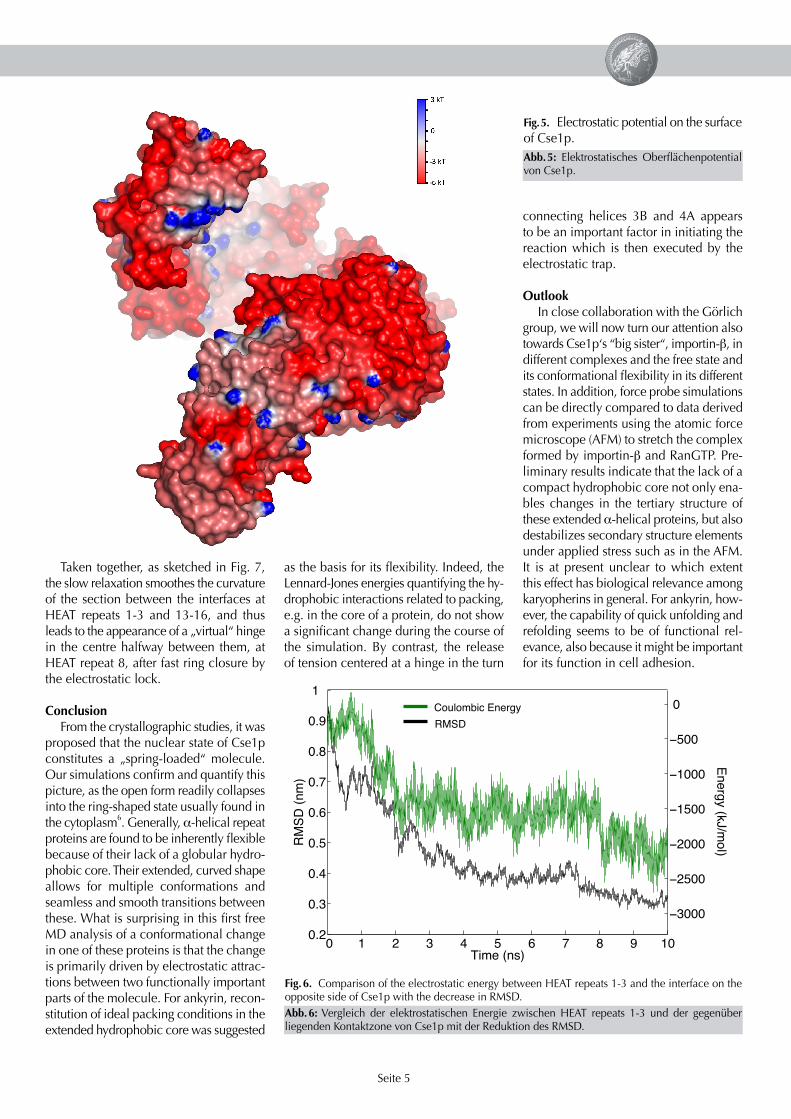
Complementing the static picture given by the crystal structures, our simulations provide a time-resolved and causal picture of the transition and the underlying driving forces. To reveal the origin of these forces, Fig. 5 shows the electrostatic potential re-sulting from the charged residues on the surface of Cse1p; red colour denotes nega-tive, blue positive potential. The entire mol-ecule has a large negative charge of -27 e,such that most parts of the surface have a strongly negative potential. A function-ally important exception can be seen near the N-terminus, at HEAT repeats 1 and 2. Here, several positively charged amino acid residues lead to a stretch exhibiting a positive potential, depicted by its blue colour. The very strong correlation between the strength of the coulombic interaction between charges on the fi rst three HEAT repeats and the opposing surface of the protein (Fig. 6, green line) and the progress on the reaction coordinate (RMSD, black line) shows that it is mainly the electrostatic attraction between the complementary charges on both interfaces which drives the conformational transition. This attrac-
100
200
300
400
500
600
700
800
900
Res
idue
Num
ber
0 1 2 3 4 5 6 7 8 9 10
0.3
0.4
0.5
0.6
0.7
0.8
0.9
1
Time (ns)
RM
SD
(nm)
0.1 0.2 0.3Deviation (nm)
Fig. 3. Spontaneous approach of the nuclear structure of Cse1p to its cytoplasmic state, as measured by RMSD (white curve), and residue-resolved regions of fl exibility contributing most to this transition (increasing deviation from blue to red).
N-terminal HEAT repeat 1 has moved by ~2 nm relative to its interface at the op-posing arch (HEAT repeats 14-16). Despite the small difference, we have thus seen the major part of the conformational change from the nuclear to the cytoplasmic state in the simulation.
The time required for the structural rearrangement is remarkably short. Fur-thermore, the conformational transition starts immediately after release of the atomic positions, taken from the 77K crystal structure in aqueous solution at 310K. Moreover, it was observed to take
place in three independent simulations at comparable time scales. This means that the open state of Cse1p is inherently unstable, which renders the population shift hypothesis rather unlikely. Instead, the open state seems to be stable only in complex with RanGTP and importin-α.Our simulations therefore suggest an in-duced fi t mechanism for RanGTP and im-portin-α binding, without any signifi cant intermediate state involved.
The colour plot underlying the RMSD curve in Fig. 3 depicts the structural de-viation of Cse1p within time windows of
Abb. 2: Schema der Transportprozesse, die am Import von denjenigen Proteinen in den Zell-kern beteiligt sind, die eine klassische Kern- Lokalisations-Sequenz (NLS) besitzen.
Abb. 3: Spontaner Übergang der im Kern vor-liegenden, offenen Struktur von Cse1p in den Zustand (weiße Kurve) und fl exible Regionen in der Sequenz von Cse1p, die zu diesem Übergang am meisten beitragen (Abweichung nimmt von blau nach rot zu).

Seite 4
Phe117, and Pro118, in the turn between helix 3B and helix 4A.
After ring closure, relaxation contin-ues on a slower time scale. This phase is characterized by slight shifts of the angles between HEAT repeats, by rearrangements of amino acid side chains, and by changes in the geometry of some helices. The A helices are longer and more curved than the B helices, and especially helices 9A and 12A contribute to the adaptation of the molecular structure to the newly formed ring of HEAT repeats by changing their bending angle or developing a new kink in the centre, respectively.
tion also locks the molecule in its fi nal closed ring form.
Whereas the electrostatics are the main driving force for closure, they are only part of the story. As the shape of the curve at the beginning of the simulation suggests (fi rst ~1 ns), the electrostatic trap does not drive the early part of the rearrangement, which is particularly fast. Instead, an energy re-laxation due to torsions around bonds is dominating during this regime. A closer look at the angles between adjacent he-lix pairs is revealing here. Whereas the angles do not differ much between the starting and the fi nal structure, the angle
Fig. 4. (A) Initial nuclear starting structure without cargo. (B) Final structure obtained from the MD simulation. (C) Crystal structure of the cytoplasmic state. (D), (E), and (F) are top views of (A), (B), and (C), respectively.
between HEAT repeats 3 and 4 shifts by over 20° - and back - in exactly this part of the simulation.
Interestingly, the region between HEAT repeats 1 and 3 is one of the main binding sites for RanGTP in the complex. Obvi-ously, this part of the molecule undergoes a rapid mechanical change when the strain induced by association with RanGTP is released. Closer analysis of this motion shows that it actually is a quasi-rigid body motion of the fi rst three HEAT repeats rela-tive to the rest of the molecule, and that it occurs around a hinge given by a con-served motif of three amino acids, Asp116,
Abb. 4: (A) Startstruktur, wie sie im Zellkern vorliegt, ohne Bindungspartner. (B) Endstruktur aus der MD-Simulation. (C) Kristallstruktur des zytoplasmatischen Zustands. (D), (E) und (F) sind um 90° gedrehte Ansichten von (A), (B), bzw. (C).
Seite 5
Taken together, as sketched in Fig. 7, the slow relaxation smoothes the curvature of the section between the interfaces at HEAT repeats 1-3 and 13-16, and thus leads to the appearance of a „virtual“ hinge in the centre halfway between them, at HEAT repeat 8, after fast ring closure by the electrostatic lock.
ConclusionFrom the crystallographic studies, it was
proposed that the nuclear state of Cse1p constitutes a „spring-loaded“ molecule. Our simulations confi rm and quantify this picture, as the open form readily collapses into the ring-shaped state usually found in the cytoplasm6. Generally, α-helical repeat proteins are found to be inherently fl exible because of their lack of a globular hydro-phobic core. Their extended, curved shape allows for multiple conformations and seamless and smooth transitions between these. What is surprising in this fi rst free MD analysis of a conformational change in one of these proteins is that the change is primarily driven by electrostatic attrac-tions between two functionally important parts of the molecule. For ankyrin, recon-stitution of ideal packing conditions in the extended hydrophobic core was suggested
as the basis for its fl exibility. Indeed, the Lennard-Jones energies quantifying the hy-drophobic interactions related to packing, e.g. in the core of a protein, do not show a signifi cant change during the course of the simulation. By contrast, the release of tension centered at a hinge in the turn
Fig. 5. Electrostatic potential on the surface of Cse1p.
0 1 2 3 4 5 6 7 8 9 100.2
0.3
0.4
0.5
0.6
0.7
0.8
0.9
1
Time (ns)
RM
SD
(nm
)
−3000
−2500
−2000
−1500
−1000
−500
0
Energy (kJ/m
ol)
Coulombic Energy
RMSD
Fig. 6. Comparison of the electrostatic energy between HEAT repeats 1-3 and the interface on the opposite side of Cse1p with the decrease in RMSD.
connecting helices 3B and 4A appears to be an important factor in initiating the reaction which is then executed by the electrostatic trap.
OutlookIn close collaboration with the Görlich
group, we will now turn our attention also towards Cse1p‘s “big sister“, importin-β, in different complexes and the free state and its conformational fl exibility in its different states. In addition, force probe simulations can be directly compared to data derived from experiments using the atomic force microscope (AFM) to stretch the complex formed by importin-β and RanGTP. Pre-liminary results indicate that the lack of a compact hydrophobic core not only ena-bles changes in the tertiary structure of these extended α-helical proteins, but also destabilizes secondary structure elements under applied stress such as in the AFM. It is at present unclear to which extent this effect has biological relevance among karyopherins in general. For ankyrin, how-ever, the capability of quick unfolding and refolding seems to be of functional rel-evance, also because it might be important for its function in cell adhesion.
Abb. 5: Elektrostatisches Oberfl ächenpotential von Cse1p.
Abb. 6: Vergleich der elektrostatischen Energie zwischen HEAT repeats 1-3 und der gegenüber liegenden Kontaktzone von Cse1p mit der Reduktion des RMSD.

Seite 6
Ulrich Zachariaestudied chemistry in Munich and Guildford and did his Ph.D. work at the Max Planck Institute of Biochemistry in Martins-ried. After postdoctoral
work at Nestlé Research Center he is now working as a Postdoc in the Department of Theoretical and Computational Biophysics since 2005.
Helmut Grubmüller1994: PhD in Theoreti-cal Physics, Technical University of Munich. 1994-1998: Postdoctoralassistant at the Theoreti-cal Biophysics Group,
University of Munich. Research visits: Laboratoire de Biophysique Moleculaire et Cellulaire, CENG, Grenoble, France; Theoretical Biophysics Group, Beckman Institute, University of Illinois at Urbana/Champaign, U.S.A.; Institute for Molecu-lar Biology and Biophysics, ETH Zürich, Switzerland. 1998-2003: Head of the Theo-retical Molecular Biophysics Group at the Max-Planck Institute for Biophysical Chem-istry. 2002: Habilitation and venia legendi in Physics at the Univ. of Göttingen. 2003: Associate Professor at the EPFL Lausanne. Since 2003: Director of the Theoretical and Computational Biophysics Department at the Max-Planck Institute for Biophysi-cal Chemistry, Göttingen, and Scientifi c Member of the Max-Planck Society. Since 2005: Honorary Professor for Physics at the Univ. of Göttingen.
t = 4 ns t = 10 nst = 0 ns
Die Zellen aller höheren Lebewesen besit-zen einen Zellkern, in dem die genetische Information in Form von DNA gespeichert ist. Eine Hülle, die aus einer Doppelmemb-ranschicht besteht, grenzt diesen Zellkern vom restlichen Volumen der Zelle ab. Da DNA nur von Proteinen abgelesen werden kann, und die abgelesene Information in Form von RNA aus dem Zellkern gebracht werden muss, existieren in dieser Hülle Kanalstrukturen, sogenannte Kernporen-Komplexe. Die Zelle hat nun einen ein-zigartigen Mechanismus entwickelt, nur diejenigen Proteine bzw. RNA durch diese Poren passieren zu lassen, die auch im Zellkern benötigt werden. Eigens dafür spe-zialisierte Proteine greifen diese Fracht auf und transportieren sie durch den Kanal, während alle anderen Proteine nicht durch
den Kanal wandern können. Darunter gibt es sogenannte Importine und Exportine, je nach Transportrichtung. Diese Transport-Proteine entstammen einer Klasse von Pro-teinen, deren Struktur eine hohe Flexibilität vermuten lässt, so dass ihre große Ober-fl äche zahlreiche Bindungen mit anderen Proteinen (bzw. RNA) ermöglicht.
Wir sind dieser Vermutung mit Molekular-dynamik-Simulationen nachgegangen undhaben die Flexibilität eines Exportins(Cse1p) untersucht; dabei wurden im Computer die Newtonschen Bewegungs-gleichungen gelöst, um die Dynamik des Proteins zu charakterisieren.
Das Exportin bindet seine Fracht (Impor-tin-α) im Zellkern zusammen mit einem weiteren Protein (RanGTP) und transpor-tiert beide durch die Kernpore. Außerhalb
des Kerns bricht dieser Komplex aus drei Proteinen auseinander und es ergibt sich eine große strukturelle Veränderung im Exportin (Ringschluss). Wir konnten mit unserer Arbeit zeigen, dass die im Zell-kern vorkommende Form von Cse1p stark vorgespannt ist und daher nur durch die Wechselwirkung mit seinen zwei Partnernstabil bleibt. Die Simulationen zeigten ferner, dass die Strukturänderung im Wesentlichen durch elektrostatische Kräfte des Proteins angetrieben wurde. Schließ-lich konnte eine mechanische Schnitt-stelle im Protein identifi ziert werden, durch die die Umlagerung eingeleitet wird. Der Ringschluss wurde durch die Computer-simulation ohne Kenntnis der Endstruktur korrekt vorhergesagt.
Zusammenfassung
Fig. 7. Schematic diagram of the closing mechanism.
References1. Weis, K. Regulating access to the genome: nucleocytoplasmic transport throughout the cell cycle. Cell 2003, 112, 441-451. 2. Chook, Y. M.; Blobel, G. Karyopherins and nuclear import. Curr. Opin. Struct. Biol. 2001, 11, 703-715. 3. Kutay, U.; Bischoff, F. R.; Kostka, S.; Kraft, R., Görlich, D. Export of importin-α from the nucleus is mediated by a specifi c nuclear transport factor. Cell 1997, 90, 1061-1071. 4. Matsuura, Y.; Stewart, M. Structural basis for the assembly of a nuclear export complex. Nature 2004, 432, 872-877. 5. Cook, A.; Fernandez, E.; Lindner, D.; Ebert, J.; Schlenstedt, G.; Conti, E. The structure of the nuclear export receptor Cse1 in its cytosolic state reveals a closed coformation incompatible with cargo binding. Mol. Cell 2005, 18, 355-367.6. Zachariae, U.; Grubmüller, H., submitted.
Abb. 7: Schematische Zeichnung des Schließmechanismus.