Embed Size (px)
Citation preview
10 MAGNETOM Flash · 2/2012 · www.siemens.com/magnetom-world
Clinical Neurology
MR Neurography – Diagnostic Criteria to Determine Lesions of Peripheral NervesPhilipp Bäumer, M.D., M.Sc.1; Sabine Heiland, Ph.D.2; Martin Bendszus, M.D.1; Mirko Pham, M.D.1
1 Department of Neuroradiology, Heidelberg University Hospital, Germany 2 Section of Experimental Radiology, Department of Neuroradiology, Heidelberg University Hospital, Germany
I) Introduction with potential clinical indications
Clinical examination and electrodiagnos-tic testing comprise the traditional meth-ods of diagnostic investigation in periph-eral nerve disease. These are excellent tools to assess function of peripheral nerves but can be limited in localizing
visualization of peripheral nerves at the fascicular level – the fascicle being the first order subunit of a peripheral nerve. The major limitation of clinical and elec-trophysiological examination is precisely the strength of MRN: the exact localiza-tion of nerve lesions. Lesion localization and spatial lesion patterns in the periph-
a lesion. Recent developments in MR scanner and coil technology and the refinement of pulse sequences for increasing structural resolution have allowed imaging of fine details in healthy and diseased peripheral nerve [1, 2]. MR Neurography (MRN) at high magnetic field strength of 3 Tesla allows for the
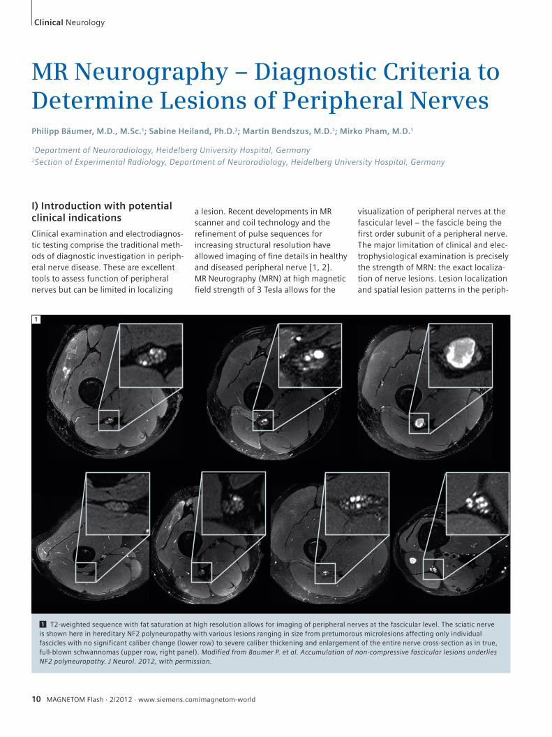
1 T2-weighted sequence with fat saturation at high resolution allows for imaging of peripheral nerves at the fascicular level. The sciatic nerve is shown here in hereditary NF2 polyneuropathy with various lesions ranging in size from pretumorous microlesions affecting only individual fascicles with no significant caliber change (lower row) to severe caliber thickening and enlargement of the entire nerve cross-section as in true, full-blown schwannomas (upper row, right panel). Modified from Baumer P. et al. Accumulation of non-compressive fascicular lesions underlies NF2 polyneuropathy. J Neurol. 2012, with permission.
1
MAGNETOM Flash · 2/2012 · www.siemens.com/magnetom-world 11
Neurology Clinical
eral nervous system are, as in many diseases of the CNS, arguably among the most important pieces of diagnostic information. One typical example for the use of MRN is the differentiation between spinal nerve root compression (radiculopathy), plexus affection and peripheral nerve lesion (peripheral neuropathy). Another frequent and important situation is the differentiation of a focal mononeuropa-thy, which is potentially surgically ame-nable, from neuropathies with involve-ment of more than one nerve. These oligo- or polyneuropathies almost exclu-sively exhibit a disseminated lesion pat-tern stemming from an inflammatory,
immune-mediated, metabolic or heredi-tary origin, so that nerve surgery is not a primary therapeutic option.In traumatic nerve injuries, exact lesion localization is the central diagnostic step in pre-surgical work-up. The assessment of nerve continuity and the identification of potential scar tissue within and around the nerve yield essential diagnostic infor-mation. In cases of true neuroma with discontinuity of the nerve, early indica-tion for reconstructive nerve surgery by MRN can improve outcome. However, in instances where MRN shows a neu-roma in continuity, surgery may become unnecessary since spontaneous recovery may occur under clinical monitoring.
II) How I do it
a) MRN protocolA magnetic field strength of 3 Tesla is advisable to achieve high-resolution images at the fascicular level of peripheral nerves. We perform clinical examinations on a Siemens MAGNETOM Verio 3 Tesla MR System (Siemens Healthcare, Erlan-gen, Germany) which allows comfortable positioning due to its wide bore diame-ter. The MRN protocol varies depending on the body region of interest and the clinical-diagnostic question. In the vast majority of patients, T2-weighted fat-saturated sequences are acquired, which show high contrast of nerve lesions to
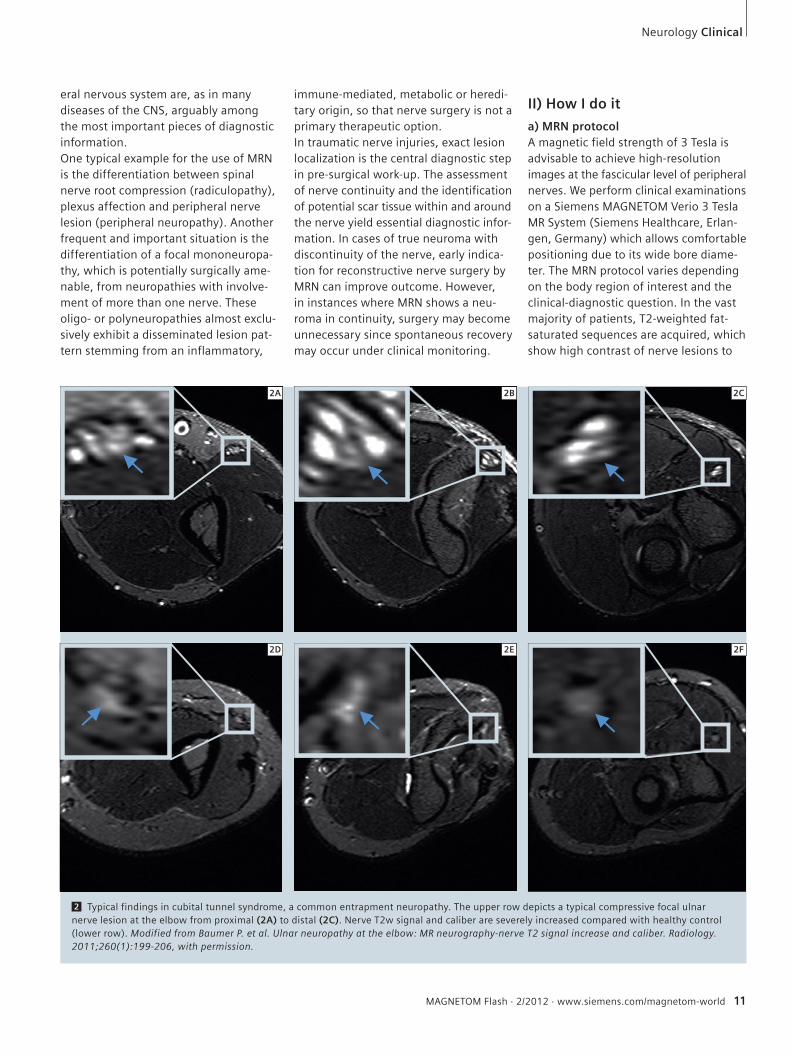
2 Typical findings in cubital tunnel syndrome, a common entrapment neuropathy. The upper row depicts a typical compressive focal ulnar nerve lesion at the elbow from proximal (2A) to distal (2C). Nerve T2w signal and caliber are severely increased compared with healthy control (lower row). Modified from Baumer P. et al. Ulnar neuropathy at the elbow: MR neurography-nerve T2 signal increase and caliber. Radiology. 2011;260(1):199-206, with permission.
2A 2B 2C
2D 2E 2F
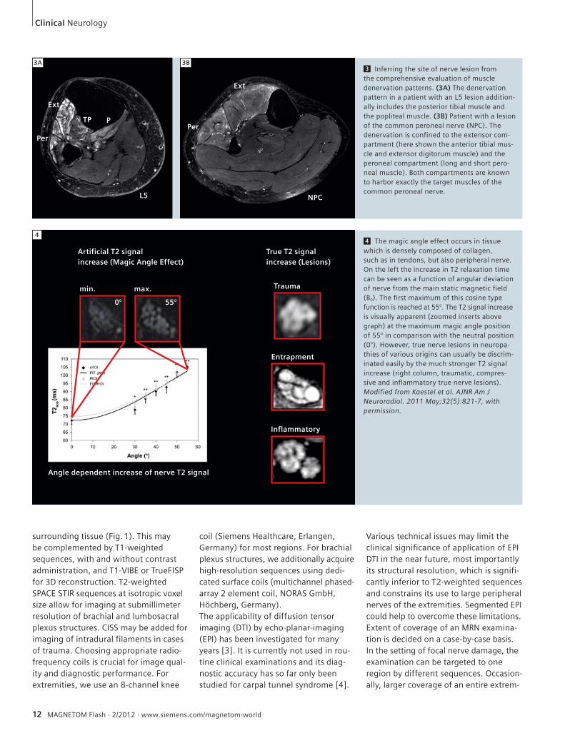
3 Inferring the site of nerve lesion from the comprehensive evaluation of muscle denervation patterns. (3A) The denervation pattern in a patient with an L5 lesion addition-ally includes the posterior tibial muscle and the popliteal muscle. (3B) Patient with a lesion of the common peroneal nerve (NPC). The denervation is confined to the extensor com-partment (here shown the anterior tibial mus-cle and extensor digitorum muscle) and the peroneal compartment (long and short pero-neal muscle). Both compartments are known to harbor exactly the target muscles of the common peroneal nerve.
12 MAGNETOM Flash · 2/2012 · www.siemens.com/magnetom-world
Clinical Neurology
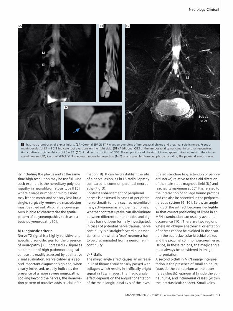
4 The magic angle effect occurs in tissue which is densely composed of collagen, such as in tendons, but also peripheral nerve. On the left the increase in T2 relaxation time can be seen as a function of angular deviation of nerve from the main static magnetic field (B0). The first maximum of this cosine type function is reached at 55°. The T2 signal increase is visually apparent (zoomed inserts above graph) at the maximum magic angle position of 55° in comparison with the neutral position (0°). However, true nerve lesions in neuropa-thies of various origins can usually be discrim-inated easily by the much stronger T2 signal increase (right column, traumatic, compres-sive and inflammatory true nerve lesions). Modified from Kaestel et al. AJNR Am J Neuroradiol. 2011 May;32(5):821-7, with permission.
4
surrounding tissue (Fig. 1). This may be complemented by T1-weighted sequences, with and without contrast administration, and T1-VIBE or TrueFISP for 3D reconstruction. T2-weighted SPACE STIR sequences at isotropic voxel size allow for imaging at submillimeter resolution of brachial and lumbosacral plexus structures. CISS may be added for imaging of intradural filaments in cases of trauma. Choosing appropriate radio-frequency coils is crucial for image qual-ity and diagnostic performance. For extremities, we use an 8-channel knee
coil (Siemens Healthcare, Erlangen, Germany) for most regions. For brachial plexus structures, we additionally acquire high-resolution sequences using dedi-cated surface coils (multichannel phased-array 2 element coil, NORAS GmbH, Höchberg, Germany). The applicability of diffusion tensor imaging (DTI) by echo-planar-imaging (EPI) has been investigated for many years [3]. It is currently not used in rou-tine clinical examinations and its diag-nostic accuracy has so far only been studied for carpal tunnel syndrome [4].
Various technical issues may limit the clinical significance of application of EPI DTI in the near future, most importantly its structural resolution, which is signifi-cantly inferior to T2-weighted sequences and constrains its use to large peripheral nerves of the extremities. Segmented EPI could help to overcome these limitations. Extent of coverage of an MRN examina-tion is decided on a case-by-case basis. In the setting of focal nerve damage, the examination can be targeted to one region by different sequences. Occasion-ally, larger coverage of an entire extrem-
3A 3B
Ext
Per
TP P
L5
Ext
Per
NPC
Artificial T2 signalincrease (Magic Angle Effect)
True T2 signalincrease (Lesions)
Trauma
Entrapment
Inflammatory
min. max.
Angle dependent increase of nerve T2 signal
0° 55°
MAGNETOM Flash · 2/2012 · www.siemens.com/magnetom-world 13
Neurology Clinical
5
ity including the plexus and at the same time high resolution may be useful. One such example is the hereditary polyneu-ropathy in neurofibromatosis type II [5] where a large number of microlesions may lead to motor and sensory loss but a single, surgically removable macrolesion must be ruled out. Also, large coverage MRN is able to characterize the spatial pattern of polyneuropathies such as dia-betic polyneuropathy [6].
b) Diagnostic criteriaNerve T2 signal is a highly sensitive and specific diagnostic sign for the presence of neuropathy [7]. Increased T2 signal as a parameter of high pathomorphogical contrast is readily assessed by qualitative visual evaluation. Nerve caliber is a sec-ond important diagnostic sign and, when clearly increased, usually indicates the presence of a more severe neuropathy. Looking beyond the nerves, the denerva-tion pattern of muscles adds crucial infor-
mation [8]. It can help establish the site of a nerve lesion, as in L5 radiculopathy compared to common peroneal neurop-athy (Fig. 3).Contrast enhancement of peripheral nerves is observed in cases of peripheral nerve sheath tumors such as neurofibro-mas, schwannomas and perineuriomas. Whether contrast uptake can discriminate between different tumor entities and dig-nities has not been formally investigated. In cases of potential nerve trauma, nerve continuity is a straightforward but essen-tial criterion when a ‘true’ neuroma has to be discriminated from a neuroma-in-continuity.
c) PitfallsThe magic angle effect causes an increase in T2 of fibrous tissue densely packed with collagen which results in artificially bright signal in T2w images. The magic angle effect depends on the angular orientation of the main longitudinal axis of the inves-
tigated structure (e.g. a tendon or periph-eral nerve) relative to the field direction of the main static magnetic field (B0) and reaches its maximum at 55°. It is related to the interaction of collage bound protons and can also be observed in the peripheral nervous system [9, 10]. Below an angle of < 30° the artifact becomes negligible so that correct positioning of limbs in an MRN examination can usually avoid its occurrence [10]. There are two regions where an oblique anatomical orientation of nerves cannot be avoided in the scan-ner: the supraclavicular brachial plexus and the proximal common peroneal nerve. Hence, in these regions, the magic angle must always be considered in image interpretation.A second pitfall in MRN image interpre-tation is the presence of small epineural (outside the epineurium as the outer nerve sheath), epineurial (inside the epi-neurium), and intraneural vessels (within the interfascicular space). Small veins
5 Traumatic lumbosacral plexus injury. (5A) Coronal SPACE STIR gives an overview of lumbosacral plexus and proximal sciatic nerve. Pseudo-meningoceles of L4 – S 2/3 indicate root avulsions on the right side. (5B) Additional CISS of the lumbosacral spinal canal in coronal reconstruc-tion confirms roots avulsions of L5 – S2. (5C) Axial reconstruction of CISS. Dorsal portions of the right L4 root appear intact at least in their intra-spinal course. (5D) Coronal SPACE STIR maximum intensity projection (MIP) of a normal lumbosacral plexus including the proximal sciatic nerve.
5A 5B 5D
5C
L5
S1
L3
L4
L5
S1
Sciaticnerve
and nerve lesions can have a similar T2 signal intensity. In most cases, small veins are readily discriminated from nerve lesions due to their winded course, strong and relatively homogeneous T2w hyperintense signal, and their eventual anatomical entry or exit into the nerve via its epineurial surface. When in doubt, one proposed method to discriminate the two is to acquire an SSFP sequence [11, 12]. Another approach is to acquire T1-weighted images with fat saturation after administration of contrast media since nerve lesions do not enhance as strongly as vessels.
III) Useful applications of MR Neurographya) Focal compressive and traumatic lesionsTypical frequent referrals for an MRN exam include suspected compressive neuro-pathies such as ulnar neuropathy at the elbow or at the wrist. MRN has a high diagnostic accuracy in confirming or excluding these [7, 13]. Main diagnostic criteria are T2 signal and caliber increase. Other frequent focal neuropathies are traumatic nerve injuries. For patients in whom the region of the nerve lesion is known, MRN is most useful in determin-
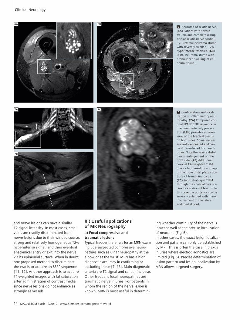
ing whether continuity of the nerve is intact as well as the precise localization of neuroma (Fig. 6).In other cases, the exact lesion localiza-tion and pattern can only be established by MRI. This is often the case in plexus injuries where electrodiagnostics are limited (Fig. 5). Precise determination of lesion pattern and lesion localization by MRN allows targeted surgery.
Clinical Neurology
6 Neuroma of sciatic nerve. (6A) Patient with severe trauma and complete disrup-tion of sciatic nerve continu-ity. Proximal neuroma stump with severely swollen, T2w hyperintense fascicles. (6B) Distal neuroma stump with pronounced swelling of epi-neural tissue.
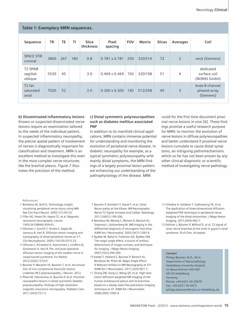
7 Confirmation and local-ization of inflammatory neu-ropathy. (7A) Composed cor-onal SPACE STIR sequence in maximum intensity projec-tion (MIP) provides an over-view of the brachial plexus on both sides. Spinal nerves are well delineated and can be differentiated from each other. Note the severe distal plexus enlargement on the right side. (7B) Additional coronal T2-weighted TIRM gives a high resolution image of the more distal plexus por-tions of truncs and cords. (7C) Sagittal-oblique TIRM through the cords allows pre-cise localization of lesions. In this case the posterior cord is severely enlarged with minor involvement of the lateral and medial cord.
6A 6B
7A
7B
7C
Ifa
v
pf
mf
14 MAGNETOM Flash · 2/2012 · www.siemens.com/magnetom-world
MAGNETOM Flash · 2/2012 · www.siemens.com/magnetom-world 15
b) Disseminated inflammatory lesionsKnown or suspected disseminated nerve lesions require an examination tailored to the needs of the individual patient. In suspected inflammatory neuropathy, the precise spatial pattern of involvement of nerves is diagnostically important for classification and treatment. MRN is an excellent method to investigate this even in the most complex nerve structures like the brachial plexus. Figure 7 illus-trates the precision of the method.
References 1 Bendszus M, Stoll G. Technology insight:
visualizing peripheral nerve injury using MRI. Nat Clin Pract Neurol. 2005;1(1):45-53.
2 Filler AG, Howe FA, Hayes CE, et al. Magnetic resonance neurography. Lancet. 1993;341(8846):659-61.
3 Hiltunen J, Suortti T, Arvela S, Seppa M, Joensuu R, Hari R. Diffusion tensor imaging and tractography of distal peripheral nerves at 3 T. Clin Neurophysiol. 2005;116(10):2315-23.
4 Hiltunen J, Kirveskari E, Numminen J, Lindfors N, Goransson H, Hari R. Pre- and post-operative diffusion tensor imaging of the median nerve in carpal tunnel syndrome. Eur Radiol. 2012;22(6):1310-9.
5 Baumer P, Mautner VF, Baumer T, et al. Accumula-tion of non-compressive fascicular lesions underlies NF2 polyneuropathy. J Neurol. 2012.
6 Pham M, Oikonomou D, Baumer P, et al. Proximal neuropathic lesions in distal symmetric diabetic polyneuropathy: findings of high-resolution magnetic resonance neurography. Diabetes Care. 2011;34(3):721-3.
Contact Philipp Bäumer, M.D., M.Sc.Department of NeuroradiologyHeidelberg University HospitalIm Neuenheimer Feld 40069120 HeidelbergGermanyPhone: +49-6221-56-39670Fax: [email protected]
7 Baumer P, Dombert T, Staub F, et al. Ulnar Neuro-pathy at the Elbow: MR Neurography-Nerve T2 Signal Increase and Caliber. Radiology. 2011;260(1):199-206.
8 Bendszus M, Wessig C, Reiners K, Bartsch AJ, Solymosi L, Koltzenberg M. MR imaging in the differential diagnosis of neurogenic foot drop. AJNR Am J Neuroradiol. 2003;24(7):1283-9.
9 Bydder M, Rahal A, Fullerton GD, Bydder GM. The magic angle effect: a source of artifact, determinant of image contrast, and technique for imaging. J Magn Reson Imaging. 2007;25(2):290-300.
10 Kastel T, Heiland S, Baumer P, Bartsch AJ, Bendszus M, Pham M. Magic Angle Effect: A Relevant Artifact in MR Neurography at 3T? AJNR Am J Neuroradiol. 2011;32(5):821-7.
11 Zhang ZW, Song LJ, Meng QF, et al. High-reso-lution diffusion-weighted MR imaging of the human lumbosacral plexus and its branches based on a steady-state free precession imaging technique at 3T. AJNR Am J Neuroradiol. 2008;29(6):1092-4.
Neurology Clinical
Table 1: Exemplary MRN sequences.
Sequence TR TE TI Slice thickness
Pixel spacing
FOV Matrix Slices Averages Coil
SPACE STIR coronal
3800 267 180 0.8 0.781 x 0.781 250 320/314 72 2 neck (Siemens)
T2 SPAIR sagittal-oblique
5530 45 3.0 0.469 x 0.469 150 320/198 51 4dedicated
surface coil (NORAS GmbH)
T2 fat-saturated axial
7020 52 3.0 0.300 x 0.300 130 512/358 45 3knee 8-channel
phased-array (Siemens)
c) Distal symmetric polyneuropathies such as diabetes mellitus associated PNPIn addition to its manifold clinical appli-cations, MRN contains immense potential for understanding and monitoring the evolution of peripheral nerve disease. In diabetic neuropathy for example, as a typical symmetric polyneuropathy with mainly distal symptoms, the MRN find-ings of a largely proximal lesion pattern are enhancing our understanding of the pathophysiology of the disease. MRN
could for the first time document proxi-mal nerve lesions in-vivo [6]. These find-ings promise a useful research purpose for MRN: to monitor the evolution of nerve lesions in diffuse polyneuropathies and better understand if proximal nerve lesions cumulate to cause distal symp-toms, an intriguing pathomechanisms which so far has not been proven by any other clinical-diagnostic or scientific method of investigating nerve pathology.
12 Chhabra A, Soldatos T, Subhawong TK, et al. The application of three-dimensional diffusion-weighted PSIF technique in peripheral nerve imaging of the distal extremities. J Magn Reson Imaging. 2011;34(4):962-7.
13 Kollmer J, Bäumer P, Milford D, et al. T2 signal of ulnar nerve branches at the wrist in guyon’s canal syndrome. PLoS One. Accepted.