Embed Size (px)
Citation preview

Mucoepidermoid carcinoma of Maxillary Sinus
ORIGINAL RESEARCH PAPER
Dr.Nishat ShahReader, Department of Oral Pathology & Microbiology, MIDSR Dental College, Ambajogai Road, Latur. Maharashra, INDIA.
ABSTRACTMalignant tumors of the paranasal sinus are uncommon, constituting less than 1% of all malignancies and 3% of all head and neck cancers. Nonsquamous cancers of the maxillary sinus are even rarer as is evident from the limited data available on the clinical characteristics and outcomes. Mucoepidermoid carcinoma (MEC) accounts for 13% of all malignancies occurring in maxillary sinus. Maxillary MEC have a worse prognosis than mandibular lesions and it should be followed-up for a longer period due to the possibility of late recurrence or regional metastasis. We report a rare case of mucoepidermoid carcinoma of maxillary sinus in a 16-year-old female with clear cell predominance.
KEYWORDS:Paranasal sinus, Nonsquamous cancers, Maxillary mucoepidermoid carcinoma, Mucous cells.
IntroductionMalignancies of the nasal cavity and paranasal sinuses constitute fewer than 1% of all malignancies and 3% of upper aerodigestive tract
[1]malignancies. The majority of these tumors are in the maxillary sinus, [2]and squamous cell carcinoma is the commonest histological type.
Mucoepidermoid carcinomas arising from mucous glands of maxillary sinus are extremely rare and accounts for 13% of all malignancies
[1]occurring in maxillary sinus. Sinonasal malignancies occur twice as often in males as in females, and are most often diagnosed in patients
[3]50 to 70 years of age. We report a rare case of mucoepidermoid carcinoma of maxillary sinus in a 16-year-old female with clear cell predominance.
Case reportA 16yr old female presented with a complaint of swelling on right side of face since 3yrs which has gradually increased to present size. The swelling was associated with the pain which was pricking type; intermittent; dull etching and aggravate on swallowing; was to be relieved by itself. Patient complained of nasal obstruction and slight difculty in breathing. There was no signicant past medical history, family history or personal history.
Extra orally a solitary diffuse swelling was noticed unilaterally on the right middle one-third of the face causing gross asymmetry of the face. Swelling was irregular, measuring about 5 cm in its greatest extension, located over the malar region, extending from infraorbital rim to the corner of mouth. Swelling resulted into drifting of the nose towards the left side of face. The skin over the swelling was shiny and inamed. On palpation the swelling was mildly tender, rm, non uctuant and non compressible [Fig 1].
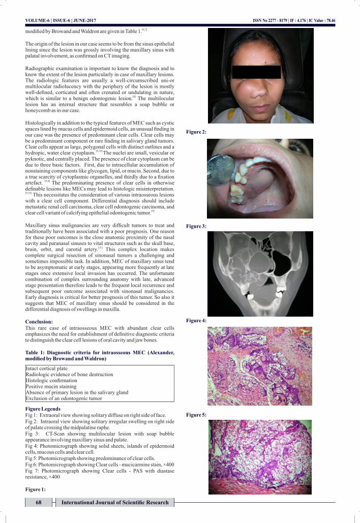
Intraorally, a diffuse swelling was seen on the right side of the palate, crossing the midline and extended from alveolar region of 14 to tuberosity of maxilla; measuring about 3× 1.5 cm in size;overlying mucosa appeared to be normal.
On palpation, site, size, extensions were conrmed; swelling was non tender; rm in consistency; with ill-dened borders; non uctuant and non compressible. With all the above mentioned clinical ndings differential diagnosis of malignancy of right maxillary sinus, mucoepidermoid carcinoma, adenoid cystic carcinoma were given.[Fig.2]
Computed tomography of right maxilla revealed an extensive contrast enhancing lesion in maxillary sinus with a soft tissue density and measuring 6 × 5.4 cm. Coronal and axial sections revealed a primarily multilocular lesion involving medial and lateral wall of maxillary sinus (medially and laterally) and hard palate inferiorly and superiorly drifting the orbital oor. Drifting of nasal septum and nasal turbinate towards the left side [Fig.3]. The H and E stained sections of the biopsy specimen showed solid sheets, islands and cords of epidermoid cells, mucus-secreting cells and abundance of clear cells in brovascular connective tissue stroma[Fig.4,5]. The sections were stained with mucicarmine and Periodic acid Schiff's reagent (PAS) to assess the nature of clear cells. The eosinophilic material in cyst like spaces was PAS and mucicarmine positive. Mucus secreting cells were visualized through mucicarmine staining [Fig.6]. The clear cells retained PAS positivity after diastase digestion [Figure 7] with a focal positivity for mucicarmine [Fig 6]. Diagnosis of low grade mucoepidermoid carcinoma of maxillary sinus with predominant clear cells was made and further conrmed by histopathological examination of excisional specimen. The patient has been on regular follow up for the last 6 years without any evidence of recurrence.
DiscussionPrimary central MEC has been reported in the rst to seventh decade; however, cases occurring in the: fourth and fth decades are most
[4] common. In children, gender ratio and the mandible to maxilla ratio is 1:1, whereas in adults: MEC is slightly more common in women and
[4]the posterior mandible. In this case the lesion was seen in 16 yr female patient.
The pathogenesis of intraosseous MEC is much debated. It may [5]originate from
Ÿ Entrapment of retromolar mucus glands within the mandible which undergo neoplastic transformation
Ÿ Neoplastic transformation of mucus secreting cells found in the pleuripotent epithelial lining of dentigerous cysts associated with impacted third molars.
Ÿ Developmentally induced embryonic remnants of the submaxillary gland within the mandible.
Ÿ Neoplastic transformation and invasion from the lining of maxillary sinus
Diagnostic criteria for intraosseous MEC proposed by Alexander and
67International Journal of Scientific Research
INTERNATIONAL JOURNAL OF SCIENTIFIC RESEARCH
Pathology
VOLUME-6 | ISSUE-6 | JUNE-2017 | ISSN No 2277 - 8179 | IF : 4.176 | IC Value : 78.46
Dr.Anuja HolaniHead of the department, Department of Oral Pathology & Microbiology, MIDSR Dental College, Ambajogai Road, Latur. Maharashra, INDIA.
Dr Varsha Ajit Sangle Sr. Lecturer, Dept. of Oral Pathology, MIDSR Dental college & hospital, Latur
Dr Abhay Kulkarni Sr. Lecturer, Dept. of Oral Medicine & Radiology, PDU Dental College, Solapur
Dr Rohit AgrawalReader, Dept. of Oral Medicine & Radiology, Maharana Pratap college of Dentistry & Research Centre, Gwalior, M.P.
Dr Shipra AgrawalSr Lecturer, Dept. of Conservative Dentistry & Endodontics, , Maharana Pratap college of Dentistry & Research Centre, Gwalior, M.P.
ISSN No 2277 - 8179 | IF : 4.176 | IC Value : 78.46VOLUME-6 | ISSUE-6 | JUNE-2017
68 International Journal of Scientific Research
[6,7]modied by Browand and Waldron are given in Table 1.
The origin of the lesion in our case seems to be from the sinus epithelial lining since the lesion was grossly involving the maxillary sinus with palatal involvement, as conrmed on CT imaging.
Radiographic examination is important to know the diagnosis and to know the extent of the lesion particularly in case of maxillary lesions. The radiologic features are usually a well-circumscribed uni-or multilocular radiolucency with the periphery of the lesion is mostly well-dened, corticated and often crenated or undulating in nature,
[8]which is similar to a benign odontogenic lesion. The multilocular lesion has an internal structure that resembles a soap bubble or honeycomb as in our case.
Histologically in addition to the typical features of MEC such as cystic spaces lined by mucus cells and epidermoid cells, an unusual nding in our case was the presence of predominant clear cells. Clear cells may be a predominant component or rare nding in salivary gland tumors. Clear cells appear as large, polygonal cells with distinct outlines and a
[9,10] hydropic, water clear cytoplasm. The nuclei are small, vesicular or pyknotic, and centrally placed. The presence of clear cytoplasm can be due to three basic factors. First, due to intracellular accumulation of nonstaining components like glycogen, lipid, or mucin. Second, due to a true scarcity of cytoplasmic organelles, and thirdly due to a xation
[9,8]artefact. The predominating presence of clear cells in otherwise denable lesions like MECs may lead to histologic misinterpretation. [9,10] This necessitates the consideration of various intraosseous lesions with a clear cell component. Differential diagnosis should include metastatic renal cell carcinoma, clear cell odontogenic carcinoma, and
[9]clear cell variant of calcifying epithelial odontogenic tumor.
Maxillary sinus malignancies are very difcult tumors to treat and traditionally have been associated with a poor prognosis. One reason for these poor outcomes is the close anatomic proximity of the nasal cavity and paranasal sinuses to vital structures such as the skull base,
[11]brain, orbit, and carotid artery. This complex location makes complete surgical resection of sinonasal tumors a challenging and sometimes impossible task. In addition, MEC of maxillary sinus tend to be asymptomatic at early stages, appearing more frequently at late stages once extensive local invasion has occurred. The unfortunate combination of complex surrounding anatomy with late, advanced stage presentation therefore leads to the frequent local recurrence and subsequent poor outcome associated with sinonasal malignancies. Early diagnosis is critical for better prognosis of this tumor. So also it suggests that MEC of maxillary sinus should be considered in the differential diagnosis of swellings in maxilla.
Conclusion:This rare case of intraosseous MEC with abundant clear cells emphasizes the need for establishment of denitive diagnostic criteria to distinguish the clear cell lesions of oral cavity and jaw bones.
Table 1: Diagnostic criteria for intraosseous MEC (Alexander, modified by Browand and Waldron)
Figure LegendsFig 1: Extraoral view showing solitary diffuse on right side of face.Fig 2: Intraoral view showing solitary irregular swelling on right side of palate crossing the midpalatine raphe.Fig 3: CT-Scan showing multilocular lesion with soap bubble appearance involving maxillary sinus and palate.Fig 4: Photomicrograph showing solid sheets, islands of epidermoid cells, mucous cells and clear cell.Fig 5: Photomicrograph showing predominance of clear cells.Fig 6: Photomicrograph showing Clear cells - mucicarmine stain, ×400 Fig 7: Photomicrograph showing Clear cells - PAS with diastase resistance, ×400
Figure 1:
Figure 2:
Figure 3:
Figure 4:
Figure 5:
Intact cortical plateRadiologic evidence of bone destructionHistologic conrmationPositive mucin stainingAbsence of primary lesion in the salivary glandExclusion of an odontogenic tumor

Figure 6:
Figure 7:
References1. Dulguerov P, Jacobsen MS, Allal AS, Lehmann W, Calcaterra T. Nasal and paranasal
sinus carcinoma: Are we making progress? A series of 220 patients and a systematic review. Cancer 2001;92:3012-29.
2. Osguthorpe JD, Richardson M. Frontal sinus malignancies. Otolaryngol Clin North Am 2001;34:269-81.
3. Carrau RL, Myers EN, Johnson JT. Paranasal sinus carcinoma-diagnosis, treatment, and prognosis. Oncology 1992;6:43-50.
4. Ezsias A, Sugar AW, Milling MAP, Ashley KF. Central mucoepidermoid carcinoma in a child. J Oral Maxillofac Surg 1994; 52: 512–515.
5. Neville BW, Damm DD, Allen CM, Bouquot JE. Oral and Maxillofacial Pathology. 2nd ed. Philadelphia, PA: Saunders; 2002. Salivary gland pathology; p. 420.
6. Alexander RW, Dupuis RH, Holton H. Central mucoepidermoid tumor (carcinoma) of the mandible. J Oral Surg 1974;32:541-7.
7. Browand BC, Waldron CA. Central mucoepidermoid tumors of the jaws. Oral Surg Oral Med Oral Pathol 1975;40:631-43.
8. Simon D, Somanathan T, Ramdas K, Pandey M. Central mucoepidermoid carcinoma of mandible: A case report and review of literature. World J Surg Oncol 2003;1:1.
9. Sivapathasundaram B, Bertin E. Clear cell variant of mucoepidermoid carcinoma –Report of a case and review of literature. Indian J Dent Res. 2003;14:23–8
10. Ellis GL, Auclair PL, Gnepp DR. In Surgical Pathology of salivary gland. 1st ed. Philadelphia: WB Saunders Company; 1991. Mucoepidermoid carcinoma; pp. 379–88.
11. Bhattacharyya N. Survival and staging characteristics for non-squamous cell malignancies of the maxillary sinus. Arch Otolaryngol Head Neck Surg 2003;129:334-7
ISSN No 2277 - 8179 | IF : 4.176 | IC Value : 78.46VOLUME-6 | ISSUE-6 | JUNE-2017
69International Journal of Scientific Research