Embed Size (px)
Citation preview
1
Multi-excitation MR elastography of the brain: wave propagation in anisotropic white matter
Daniel R. Smith1, Charlotte A. Guertler2, Ruth J. Okamoto2, Anthony J. Romano3,
Philip V. Bayly2, Curtis L. Johnson1
1 Department of Biomedical Engineering, University of Delaware; Newark, DE 19716
2 Department of Mechanical Engineering & Material Science, Washington University in
St. Louis; St. Louis, MO 63130
3 Naval Research Laboratory, Code 7160; Washington D.C. 20375
Correspondence To: Curtis L. Johnson, PhD
150 Academy St
Newark, DE 19716
Approximate Word Count: 3500
2
Abstract
Magnetic resonance elastography (MRE) has emerged as a sensitive imaging
technique capable of providing a quantitative understanding of neural microstructural
integrity. However, a reliable method for the quantification of the anisotropic mechanical
properties of human white matter is currently lacking, despite the potential to illuminate
the pathophysiology behind neurological disorders and traumatic brain injury. In this
study, we examine the use of multiple excitations in MRE to generate wave
displacement data sufficient for anisotropic inversion in white matter. We show the
presence of multiple unique waves from each excitation which we combine to solve for
parameters of an incompressible, transversely isotropic material: shear modulus, 𝜇,
shear anisotropy, 𝜙, and tensile anisotropy, 𝜁. We calculate these anisotropic
parameters in the corpus callosum body and find the mean values as 𝜇 = 3.78 kPa, 𝜙 =
0.151, and 𝜁 = 0.099 (at 50 Hz vibration frequency). This study demonstrates that multi-
excitation MRE provides displacement data sufficient for the evaluation of the
anisotropic properties of white matter.
Keywords: Magnetic Resonance Elastography, Brain, Stiffness, Anisotropy, White Matter
3
Introduction
Imaging methods for noninvasively characterizing the microstructural health and
integrity of white matter make up an important area of research for understanding
various neuropathologies. These include traumatic brain injury, which often involves
diffuse axonal injury caused by shearing of white matter due to linear or rotational
accelerations of the head. In addition to common metrics of damage or degeneration of
white matter tracts, most notably from diffusion magnetic resonance imaging (MRI) [1–
4], mechanical properties of the brain measured with magnetic resonance elastography
(MRE) [5–9] also appear sensitive to white matter tissue health. Previous work has
shown that MRE can detect mechanical alterations in several neurological diseases
including multiple sclerosis [10–12], Alzheimer’s disease [13,14], and Parkinson’s
disease [15], and animal studies have demonstrated correlations between these
properties with tissue microstructure such as axonal myelination [16,17]. However, most
MRE studies use methods that assume the brain is mechanically isotropic [6,18,19],
though the fibrous nature of white matter, comprising bundles of aligned axons, gives
rise to anisotropic, directionally-dependent mechanical properties [20–23]. As such,
anisotropic MRE methods are likely needed to improve accuracy, sensitivity, and
reliability of white matter mechanical property measurements.
Several anisotropic MRE analysis methods have been proposed, which generally
vary in the underlying anisotropic material model and thus the number of parameters to
be estimated. One anisotropic model used in MRE considers white matter as an
incompressible, transversely isotropic (ITI) material with both shear and tensile
anisotropy [24–26]. The three parameters of this model can be estimated through the
4
speed of waves propagating in different directions relative to the fiber direction, which
can be separated into “slow” and “fast” components based on their propagation and
polarization [27]. Two separate methods have been proposed to estimate these
parameters from MRE data: one based on filtering data based on the presence of slow
or fast waves [27] and a separate method using displacements at multiple frequencies
[28]. The three-parameter ITI model provides a minimal model to accurately describe
the behavior of white matter in a small number of parameters to be estimated.
Additional anisotropic MRE methods include a two-parameter model that includes only
shear anisotropy [29], which has been used to characterize breast tissue [29] and
skeletal muscle [30], and a nine-parameter model that does not assume near-
incompressibility of tissue [31], which has been used to examine white matter tracts
[32].
These anisotropic MRE methods all vary in the complexity of their material model
and details of their inversion algorithm, however, they all depend on having sufficient
displacement data to estimate parameters. In general, there must be sufficient
deformation in multiple directions to calculate the direction-dependent mechanical
behavior of white matter [31]. Tweten et al. [33] described requirements for accurate
estimation of the three parameters of the ITI model that include having slow and fast
waves present in multiple propagation directions. To ensure that these requirements are
met for anisotropic MRE of white matter, we propose to use multi-excitation MRE to
capture distinct displacement fields [34]. Anderson et al. [34] first used multi-excitation
MRE in the human brain and observed differences in recovered properties up to
approximately 25% in several white matter tracts, which is likely due to differences in
5
wave propagation arising from excitation location, making this a promising approach for
use with anisotropic inversion.
The primary objective of this paper is to measure wave propagation in the brain
from multi-excitation MRE to assess whether data requirements for anisotropic property
estimation in white matter are met. By directionally filtering the MRE displacement data,
we isolate distinct, independent propagating shear waves. We then identify the
dominant (primary) and secondary directions of wave propagation, based on relative
amplitudes of filtered components. We classify voxels in white matter based on whether
they meet minimum data requirements and show that multi-excitation MRE results in
more voxels with sufficient displacement information. Finally, we demonstrate that
combining wave information from multiple excitations allows us to estimate the three
parameters of the ITI model.
Methods
Slow and Fast Wave Propagation in ITI Materials
The three independent material parameters that determine the behavior of ITI
materials are substrate shear modulus, 𝜇, shear anisotropy, 𝜙, and tensile anisotropy, 𝜁,
which are based upon the shear modulus and tensile modulus of the material in two
directions: parallel (𝜇$ and 𝐸$) and perpendicular (𝜇& and 𝐸&) to the fibers. These
parameters are described by Eqs. 1-3:
𝜇 = 𝜇&, (1)
𝜙 = ()(*− 1, (2)
𝜁 = -)-*− 1. (3)
6
In an ITI material, shear waves with a given propagation direction, 𝑁/, can be
either “slow” (transverse) or “fast” (quasi-transverse) waves, which have separate
polarization directions. These components are determined by 𝑁/ relative to the fiber
direction, 𝐴1, and the angle between them, 𝜃 = cos6$7𝑁/ ∙ 𝐴19. The polarization direction of
the slow wave, 𝑚;<, is perpendicular to 𝑁/ and 𝐴1 (Eq. 4), while the polarization of the fast
wave, 𝑚;=, is perpendicular to 𝑁/ and 𝑚;< (Eq. 5):
𝑚;< =>/×@A|>/×@A|
, (4)
𝑚;= = 𝑁/ ×𝑚;<. (5)
The slow and fast wave speeds, 𝑐< and 𝑐=, are determined from the three
material parameters and the angle of propagation relative to fiber direction (q). The slow
wave speed depends on the shear modulus, 𝜇, material density, r, and shear
anisotropy, 𝜙 (Eq. 6), while the fast wave speed also depends on tensile anisotropy, 𝜁
(Eq. 7):
𝑐<& = (G(1 +𝜙cos& 𝜃), (6)
𝑐=& = (G(1 +𝜙cos& 2𝜃 + 𝜁 sin& 2𝜃). (7)
MRE is potentially well-suited to quantify the three anisotropic material
parameters, as wave speed, propagation direction, and fiber direction are obtainable
through MRE and MRI (i.e. using diffusion tensor imaging, DTI). However, solving for
these parameters requires sufficient displacement data, as described by Tweten et al.
[33]. Specifically, there should be (1) multiple slow and fast waves in multiple directions
and (2) each wave amplitude must have sufficient signal, i.e. at least 20% of the total
7
original amplitude. Voxels with displacement data that meet these criteria should
provide sufficient information for estimating the anisotropic parameters.
Data Acquisition
Four healthy subjects (3/1 M/F; 22-32 years old) provided informed, written
consent and participated in the study approved by our Institutional Review Board. Each
participant was scanned using a Siemens 3T Prisma MRI scanner with 20-channel head
coil. Vibrations at 50 Hz were generated by an active pneumatic driver (Resoundant,
Inc., Rochester, MN) and delivered to the head with two passive drivers: a pillow-driver
placed behind the head for anterior-posterior (AP) excitation, and a paddle-driver placed
against the temple for left-right (LR) excitation, as shown in Figure 1. Separate MRE
scans were acquired for AP and LR excitations. The acquisition employed an
echoplanar-imaging (EPI) sequence with the following parameters: 3 x 3 x 3 mm3
isotropic voxels; field-of-view = 240 x 240 mm2; 48 slices; repetition time (TR)/echo time
(TE) = 6720/65 ms. Auxiliary scans included DTI with resolution and field-of-view
matched to MRE to estimate white matter fiber direction and T1-weighted anatomical
image at 0.9 mm3 isotropic resolution to localize white matter tracts. DTI scan
parameters included: TR/TE = 4800/60 ms; b = 1000 s/mm2; 30 non-colinear direction.
T1-weighted anatomical scan parameters included: field-of-view = 256 x 256 mm2; slices
= 176; TR/TE/inversion time (TI) = 2080/4.45/1050 ms.
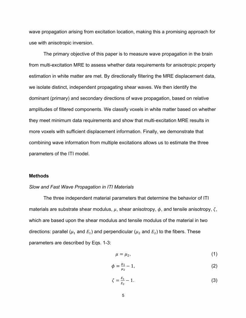
8
Figure 1. Overview of multi-excitation MRE. (A) Positioning of pillow driver for AP excitation and paddle driver for LR excitation. (B) Representative wave fields from AP and LR excitations, shown as their x, y, and z components.
Individual Wave Identification
We directionally-filtered [35] each MRE displacement field to determine dominant
directions of wave propagation [36,37]. Each wave field was directionally filtered in 300
directions across the surface of a sphere, with a spherical bandpass filter (4.2 to 166.7
m-1) applied in all directions. In each voxel, the primary wave direction was determined
as the filter direction in which the filtered field retained the highest energy. We also
identified a secondary wave direction as the filter direction which retained the next
greatest energy in the filtered field, and that differed by at least 30° from the primary
direction. From this analysis, we identified primary (𝑁/$) and secondary (𝑁/&) wave
propagation directions at every voxel for both AP and LR excitations.
We then isolated the slow and fast shear waves in each of the primary and
secondary directions. We determined the polarization directions of each slow and fast
wave based on propagation direction (primary and secondary, n = 1,2) and fiber
9
direction (Eqs. 8 and 9). Slow and fast waves were then reconstructed by projecting the
directionally filtered wave field, 𝑈OO⃑=QRS,T, onto the polarization direction (Eqs. 10 and 11):
𝑚;<,T =>/U×@A|>/U×@A|
; (8)
𝑚;=,T = 𝑁/T × 𝑚;<,T; (9)
𝑈OO⃑ <,T = V𝑈OO⃑=QRS,T ∙ 𝑚;<,TW𝑚;<,T; (10)
𝑈OO⃑=,T = V𝑈OO⃑=QRS,T ∙ 𝑚;=,TW𝑚;=,T. (11)
Using the analysis above, there are eight possible wave fields (AP/LR,
primary/secondary, slow/fast) that can be used to meet the criteria for successful
anisotropic inversion. We determined if a voxel had sufficient data to meet these criteria
if there were at least two slow waves and two fast waves with amplitude greater than
20% of the original motion amplitude and with propagation directions different by at
least 15° [33]. We considered the number of voxels that met these criteria using both
single excitations (AP or LR) and multiple excitations together (AP+LR).
Anisotropic Parameter Estimation
In order to demonstrate that individual waves from multi-excitation MRE can be
used to estimate anisotropic material parameters, we estimated 𝜇, 𝜙, and 𝜁 using an
overdetermined system of equations (Eq. 12). Specifically, we considered the
relationships for both slow and fast wave speeds (Eqs. 6 and 7) separately for each of
the primary and secondary waves included in each excitation. This resulted in a system
of eight equations comprising two polarizations (slow and fast) for each of two wave
propagation directions (primary and secondary) for each of two excitations (AP and LR).
10
Since the material parameters 𝜇, 𝜙, and 𝜁 are the same in each case, this system of
equations can be solved to generate property estimates.
(12)
Solving this system of equations requires estimates of the wave speed for each
individual wave field, which we perform for each voxel. First, for a given voxel – i.e.
(𝑥Y, 𝑦Y, 𝑧Y) – we directionally-filtered the wave field from AP or LR excitation in the
direction of the primary or secondary wave at that voxel (Figure 2A), and then projected
onto the slow or fast polarization direction to create an isolated wave field (Figure 2B) –
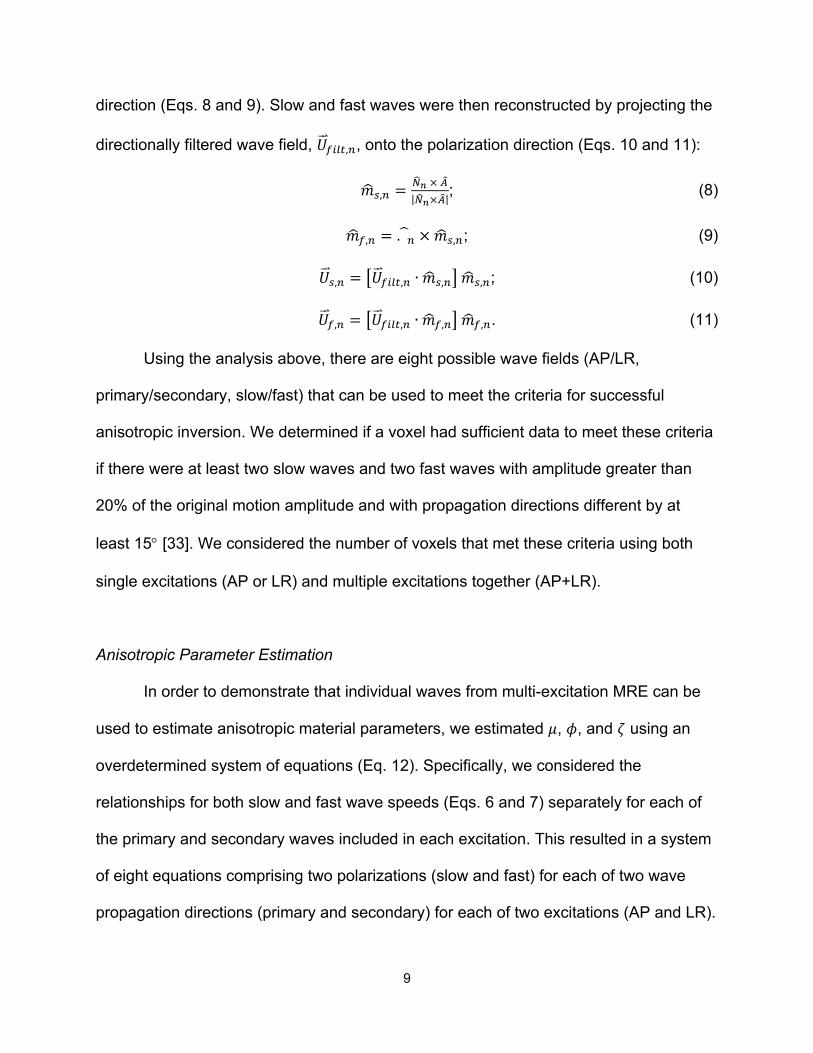
e.g. 𝑈OO⃑ <,$,@\(𝑥, 𝑦, 𝑧). This wave field is input to the local direct inversion (LDI) algorithm
[38] to estimate the wave speed – e.g. 𝑐<,$,@\(𝑥, 𝑦, 𝑧) – from which we extract the value
only at the voxel of interest (𝑥Y, 𝑦Y, 𝑧Y). This process is repeated for each voxel and for
each of the additional wave fields, as seen in Figure 2C. Each of the angles, q,
describing the relationship between propagation direction and fiber angle were
computed for each individual primary and secondary wave by 𝜃 = cos6$7𝑁/ ∙ 𝐴19. This
results in a system of equations (Eq. 12) at each voxel. We use a least squares solution
of this system at each voxel, including only the isolated wave fields that meet the
amplitude threshold (see above), to estimate 𝜇, 𝜙, and 𝜁 at each voxel. We constrained
11
values of 𝜇 to be positive between 0 and 8 kPa and left the other parameters
unconstrained in the solution.
Figure 2. The approach used to determine voxelwise stiffness estimates from individual wave fields. (A) Directional filters, based on the primary and secondary directions of a specific voxel, are applied to the wave field over the entire brain, providing two (primary
and secondary) filtered fields. (B) Slow and fast shear waves in those primary and secondary directions. (C) Isolated section, outlined by the white box, of the stiffness map estimated for each of the slow and fast waves, at every voxel, and the stiffness
map created by compiling results from individual inversions.
Analysis
We analyzed wave propagation throughout the brain, in white matter, and in an
individual white matter tract – the body of the corpus callosum, which was chosen as it
12
has highly aligned fibers and thus is likely to behave like an ITI material. The white
matter mask was created using the FMRIB Automated Segmentation Tool (FAST) in the
FMRIB Software Library (FSL) [39] to segment the T1-weighted MPRAGE, which was
then registered to the MRE data. Only voxels with fractional anisotropy (FA) > 0.25 from
DTI were included. A white matter atlas in standard-space [40] was registered to the
MRE data using the FMRIB Linear Image Registration Tool (FLIRT) in FSL [41] to
create a mask of the corpus callosum body.
Results
13
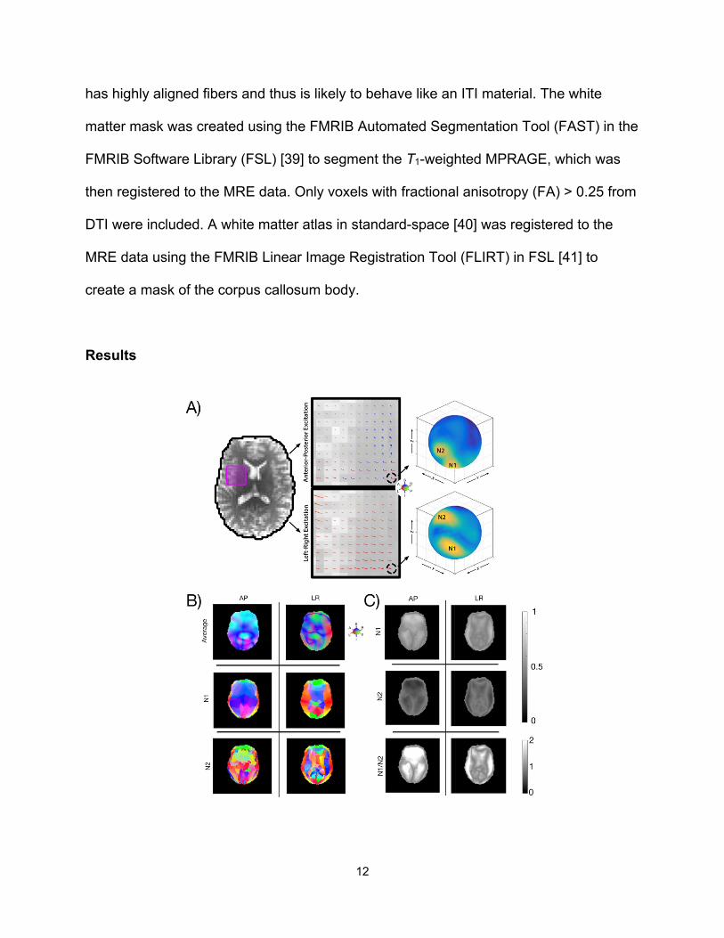
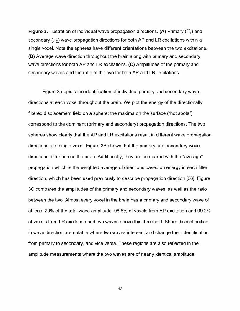
Figure 3. Illustration of individual wave propagation directions. (A) Primary (𝑁/$) and
secondary (𝑁/&) wave propagation directions for both AP and LR excitations within a
single voxel. Note the spheres have different orientations between the two excitations.
(B) Average wave direction throughout the brain along with primary and secondary wave directions for both AP and LR excitations. (C) Amplitudes of the primary and secondary waves and the ratio of the two for both AP and LR excitations.
Figure 3 depicts the identification of individual primary and secondary wave
directions at each voxel throughout the brain. We plot the energy of the directionally
filtered displacement field on a sphere; the maxima on the surface (“hot spots”),
correspond to the dominant (primary and secondary) propagation directions. The two
spheres show clearly that the AP and LR excitations result in different wave propagation
directions at a single voxel. Figure 3B shows that the primary and secondary wave
directions differ across the brain. Additionally, they are compared with the “average”
propagation which is the weighted average of directions based on energy in each filter
direction, which has been used previously to describe propagation direction [36]. Figure
3C compares the amplitudes of the primary and secondary waves, as well as the ratio
between the two. Almost every voxel in the brain has a primary and secondary wave of
at least 20% of the total wave amplitude: 98.8% of voxels from AP excitation and 99.2%
of voxels from LR excitation had two waves above this threshold. Sharp discontinuities
in wave direction are notable where two waves intersect and change their identification
from primary to secondary, and vice versa. These regions are also reflected in the
amplitude measurements where the two waves are of nearly identical amplitude.

14
Figure 4. Overview of displacement data sufficient for anisotropic inversion in white matter. (A) Number of slow and fast waves in each voxel for single excitations (AP or LR) and multiple excitations (AP+LR), and (B) white matter voxels with data meeting the minimum inversion criteria from both excitations. (C) Percentage of voxels across white matter and in the corpus callosum body, an individual white matter tract, that fit the
criteria for each excitation.
Slow and fast waves were isolated in each of the individual primary and
secondary wave directions. Figure 4 shows which voxels fit the criteria for anisotropic
inversion based on the slow and fast waves from single (AP or LR) and multiple
(AP+LR) excitations. Combining both excitations results in a substantially greater
number of voxels that meet the minimum criteria compared to single excitations (Figure
4C). Overall, 68.7% of white matter voxels meet the criteria from multiple excitations,
15
and an even higher 73.8% meet the criteria in the corpus callosum body, while many
fewer voxels met these criteria for single excitations (29-37% in each region for both AP
and LR).
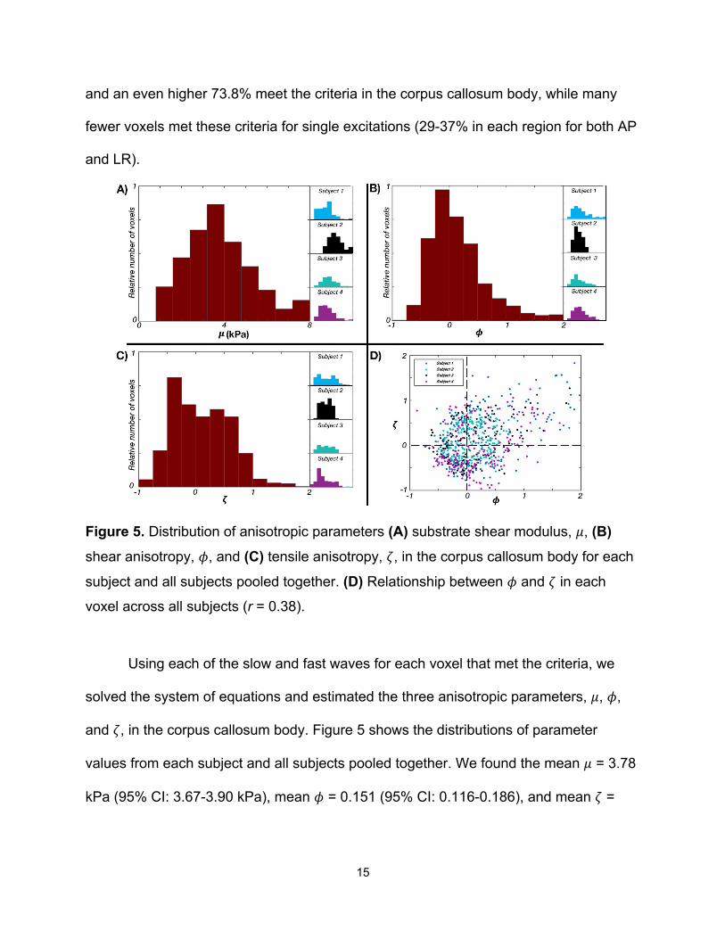
Figure 5. Distribution of anisotropic parameters (A) substrate shear modulus, 𝜇, (B) shear anisotropy, 𝜙, and (C) tensile anisotropy, 𝜁, in the corpus callosum body for each subject and all subjects pooled together. (D) Relationship between 𝜙 and 𝜁 in each voxel across all subjects (r = 0.38).
Using each of the slow and fast waves for each voxel that met the criteria, we
solved the system of equations and estimated the three anisotropic parameters, 𝜇, 𝜙,
and 𝜁, in the corpus callosum body. Figure 5 shows the distributions of parameter
values from each subject and all subjects pooled together. We found the mean 𝜇 = 3.78
kPa (95% CI: 3.67-3.90 kPa), mean 𝜙 = 0.151 (95% CI: 0.116-0.186), and mean 𝜁 =

16
0.099 (95% CI: 0.063-0.134). Figure 5D illustrates the relationship between 𝜙 and 𝜁 in
each voxel, showing a weak positive correlation between the two (r = 0.38).
Discussion
In this paper we examined the use of multi-excitation MRE to provide sufficient
displacement data to estimate anisotropic mechanical parameters of the brain in vivo.
This study builds on, and quantitatively extends, our previous observation that separate
excitation directions in MRE give rise to different property estimates in white matter [34],
which are presumably due to different propagation and polarization directions from each
excitation.
By directionally filtering the MRE displacement fields in the brain, we were able
to identify multiple shear waves with different propagation directions in most voxels.
This is the first study to demonstrate the presence of at least two (primary and
secondary) directions of wave propagation throughout the brain. In brain MRE, shear
deformations are generated from the skull [36], and thus the existence of many different
source points likely gives rise to the multiple waves. Shear waves are also likely to
originate from the falx and tentorium [36], which are stiff membranes between the
cerebral hemispheres and the cerebrum and cerebellum, and perhaps even from the
brainstem, which can move independently of the brain [42].
The two different excitation methods, AP and LR, lead to motion patterns that are
distinct but share some common features. AP-excited primary wave propagation is
predominantly in the z-direction (superior-inferior), while LR motion excites primary
wave propagation predominantly in the x-direction (left-right). In both cases there are
17
sharp discontinuities in propagation direction away from the skull where the primary and
secondary waves cross (and thus swap their identification). At these crossover regions,
two waves exist with similar amplitude. The two excitations result in greater differences
in secondary wave direction at the center of the brain where the propagating waves
excited by contact with the cranium have converged and mix with waves from internal
sources. These waves also dampen and lose amplitude causing the estimation of
secondary waves in the center of the brain to be more affected by noise. In future work,
these waves might be tracked individually back to their source points to better identify
and characterize propagating waves, and potentially identify additional waves that may
be useful in improving anisotropic MRE inversion.
By using multiple excitations, more propagation directions are generated, which
increases the number of voxels that meet the minimum criteria for anisotropic inversion.
Single excitations (AP or LR) led to a much lower percentage of voxels that met the
criteria as compared to the combination of multiple excitations (AP+LR). Thus, using
multiple excitations likely increases the ability to determine the anisotropic properties of
white matter at the individual tract or voxel level. However, two excitations did not
provide sufficient data in every white matter voxel or across an entire white matter tract,
and thus additional excitations providing unique displacement data may be required to
completely map anisotropic tissue parameters at the voxel level throughout the brain.
To combat the commensurate increase in scan time necessary for additional
excitations, accelerated data collection may be required, perhaps through simultaneous
acquisition of MRE displacement directions [43–45].
18
We demonstrated that multiple wave fields may be used to estimate anisotropic
parameters in white matter by determining the wave speeds of individual slow and fast
waves. In the corpus callosum body, we found the mean shear stiffness 𝜇 of 3.78 kPa.
This estimate is slightly higher than two other recent estimates that used a different
inversion technique without considering anisotropy [34,46], which may account for
differences we observe in this work. We found mean shear anisotropy 𝜙 of 0.151,
indicating that the shear modulus is 15.1% higher for shear in planes parallel to the fiber
direction compared to planes normal to the fibers. We also found mean tensile
anisotropy 𝜁 of 0.099, indicating that the elastic modulus is 9.9% higher parallel to the
fiber direction. The recovered 𝜙 and 𝜁 values in each voxel were positively correlated
indicating that the degree of shear and tensile anisotropy increases together, as we may
expect for fibrous material like brain white matter.
Our estimate of 𝜙 is smaller than a similar report of 𝜙 in porcine white matter
between 0.27-0.34 as reported by Schmidt, et al. [24]. However, both 𝜙 and 𝜁 are more
similar to equivalent parameters in the in vivo human corticospinal tract of 𝜙 = 0.14 and
𝜁= 0.06 as reported by Romano, et al. [31]. The true in vivo parameters are potentially
higher, indicating greater anisotropy, based on ex vivo mechanical testing [20], and the
incomplete contrast recovery in our preliminary estimates is likely due to methodological
factors. For instance, the resolution used in this study is lower than used in previous
studies of white matter tracts [34,46,47], which likely impacts the accuracy and reliability
of property maps in these small regions [48,49]. Ideally, future investigations should
adopt higher resolution imaging methods to better resolve individual white matter tracts.
19
Additionally, while this initial attempt to estimate anisotropic parameters showed
promising results, we also acknowledge several limitations. First, we used a direct
inversion method to process individual wave fields. Direct inversion methods assume
local tissue homogeneity [6] and thus introduce uncertainty at tissue boundaries, which
is exacerbated by lower spatial resolution. Inversion methods specifically designed to
model white matter anisotropy, such as the waveguide MRE method [31], may be
potentially combined with multi-excitation MRE to provide more robust parameter
estimates. Additionally, the nonlinear inversion (NLI) method [19,50], which explicitly
models tissue as a heterogeneous material and has been applied successfully to
estimate local brain properties [46,49], may improve accuracy of measures in specific
white matter tracts if formulated to include anisotropy. Advanced inversion methods will
also allow us to incorporate data from multiple frequencies and model viscoelastic
behavior of the anisotropic white matter [28,51]. More robust methods for LR excitation
or additional excitations may improve feasibility and comfort in participants. Yin et al.
[52] have previously used multiple active pneumatic drivers, in addition to multiple
passive drivers arrayed around the head, in order to excite the brain in several
directions; though the use of multiple active drivers can potentially lead to prohibitive
equipment costs. Finally, future studies are needed to evaluate the consistency and
reliability of parameter estimates through phantom studies, repeated measurements in
healthy brains [46,49,53], and studies with a larger number of subjects to determine
sensitivity to pathology.
Conclusions
20
This study is the first to investigate the use of multi-excitation MRE to estimate
anisotropic parameters (𝜇, 𝜙, and 𝜁) of an ITI material model of white matter. We found
that each excitation results in distinct shear waves, in at least two propagation
directions, and both “slow” (pure shear) and “fast” (quasi-shear) polarizations. Thus, by
combining AP and LR excitations, shear wave data from most voxels in white matter
was sufficient for anisotropic inversion. We demonstrated that by isolating individual
waves and calculating their wave speeds we could estimate the three anisotropic
parameters and recover values consistent with previous reports in white matter. Overall,
these results suggest that multi-excitation MRE is a promising technique for providing
data for anisotropic inversion and that this approach can be used in future work to more
accurately and reliably map the mechanical properties of brain white matter in vivo.
Acknowledgments
Support for this study was provided by the Office of Naval Research (N00014-17-
P-7001, N00014-18-P-7004, and N00014-18-1-2018), the Delaware INBRE Program
through NIH/NIGMS (P20-GM103446), NIH/NIBIB (R01-EB027577), NIH/NINDS (R01-
NS055951), and NSF (CMMI-1727412).
References [1] Aoki, Y., Inokuchi, R., Gunshin, M., Yahagi, N., and Suwa, H., 2012, “Diffusion
Tensor Imaging Studies of Mild Traumatic Brain Injury: A Meta-Analysis,” J. Neurol. Neurosurg. Psychiatry, 83(9), pp. 870–876.
[2] Wozniak, J. R., Krach, L., Ward, E., Mueller, B. A., Muetzel, R., Schnoebelen, S., Kiragu, A., and Lim, K. O., 2007, “Neurocognitive and Neuroimaging Correlates of Pediatric Traumatic Brain Injury: A Diffusion Tensor Imaging (DTI) Study,” Arch. Clin. Neuropsychol., 22(5), pp. 555–568.
[3] Rutgers, D. R., Fillard, P., Paradot, G., Tadié, M., Lasjaunias, P., and Ducreux,
21
D., 2008, “Diffusion Tensor Imaging Characteristics of the Corpus Callosum in Mild, Moderate, and Severe Traumatic Brain Injury,” Am. J. Neuroradiol., 29(9), pp. 1730–1735.
[4] Niogi, S. N., Mukherjee, P., Ghajar, J., Johnson, C., Kolster, R. A., Sarkar, R., Lee, H., Meeker, M., Zimmerman, R. D., Manley, G. T., and McCandliss, B. D., 2008, “Extent of Microstructural White Matter Injury in Postconcussive Syndrome Correlates with Impaired Cognitive Reaction Time: A 3T Diffusion Tensor Imaging Study of Mild Traumatic Brain Injury,” Am. J. Neuroradiol., 29(5), pp. 967–973.
[5] Muthupillai, R., Lomas, D. J., Rossman, P. J., Greenleaf, J. F., Manduca, A., and Ehman, R. L., 1995, “Magnetic Resonance Elastography by Direct Visualization of Propagating Acoustic Strain Waves.,” Science, 269(5232), pp. 1854–7.
[6] Manduca, A., Oliphant, T. E. E., Dresner, M. A. A., Mahowald, J. L. L., Kruse, S. A. A., Amromin, E., Felmlee, J. P. P., Greenleaf, J. F. F., and Ehman, R. L. L., 2001, “Magnetic Resonance Elastography: Non-Invasive Mapping of Tissue Elasticity,” Med. Image Anal., 5(4), pp. 237–254.
[7] Mariappan, Y. K., Glaser, K. J., and Ehman, R. L., 2010, “Magnetic Resonance Elastography: A Review,” Clin. Anat., 23(5), pp. 497–511.
[8] Sack, I., Beierbach, B., Hamhaber, U., Klatt, D., and Braun, J., 2008, “Non-Invasive Measurement of Brain Viscoelasticity Using Magnetic Resonance Elastography,” NMR Biomed., 21(3), pp. 265–271.
[9] Hiscox, L. V, Johnson, C. L., Barnhill, E., McGarry, M. D. J., Huston, J., van Beek, E. J. R., Starr, J. M., and Roberts, N., 2016, “Magnetic Resonance Elastography (MRE) of the Human Brain: Technique, Findings and Clinical Applications,” Phys. Med. Biol., 61(24), pp. R401–R437.
[10] Sandroff, B. M., Johnson, C. L., and Motl, R. W., 2017, “Exercise Training Effects on Memory and Hippocampal Viscoelasticity in Multiple Sclerosis: A Novel Application of Magnetic Resonance Elastography,” Neuroradiology, 59(1), pp. 61–67.
[11] Streitberger, K.-J., Sack, I., Krefting, D., Pfüller, C., Braun, J., Paul, F., and Wuerfel, J., 2012, “Brain Viscoelasticity Alteration in Chronic-Progressive Multiple Sclerosis,” PLoS One, 7(1), p. e29888.
[12] Wuerfel, J., Paul, F., Beierbach, B., Hamhaber, U., Klatt, D., Papazoglou, S., Zipp, F., Martus, P., Braun, J., and Sack, I., 2010, “MR-Elastography Reveals Degradation of Tissue Integrity in Multiple Sclerosis,” Neuroimage, 49(3), pp. 2520–2525.
[13] Murphy, M. C., Curran, G. L., Glaser, K. J., Rossman, P. J., Huston, J., Poduslo, J. F., Jack, C. R., Felmlee, J. P., and Ehman, R. L., 2012, “Magnetic Resonance Elastography of the Brain in a Mouse Model of Alzheimer’s Disease: Initial Results,” Magn. Reson. Imaging, 30(4), pp. 535–539.
[14] Murphy, M. C., Huston, J., Jack, C. R., Glaser, K. J., Manduca, A., Felmlee, J. P., and Ehman, R. L., 2011, “Decreased Brain Stiffness in Alzheimer’s Disease Determined by Magnetic Resonance Elastography,” J. Magn. Reson. Imaging, 34(3), pp. 494–498.
[15] Lipp, A., Trbojevic, R., Paul, F., Fehlner, A., Hirsch, S., Scheel, M., Noack, C., Braun, J., and Sack, I., 2013, “Cerebral Magnetic Resonance Elastography in Supranuclear Palsy and Idiopathic Parkinson’s Disease,” NeuroImage Clin., 3, pp.
22
381–387. [16] Schregel, K., Wuerfel, E., Garteiser, P., Gemeinhardt, I., Prozorovski, T., Aktas,
O., Merz, H., Petersen, D., Wuerfel, J., and Sinkus, R., 2012, “Demyelination Reduces Brain Parenchymal Stiffness Quantified in Vivo by Magnetic Resonance Elastography.,” Proc. Natl. Acad. Sci. U. S. A., 109(17), pp. 6650–5.
[17] Weickenmeier, J., de Rooij, R., Budday, S., Steinmann, P., Ovaert, T. C., and Kuhl, E., 2016, “Brain Stiffness Increases with Myelin Content,” Acta Biomater., 42, pp. 265–272.
[18] Oliphant, T. E., Manduca, A., Ehman, R. L., and Greenleaf, J. F., 2001, “Complex-Valued Stiffness Reconstruction for Magnetic Resonance Elastography by Algebraic Inversion of the Differential Equation,” Magn. Reson. Med., 45(2), pp. 299–310.
[19] McGarry, M. D. J., Van Houten, E. E. W., Johnson, C. L., Georgiadis, J. G., Sutton, B. P., Weaver, J. B., and Paulsen, K. D., 2012, “Multiresolution MR Elastography Using Nonlinear Inversion,” Med. Phys., 39(10), pp. 6388–6396.
[20] Feng, Y., Okamoto, R. J., Namani, R., Genin, G. M., and Bayly, P. V., 2013, “Measurements of Mechanical Anisotropy in Brain Tissue and Implications for Transversely Isotropic Material Models of White Matter,” J. Mech. Behav. Biomed. Mater., 23, pp. 117–132.
[21] Feng, Y., Okamoto, R. J., Genin, G. M., and Bayly, P. V, 2016, “On the Accuracy and Fitting of Transversely Isotropic Material Models,” J. Mech. Behav. Biomed. Mater., 61, pp. 554–566.
[22] Feng, Y., Lee, C.-H., Sun, L., Ji, S., and Zhao, X., 2017, “Characterizing White Matter Tissue in Large Strain via Asymmetric Indentation and Inverse Finite Element Modeling,” J. Mech. Behav. Biomed. Mater., 65, pp. 490–501.
[23] Feng, Y., Qiu, S., Xia, X., Ji, S., and Lee, C.-H., 2017, “A Computational Study of Invariant I5 in a Nearly Incompressible Transversely Isotropic Model for White Matter,” J. Biomech., 57, pp. 146–151.
[24] Schmidt, J. L., Tweten, D. J., Badachhape, A. A., Reiter, A. J., Okamoto, R. J., Garbow, J. R., and Bayly, P. V., 2018, “Measurement of Anisotropic Mechanical Properties in Porcine Brain White Matter Ex Vivo Using Magnetic Resonance Elastography,” J. Mech. Behav. Biomed. Mater., 79, pp. 30–37.
[25] Feng, Y., Clayton, E. H., Chang, Y., Okamoto, R. J., and Bayly, P. V., 2013, “Viscoelastic Properties of the Ferret Brain Measured in Vivo at Multiple Frequencies by Magnetic Resonance Elastography,” J. Biomech., 46(5), pp. 863–870.
[26] Schmidt, J. L., Tweten, D. J., Benegal, A. N., Walker, C. H., Portnoi, T. E., Okamoto, R. J., Garbow, J. R., and Bayly, P. V., 2016, “Magnetic Resonance Elastography of Slow and Fast Shear Waves Illuminates Differences in Shear and Tensile Moduli in Anisotropic Tissue,” J. Biomech., 49(7), pp. 1042–1049.
[27] Tweten, D. J., Okamoto, R. J., Schmidt, J. L., Garbow, J. R., and Bayly, P. V., 2015, “Estimation of Material Parameters from Slow and Fast Shear Waves in an Incompressible, Transversely Isotropic Material,” J. Biomech., 48(15), pp. 4002–4009.
[28] Guo, J., Hirsch, S., Scheel, M., Braun, J., and Sack, I., 2016, “Three-Parameter Shear Wave Inversion in MR Elastography of Incompressible Transverse Isotropic
23
Media: Application to in Vivo Lower Leg Muscles,” Magn. Reson. Med., 75(4), pp. 1537–1545.
[29] Sinkus, R., Tanter, M., Catheline, S., Lorenzen, J., Kuhl, C., Sondermann, E., and Fink, M., 2005, “Imaging Anisotropic and Viscous Properties of Breast Tissue by Magnetic Resonance-Elastography,” Magn. Reson. Med., 53(2), pp. 372–387.
[30] Green, M. A., Geng, G., Qin, E., Sinkus, R., Gandevia, S. C., and Bilston, L. E., 2013, “Measuring Anisotropic Muscle Stiffness Properties Using Elastography,” NMR Biomed., 26(11), pp. 1387–1394.
[31] Romano, A., Scheel, M., Hirsch, S., Braun, J., and Sack, I., 2012, “In Vivo Waveguide Elastography of White Matter Tracts in the Human Brain,” Magn. Reson. Med., 68(5), pp. 1410–1422.
[32] Romano, A., Guo, J., Prokscha, T., Meyer, T., Hirsch, S., Braun, J., Sack, I., and Scheel, M., 2014, “In Vivo Waveguide Elastography: Effects of Neurodegeneration in Patients with Amyotrophic Lateral Sclerosis,” Magn. Reson. Med., 72(6), pp. 1755–1761.
[33] Tweten, D. J., Okamoto, R. J., and Bayly, P. V., 2017, “Requirements for Accurate Estimation of Anisotropic Material Parameters by Magnetic Resonance Elastography: A Computational Study,” Magn. Reson. Med.
[34] Anderson, A. T., Van Houten, E. E., McGarry, M. D., Paulsen, K. D., Holtrop, J. L., Sutton, B. P., Georgiadis, J. G., and Johnson, C. L., 2016, “Observation of Direction-Dependent Mechanical Properties in the Human Brain with Multi-Excitation MR Elastography.”
[35] Manduca, A., Lake, D. S., Kruse, S. A., and Ehman, R. L., 2003, “Spatio-Temporal Directional Filtering for Improved Inversion of MR Elastography Images,” Med. Image Anal., 7(4), pp. 465–473.
[36] Clayton, E. H., Genin, G. M., and Bayly, P. V, 2012, “Transmission, Attenuation and Reflection of Shear Waves in the Human Brain.,” J. R. Soc. Interface, 9(76), pp. 2899–910.
[37] Okamoto, R. J., Romano, A. J., Johnson, C. L., and Bayly, P. V, 2019, “Insights Into Traumatic Brain Injury From MRI of Harmonic Brain Motion,” J. Exp. Neurosci., 13, p. 117906951984044.
[38] Okamoto, R. J., Clayton, E. H., and Bayly, P. V, 2011, “Viscoelastic Properties of Soft Gels: Comparison of Magnetic Resonance Elastography and Dynamic Shear Testing in the Shear Wave Regime,” Phys. Med. Biol., 56(19), pp. 6379–6400.
[39] Jenkinson, M., Beckmann, C. F., Behrens, T. E. J., Woolrich, M. W., and Smith, S. M., 2012, FSL, Academic Press.
[40] Mori, S., Oishi, K., Jiang, H., Jiang, L., Li, X., Akhter, K., Hua, K., Faria, A. V., Mahmood, A., Woods, R., Toga, A. W., Pike, G. B., Neto, P. R., Evans, A., Zhang, J., Huang, H., Miller, M. I., van Zijl, P., and Mazziotta, J., 2008, “Stereotaxic White Matter Atlas Based on Diffusion Tensor Imaging in an ICBM Template,” Neuroimage, 40(2), pp. 570–582.
[41] Jenkinson, M., Bannister, P., Brady, M., and Smith, S., 2002, “Improved Optimization for the Robust and Accurate Linear Registration and Motion Correction of Brain Images.,” Neuroimage, 17(2), pp. 825–41.
[42] Ji, S., and Margulies, S. S., 2007, “In Vivo Pons Motion within the Skull,” J. Biomech., 40(1), pp. 92–99.
24
[43] Klatt, D., Johnson, C. L., and Magin, R. L., 2015, “Simultaneous, Multidirectional Acquisition of Displacement Fields in Magnetic Resonance Elastography of the in Vivo Human Brain,” J. Magn. Reson. Imaging, 42(2), pp. 297–304.
[44] Nir, G., Sahebjavaher, R. S., Sinkus, R., and Salcudean, S. E., 2015, “A Framework for Optimization-Based Design of Motion Encoding in Magnetic Resonance Elastography,” Magn. Reson. Med., 73(4), pp. 1514–1525.
[45] Guenthner, C., Runge, J. H., Sinkus, R., and Kozerke, S., 2018, “Analysis and Improvement of Motion Encoding in Magnetic Resonance Elastography,” NMR Biomed., 31(5), p. e3908.
[46] Johnson, C. L., McGarry, M. D. J., Gharibans, A. A., Weaver, J. B., Paulsen, K. D., Wang, H., Olivero, W. C., Sutton, B. P., and Georgiadis, J. G., 2013, “Local Mechanical Properties of White Matter Structures in the Human Brain,” Neuroimage, 79, pp. 145–152.
[47] Guo, J., Hirsch, S., Fehlner, A., Papazoglou, S., Scheel, M., Braun, J., and Sack, I., 2013, “Towards an Elastographic Atlas of Brain Anatomy,” PLoS One, 8(8), p. e71807.
[48] Johnson, C. L., McGarry, M. D. J., Van Houten, E. E. W., Weaver, J. B., Paulsen, K. D., Sutton, B. P., and Georgiadis, J. G., 2013, “Magnetic Resonance Elastography of the Brain Using Multishot Spiral Readouts with Self-Navigated Motion Correction,” Magn. Reson. Med., 70(2), pp. 404–412.
[49] Johnson, C. L., Schwarb, H., D.J. McGarry, M., Anderson, A. T., Huesmann, G. R., Sutton, B. P., and Cohen, N. J., 2016, “Viscoelasticity of Subcortical Gray Matter Structures,” Hum. Brain Mapp., 37(12), pp. 4221–4233.
[50] Solamen, L. M., McGarry, M. D., Tan, L., Weaver, J. B., and Paulsen, K. D., 2018, “Phantom Evaluations of Nonlinear Inversion MR Elastography,” Phys. Med. Biol., 63(14), p. 145021.
[51] Testu, J., McGarry, M. D. J., Dittmann, F., Weaver, J. B., Paulsen, K. D., Sack, I., and Van Houten, E. E. W., 2017, “Viscoelastic Power Law Parameters of in Vivo Human Brain Estimated by MR Elastography,” J. Mech. Behav. Biomed. Mater., 74, pp. 333–341.
[52] Yin, Z., Sui, Y., Trzasko, J. D., Rossman, P. J., Manduca, A., Ehman, R., and Huston, J., 2018, “In Vivo Charazterization of 3D Skull and Brain Motion Using MR Elastography with Multi-Excitation Head Driver,” 2018 Joint Annual Meeting ISMRM-ESMRMB, Paris, France. Abstract 1072
[53] Murphy, M. C., Huston, J., Jack, C. R., Glaser, K. J., Senjem, M. L., Chen, J., Manduca, A., Felmlee, J. P., and Ehman, R. L., 2013, “Measuring the Characteristic Topography of Brain Stiffness with Magnetic Resonance Elastography,” PLoS One, 8(12), p. e81668.