Embed Size (px)
Citation preview

The Plant Cell, Vol. 11, 887–900, May 1999, www.plantcell.org © 1999 American Society of Plant Physiologists
Multicellular Compartmentation of
Catharanthus roseus
Alkaloid Biosynthesis Predicts Intercellular Translocation of a Pathway Intermediate
Benoit St-Pierre, Felipe A. Vazquez-Flota,
1
and Vincenzo De Luca
2,3
Institut de Recherche en Biologie Végétale, Département de Sciences Biologiques, Université de Montréal,4101 rue Sherbrooke est, Montréal, Québec H1X 2B2, Canada
In situ RNA hybridization and immunocytochemistry were used to establish the cellular distribution of monoterpenoidindole alkaloid biosynthesis in Madagascar periwinkle (
Catharanthus roseus
). Tryptophan decarboxylase (TDC) andstrictosidine synthase (STR1), which are involved in the biosynthesis of the central intermediate strictosidine, anddesacetoxyvindoline 4-hydroxylase (D4H) and deacetylvindoline 4-
O
-acetyltransferase (DAT), which are involved in theterminal steps of vindoline biosynthesis, were localized.
tdc
and
str1
mRNAs were present in the epidermis of stems,leaves, and flower buds, whereas they appeared in most protoderm and cortical cells around the apical meristem ofroot tips. In marked contrast,
d4h
and
dat
mRNAs were associated with the laticifer and idioblast cells of leaves, stems,and flower buds. Immunocytochemical localization for TDC, D4H, and DAT proteins confirmed the differential localiza-tion of early and late stages of vindoline biosynthesis. Therefore, we concluded that the elaboration of the major leaf al-kaloids involves the participation of at least two cell types and requires the intercellular translocation of a pathwayintermediate. A basipetal gradient of expression in maturing leaves also was shown for all four genes by in situ RNA hy-bridization studies and by complementary studies with dissected leaves, suggesting that expression of the vindolinepathway occurs transiently during early leaf development. These results partially explain why attempts to produce vin-doline by cell culture technology have failed.
INTRODUCTION
The organs forming the plant body consist of several differ-ent cell types that are organized in relation to each other andthat confer specific functions to the resulting organ. Eachcell type emerges from an undifferentiated meristem ac-cording to a sophisticated and partially understood develop-mental program (Sylvester et al., 1996; von Arnim and Deng,1996). The commitment to differentiate into specializedstructures involves the perception by cells in the meristemof a complex array of signals, which communicate cellularage, position in relation to other cells, and hormonal bal-ance. Environmental factors, such as light and temperature,also play a critical role in modulating these signals through-out the process of organogenesis (Bernier, 1988; Dale,1988; Sylvester et al., 1996).
In addition to morphogenesis, developmental processesresult in biochemical specialization of cells for the biosyn-
thesis and/or accumulation of secondary metabolites, suchas phenylpropanoids (Ibrahim et al., 1987; Reinold andHahlbrock, 1997), monoterpenoids (Fahn, 1988; McCaskill etal., 1992), and alkaloids (Robinson, 1974, 1981; Nessler andMahlberg, 1977; Eilert et al., 1985; Hashimoto and Yamada,1994; Facchini and De Luca, 1995). Studies with germinat-ing seedlings have suggested that alkaloid biosynthesisand accumulation are associated with seedling development(Weeks and Bush, 1974; De Luca et al., 1986; Aerts et al.,1994). Studies with mature plants also reveal this type of de-velopmental control (Westekemper et al., 1980; Frischknechtet al., 1986). Furthermore, alkaloid biosynthesis in cell sus-pension cultures appears to be coordinated with cytodiffer-entiation (Kutchan et al., 1983; Lindsey and Yeoman, 1983).
Vindoline biosynthesis in
Catharanthus roseus
also ap-pears to be under this type of developmental control(Westekemper et al., 1980; Constabel et al., 1982). In theleaves of
C. roseus
, vindoline is enzymatically coupled withcatharanthine to produce the powerful cytotoxic dimericalkaloids vinblastine and vincristine (Svoboda and Blake,1975). Vindoline as well as the dimeric alkaloids are re-stricted to leaves and stems, whereas catharanthine is dis-tributed equally throughout the aboveground and undergroundtissues (Westekemper et al., 1980; Deus-Neumann et al., 1987;
1
Current address: Unidad de Biologia Experimental, Centro de In-vestigacion Cientifica de Yucatan, Apdo. Postal 87 Cordemex 97310Merida, Yucatan, Mexico.
2
Current address: Novartis Seeds Inc., Seed Biotechnology ResearchUnit, 3054 Cornwallis Road, Research Triangle Park, NC 27709.
3
To whom correspondence should be addressed. E-mail [email protected]; fax 514-872-9406.
Dow
nloaded from https://academ
ic.oup.com/plcell/article/11/5/887/6008551 by guest on 12 O
ctober 2021

888 The Plant Cell
Balsevich and Bishop, 1989). The developmental regulation ofvindoline biosynthesis has been well documented in
C. roseus
seedlings, in which it is light inducible. This is in contrast tocatharanthine, which also accumulates in etiolated seedlings(Balsevich et al., 1986; De Luca et al., 1986). Furthermore,cell cultures that accumulate catharanthine but not vindoline(van der Heijden et al., 1989) recover this ability upon redif-ferentiation of shoots (Constabel et al., 1982). These obser-vations suggest that the biosynthesis of catharanthine andvindoline is differentially regulated and that vindoline biosyn-thesis is under more rigid tissue-, development-, and envi-ronment-specific control than is that of catharanthine.
More than 100
C. roseus
alkaloids that have been identi-fied share many common biosynthetic steps. The earlystages of alkaloid biosynthesis in
C. roseus
involve the for-mation of tryptamine from tryptophan and its condensationwith secologanin to produce the central intermediate stricto-sidine, the common precursor for the monoterpenoid indolealkaloids (Figure 1). The enzymes catalyzing these two re-actions are tryptophan decarboxylase (TDC) and strictosi-dine synthase (STR1), respectively (reviewed in De Luca,1993). Strictosidine is the precursor for both the
Iboga
(ca-tharanthine) and
Aspidosperma
(tabersonine and vindoline)types of alkaloids (Figure 1). According to this biosyntheticscheme, further enzymatic reactions would transform taber-sonine into vindoline, including hydroxylation at C-16, 16-
O
-methylation, hydration of the 2,3-double bond,
N
-meth-ylation, hydroxylation at C-4, and 4-
O
-acetylation (De Lucaet al., 1986, 1988). The first pair of reactions are catalyzedby tabersonine 16-hydroxylase and
S
-adenosyl-
L
-methionine:16-hydroxytabersonine-
O
-methyltransferase, respectively (St-Pierre and De Luca, 1995). The third to the last reaction iscatalyzed by an
S
-adenosyl-
L
-methionine:2,3-dihydro-3-hydroxytabersonine-
N
-methyltransferase (De Luca et al.,1987; Dethier and De Luca, 1993), whereas 4-hydroxylationis catalyzed by desacetoxyvindoline 4-hydroxylase (D4H), a2-oxoglutarate–dependent dioxygenase (De Carolis et al.,1990; De Carolis and De Luca, 1993). The final reaction iscatalyzed by acetyl coenzyme A (CoA):deacetylvindoline4-
O
-acetyltransferase (DAT; De Luca et al., 1985; Power etal., 1990). Some of these enzymes are not expressed in cellcultures or in tissues unable to produce vindoline (De Lucaet al., 1986, 1988; De Carolis et al., 1990).
It has been suggested that certain tissues are requiredfor alkaloid biosynthesis and/or accumulation. Yoder andMahlberg (1976) used chemical indicators to identify latici-fers and “specialized parenchyma cells” as the sites of alka-loid accumulation in
C. roseus.
Latex could be collected from
C. roseus
fruits and was shown to contain various monoter-penoid indole alkaloids (Eilert et al., 1985). Direct observa-tion of
C. roseus
leaves by epifluorescence microscopyshowed the random distribution of cells throughout themesophyll that displayed distinctive autofluorescent proper-ties (Mersey and Cutler, 1986). Leaf sections and protoplastpreparations revealed the presence of larger yellow autoflu-orescent cells with few chloroplasts, compared with the
surrounding red autofluorescent mesophyll cells. These“idioblast” cells (Mersey and Cutler, 1986), which occur inseveral plant families, are probably morphologically relatedto laticifers (Fahn, 1988), and they may be associated withthe biosynthesis and accumulation of secondary products(Postek and Tucker, 1983; Platt and Thomson, 1992).
In this study, we have used in situ RNA hybridization andimmunocytochemistry to localize the cell-specific expres-sion of
tdc
,
str1
,
d4h
, and
dat
in different tissues of
C. ro-seus
plants. The results suggest that the early (TDC andSTR1) and late (D4H and DAT) reactions of vindoline biosyn-thesis occur in different cells.
RESULTS
Previous studies with
C. roseus
have illustrated that genesinvolved in indole alkaloid biosynthesis are preferentially ex-
Figure 1. Terpenoid Indole Alkaloid Biosynthetic Pathway in C. ro-seus.
TDC converts tryptophan to tryptamine, and several enzymes con-vert geraniol into secologanin. STR1 converts tryptamine and sec-ologanin into the central intermediate strictosidine, from which arederived the Iboga, Corynanthe, and Aspidosperma alkaloids, suchas ajmalicine, catharanthine, and vindoline, respectively. The con-densation of vindoline and catharanthine leads to the biosynthesis ofthe bisindole alkaloid vinblastine. Single and multiple enzymaticsteps are shown as solid and dashed arrows, respectively.
Dow
nloaded from https://academ
ic.oup.com/plcell/article/11/5/887/6008551 by guest on 12 O
ctober 2021

Cells Specialized for Making Alkaloids 889
pressed in young actively growing tissues. Very young leaveswere fixed, embedded, sectioned, and prepared for in situRNA hybridization studies to localize transcripts of
tdc
,
str1
,
d4h
, and
dat
, which catalyze early (
tdc
and
str1
) and late(
d4h
and
dat
) steps in vindoline biosynthesis, respectively(Figure 1). The longitudinal section of a young leaf (Figure 2,diagram at top) shows that the basal end is curled and sug-gests that cell expansion leading to leaf uncurling has yet tobe completed because the bases of angiosperm leaves aredevelopmentally younger than are the more distal parts.
C. roseus
has simple, elliptical mesomorphic leaves(Mersey and Cutler, 1986) that are composed of severaltypes of cells. The upper and lower epidermis are com-posed of thin-walled cells arranged in a single layer,whereas the mesophyll is arranged into a single layer ofelongated palisade parenchyma on the adaxial side and athicker multicellular spongy parenchyma on the abaxialside of the leaf (Figure 2). In addition, unbranched, nonar-ticulated laticifers are associated with the veins (Yoder andMahlberg, 1976), which are curved and diverge from themidrib at a 35 to 45
8
angle (Mersey and Cutler, 1986).Branching from these are smaller veins generally com-posed of a tracheid and a laticifer. The palisade andspongy mesophyll also contain idioblasts, which can beidentified by their distinctive yellow autofluorescence,when leaves are illuminated by blue light (Figures 3F and3H), and by their distinctively larger size than surroundingmesophyll cells (Mersey and Cutler, 1986).
Cell-Specific Distribution of
tdc
,
str1
,
d4h
, and
dat
Transcripts in Developing Leaves
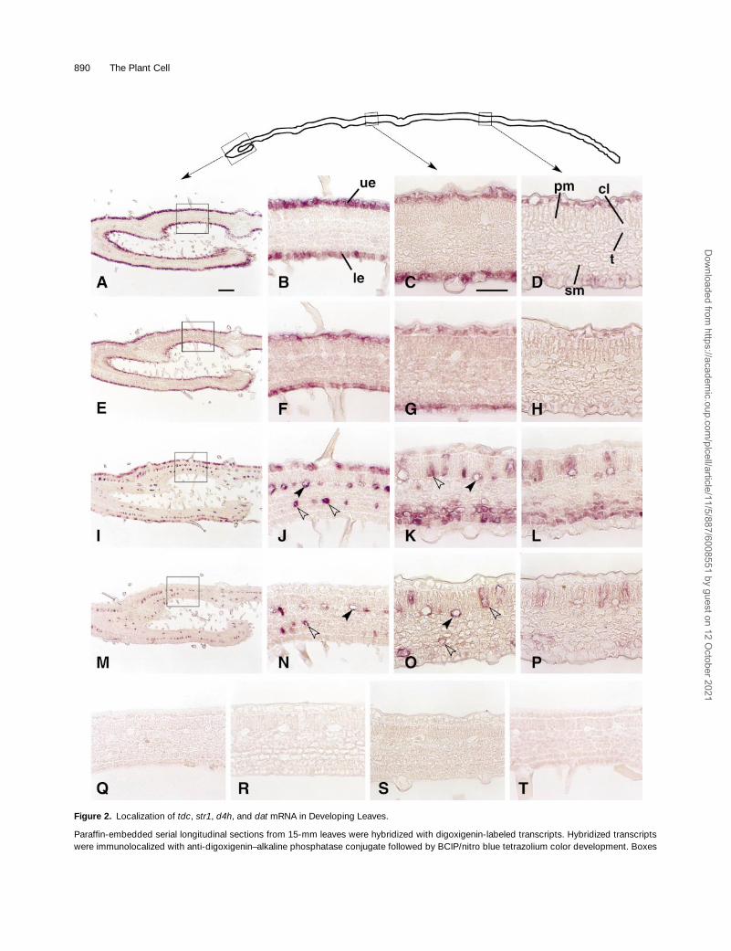
In situ RNA hybridizations of longitudinal sections with
tdc
and
str1
sense (Figures 2Q and 2R) and antisense (Figures2A to 2H) transcripts revealed that expression of these twogenes is completely restricted to the upper and lower leafepidermis. In contrast, expression of
d4h
(Figures 2I to 2L)and
dat
(Figures 2M to 2P) mRNAs (cf. sense hybridization inFigures 2S and 2T to antisense hybridization in Figures 2I to2P) is restricted almost exclusively to idioblasts occurring inthe palisade and spongy mesophyll layers and to cross-con-necting laticifers occurring at the interface between the pali-sade and spongy mesophyll. Cell-specific expression ofeach pair of transcripts was retained in the distal part of theleaf, but the levels of each transcript appeared to decreasewith leaf maturation (cf. Figures 2B, 2F, 2J, and 2N with Fig-ures 2C, 2D, 2G, 2H, 2K, 2L, 2O, and 2P). Visual inspectionof Figure 2I suggests that
d4h
also is partially expressed inthe upper epidermis of developmentally younger leaf tissue(Figures 2I and 2J), whereas it disappears in developmen-tally older upper epidermis while being retained in idioblastsand laticifers (Figures 2K and 2L).
Similar in situ RNA hybridization studies with older leavesalso were performed (data not shown). Expression of all fourgenes decreased significantly in older leaves, but the same
epidermis-, idioblast-, and laticifer-specific expression as inyounger tissues was observed.
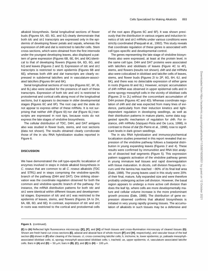
Laticifer- and Idioblast-Specific Expression of DAT
Fresh hand-cut longitudinal sections of young leaves werephotographed under blue light illumination (390 to 490 nm).The upper epidermis, the lower epidermis, the yellow fluo-rescing cross-connecting laticifers with underlying trache-ids, and the yellow fluorescing spongy mesophyll idioblastcells are clearly identified in the red fluorescent backgroundof chlorophyll-rich mesophyll (Figure 3E). Palisade meso-phyll–associated idioblasts, which also should display a yel-low fluorescence, are not clearly identified in this longitudinalsection, but many individual idioblasts are seen betweencross-connecting laticifer cells on the adaxial face of
C. ro-seus
leaves (Figure 3F). Time-course studies have shownthat the components that are responsible for the yellow flu-orescence quickly diffuse from sectioned idioblast cells,which readily explains the lack of fluorescence observed forpalisade mesophyll idioblasts in longitudinal leaf sections(Figure 3E). Longer incubation times also resulted in loss ofthe yellow fluorescent compounds occurring in laticifers(data not shown).
A comparison of longitudinal leaf sections hybridized withantisense
dat
probe (Figure 3A) to the hand-cut sectionsshows a clear correlation between the expression of
dat
andthe greenish yellow fluorescence that is characteristic of la-ticifer and spongy mesophyll–associated idioblast cells (Fig-ure 3E). This correlation also was demonstrated for palisademesophyll–associated idioblast cells; their characteristic flu-orescence is best viewed on the adaxial faces of a wholemount (Figure 3F). Additional in situ RNA hybridization stud-ies were conducted with paradermal sections prepared bystarting from the adaxial side of the leaves. Palisade meso-phyll idioblasts and vasculature-associated laticifer cells arevisualized first (Figure 3B), whereas cross-connecting latici-fer cells become apparent in the paradermal section 20
m
mfurther into the leaf (Figure 3C). The palisade mesophyll idio-blasts (Figure 3B) can be distinguished from other palisadecells because of their larger size (Mersey and Cutler, 1986).The detection of
dat
transcripts within these cell types (Fig-ures 3B and 3C) corroborates the results obtained from theanalysis of longitudinal sections (Figures 2O and 3A). Amagnified view (Figure 3D) of part of Figure 3C confirms theclose relationship observed between the presence of
dat
-expressing laticifers and the underlying tracheids. This closerelationship is clearly observed in the precise alignment ofgreenish yellow laticifers seen from the upper leaf surface(Figure 3F) with the vascular structures seen from the samesurface cleared of pigments after treatment with organic sol-vents (Figure 3G). The abaxial side of the
C. roseus
leafclearly reveals the greenish yellow fluorescence of spongymesophyll–associated idioblast cells (Figure 3H) that alsoexpress
dat
(Figure 3A).
Dow
nloaded from https://academ
ic.oup.com/plcell/article/11/5/887/6008551 by guest on 12 O
ctober 2021
890 The Plant Cell
Figure 2.
Localization of
tdc
,
str1
,
d4h
, and
dat
mRNA in Developing Leaves.
Paraffin-embedded serial longitudinal sections from 15-mm leaves were hybridized with digoxigenin-labeled transcripts. Hybridized transcriptswere immunolocalized with anti-digoxigenin–alkaline phosphatase conjugate followed by BCIP/nitro blue tetrazolium color development. Boxes
Dow
nloaded from https://academ
ic.oup.com/plcell/article/11/5/887/6008551 by guest on 12 O
ctober 2021

Cells Specialized for Making Alkaloids 891
Cell-Specific Distribution of TDC, D4H, and DAT Proteins in Developing Leaves
Longitudinal sections of the same
C. roseus
leaf shown inFigure 2 were treated with polyclonal rabbit antibodiesraised against TDC (Fernandez et al., 1989), D4H (Vazquez-Flota and De Luca, 1998), and DAT (St-Pierre et al., 1998),respectively, to identify which leaf cells produce these pro-teins. Expression of the TDC protein is completely restrictedto the upper and lower leaf epidermis (Figures 4A and 4B),as was the case for
tdc
transcript (Figures 2A to 2H). Simi-larly, expression of D4H (Figures 4C and 4D) and DAT (Fig-ures 4E and 4F) proteins appeared to be restricted toidioblasts and laticifers, which are also the sites of expres-sion for the respective mRNAs (Figures 2I to 2P). In contrastto the expression of
d4h
transcripts, which also was ob-served in the upper epidermis of developmentally youngerleaf tissue in Figure 2I, the D4H protein was detected onlyin idioblasts and laticifers throughout the leaf section (Fig-ures 4C and 4D; data not shown). Treatment of sectionswith preimmune serum did not produce any immune reac-tions to any cell type in these sections, even when applied ata low dilution (Figures 4G and 4H). Visual inspection of lon-gitudinal sections also confirmed that the patterns of TDC,D4H, and DAT protein expression coincided with those oftranscript accumulation and that the levels of each proteinappeared to decrease with developmental age. The identicalresults obtained for the localization of
tdc
,
d4h
, and
dat
tran-scripts compared with TDC, D4H, and DAT proteins stronglysuggest that at least two different cell types within the sametissue are involved in the biosynthesis of vindoline in
C. roseus.
Differential Accumulation of TDC, STR1, D4H, and DAT in the Leaf Blade
Visual inspection of longitudinal sections of
C. roseus
leavessuggested that the leaf base contained the highest levels ofall four mRNAs (Figure 2) and of the three enzymes (Figure4). The middle portion of the leaf expressed lower levels of
this pathway, whereas virtually no expression was observednear the tip of the leaf blade. These results suggest a basi-petal gradient of expression for the genes involved in indolealkaloid biosynthesis. This pattern of expression also corre-lates with the denser distribution of laticifers characteristicfor the basal area compared with those found in the middlesection or at the tip of the leaf. It also correlates with a basi-petal gradient of the yellow autofluorescent compounds inthe laticifer and idioblast cells (result not shown).
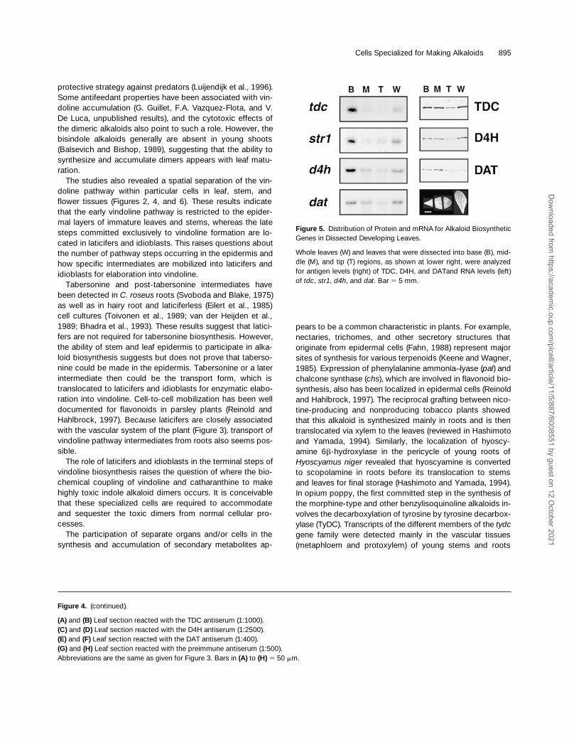
To confirm this general trend, we dissected immatureleaves (Figure 5, lower right) into base, middle, and tip areasto determine the levels of these four transcripts and of TDC,D4H, and DAT enzymes. RNA and protein were extractedseparately from whole leaves or their components, asshown in Figure 5. RNA gel blot analysis (Figure 5, left)clearly showed the basipetal distribution of all four tran-scripts, with the highest, intermediate, and lowest levels oc-curring in the basal, middle, and tip regions of the leaf,respectively. However, immunoblot analysis (Figure 5, right)revealed that the levels of the three proteins were almostequivalent in the basal and middle regions of the leaves.Their levels declined only in the tip region of the leaf, sug-gesting that these proteins were more stable than were theircorresponding mRNAs during leaf maturation. These results,which clearly suggest that the pathway leading to vindolinebiosynthesis appears very early in leaf development,corroborate the qualitative differences in the levels of thecorresponding transcripts as observed by in situ RNA hy-bridization of
C. roseus
leaves (Figure 2).
Sites of Indole Alkaloid Biosynthesis in Flower Primordia, Stems, and Roots Tips
Previous studies using various techniques have demon-strated that most
C. roseus
tissues are active in the biosyn-thesis of indole alkaloids and that each organ accumulates acharacteristic spectrum of alkaloids. In situ RNA hybridiza-tion studies were conducted to identify which cells in devel-oping flowers, stems, and root tips actually were involved in
in the profile of a longitudinal section at the top show the photographed area: the revoluted leaf base (first box;
[A]
,
[E]
,
[I]
, and
[M]
), the middlearea of the leaf at a distance of 4.5 mm from the base (second box;
[C]
,
[G]
,
[K]
, and
[O]
), and the tip portion of the leaf at 8.5 mm from the base(third box;
[D]
,
[H]
,
[L]
, and
[P]
). Boxed areas in
(A)
,
(E)
,
(I)
, and
(M)
are magnified in
(B)
,
(F)
,
(J)
, and
(N)
, respectively. A magnification of the leafbase also is shown in
(Q)
to
(T)
.
(A)
to
(D)
Sections were hybridized with antisense RNA for
tdc.
(E)
to
(H)
Sections were hybridized with antisense RNA for
str1.
(I)
to
(L)
Sections were hybridized with antisense RNA for d4h.(M) to (P) Sections were hybridized with antisense RNA for dat.(Q) to (T) Sections were hybridized with sense RNA for tdc (Q), str1 (R), d4h (S), and dat (T).cl, cross-connecting laticifer cells; le, lower epidermis; pm, palisade mesophyll cells; sm, spongy mesophyll cells; t, tracheid; ue, upper epider-mis. Solid arrowheads show laticifer cells; open arrowheads point to idioblast cells. Bar in (A) 5 100 mm for panels (A), (E), (I), and (M); bar in (C) 550 mm for (B) to (D), (F) to (H), (J) to (L), and (N) to (T).
Figure 2. (continued).
Dow
nloaded from https://academ
ic.oup.com/plcell/article/11/5/887/6008551 by guest on 12 O
ctober 2021

892 The Plant Cell
Figure 3. Localization of dat mRNA, Fluorescent Compounds, and Vascular Tissue in Developing Leaves.
(A) to (D) Paraffin-embedded sections were hybridized with antisense RNA for dat. (A) shows a longitudinal section of the leaf base, (B) a para-dermal section mainly through the palisadic mesophyll, (C) a paradermal section 20 mm under the section shown in (B), and (D) magnification ofthe boxed area shown in (C).
Dow
nloaded from https://academ
ic.oup.com/plcell/article/11/5/887/6008551 by guest on 12 O
ctober 2021
Cells Specialized for Making Alkaloids 893
alkaloid biosynthesis. Serial longitudinal sections of flowerbuds (Figures 6A, 6D, 6G, and 6J) clearly demonstrate thatboth tdc and str1 transcripts are expressed only in the epi-dermis of developing flower petals and sepals, whereas theexpression of d4h and dat is restricted to laticifer cells. Stemcross-sections, which were obtained from the first internodeunder the youngest developing leaves, also displayed a pat-tern of gene expression (Figures 6B, 6E, 6H, and 6K) identi-cal to that of developing flowers (Figures 6A, 6D, 6G, and6J) and leaves (Figures 2 and 3). Expression of tdc and str1transcripts is restricted to stem epidermis (Figures 6B and6E), whereas both d4h and dat transcripts are clearly ex-pressed in subdermal laticifers and in vasculature-associ-ated laticifers (Figures 6H and 6K).
Serial longitudinal sections of root tips (Figures 6C, 6F, 6I,and 6L) also were studied for the presence of each of thesetranscripts. Expression of both tdc and str1 is restricted toprotodermal and cortical cells along most of the longitudinalsections, but it appears to decrease in older developmentalstages (Figures 6C and 6F). The root cap and the stele donot appear to express either of these mRNAs. It is not sur-prising that neither d4h (Figure 6I) nor dat (Figure 6L) tran-scripts are expressed in root tips, because roots do notexpress the late stages of vindoline biosynthesis.
The cellular distribution of TDC, D4H, and DAT antigensalso was studied in flower buds, stems, and root sections(data not shown). The results obtained clearly corroboratethose of the in situ RNA hybridization studies reported inFigure 6.
DISCUSSION
We have demonstrated the cell type–specific localization ofenzymes involved in steps in indole alkaloid biosynthesis inC. roseus that are common to all C. roseus alkaloids (TDCand STR1) and in steps comprising the vindoline-specificbranch of the pathway (D4H and DAT). One striking obser-vation was the coordinate regulation observed for both thecommon and vindoline-specific branch of the pathway. Forinstance, the mRNA distribution patterns for both tdc andstr1 were identical within different tissues and developmen-tal stages. Expression of tdc and str1 was restricted to theepidermis of leaves, stems, and flowers (Figures 2A to 2H,6A, 6B, 6D, and 6E). In contrast, expression of tdc and str1in roots was restricted to the cortical and protodermal cells
of the root apex (Figures 6C and 6F). It was shown previ-ously that the distribution in various organs and induction ki-netics of tdc and str1 mRNAs under various conditions werestrictly coordinated (Pasquali et al., 1992). This study revealsthat coordinate regulation of these genes is associated withcell type–specific and developmental control.
The genes representing the late stage of vindoline biosyn-thesis also were expressed, at least at the protein level, inthe same cell type. D4H and DAT proteins were associatedwith laticifers and idioblasts of leaves (Figure 4C to 4F),stems, and flowers (results not shown). d4h and dat mRNAsalso were colocalized in idioblast and laticifer cells of leaves,stems, and flower buds (Figures 2I to 2P, 6G, 6H, 6J, and6K), and there was no detectable expression of either genein roots (Figures 6I and 6L). However, ectopic accumulationof d4h mRNA was observed in upper epidermal cells and insome spongy mesophyll cells in the vicinity of idioblast cells(Figures 2I to 2L) without the concomitant accumulation ofD4H protein (Figures 4C and 4D). Whereas coordinate regu-lation of d4h and dat was expected from many lines of evi-dence, particularly from their induction kinetics and light-dependent regulation in developing seedlings, as well astheir distribution patterns in mature plants, some data sug-gested specific mechanism of regulation for d4h. For in-stance, d4h mRNAs (Vazquez-Flota and De Luca, 1998), incontrast to those of dat (St-Pierre et al., 1998), rose to signif-icant levels in dark-grown seedlings.
The in situ RNA hybridization and immunocytochemicallocalization studies presented in this report revealed that ex-pression of the vindoline pathway follows a basipetal distri-bution in young expanding leaves (Figures 2 and 4). Theseresults were confirmed by immunoblot and RNA blot analy-sis of dissected leaf segments (Figure 5). This expressionpattern suggests activation of the vindoline pathway genesin young immature leaf tissues and rapid downregulationwith tissue maturation. In dicots, cell division frequently oc-curs until the lamina has reached z90% of its final leaf area(Dale, 1988). The young leaves used in this study were 20%of their final, mature, fully expanded size and were thereforeprobably undergoing active cell division. However, the basalregion appears to undergo a more active cell division thandoes the leaf tip, where cells are more developmentally ma-ture and cellular volume increase is the more predominantgrowth process (Dale, 1988). The distribution of gene ex-pression observed confirms that alkaloid biosynthesis isinitiated in very young rapidly growing tissues. The accumu-lation of alkaloids in such tissues may be a defensive and
Figure 3. (continued).
(E) to (H) Reflected light fluorescence microscopy ([E], [F], and [H]) of fresh tissues and cross-illumination microscopy of cleared tissues (G).Shown are fresh hand-cut cross-sections (E), adaxial and abaxial face of whole mount ([F] and [H], respectively), and vascular tissue of the leafsection (G) shown in (F) after clearing of the tissues. cl, cross-connecting laticifer cells; h, trichome; le, lower epidermis; pi, palisade mesophyll–associated idioblast cells; si, spongy mesophyll–associated idioblast cells; t, tracheid; ue, upper epidermis; vl, vasculature-associated laticifercells. Bars in (A) and (D) 5 50 mm; bars in (B), (C), and (E) to (H) 5 100 mm.
Dow
nloaded from https://academ
ic.oup.com/plcell/article/11/5/887/6008551 by guest on 12 O
ctober 2021

894 The Plant Cell
Figure 4. Immunolocalization of TDC, D4H, and DAT Proteins in Developing Leaves.
Longitudinal sections of the same leaf shown in Figure 2. Microscopy is from the base ([A], [C], [E], and [G]) and from the middle portion (4.5mm from the base) of the leaf ([B], [D], [F], and [H]).
Dow
nloaded from https://academ
ic.oup.com/plcell/article/11/5/887/6008551 by guest on 12 O
ctober 2021
Cells Specialized for Making Alkaloids 895
protective strategy against predators (Luijendijk et al., 1996).Some antifeedant properties have been associated with vin-doline accumulation (G. Guillet, F.A. Vazquez-Flota, and V.De Luca, unpublished results), and the cytotoxic effects ofthe dimeric alkaloids also point to such a role. However, thebisindole alkaloids generally are absent in young shoots(Balsevich and Bishop, 1989), suggesting that the ability tosynthesize and accumulate dimers appears with leaf matu-ration.
The studies also revealed a spatial separation of the vin-doline pathway within particular cells in leaf, stem, andflower tissues (Figures 2, 4, and 6). These results indicatethat the early vindoline pathway is restricted to the epider-mal layers of immature leaves and stems, whereas the latesteps committed exclusively to vindoline formation are lo-cated in laticifers and idioblasts. This raises questions aboutthe number of pathway steps occurring in the epidermis andhow specific intermediates are mobilized into laticifers andidioblasts for elaboration into vindoline.
Tabersonine and post-tabersonine intermediates havebeen detected in C. roseus roots (Svoboda and Blake, 1975)as well as in hairy root and laticiferless (Eilert et al., 1985)cell cultures (Toivonen et al., 1989; van der Heijden et al.,1989; Bhadra et al., 1993). These results suggest that latici-fers are not required for tabersonine biosynthesis. However,the ability of stem and leaf epidermis to participate in alka-loid biosynthesis suggests but does not prove that taberso-nine could be made in the epidermis. Tabersonine or a laterintermediate then could be the transport form, which istranslocated to laticifers and idioblasts for enzymatic elabo-ration into vindoline. Cell-to-cell mobilization has been welldocumented for flavonoids in parsley plants (Reinold andHahlbrock, 1997). Because laticifers are closely associatedwith the vascular system of the plant (Figure 3), transport ofvindoline pathway intermediates from roots also seems pos-sible.
The role of laticifers and idioblasts in the terminal steps ofvindoline biosynthesis raises the question of where the bio-chemical coupling of vindoline and catharanthine to makehighly toxic indole alkaloid dimers occurs. It is conceivablethat these specialized cells are required to accommodateand sequester the toxic dimers from normal cellular pro-cesses.
The participation of separate organs and/or cells in thesynthesis and accumulation of secondary metabolites ap-
pears to be a common characteristic in plants. For example,nectaries, trichomes, and other secretory structures thatoriginate from epidermal cells (Fahn, 1988) represent majorsites of synthesis for various terpenoids (Keene and Wagner,1985). Expression of phenylalanine ammonia–lyase (pal) andchalcone synthase (chs), which are involved in flavonoid bio-synthesis, also has been localized in epidermal cells (Reinoldand Hahlbrock, 1997). The reciprocal grafting between nico-tine-producing and nonproducing tobacco plants showedthat this alkaloid is synthesized mainly in roots and is thentranslocated via xylem to the leaves (reviewed in Hashimotoand Yamada, 1994). Similarly, the localization of hyoscy-amine 6b-hydroxylase in the pericycle of young roots ofHyoscyamus niger revealed that hyoscyamine is convertedto scopolamine in roots before its translocation to stemsand leaves for final storage (Hashimoto and Yamada, 1994).In opium poppy, the first committed step in the synthesis ofthe morphine-type and other benzylisoquinoline alkaloids in-volves the decarboxylation of tyrosine by tyrosine decarbox-ylase (TyDC). Transcripts of the different members of the tydcgene family were detected mainly in the vascular tissues(metaphloem and protoxylem) of young stems and roots
Figure 4. (continued).
(A) and (B) Leaf section reacted with the TDC antiserum (1:1000).(C) and (D) Leaf section reacted with the D4H antiserum (1:2500).(E) and (F) Leaf section reacted with the DAT antiserum (1:400).(G) and (H) Leaf section reacted with the preimmune antiserum (1:500).Abbreviations are the same as given for Figure 3. Bars in (A) to (H) 5 50 mm.
Figure 5. Distribution of Protein and mRNA for Alkaloid BiosyntheticGenes in Dissected Developing Leaves.
Whole leaves (W) and leaves that were dissected into base (B), mid-dle (M), and tip (T) regions, as shown at lower right, were analyzedfor antigen levels (right) of TDC, D4H, and DATand RNA levels (left)of tdc, str1, d4h, and dat. Bar 5 5 mm.
Dow
nloaded from https://academ
ic.oup.com/plcell/article/11/5/887/6008551 by guest on 12 O
ctober 2021

896 The Plant Cell
Figure 6. Localization of tdc, str1, d4h, and dat mRNA in Flower Primordia, Stems, and Roots.
Dow
nloaded from https://academ
ic.oup.com/plcell/article/11/5/887/6008551 by guest on 12 O
ctober 2021

Cells Specialized for Making Alkaloids 897
(Facchini and De Luca, 1995). The present study suggeststhat the precise cell-, tissue-, and organ-specific compart-ment of monoterpenoid indole alkaloid biosynthesis, and ofsecondary metabolism in general, is regulated by differentialexpression of biosynthetic pathways and by a controlledtransport of intermediates to the most appropriate sites foraccumulation.
It is not clear if a similar root-to-shoot alkaloid transportsystem exists in C. roseus. Catharanthine and advancedprecursors of vindoline biosynthesis, such as tabersonine,may be synthesized in roots to be transported to laticifersand idioblasts in leaves and stem, for elaboration intodimeric alkaloids and vindoline, respectively. However, theability of in vitro leaf cultures, which have no roots, to makevindoline suggests that roots are not absolutely required tomake this possible (Endo et al., 1987). The available evi-dence suggests that tabersonine and catharanthine may beproduced in more than one location, and it remains to beelucidated how substrates for vindoline and dimer produc-tion are made available.
The clear differential cell localization of the early and latestages of vindoline biosynthesis in leaves strongly suggeststhat undetermined poststrictosidine compounds are mobi-lized from the epidermis to laticifers and idioblasts, where atleast the last two reactions of vindoline biosynthesis takeplace. Differential cell-specific expression and intercellulartransport of metabolites also have been suggested for thephenylpropanoid pathway (Reinold and Hahlbrock, 1997). Inparsley leaves, the genes involved in general phenylpro-panoid metabolism, such as pal and 4-coumarate:CoA li-gase (4cl), are expressed mainly in the vascular bundle andin epithelial cells surrounding oil ducts. Lower levels of paland 4cl transcripts also were detected in the leaf epidermis,palisade cells, and spongy mesophyll cells and within the oilduct surrounding cells (Reinold and Hahlbrock, 1997).
However, the locations of the pathways leading to the for-mation of flavonoids or furanocoumarins were mutually ex-clusive. Flavonoids and furanocoumarins are two majorproducts derived from different branches of the phenylpro-panoid pathway, downstream from 4-coumaryl–CoA. chs,which is involved specifically in flavonoid biosynthesis, wasrestricted to the epidermis, spongy mesophyll, and oil ductsurrounding cells. S-adenosyl-L-methionine:bergaptol O-meth-yltransferase, which is involved in furanocoumarin biosyn-
thesis, occurred in the vascular bundle, palisade parenchyma,and the oil duct epithelial cells (Reinold and Hahlbrock,1997). Interestingly, flavonoids were detected in vascularbundles and oil duct epithelial cells that do not express chs,suggesting the mobilization of these compounds from otherexpressing cells (Reinold and Hahlbrock, 1997).
A similar cellular distribution to those of leaves also wasfound for TDC, STR1, and D4H (F.A. Vazquez-Flota, B.St-Pierre, and V. De Luca, manuscript in preparation) in cot-yledons of developing seedlings. No differences in the cellu-lar distribution of D4H were found in the cotyledons of dark- orlight-grown seedlings. The presence of inactive D4H proteinin idioblasts and laticifers of etiolated seedlings indicatesthat this isoform of D4H is expressed properly in laticifersand idioblasts from the early stages of seedling develop-ment. The results suggest that light activates d4h and datexpression (F.A. Vazquez-Flota, B. St-Pierre, and V. De Luca,manuscript in preparation) rather than inducing the produc-tion of particular cells, such as idioblasts or laticifers. It re-mains to be established, however, whether light triggers theformation/proliferation of particular subcellular structures re-quired for vindoline biosynthesis.
Because no clear physiological roles for vindoline havebeen described, this alkaloid could be considered both as afinal product as well as a precursor in the formation of thedimeric alkaloids. In the latter event, the suggested spatiallyseparate origin of the catharanthine subunit may play a criti-cal role in regulating the formation of dimers, which havebeen shown to be toxic when applied to cell suspensions ofC. roseus (V. De Luca, unpublished results).
The distribution of tdc and str1 in actively growing shootsand roots of C. roseus plants (Figures 2 and 6; Fernandez etal., 1989) suggests that the meristems of different organsare capable of making tryptamine and monoterpenoid indolealkaloids. The actual presence of different alkaloids in bothaboveground and underground tissues suggests that dis-tinct tissue-specific biosynthetic pathways are expressed.High levels of catharanthine have been found in roots of ma-ture plants, whereas shoots, which accumulate lower levelsof catharanthine, appear to be the exclusive sites of vindo-line accumulation (Westekemper et al., 1980; Deus-Neumannet al., 1987). This conclusion is clearly supported by theflower bud–, leaf-, and stem-specific distribution of d4h anddat (Figures 2 and 6).
Figure 6. (continued).
Serial longitudinal sections of flower buds ([A], [D], [G], and [J]) and root apices ([C], [F], [I], and [L]) and cross-sections of first internodes un-der the youngest developing leaf ([B], [E], [H], and [K]) were hybridized with digoxigenin-labeled transcripts.(A) to (C) Sections were hybridized with antisense RNA for tdc.(D) to (F) Sections were hybridized with antisense RNA for str1.(G) to (I) Sections were hybridized with antisense RNA for d4h.(J) to (L) Sections were hybridized with antisense RNA for dat.e, epidermal cells; la, laticifer cells; pt, petals; sp, sepals. Arrowheads point to laticifer cells in stem. Bars in (A) to (L) 5 100 mm.
Dow
nloaded from https://academ
ic.oup.com/plcell/article/11/5/887/6008551 by guest on 12 O
ctober 2021

898 The Plant Cell
METHODS
Tissue Fixation and Embedding
Tissue samples from mature Catharanthus roseus plants were infil-trated in vacuo in FAA (50% ethanol, 5% acetic acid, and 5% form-aldehyde) for 30 min and transferred to fresh FAA at 48C for 16 hr.After ethanol and tert-butanol series, the samples were incubatedovernight at 608C, first in Paraplast:tert-butanol (1:1) and then in pureParaplast (Oxford Labware, St. Louis, MO). The Paraplast-embed-ded samples were sectioned to a thickness of 10 mm by using a ro-tary microtome. Sections were spread on slides pretreated with 2%(v/v) 3-aminopropyltriethoxysilane (Sigma) in acetone, dried for 24 hrat 408C, and stored until use. Two 15-min incubations in xylene wereused to remove paraffin from the samples, and an ethanol series upto water was used to rehydrate the sections.
In Situ RNA Hybridization
The following plasmids were used for generating sense and anti-sense RNA probes. All inserts were in pBluescript SK2 or SK1
(Stratagene, La Jolla, CA). For tryptophan decarboxylase ( tdc),pTDC5 contains a 1.7-kb full-length cDNA fragment (De Luca etal., 1989). For strictosidine synthase (str1), a 0.9-kb fragment fromthe str1 cDNA clone (McKnight et al., 1990) was subcloned intopBluescript SK1. For desacetoxyvindoline 4-hydroxylase (d4h),cD4H-3A contains a 1.2-kb fragment of d4h cDNA (Vazquez-Flota etal., 1997). For deacetylvindoline 4-O-acetyltransferase (dat), pBSDAT3contains a 1.3-kb fragment representing the coding region of datobtained by polymerase chain reaction amplification of gDAT-6(St-Pierre et al., 1998). RNA probes were synthesized by in vitro tran-scription with digoxigenin–UTP and T7 or T3 RNA polymerase, ac-cording to the manufacturer’s instructions (Boehringer Mannheim).RNA probes were submitted to partial alkaline hydrolysis for 20 minat 608C (Jackson, 1992).
Rehydrated sections were prepared for in situ hybridization bytreatment with proteinase K (2 mg/mL in 100 mM Tris-HCl and 50 mMEDTA, pH 8.0) for 30 min at 378C, followed by two rinses with TBSbuffer (150 mM NaCl and 10 mM Tris-HCl, pH 7.5), by blocking ofproteinase K with glycine (2 mg/mL in TBS) for 2 min, and by tworinses in TBS buffer. Sections were postfixed with 3.7% formalde-hyde in PBS buffer for 20 min and washed in TBS for 5 min. Finally,sections were acetylated with acetic anhydride (0.25% in 0.1 M tri-ethanolamine–HCl, pH 8.0) for 10 min, washed with TBS, dehydratedin an ethanol series, and air dried.
For hybridization, portions of hybridization mixture (60 mL) weredispersed on a cover slip (22 3 50 mm), and the slides were invertedonto the droplet of probe. Hybridization mixture included 200 ng/mLof hydrolyzed digoxigenin–labeled RNA transcripts, 40% formamide,10% dextran sulfate, 1 mg/mL yeast tRNA, 0.5 mg/mL polyadenylicacid, 0.3 M NaCl, 0.01 M Tris-HCl, pH 6.8, 0.01 M Na phosphate, pH6.8, 5 mM EDTA, and 40 units per mL RNasin ribonuclease inhibitor(Promega). Hybridization was for 16 to 18 hr at 508C in an atmo-sphere of 50% formamide. Cover slips were then detached by soak-ing in 2 3 SSC at 378C (1 3 SSC is 0.15 M NaCl and 0.015 M sodiumcitrate). Slides were treated with RNase A (50 mg/mL in 0.5 M NaCl,10 mM Tris-HCl, pH 7.5, and 1 mM EDTA) for 30 min at 378C andthen washed in 2 3 SSC for 1 hr, in 1 3 SSC for 1 hr, and in 0.1 3SSC for 1 hr at 658C.
For immunolocalization of hybridized transcripts, slides werewashed in TBST (0.1 M Tris-HCl, pH 8.0, 0.15 M NaCl, and 0.3% Tri-ton X-100) for 10 min and blocked with 2% BSA fraction V (Boeh-ringer Mannheim) in TBST for 16 hr at 48C. Portions (60 mL) of sheepanti-digoxigenin–alkaline phosphatase conjugate (Boehringer Mann-heim) at a 1:200 dilution in a solution of 1% BSA in TBST were dis-pensed onto cover slips, and the slides were inverted onto thedroplet. After a 2-hr incubation at room temperature, the unboundconjugates were washed twice for 15 min with TBST and twice for 10min with AP buffer (0.1 M Tris-HCl, pH 9.5, 0.1 M NaCl, and 10 mMMgCl2). For color development, slides were immerged in 175 mg/mL5-bromo-4-chloro-3-indolyl phosphate (BCIP) and 350 mg/mL nitroblue tetrazolium chloride in AP buffer for 8 to 10 hr at 228C. After de-velopment, slides were washed in water and mounted with 50%glycerol, 7% gelatin, and 1% phenol and covered with a cover slip.
Immunocytochemical Localization of TDC, D4H, and DAT
For immunolocalization of proteins, sections were rehydrated, as de-scribed above, washed in TBSW (10 mM Tris-HCl, pH 7.5, 150 mMNaCl, and 0.05% Tween 20) for 10 min, and treated with blocking so-lution (3% BSA in TBSW) for 16 hr at 48C. After washing with TBSW,sections were incubated with primary crude antisera diluted withblocking solution (see legend to Figure 4) for 2 to 4 hr at room tem-perature in a humid chamber. After four 15-min washes in TBSW, thesections were incubated with goat anti–rabbit IgG–alkaline phos-phatase conjugate (Bio-Rad) at a 1:1000 dilution in blocking solution.After a 2-hr incubation at room temperature, unbound conjugateswere washed twice for 15 min with TBSW and twice for 15 min withcarbonate buffer (100 mM NaHCO3, pH 9.8, and 1 mM MgCl2). Theslides were immerged with 150 mg/mL BCIP and 300 mg/mL nitroblue tetrazolium chloride in carbonate buffer for 1 hr at 228C.
Fluorescence Microscopy
Fresh hand-cut sections of developing leaves were observed by areflected light fluorescence microscope (model BH-2 RFCA; Olym-pus Optical Co., Tokyo, Japan), equipped with blue excitation (wideband) dichroic mirror/filter combination, and photographed withKodak Ektachrome 400X film (Kodak Canada, Toronto, Canada). Theintensity of the green channel of the images in RGB (red green blue)mode was enhanced with Photoshop 3.0. After clearing of the tissuewith Herr’s buffer (Herr, 1971), the leaf vascular tissue was observedby using an Olympus microscope whose bright-field condenser(BH2-UCD) position was off axis. The magenta channel of the imagein CMYK mode was converted to a grayscale image.
Protein and RNA Gel Blot Analysis
Proteins were extracted from frozen leaves (15 mm in length) andsubmitted to immunoblotting, as described previously (Vazquez-Flota and De Luca, 1998). TDC, D4H, and DAT antigens were deco-rated with the polyclonal antisera H95 (Fernandez et al., 1989), D4H-ab(Vazquez-Flota and De Luca, 1998), and affinity-purified DAT anti-body (St-Pierre et al., 1998), respectively.
Procedures for the isolation, electrophoresis, and blotting of RNA, aswell as the conditions for hybridization, posthybridization washes, andautoradiography, have been reported elsewhere (Vazquez-Flota andDe Luca, 1998). tdc, str1, d4h, and dat transcripts were hybridized to
Dow
nloaded from https://academ
ic.oup.com/plcell/article/11/5/887/6008551 by guest on 12 O
ctober 2021

Cells Specialized for Making Alkaloids 899
clones TDC-5 (De Luca et al., 1989), str1 cDNA clone (McKnight etal., 1990), cD4H-3A (Vazquez-Flota et al., 1997), and pBSDAT3 (seeabove), respectively.
ACKNOWLEDGMENTS
We thank Dwight Beebe for access to the Olympus BH-2 micro-scope. We also thank Sylvain Lebeurier for maintenance of plants.This work was supported by the Natural Sciences and EngineeringResearch Council of Canada and Le Fonds pour la Formation deChercheurs et l’Aide à la Recherche.
Received November 30, 1998; accepted March 9, 1999.
REFERENCES
Aerts, R., Gisi, D., De Carolis, E., De Luca, V., and Baumann,T.W. (1994). Methyl jasmonate vapor increases the developmen-tally controlled synthesis of alkaloids in Catharanthus and Cin-chona seedlings. Plant J. 5, 635–643.
Balsevich, J., and Bishop, G. (1989). Distribution of catharanthine,vindoline and 39,49-anhydrovinblastine in the aerial parts of someCatharanthus roseus plants and the significance thereof in relationto alkaloid production in cultured cells. In Primary and SecondaryMetabolism of Plant Cell Cultures, W.G.W. Kurz, ed (Berlin:Springer-Verlag), pp. 149–153.
Balsevich, J., De Luca, V., and Kurz, W.G.W. (1986). Altered alka-loid pattern in dark grown seedlings of Catharanthus roseus. Theisolation and characterization of 4-desacetoxyvindoline: A novelindole alkaloid and proposed precursor of vindoline. Heterocycles24, 2415–2421.
Bernier, G. (1988). The control of floral evocation and morphogene-sis. Annu. Rev. Plant Physiol. Plant Mol. Biol. 39, 175–219.
Bhadra, R., Vani, S., and Shanks, J.V. (1993). Production of indolealkaloids by selected hairy root lines of Catharanthus roseus. Bio-technol. Bioeng. 41, 581–592.
Constabel, F., Gaudet-LaPrairie, P., Kurz, W.G.W., and Kutney,J.P. (1982). Alkaloid production in Catharanthus roseus cell cul-tures. XII. Biosynthetic capacity of callus from original explantsand regenerated shoots. Plant Cell Rep. 1, 139–142.
Dale, J.E. (1988). The control of leaf expansion. Annu. Rev. PlantPhysiol. Plant Mol. Biol. 39, 267–295.
De Carolis, E., and De Luca, V. (1993). Purification, characteriza-tion, and kinetic analysis of a 2-oxoglutarate–dependent dioxyge-nase involved in vindoline biosynthesis from Catharanthus roseus.J. Biol. Chem. 268, 5504–5511.
De Carolis, E., Chan, F., Balsevich, J., and De Luca, V. (1990). Iso-lation and characterization of a 2-oxoglutarate dependent dioxygen-ase involved in the second-to-last step in vindoline biosynthesis.Plant Physiol. 94, 1323–1329.
De Luca, V. (1993). Enzymology of indole alkaloid biosynthesis. InMethods in Plant Biochemistry, Enzymes of Secondary Metabo-lism, P.J. Lea, ed (London: Academic Press), pp. 345–368.
De Luca, V., Balsevich, J., and Kurz, W.G.W. (1985). Acetyl coen-zyme A:deacetylvindoline O-acetyltransferase, a novel enzymefrom Catharanthus. J. Plant Physiol. 121, 417–428.
De Luca, V., Balsevich, J., Tyler, R.T., Eilert, U., Panchuk, B.D.,and Kurz, W.G.W. (1986). Biosynthesis of indole alkaloids: Devel-opmental regulation of the biosynthetic pathway from tabersonine tovindoline in Catharanthus roseus. J. Plant Physiol. 125, 147–156.
De Luca, V., Balsevich, J., Tyler, R.T., and Kurz, W.G.W. (1987).Characterization of a novel N-methyltransferase (NMT) from Catha-ranthus roseus plants. Detection of NMT and other enzymes ofthe indole alkaloid biosynthetic pathway in different cell suspen-sion culture systems. Plant Cell Rep. 6, 458–461.
De Luca, V., Alvarez Fernandez, J., Campbell, D., and Kurz,W.G.W. (1988). Developmental regulation of enzymes of indolealkaloid biosynthesis in Catharanthus roseus. Plant Physiol. 86,447–450.
De Luca, V., Marineau, C., and Brisson, N. (1989). Molecular clon-ing and analysis of cDNA encoding a plant tryptophan decarboxyl-ase: Comparison with animal dopa decarboxylases. Proc. Natl.Acad. Sci. USA 86, 2582–2586.
Dethier, M., and De Luca, V. (1993). Partial purification of anN-methyltransferase involved in vindoline biosynthesis in Catha-ranthus roseus. Phytochemistry 32, 673–678.
Deus-Neumann, B., Stöckigt, J., and Zenk, M.H. (1987). Radioim-munoassay for the quantitative determination of catharanthine.Planta Med. 53, 184–188.
Eilert, U., Nesbitt, L.R., and Constabel, F. (1985). Laticifers andlatex in fruits of periwinkle, Catharanthus roseus. Can. J. Bot. 63,1540–1546.
Endo, T., Goodbody, A., and Misawa, M. (1987). Alkaloid produc-tion in root and shoot cultures of Catharanthus roseus. PlantaMed. 53, 479–482.
Facchini, P.J., and De Luca, V. (1995). Phloem-specific expressionof tyrosine/dopa decarboxylase genes and the biosynthesis ofisoquinoline alkaloids in opium poppy. Plant Cell 7, 1811–1821.
Fahn, A. (1988). Secretory tissues in vascular plants. New Phytol.108, 229–257.
Fernandez, J.A., Owen, T.G., Kurz., W.G.W., and De Luca, V.(1989). Immunological detection and quantification of tryptophandecarboxylase in developing Catharanthus roseus seedlings.Plant Physiol. 91, 79–84.
Frischknecht, P.M., Ulmer-Dufek, J., and Baumann, T.W. (1986).Purine alkaloid formation in buds and developing leaflets of Cof-fea arabica: Expression of an optimal defense strategy? Phy-tochemistry 25, 613–616.
Hashimoto, T., and Yamada, Y. (1994). Alkaloid biogenesis: Molecu-lar aspects. Annu. Rev. Plant Physiol. Plant Mol. Biol. 45, 257–285.
Herr, J.M. (1971). A new clearing-squash technique for the study ofovule development in angiosperms. Am. J. Bot. 58, 785–790.
Ibrahim, R.K., De Luca, V., Khouri, H., Latchinian, L., Brisson, L.,and Charest, P.M. (1987). Enzymology and compartmentation ofpolymethylated flavonol glucosides in Chrysosplenium ameri-canum. Phytochemistry 26, 1237–1245.
Jackson, D. (1992). In situ hybridization in plants. In Molecular PlantPathology: A Practical Approach, S.J. Gurr, M.J. McPherson, andD.J. Bowles, eds (Oxford, UK: IRL Press at Oxford University), pp.163–174.
Dow
nloaded from https://academ
ic.oup.com/plcell/article/11/5/887/6008551 by guest on 12 O
ctober 2021

900 The Plant Cell
Keene, C.K., and Wagner, G.J. (1985). Direct demonstration ofduvatrienediol biosynthesis in glandular heads of tobacco tri-chomes. Plant Physiol. 79, 1026–1032.
Kutchan, T.M., Ayabe, S., Krueger, R.J., Coscia, E.M., and Coscia,C.J. (1983). Cytodifferentiation and alkaloid accumulation in cul-tured cells of Papaver bracteatum. Plant Cell Rep. 2, 281–284.
Lindsey, K., and Yeoman, M.M. (1983). The relationship betweengrowth rate, differentiation and alkaloid accumulation in cell cul-tures. J. Exp. Bot. 34, 1055–1065.
Luijendijk, T.J.C., Vandermeijden, E., and Verpoorte, R. (1996).Involvement of strictosidine as a defensive chemical in Catharan-thus roseus. J. Chem. Ecol. 22, 1355–1366.
McCaskill, D., Gershenzon, J., and Croteau, R. (1992). Morphol-ogy and monoterpene biosynthetic capabilities of secretory cellclusters isolated from glandular trichomes of peppermint (Menthapiperita L.). Planta 187, 445–454.
McKnight, T.D., Roessner, C.A., Devagupta, R., Scott, A.I., andNessler, C.L. (1990). Nucleotide sequence of a cDNA encodingthe vacuolar protein strictosidine synthase from Catharanthusroseus. Nucleic Acids Res. 18, 4939.
Mersey, B.G., and Cutler, A.J. (1986). Differential distribution ofspecific indole alkaloids in leaves of Catharanthus roseus. Can. J.Bot. 64, 1039–1045.
Nessler, C.L., and Mahlberg, P.G. (1977). Ontogeny andcytochemistry of alkaloidal vesicles in laticifers of Papaver som-niferum L. (Papaveraceae). Am. J. Bot. 64, 541–551.
Pasquali, G., Goddijn, O.J.M., De Waal, A., Verpoorte, R.,Schilperoort, R.A., Hoge, J.H.C., and Memelink, J. (1992).Coordinated regulation of two indole alkaloid biosynthetic genesfrom Catharanthus roseus by auxin and elicitors. Plant Mol. Biol.18, 1121–1131.
Platt, K.A., and Thomson, W.W. (1992). Idioblast oil cells of avo-cado: Distribution, isolation, ultrastructure, histochemistry, andbiochemistry. Int. J. Plant Sci. 153, 301–310.
Postek, M.T., and Tucker, S.C. (1983). Ontogeny and ultrastructure ofsecretory oil cells in Magnolia grandiflora L. Bot. Gaz. 144, 501–512.
Power, R., Kurz, W.G.W., and De Luca, V. (1990). Purification andcharacterization of acetylcoenzyme A:deacetylvindoline 4-O-acetyl-transferase from Catharanthus roseus. Arch. Biochem. Biophys.279, 370–376.
Reinold, S., and Hahlbrock, K. (1997). In situ localization of phenyl-propanoid biosynthetic mRNAs and proteins in parsley (Petroseli-num crispum). Bot. Acta 110, 431–443.
Robinson, T. (1974). Metabolism and function of alkaloids in plants.Science 184, 430–435.
Robinson, T. (1981). The Biochemistry of Alkaloids, 2nd ed. (NewYork: Springer-Verlag).
St-Pierre, B., and De Luca, V. (1995). A cytochrome P-450monooxygenase catalyzes the first step in the conversion of tab-ersonine to vindoline in Catharanthus roseus. Plant Physiol. 109,131–139.
St-Pierre, B., Laflamme, P., Alarco, A.-M., and De Luca, V. (1998).The terminal O-acetyltransferase involved in vindoline biosynthesisdefines a new class of proteins responsible for coenzyme A–depen-dent acyl transfer. Plant J. 14, 703–713.
Svoboda, G.H., and Blake, D.A. (1975). The phytochemistry andpharmacology of Catharanthus roseus (L.) G. Don. In The Catha-ranthus Alkaloids: Botany, Chemistry, Pharmacology and ClinicalUses, W.I. Taylor and N.R. Farnsworth, eds (New York: MarcelDekker Inc.), pp. 45–83.
Sylvester, A.W., Smith, L., and Freeling, M. (1996). Acquisition ofidentity in the developing leaf. Annu. Rev. Cell Dev. Biol. 12,257–304.
Toivonen, L., Balsevich, J., and Kurz, W.G.W. (1989). Indole alka-loid production by hairy root cultures of Catharanthus roseus.Plant Cell Tissue Organ Cult. 18, 79–93.
van der Heijden, R., Verpoorte, R., and Ten Hoopen, H.J.G.(1989). Cell and tissue cultures of Catharanthus roseus (L.) G. Don:A literature survey. Plant Cell Tissue Organ Cult. 18, 231–280.
Vazquez-Flota, F., and De Luca, V. (1998). Developmental and lightregulation of desacetoxyvindoline 4-hydroxylase in Catharanthusroseus (L.) G. Don. Plant Physiol. 117, 1351–1361.
Vazquez-Flota, F., De Carolis, E., Alarco, A.-M., and De Luca, V.(1997). Molecular cloning and characterization of desacetoxyvin-doline-4-hydroxylase, a 2-oxoglutarate–dependent dioxygenaseinvolved in the biosynthesis of vindoline in Catharanthus roseus(L.) G. Don. Plant Mol. Biol. 34, 935–948.
von Arnim, A., and Deng, X.W. (1996). Light control of seedlingdevelopment. Annu. Rev. Plant Physiol. Plant Mol. Biol. 47, 215–243.
Weeks, W.W., and Bush, L.P. (1974). Alkaloid changes in tobaccoseeds during germination. Plant Physiol. 53, 73–75.
Westekemper, P., Wieczorek, U., Gueritte, F., Langlois, N.,Langlois, Y., Potier, P., and Zenk, M.H. (1980). Radioimmunoas-say for the determination of the indole alkaloid vindoline in Catha-ranthus. Planta Med. 39, 24–37.
Yoder, L.R., and Mahlberg, P.G. (1976). Reactions of alkaloid and his-tochemical indicators in laticifers and specialized parenchyma cellsof Catharanthus roseus (Apocynaceae). Am. J. Bot. 63, 1167–1173.
Dow
nloaded from https://academ
ic.oup.com/plcell/article/11/5/887/6008551 by guest on 12 O
ctober 2021