Embed Size (px)
Citation preview
Multidirectional
Instability of the Shoulder
Brian D. Busconi, MD Chief of Sports Medicine & Arthroscopy
UMass Memorial Medical Center
281 Lincoln Street
Worcester, MA 01605

We can sum up MDI in
3 Slides…
REHAB
REHAB
REHAB
REHABILITACIÓN
RIABILITAZIONE!!!
And if all else fails…
SURGERY!
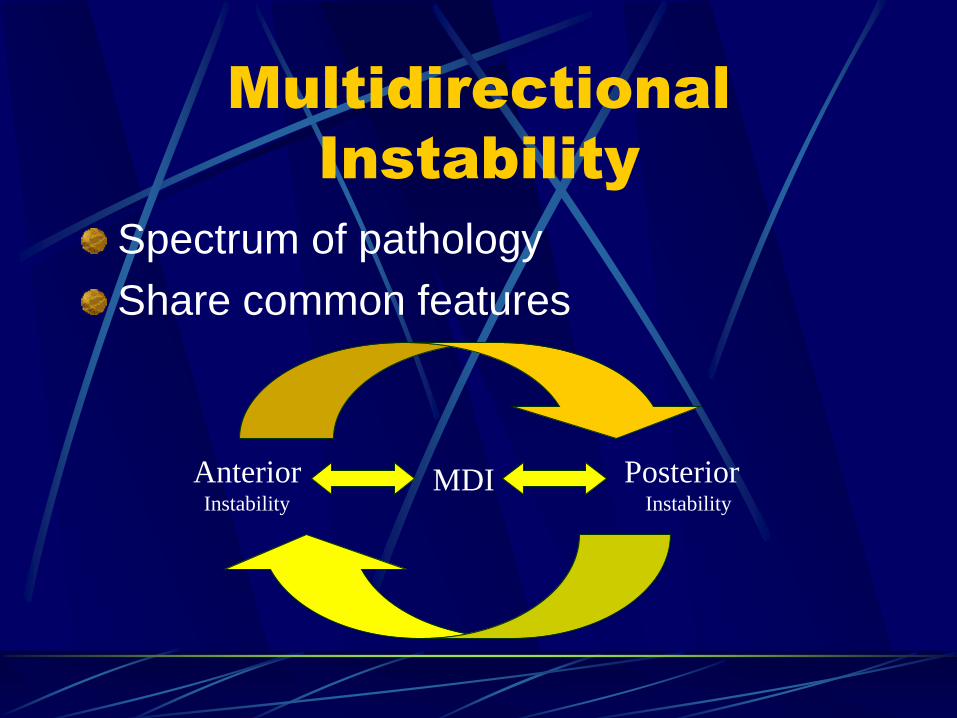
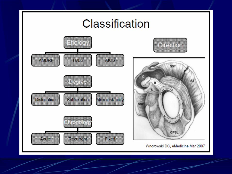
Multidirectional
Instability
Spectrum of pathology
Share common features
MDI Anterior Instability
Posterior Instability
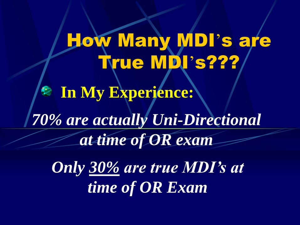
How Many MDI’s are
True MDI’s???
70% are actually Uni-Directional
at time of OR exam
Only 30% are true MDI’s at
time of OR Exam
In My Experience:
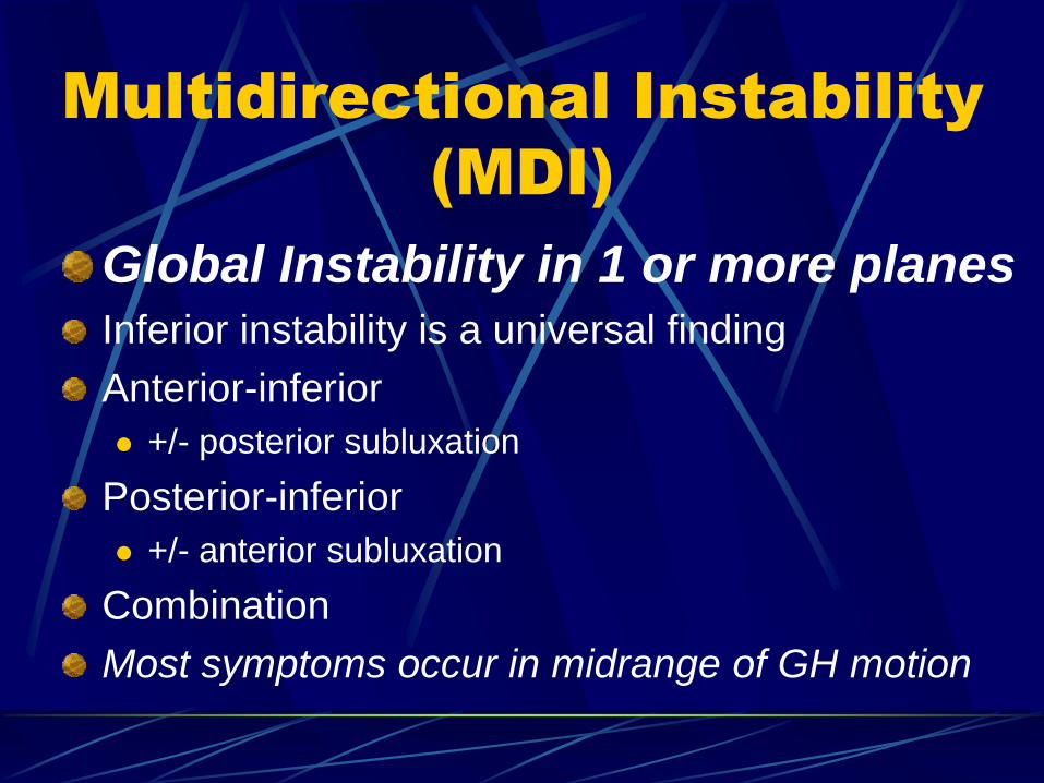
Multidirectional Instability
(MDI)
Global Instability in 1 or more planes Inferior instability is a universal finding
Anterior-inferior
+/- posterior subluxation
Posterior-inferior
+/- anterior subluxation
Combination
Most symptoms occur in midrange of GH motion
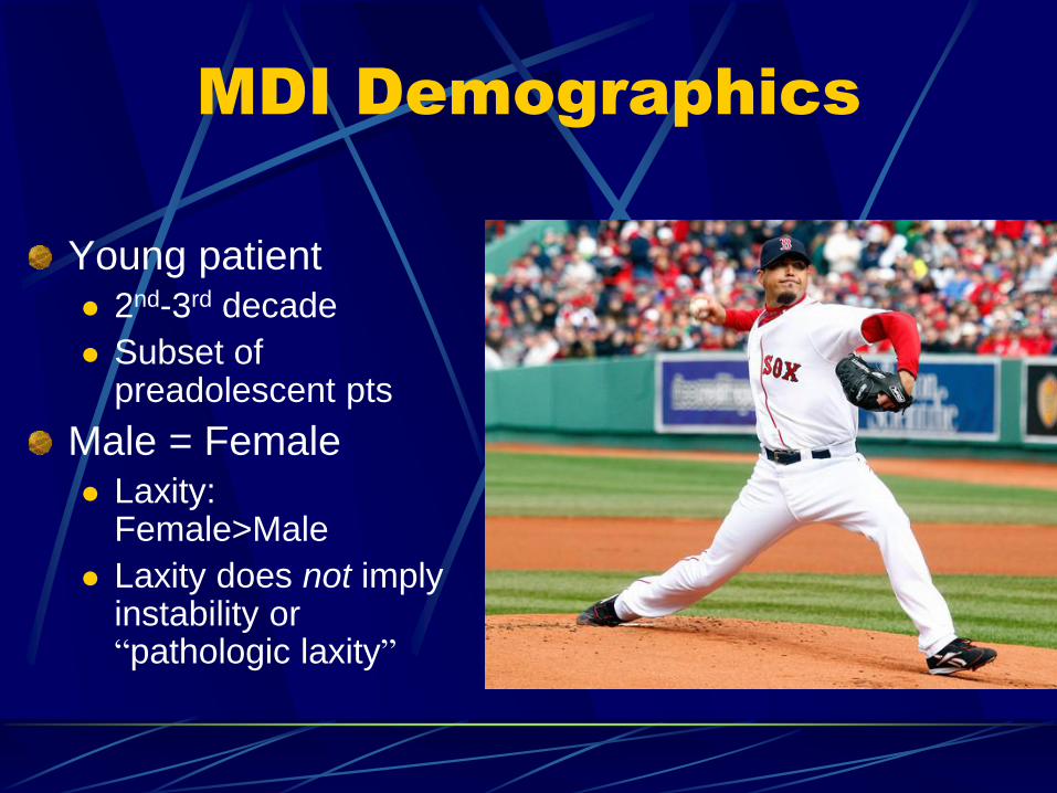
MDI Demographics
Young patient 2nd-3rd decade
Subset of preadolescent pts
Male = Female Laxity:
Female>Male
Laxity does not imply instability or “pathologic laxity”
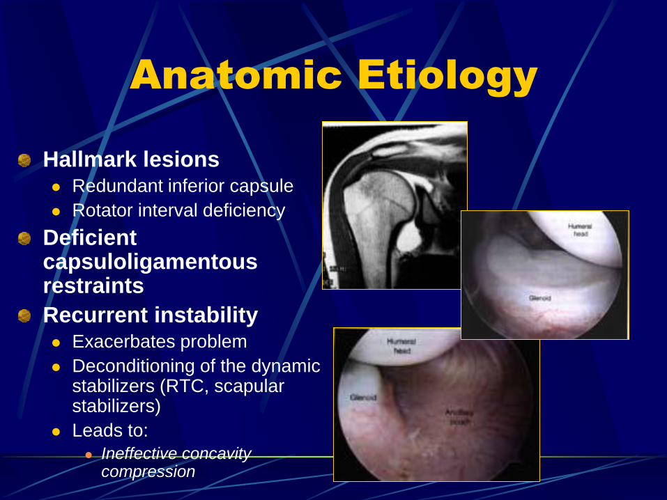
Anatomic Etiology
Hallmark lesions Redundant inferior capsule
Rotator interval deficiency
Deficient capsuloligamentous restraints
Recurrent instability Exacerbates problem
Deconditioning of the dynamic stabilizers (RTC, scapular stabilizers)
Leads to:
Ineffective concavity compression
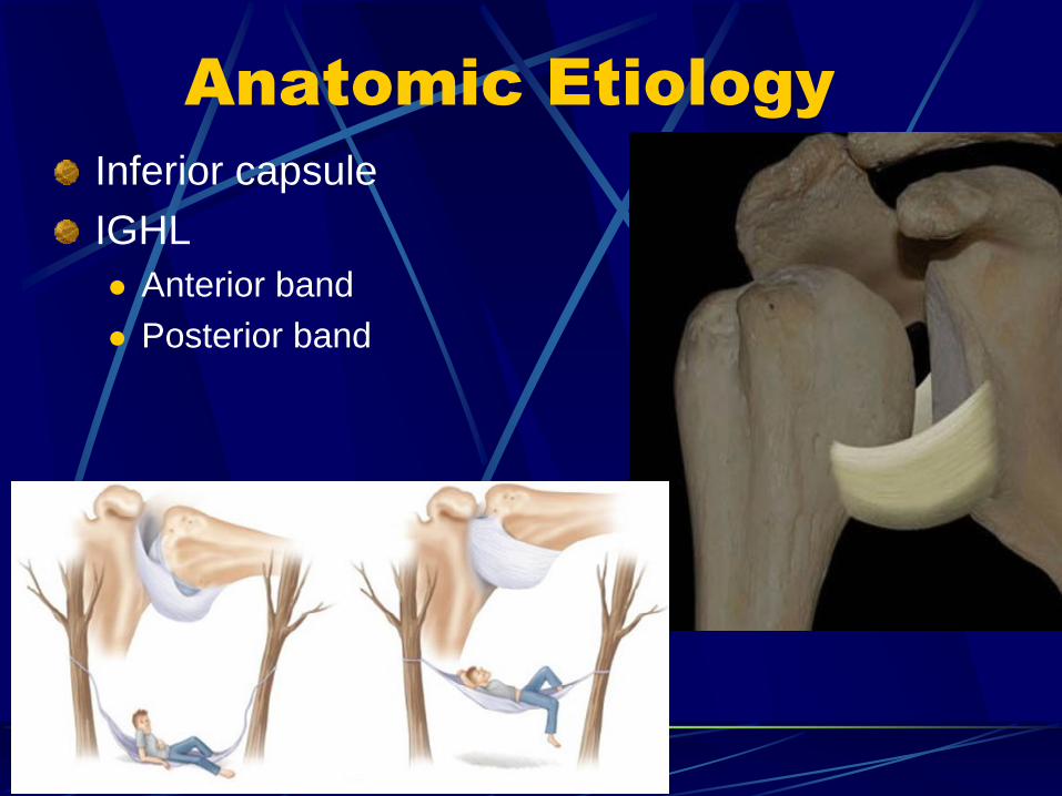
Anatomic Etiology
Inferior capsule
IGHL
Anterior band
Posterior band
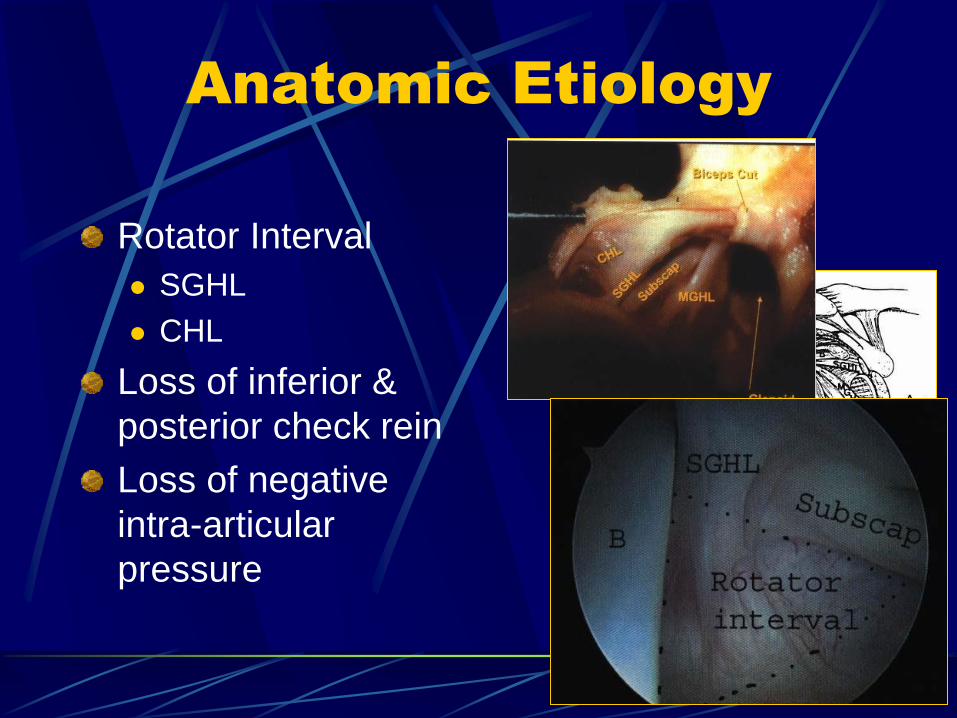
Anatomic Etiology
Rotator Interval
SGHL
CHL
Loss of inferior &
posterior check rein
Loss of negative
intra-articular
pressure
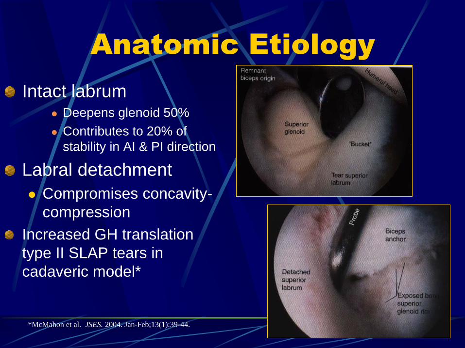
Anatomic Etiology
Intact labrum Deepens glenoid 50%
Contributes to 20% of
stability in AI & PI direction
Labral detachment
Compromises concavity-
compression
Increased GH translation
type II SLAP tears in
cadaveric model*
*McMahon et al. JSES. 2004. Jan-Feb;13(1):39-44.
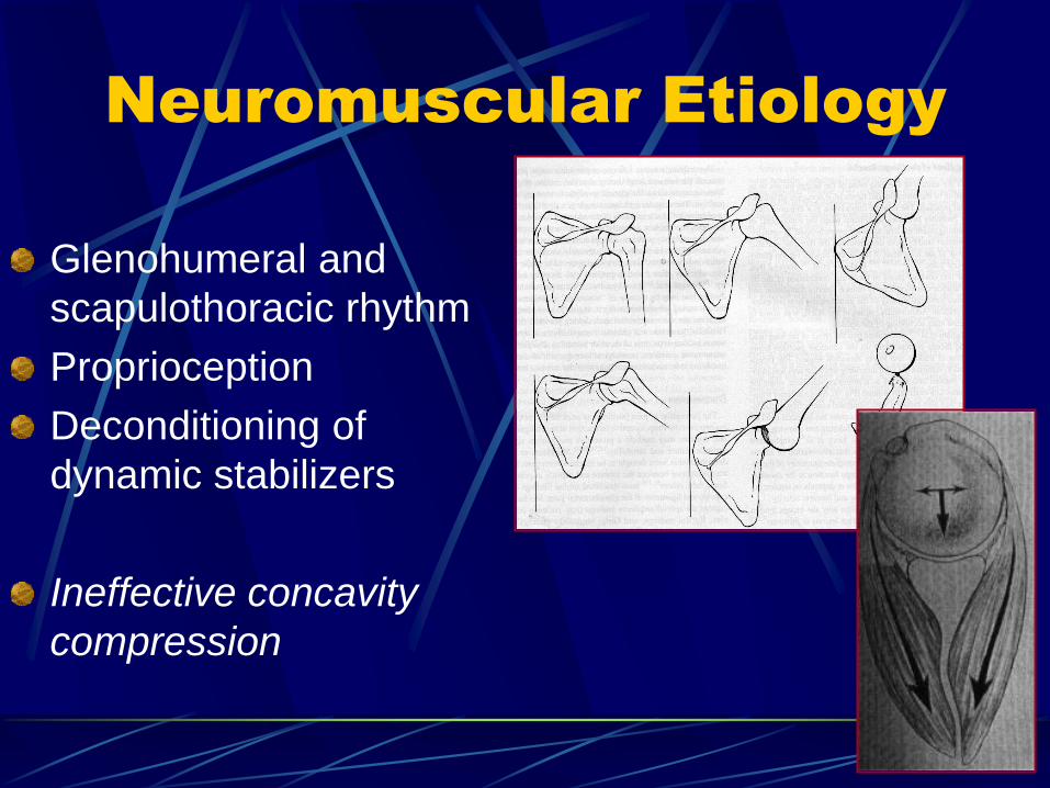
Neuromuscular Etiology
Glenohumeral and
scapulothoracic rhythm
Proprioception
Deconditioning of
dynamic stabilizers
Ineffective concavity
compression

History/Presentation
Varied symptoms
Pain
Instability
(dislocation/subluxation
Looseness
Weakness
Parasthesias
Fatigue
Popping,clicking,grinding
Dead arm syndrome
“Traction paresthesias”
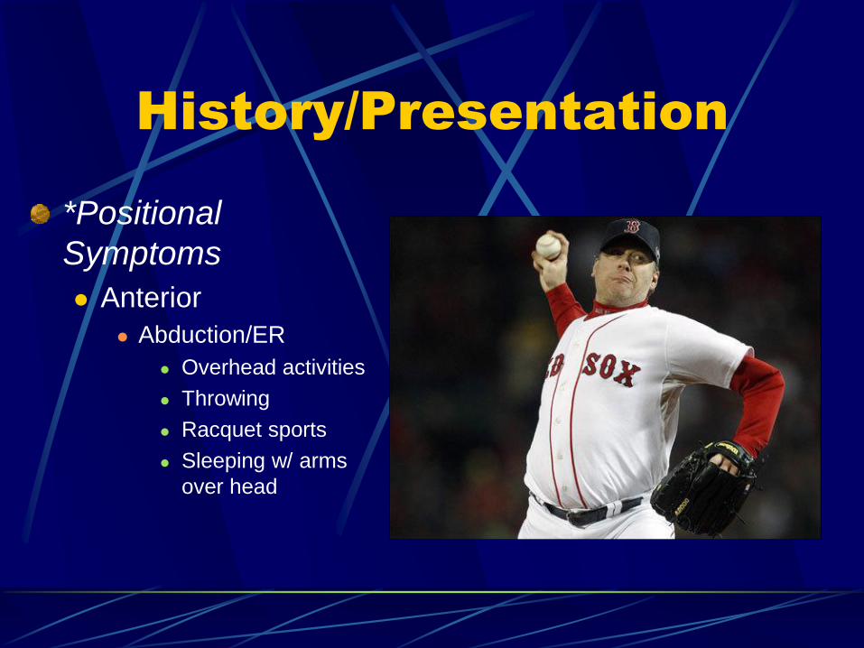
History/Presentation
*Positional
Symptoms
Anterior
Abduction/ER
Overhead activities
Throwing
Racquet sports
Sleeping w/ arms
over head
History/Presentation
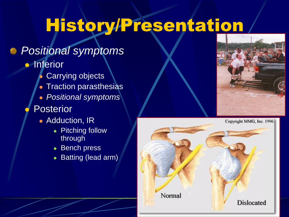
Positional symptoms Inferior
Carrying objects
Traction parasthesias
Positional symptoms
Posterior Adduction, IR
Pitching follow through
Bench press
Batting (lead arm)
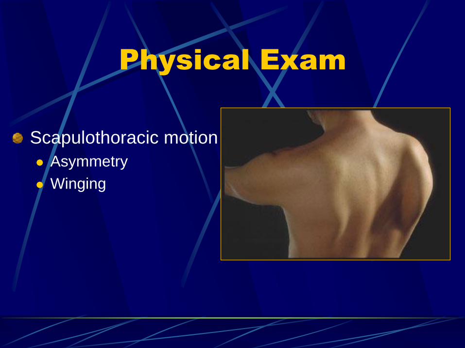
Physical Exam
Scapulothoracic motion
Asymmetry
Winging
Physical Exam
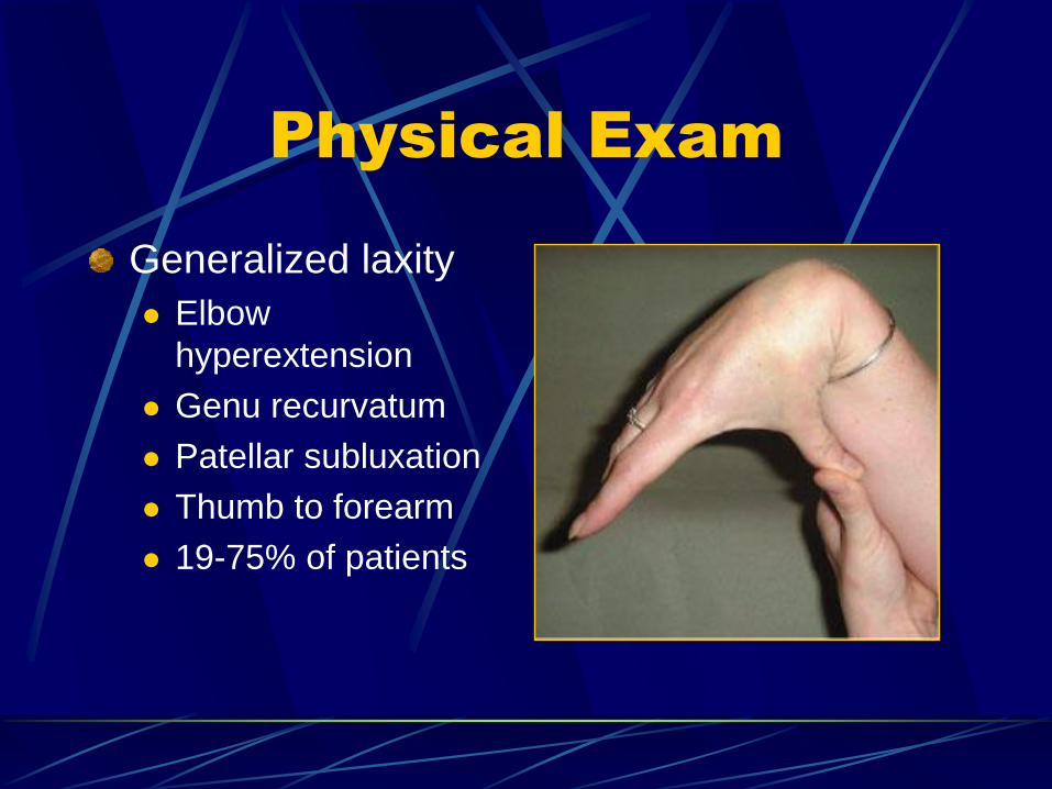
Generalized laxity
Elbow
hyperextension
Genu recurvatum
Patellar subluxation
Thumb to forearm
19-75% of patients
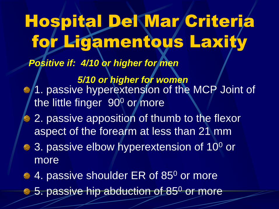
Hospital Del Mar Criteria
for Ligamentous Laxity
1. passive hyperextension of the MCP Joint of
the little finger 900 or more
2. passive apposition of thumb to the flexor
aspect of the forearm at less than 21 mm
3. passive elbow hyperextension of 100 or
more
4. passive shoulder ER of 850 or more
5. passive hip abduction of 850 or more
Positive if: 4/10 or higher for men
5/10 or higher for women
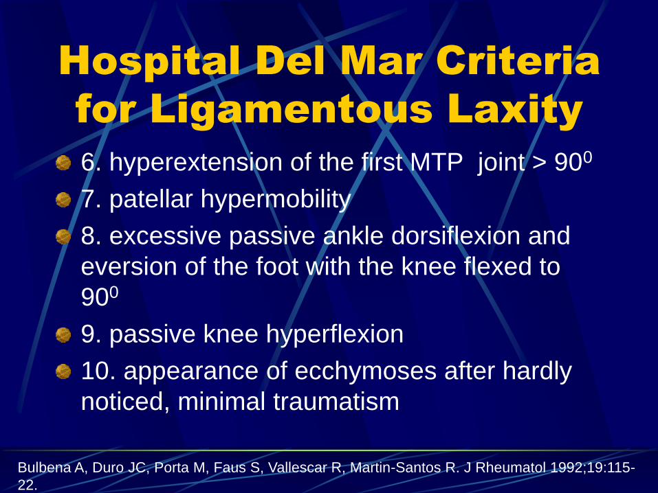
Hospital Del Mar Criteria
for Ligamentous Laxity
6. hyperextension of the first MTP joint > 900
7. patellar hypermobility
8. excessive passive ankle dorsiflexion and
eversion of the foot with the knee flexed to
900
9. passive knee hyperflexion
10. appearance of ecchymoses after hardly
noticed, minimal traumatism
Bulbena A, Duro JC, Porta M, Faus S, Vallescar R, Martin-Santos R. J Rheumatol 1992;19:115-
22.
Physical Exam
Physical Exam
Sulcus sign
Inferior laxity
Adduction/neutral rotation
Adduction/external
rotation
Grade
1+=1cm
2+=1-2cm
3+=>3cm
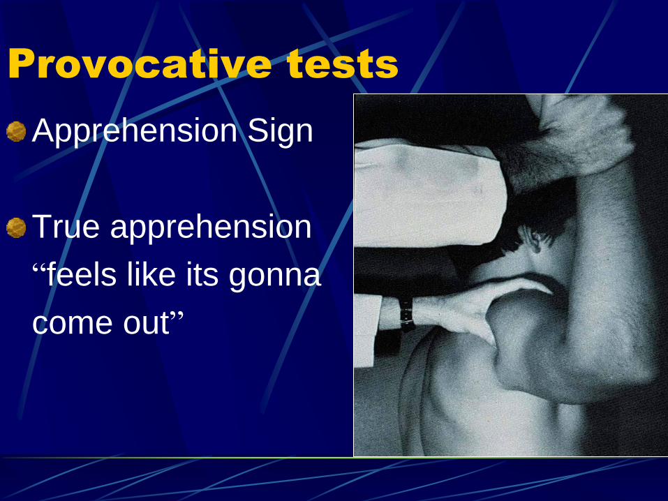
Provocative tests
Apprehension Sign
True apprehension
“feels like its gonna
come out”
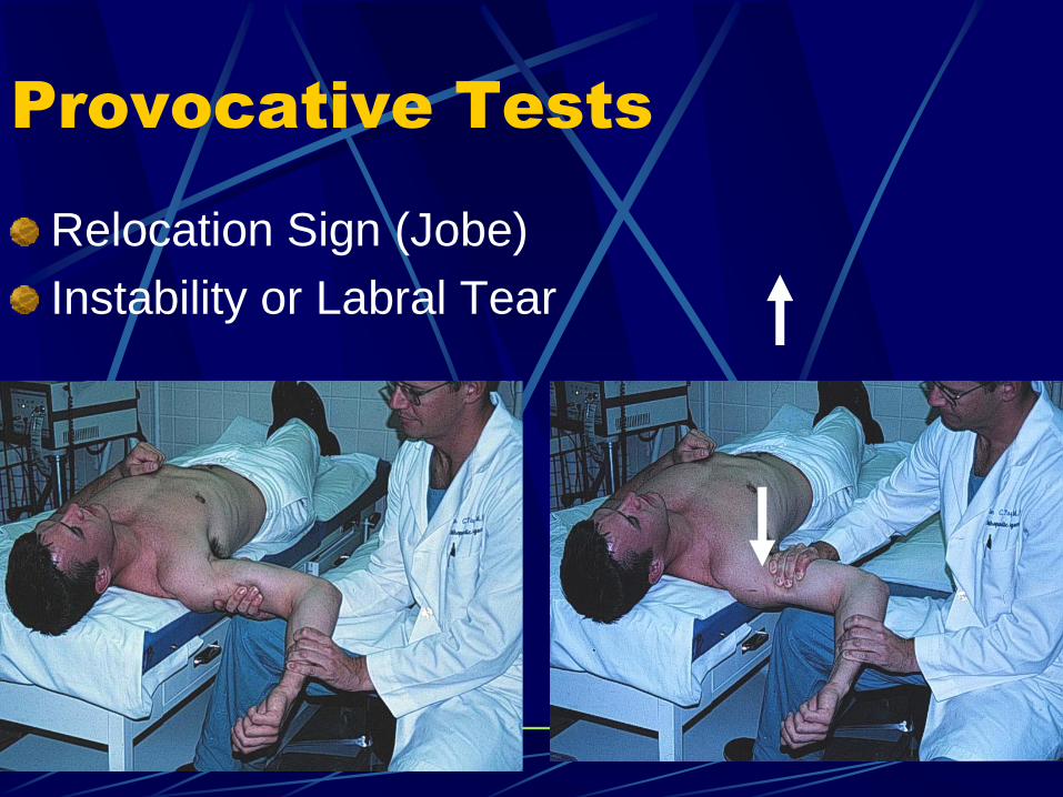
Provocative Tests
Relocation Sign (Jobe)
Instability or Labral Tear
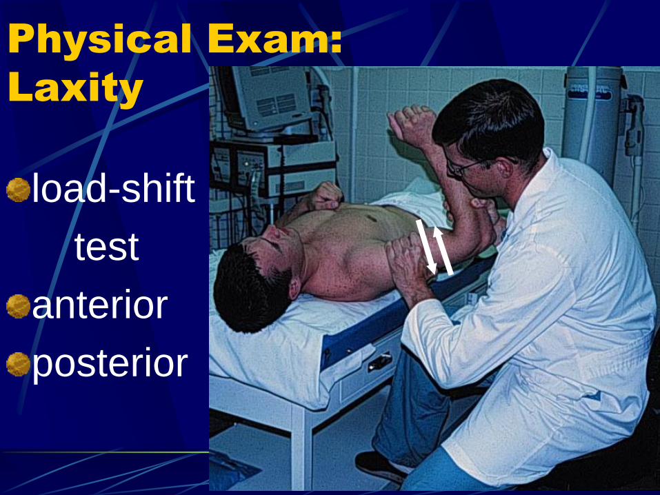
Physical Exam:
Laxity
load-shift
test
anterior
posterior
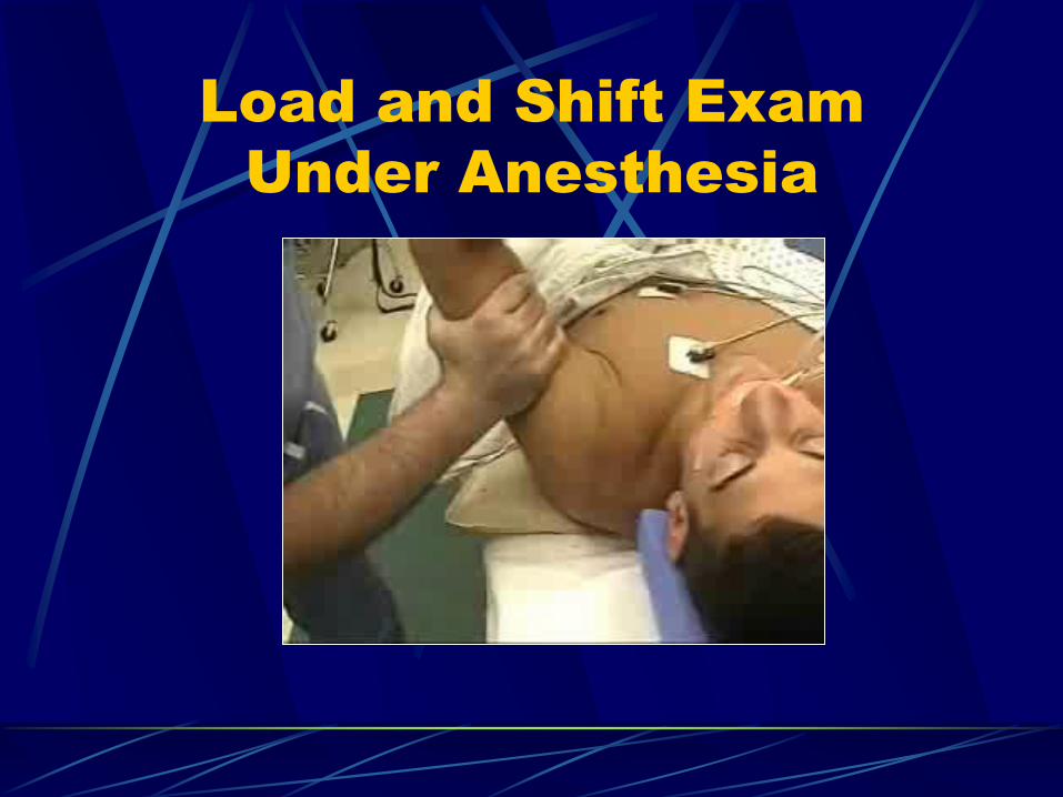
Load and Shift Exam
Under Anesthesia
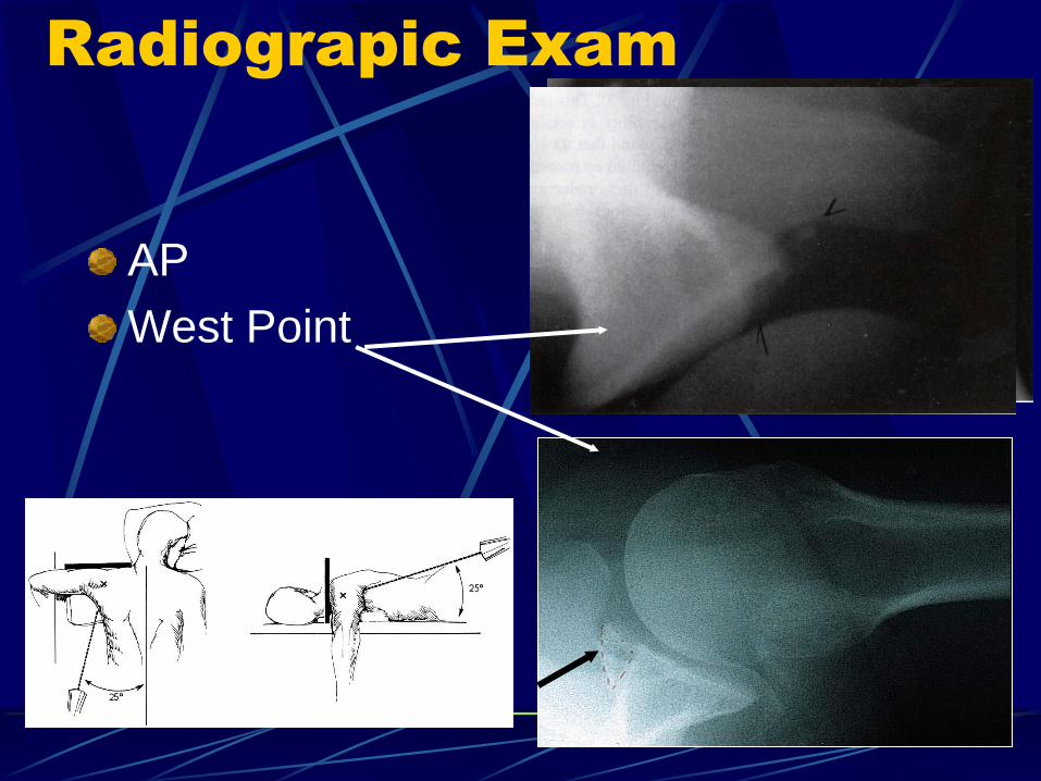
Radiograpic Exam
AP
West Point
Rim fx.
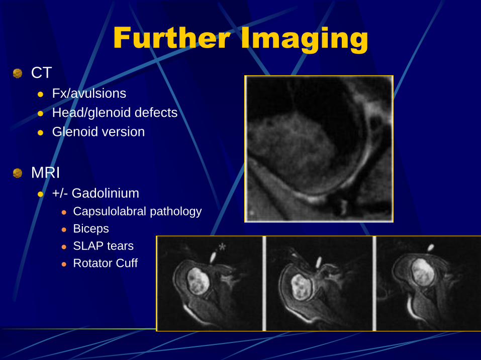
Further Imaging
CT
Fx/avulsions
Head/glenoid defects
Glenoid version
MRI
+/- Gadolinium
Capsulolabral pathology
Biceps
SLAP tears
Rotator Cuff
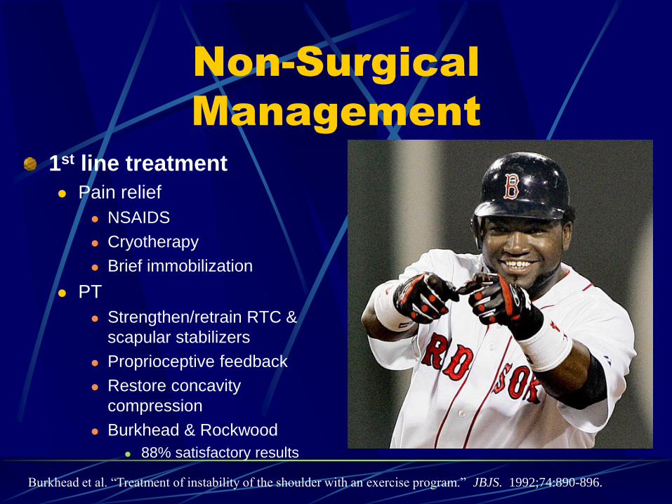
Non-Surgical
Management
1st line treatment
Pain relief
NSAIDS
Cryotherapy
Brief immobilization
PT
Strengthen/retrain RTC &
scapular stabilizers
Proprioceptive feedback
Restore concavity
compression
Burkhead & Rockwood
88% satisfactory results
Burkhead et al. “Treatment of instability of the shoulder with an exercise program.” JBJS. 1992;74:890-896.
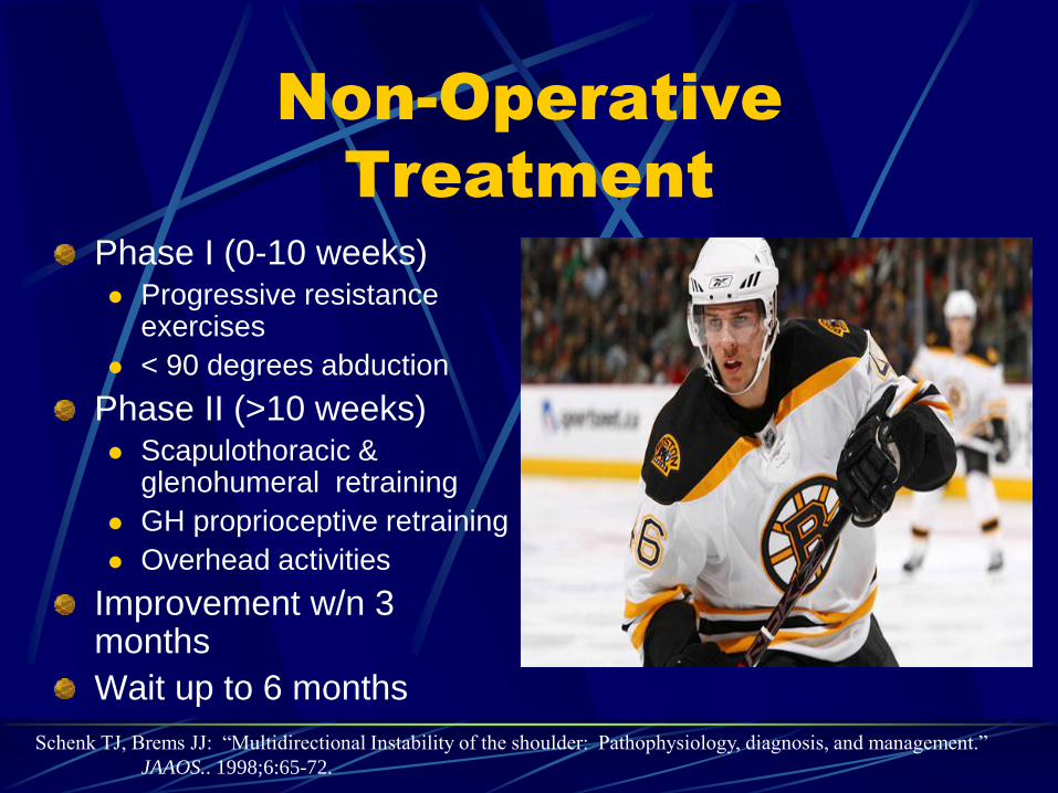
Non-Operative
Treatment
Phase I (0-10 weeks) Progressive resistance
exercises
< 90 degrees abduction
Phase II (>10 weeks) Scapulothoracic &
glenohumeral retraining
GH proprioceptive retraining
Overhead activities
Improvement w/n 3 months
Wait up to 6 months
Schenk TJ, Brems JJ: “Multidirectional Instability of the shoulder: Pathophysiology, diagnosis, and management.”
JAAOS.. 1998;6:65-72.

If non-operative
treatment fails…
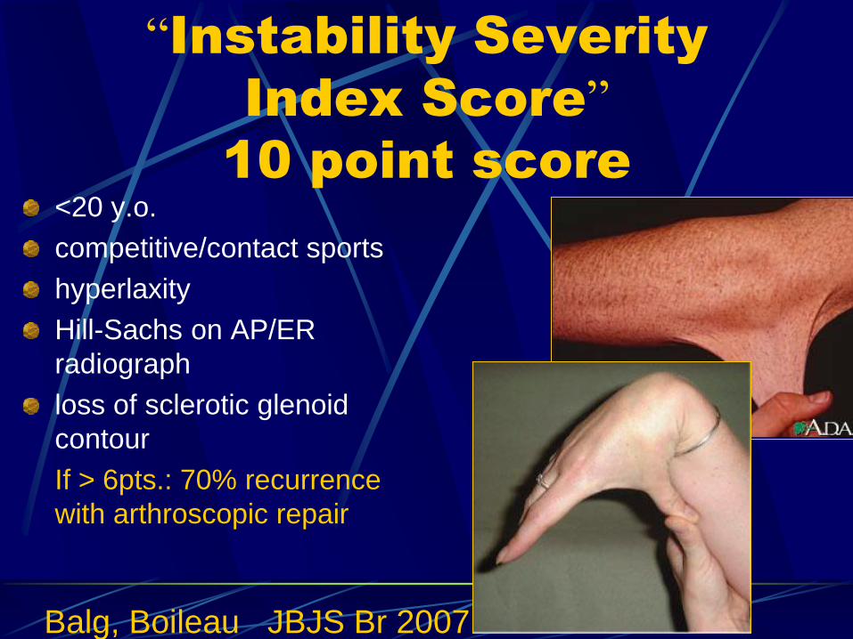
“Instability Severity
Index Score”
10 point score
<20 y.o.
competitive/contact sports
hyperlaxity
Hill-Sachs on AP/ER
radiograph
loss of sclerotic glenoid
contour
If > 6pts.: 70% recurrence
with arthroscopic repair
Balg, Boileau JBJS Br 2007
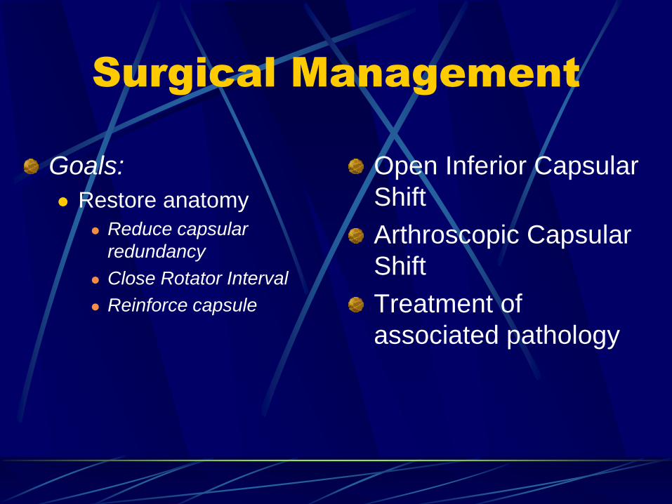
Surgical Management
Goals:
Restore anatomy
Reduce capsular
redundancy
Close Rotator Interval
Reinforce capsule
Open Inferior Capsular
Shift
Arthroscopic Capsular
Shift
Treatment of
associated pathology
Open Shoulder
Stabilization
Advantages of Open
Shift
Adjust capsular tension
Reduces capsular volume
Lateral “T” capsulotomy
“Greater shift”
Axillary nerve exposure
Thickens capsule
Bankart easily repaired
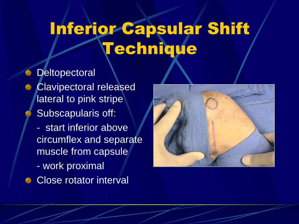
Inferior Capsular Shift
Technique
Deltopectoral
Clavipectoral released
lateral to pink stripe
Subscapularis off:
- start inferior above
circumflex and separate
muscle from capsule
- work proximal
Close rotator interval
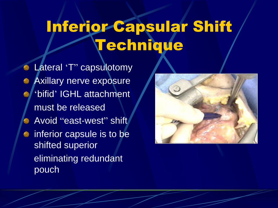
Inferior Capsular Shift
Technique
Lateral ‘T” capsulotomy
Axillary nerve exposure
‘bifid’ IGHL attachment
must be released
Avoid “east-west” shift
inferior capsule is to be
shifted superior
eliminating redundant
pouch
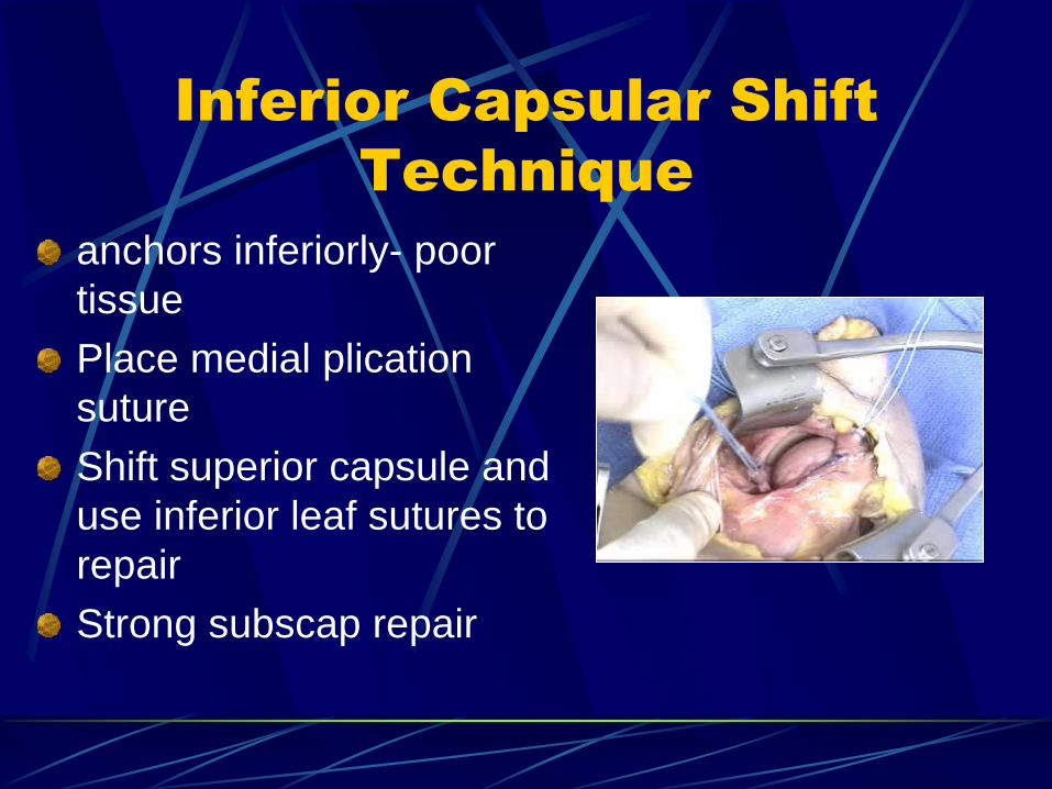
Inferior Capsular Shift
Technique
anchors inferiorly- poor
tissue
Place medial plication
suture
Shift superior capsule and
use inferior leaf sutures to
repair
Strong subscap repair
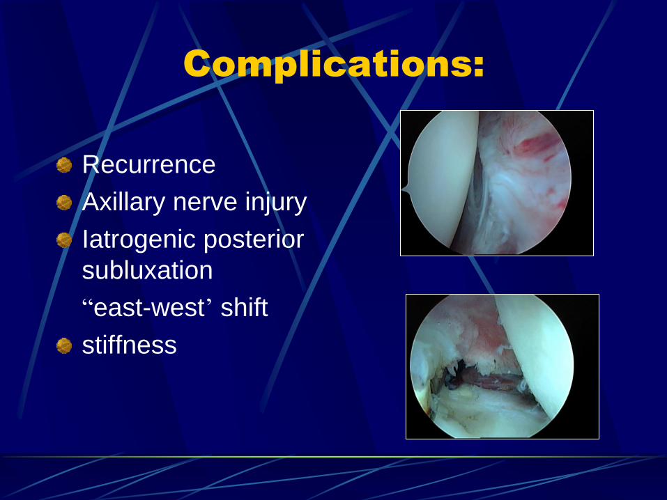
Complications:
Recurrence
Axillary nerve injury
Iatrogenic posterior
subluxation
“east-west’ shift
stiffness
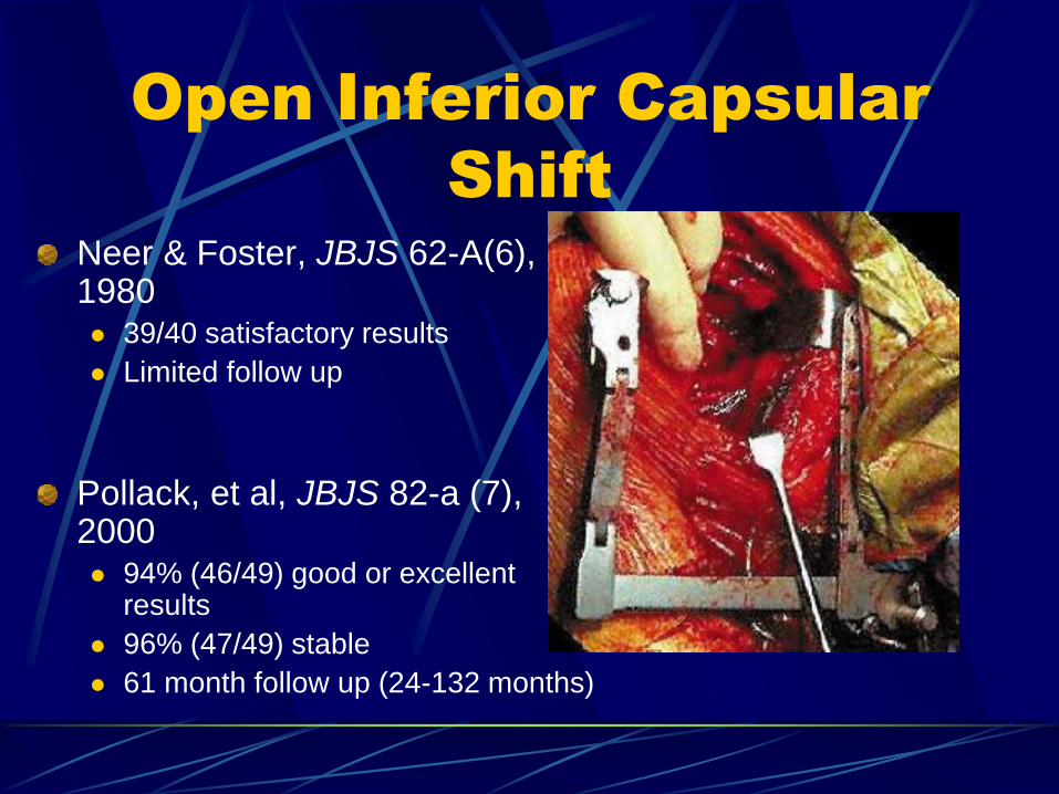
Open Inferior Capsular
Shift
Neer & Foster, JBJS 62-A(6), 1980 39/40 satisfactory results
Limited follow up
Pollack, et al, JBJS 82-a (7), 2000 94% (46/49) good or excellent
results
96% (47/49) stable
61 month follow up (24-132 months)
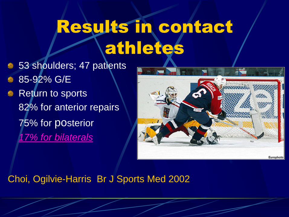
Results in contact
athletes
53 shoulders; 47 patients
85-92% G/E
Return to sports
82% for anterior repairs
75% for posterior
17% for bilaterals
Choi, Ogilvie-Harris Br J Sports Med 2002
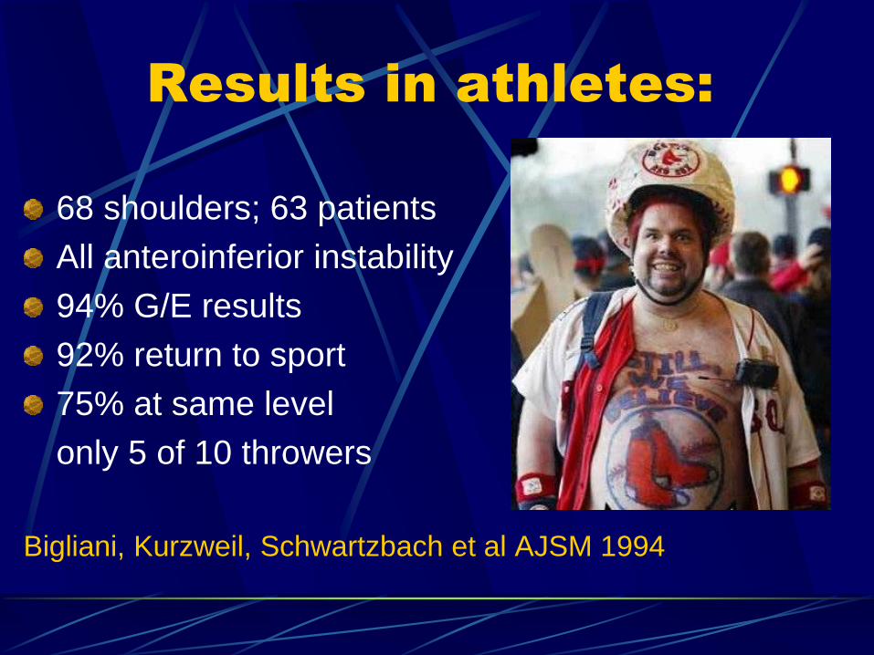
Results in athletes:
68 shoulders; 63 patients
All anteroinferior instability
94% G/E results
92% return to sport
75% at same level
only 5 of 10 throwers
Bigliani, Kurzweil, Schwartzbach et al AJSM 1994
Results: Open Inferior Capsular
Shift
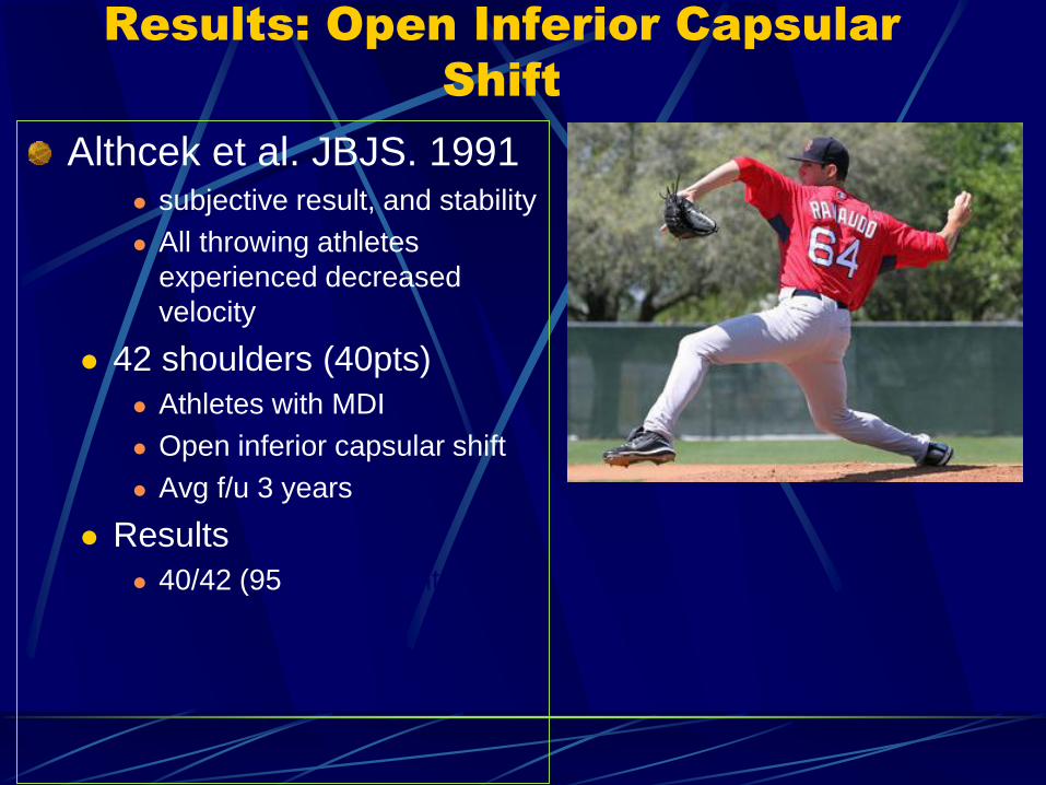
Althcek et al. JBJS. 1991 subjective result, and stability
All throwing athletes
experienced decreased
velocity
42 shoulders (40pts)
Athletes with MDI
Open inferior capsular shift
Avg f/u 3 years
Results
40/42 (95%) excellent
Arthroscopic Shoulder
Stabilization
MDI Tx Options
Capsulorrhaphy (capsular plication)
Vs
“Combi- Stitch” (capsular plication with labral imbrication)
Vs
“Combi-Repair” (capsular plication with labral repair)
Anterior vs Posterior
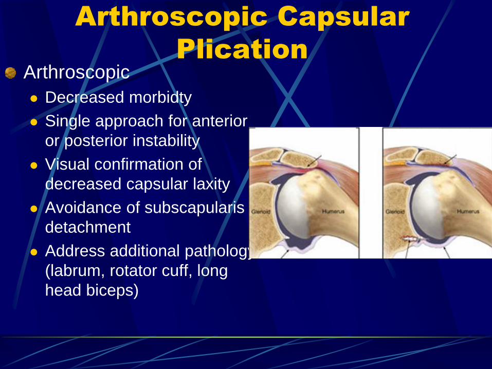
Arthroscopic Capsular
Plication
Arthroscopic
Decreased morbidty
Single approach for anterior
or posterior instability
Visual confirmation of
decreased capsular laxity
Avoidance of subscapularis
detachment
Address additional pathology
(labrum, rotator cuff, long
head biceps)
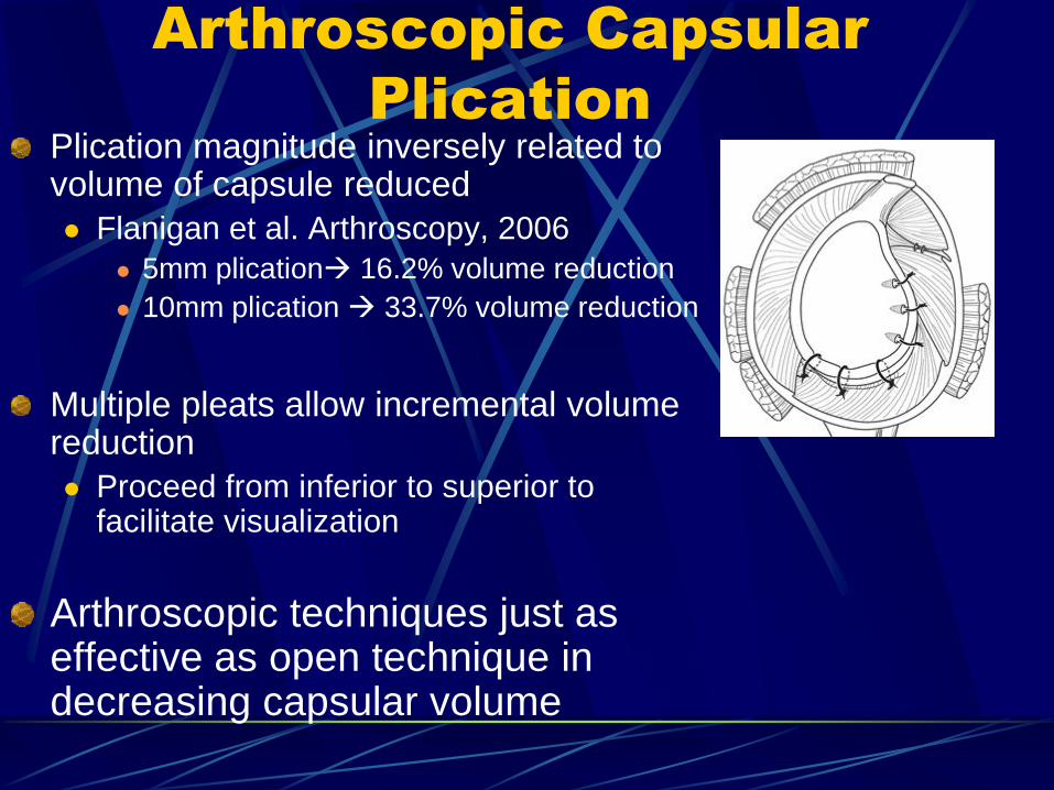
Arthroscopic Capsular
Plication
Plication magnitude inversely related to volume of capsule reduced
Flanigan et al. Arthroscopy, 2006
5mm plication 16.2% volume reduction
10mm plication 33.7% volume reduction
Multiple pleats allow incremental volume reduction
Proceed from inferior to superior to facilitate visualization
Arthroscopic techniques just as effective as open technique in decreasing capsular volume
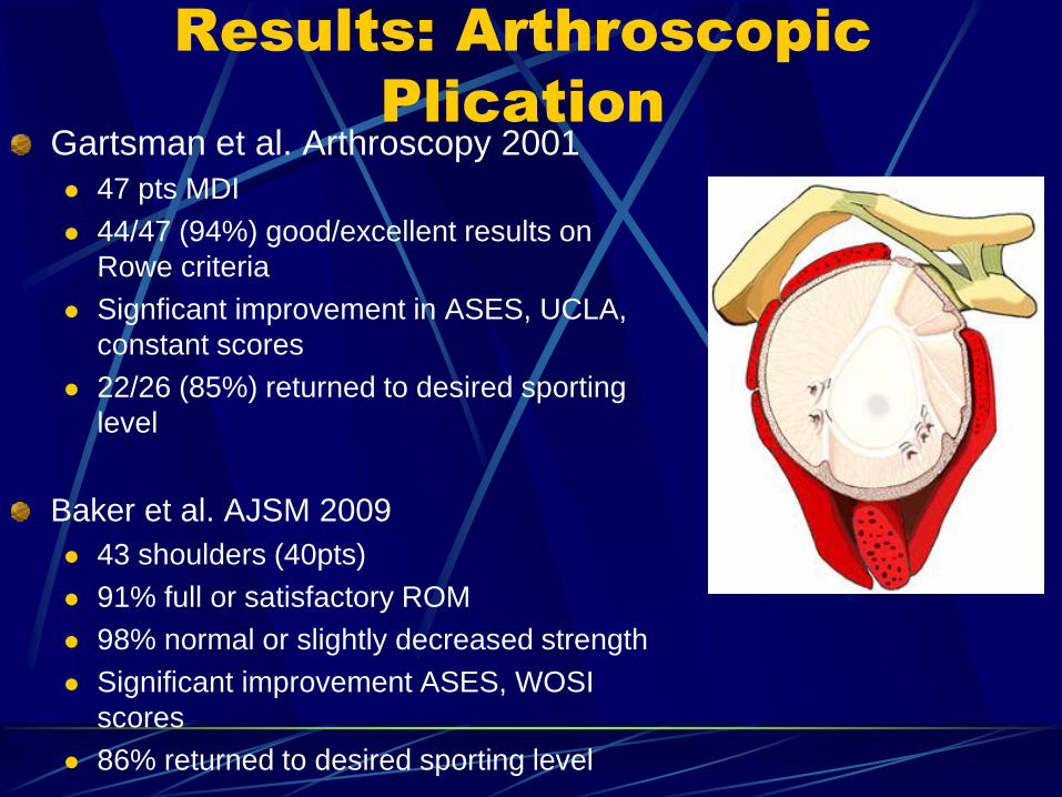
Results: Arthroscopic
Plication Gartsman et al. Arthroscopy 2001
47 pts MDI
44/47 (94%) good/excellent results on
Rowe criteria
Signficant improvement in ASES, UCLA,
constant scores
22/26 (85%) returned to desired sporting
level
Baker et al. AJSM 2009
43 shoulders (40pts)
91% full or satisfactory ROM
98% normal or slightly decreased strength
Significant improvement ASES, WOSI
scores
86% returned to desired sporting level
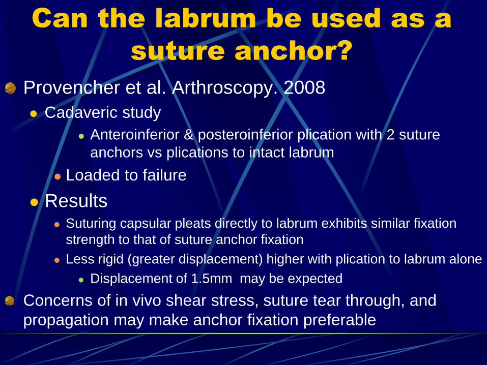
Can the labrum be used as a
suture anchor?
Provencher et al. Arthroscopy. 2008
Cadaveric study
Anteroinferior & posteroinferior plication with 2 suture
anchors vs plications to intact labrum
Loaded to failure
Results Suturing capsular pleats directly to labrum exhibits similar fixation
strength to that of suture anchor fixation
Less rigid (greater displacement) higher with plication to labrum alone
Displacement of 1.5mm may be expected
Concerns of in vivo shear stress, suture tear through, and
propagation may make anchor fixation preferable.
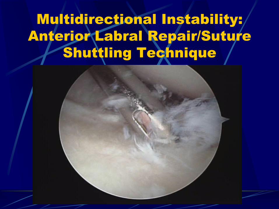
Multidirectional Instability:
Anterior Labral Repair/Suture
Shuttling Technique
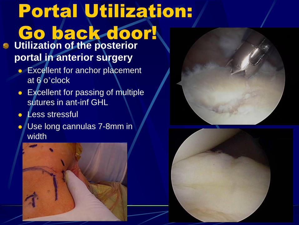
Portal Utilization:
Go back door! Utilization of the posterior
portal in anterior surgery
Excellent for anchor placement
at 6 o’clock
Excellent for passing of multiple
sutures in ant-inf GHL
Less stressful
Use long cannulas 7-8mm in
width
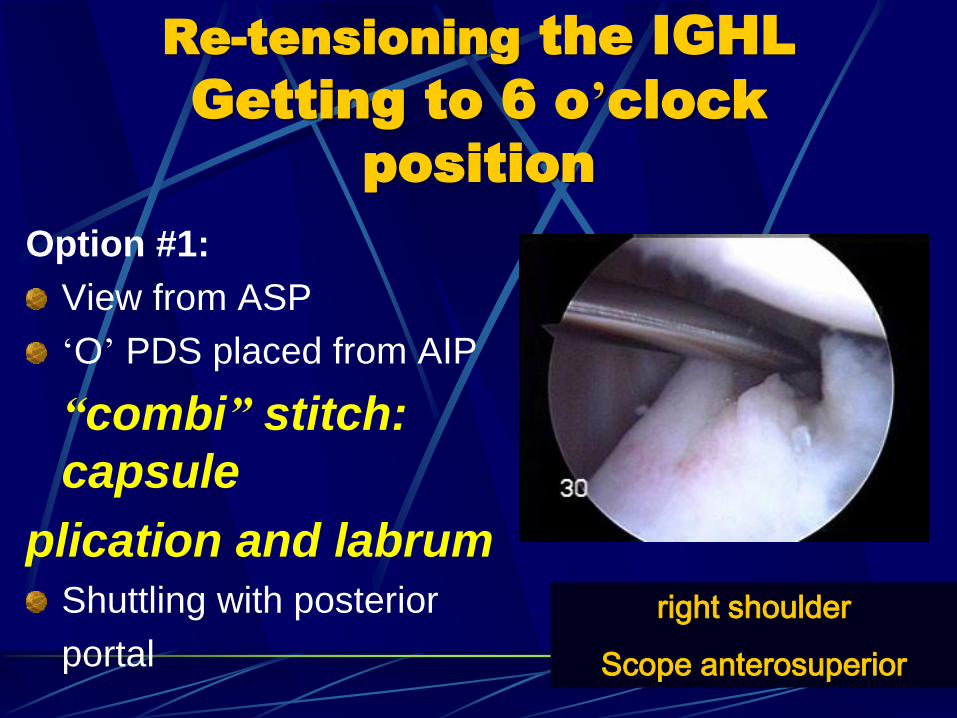
Re-tensioning the IGHL
Getting to 6 o’clock
position
Option #1:
View from ASP
‘O’ PDS placed from AIP
“combi” stitch:
capsule
plication and labrum Shuttling with posterior
portal
right shoulder
Scope anterosuperior
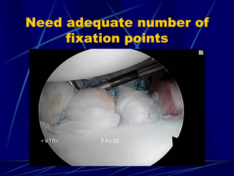
Need adequate number of
fixation points
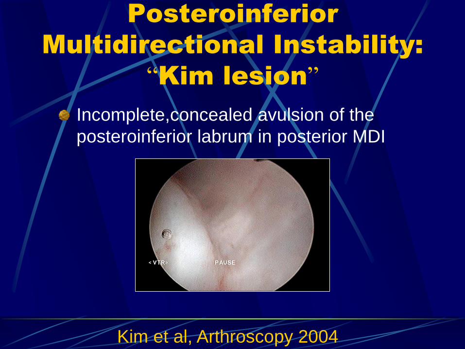
Posteroinferior
Multidirectional Instability:
“Kim lesion”
Incomplete,concealed avulsion of the
posteroinferior labrum in posterior MDI
Kim et al, Arthroscopy 2004
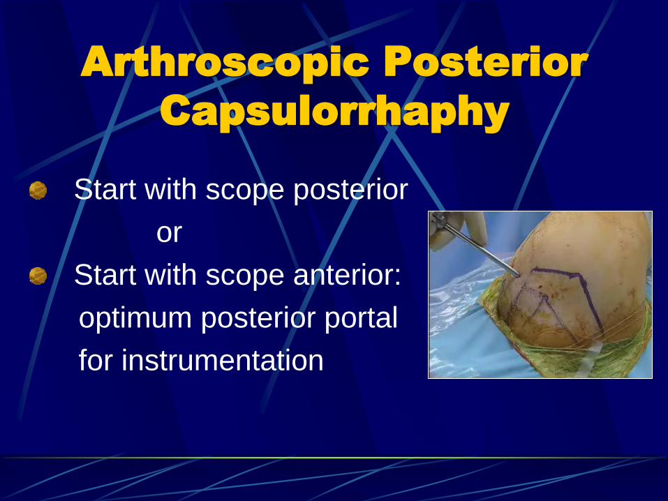
Arthroscopic Posterior
Capsulorrhaphy
Start with scope posterior
or
Start with scope anterior:
optimum posterior portal
for instrumentation
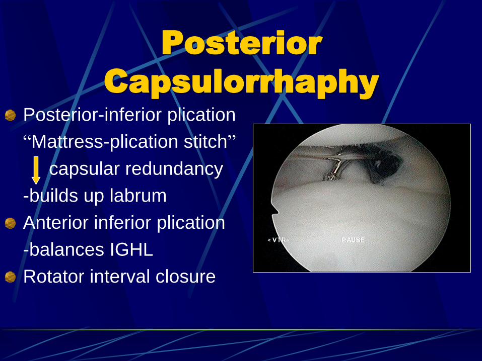
Posterior
Capsulorrhaphy
Posterior-inferior plication
“Mattress-plication stitch”
capsular redundancy
-builds up labrum
Anterior inferior plication
-balances IGHL
Rotator interval closure
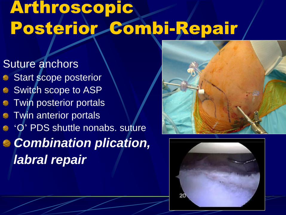
Arthroscopic
Posterior Combi-Repair
Suture anchors Start scope posterior
Switch scope to ASP
Twin posterior portals
Twin anterior portals
‘O’ PDS shuttle nonabs. suture
Combination plication,
labral repair
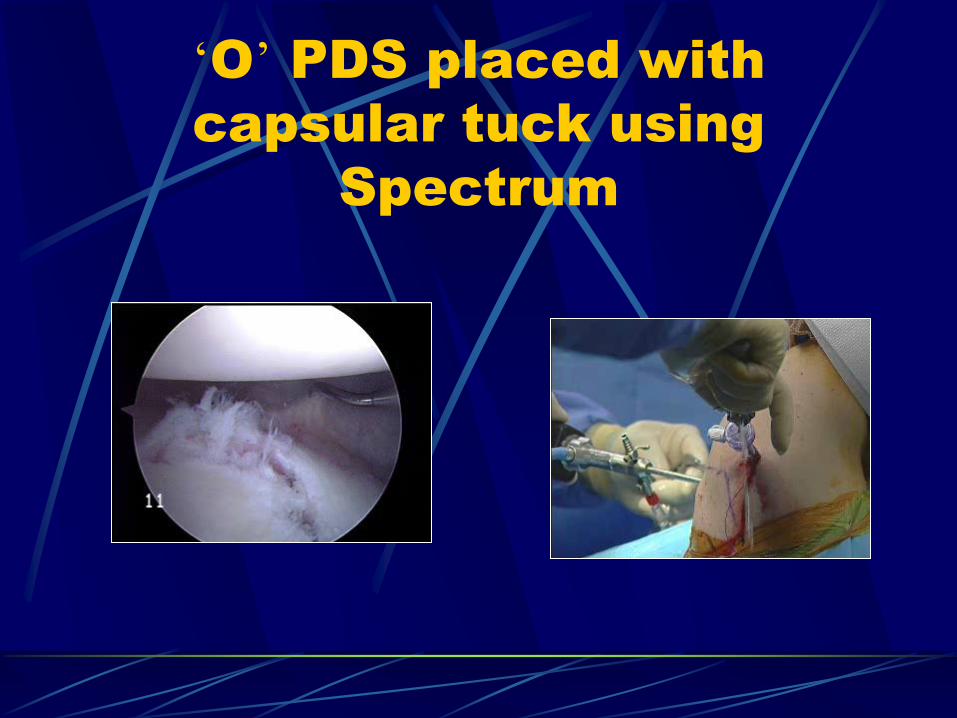
‘O’ PDS placed with
capsular tuck using
Spectrum
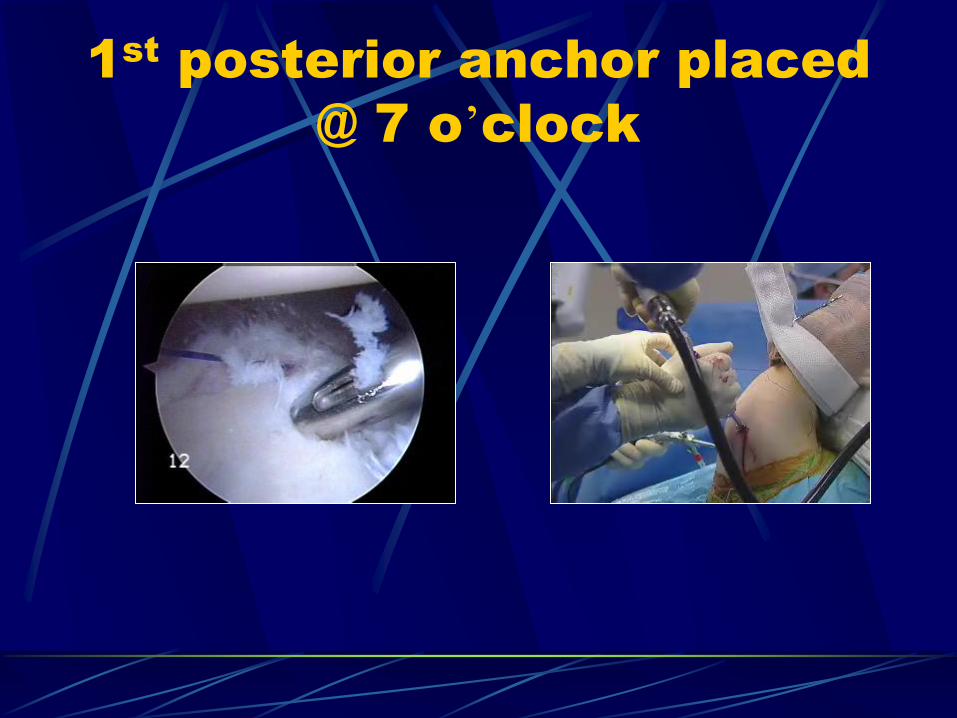
1st
posterior anchor placed
@ 7 o’clock
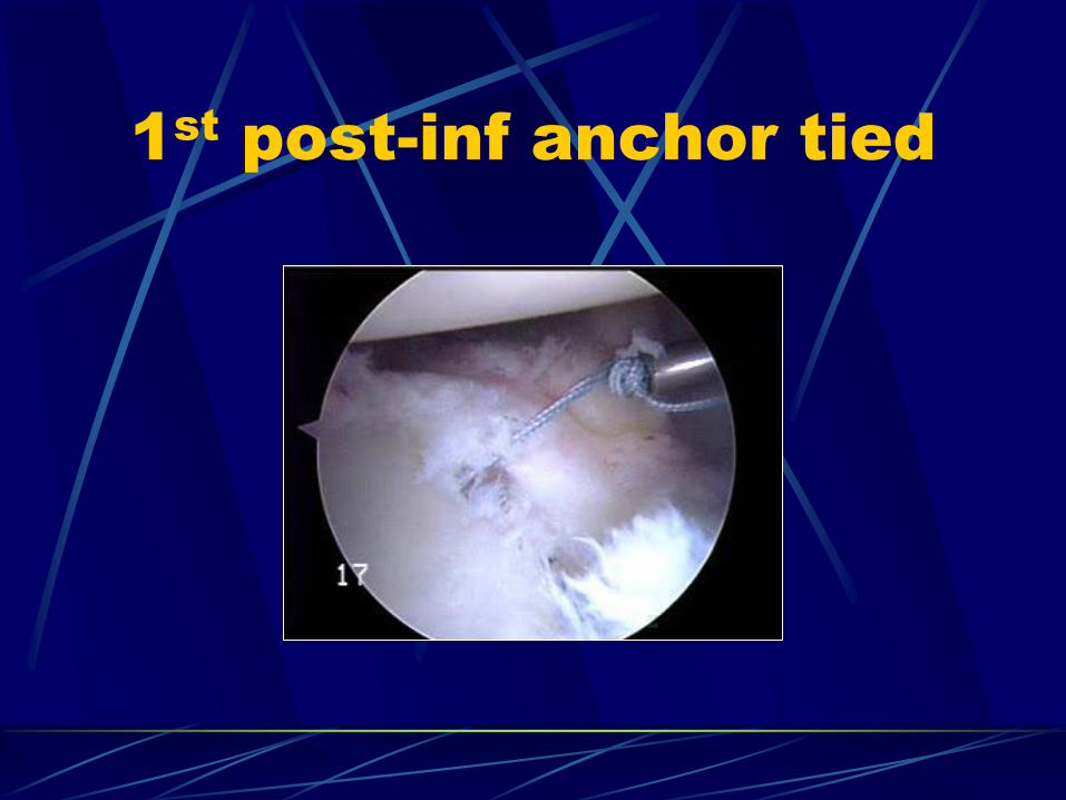
1st
post-inf anchor tied
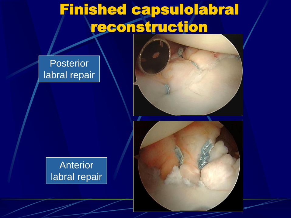
Finished capsulolabral
reconstruction
Posterior
labral repair
Anterior
labral repair
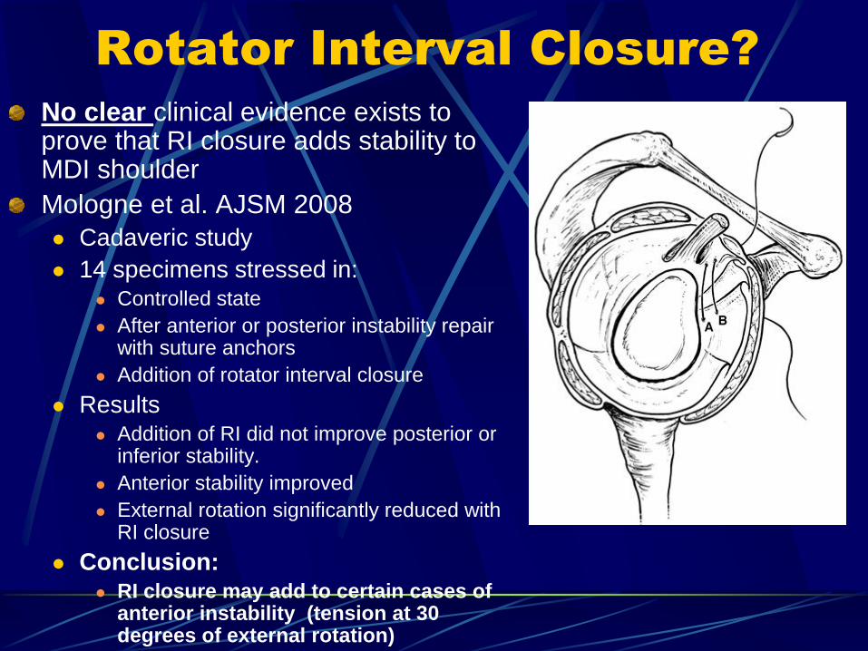
Rotator Interval Closure?
No clear clinical evidence exists to prove that RI closure adds stability to MDI shoulder
Mologne et al. AJSM 2008
Cadaveric study
14 specimens stressed in: Controlled state
After anterior or posterior instability repair with suture anchors
Addition of rotator interval closure
Results Addition of RI did not improve posterior or
inferior stability.
Anterior stability improved
External rotation significantly reduced with RI closure
Conclusion: RI closure may add to certain cases of
anterior instability (tension at 30 degrees of external rotation)
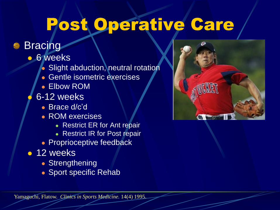
Post Operative Care
Bracing 6 weeks
Slight abduction, neutral rotation
Gentle isometric exercises
Elbow ROM
6-12 weeks Brace d/c’d
ROM exercises Restrict ER for Ant repair
Restrict IR for Post repair
Proprioceptive feedback
12 weeks Strengthening
Sport specific Rehab
Yamaguchi, Flatow. Clinics in Sports Medicine. 14(4) 1995.

THANK YOU