Embed Size (px)
Citation preview
Multilayer DNA Origami Packed on Hexagonal and Hybrid LatticesYonggang Ke,† Niels V. Voigt,‡ Kurt V. Gothelf,‡ and William M. Shih*,†,§
†Department of Cancer Biology, Dana-Farber Cancer Institute, and Department of Biological Chemistry and MolecularPharmacology, Harvard Medical School, Boston, Massachusetts 02115, United States§Wyss Institute for Biologically Inspired Engineering at Harvard, Cambridge, Massachusetts 02138, United States‡Danish National Research Foundation: Centre for DNA Nanotechnology at Interdisciplinary Nanoscience Center (iNANO), andDepartment of Chemistry, Aarhus University, DK-8000 Aarhus, Denmark
*S Supporting Information
ABSTRACT: “Scaffolded DNA origami” has been proven tobe a powerful and efficient approach to construct two-dimensional or three-dimensional objects with great complex-ity. Multilayer DNA origami has been demonstrated withhelices packing along either honeycomb-lattice geometry orsquare-lattice geometry. Here we report successful folding ofmultilayer DNA origami with helices arranged on a close-packed hexagonal lattice. This arrangement yields a higherdensity of helical packing and therefore higher resolution ofspatial addressing than has been shown previously. We alsodemonstrate hybrid multilayer DNA origami with honeycomb-lattice, square-lattice, and hexagonal-lattice packing of helices all inone design. The availability of hexagonal close-packing of helices extends our ability to build complex structures using DNAnanotechnology.
■ INTRODUCTIONThe past two decades have witnessed the rapid growth ofstructural DNA nanotechnology as a field. A variety of one-dimensional (1D), two-dimensional (2D), and three-dimen-sional (3D) DNA nanostructures have been developed.1−3
These versatile DNA nanostructures offer unique advantagesfor nanoscale patterning4−8 and construction of nanoscaledevices9−13 with applications such as tools for molecularbiophysics.14−17 Nonetheless, rational design of complicated3D DNA objects with high accuracy still poses great challenges.Recently, great progress has been achieved in this realm byusing strategies based on scaffolded DNA origami.18 Forexample, two origami boxes19,20 and a hollow tetrahedralshape21 have been demonstrated by linking discrete 2D origamiassembled on a single scaffold. More generalized methods toconstruct 3D DNA origami packed on honeycomb-latticegeometry22 or square-lattice geometry23 also have beenreported. Seven helices in a close-packed hexagonal latticehave been demonstrated for a non-origami DNA system.24
Here we generalize hexagonal-lattice close-packing for helices in3D DNA origami, and also demonstrate hybrid 3D DNAorigami packed on mixed geometries of honeycomb lattice,square lattice, and hexagonal lattice.
■ DESIGNHexagonal-Lattice Origami. The design strategy used in
this study for multilayer hexagonal-lattice DNA origami isillustrated in Figure 1. DNA origami conventionally aredesigned with a long single “scaffold” strand, typically the 7-
kilobase (kb) genome of the M13 bacteriophage, that is foldedwith hundreds of short “staple” strands into a parallel array ofdouble helices. Viewed down the axis parallel to the helices ofthe bundle (i.e., Z axis), every double helix can be conceived asof two strands that each are characterized by a polarity (i.e., 5′to 3′ away from the viewer versus toward the viewer) and aparity (made of scaffold DNA versus staple DNA). In thehoneycomb- or square-lattice designs, like-parity strands onnearest-neighbor helices all are arranged with antiparallelpolarity. As a result, adjacent helices are held together byantiparallel scaffold crossovers and antiparallel staple cross-overs. In contrast, it is impossible to arrange helices in ahexagonal-lattice design such that like-parity strands on nearest-neighbor helices all are antiparallel. For example, if the sixnearest-neighbor scaffold-type strands are all antiparallel to thecentral strand, then they must all be parallel to each other.For our design, we chose to arrange strand polarity in the
pattern shown in Figure 1B. Each double helix is connected bystaple-strand antiparallel crossovers to three, four, or five of itssix potential neighbors. Alternative crossover patterns arepossible, although near equal representation of polarity forscaffold-parity strands has to be present to enable a singlescaffold to traverse back and forth through the entire structure.Two versions of hexagonal-lattice origami were designed: long(L) with staple crossovers occurring at 13-base-pair (bp)intervals (every 7/6 helical turns), and short (S) with staple
Received: October 16, 2011
Article
pubs.acs.org/JACS
© XXXX American Chemical Society A dx.doi.org/10.1021/ja209719k | J. Am. Chem.Soc. XXXX, XXX, XXX−XXX
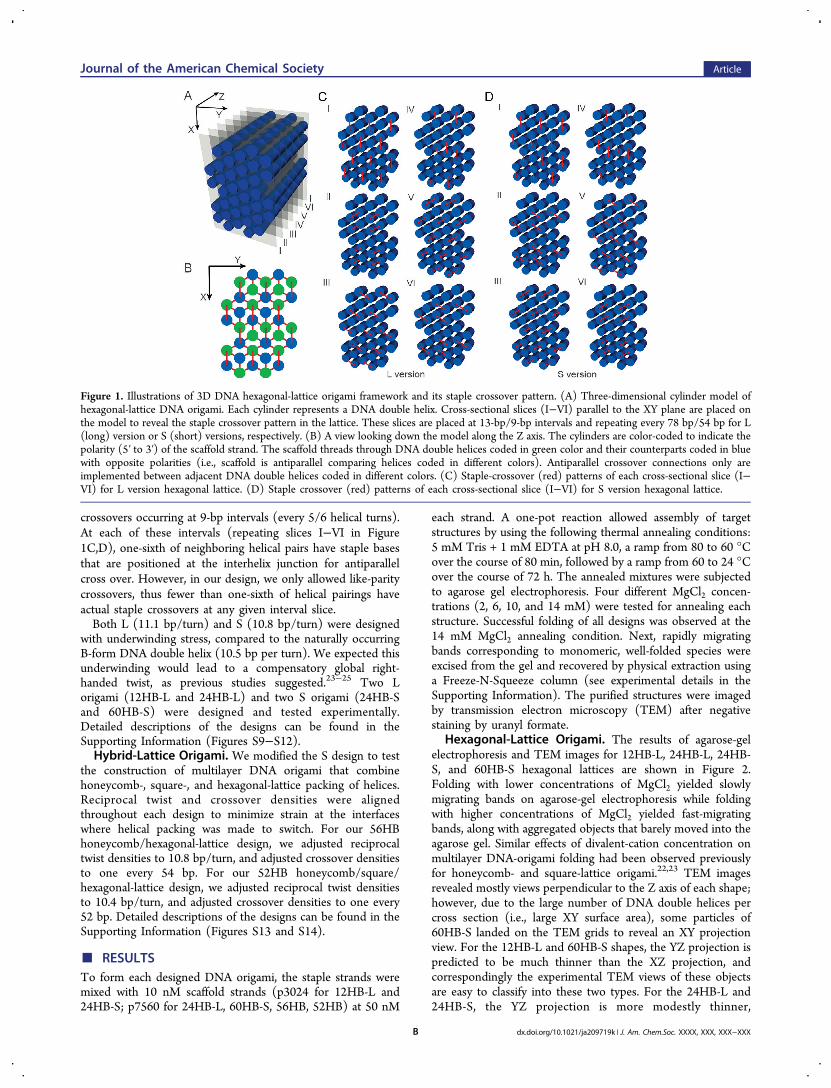
crossovers occurring at 9-bp intervals (every 5/6 helical turns).At each of these intervals (repeating slices I−VI in Figure1C,D), one-sixth of neighboring helical pairs have staple basesthat are positioned at the interhelix junction for antiparallelcross over. However, in our design, we only allowed like-paritycrossovers, thus fewer than one-sixth of helical pairings haveactual staple crossovers at any given interval slice.Both L (11.1 bp/turn) and S (10.8 bp/turn) were designed
with underwinding stress, compared to the naturally occurringB-form DNA double helix (10.5 bp per turn). We expected thisunderwinding would lead to a compensatory global right-handed twist, as previous studies suggested.23−25 Two Lorigami (12HB-L and 24HB-L) and two S origami (24HB-Sand 60HB-S) were designed and tested experimentally.Detailed descriptions of the designs can be found in theSupporting Information (Figures S9−S12).Hybrid-Lattice Origami. We modified the S design to test
the construction of multilayer DNA origami that combinehoneycomb-, square-, and hexagonal-lattice packing of helices.Reciprocal twist and crossover densities were alignedthroughout each design to minimize strain at the interfaceswhere helical packing was made to switch. For our 56HBhoneycomb/hexagonal-lattice design, we adjusted reciprocaltwist densities to 10.8 bp/turn, and adjusted crossover densitiesto one every 54 bp. For our 52HB honeycomb/square/hexagonal-lattice design, we adjusted reciprocal twist densitiesto 10.4 bp/turn, and adjusted crossover densities to one every52 bp. Detailed descriptions of the designs can be found in theSupporting Information (Figures S13 and S14).
■ RESULTSTo form each designed DNA origami, the staple strands weremixed with 10 nM scaffold strands (p3024 for 12HB-L and24HB-S; p7560 for 24HB-L, 60HB-S, 56HB, 52HB) at 50 nM
each strand. A one-pot reaction allowed assembly of targetstructures by using the following thermal annealing conditions:5 mM Tris + 1 mM EDTA at pH 8.0, a ramp from 80 to 60 °Cover the course of 80 min, followed by a ramp from 60 to 24 °Cover the course of 72 h. The annealed mixtures were subjectedto agarose gel electrophoresis. Four different MgCl2 concen-trations (2, 6, 10, and 14 mM) were tested for annealing eachstructure. Successful folding of all designs was observed at the14 mM MgCl2 annealing condition. Next, rapidly migratingbands corresponding to monomeric, well-folded species wereexcised from the gel and recovered by physical extraction usinga Freeze-N-Squeeze column (see experimental details in theSupporting Information). The purified structures were imagedby transmission electron microscopy (TEM) after negativestaining by uranyl formate.
Hexagonal-Lattice Origami. The results of agarose-gelelectrophoresis and TEM images for 12HB-L, 24HB-L, 24HB-S, and 60HB-S hexagonal lattices are shown in Figure 2.Folding with lower concentrations of MgCl2 yielded slowlymigrating bands on agarose-gel electrophoresis while foldingwith higher concentrations of MgCl2 yielded fast-migratingbands, along with aggregated objects that barely moved into theagarose gel. Similar effects of divalent-cation concentration onmultilayer DNA-origami folding had been observed previouslyfor honeycomb- and square-lattice origami.22,23 TEM imagesrevealed mostly views perpendicular to the Z axis of each shape;however, due to the large number of DNA double helices percross section (i.e., large XY surface area), some particles of60HB-S landed on the TEM grids to reveal an XY projectionview. For the 12HB-L and 60HB-S shapes, the YZ projection ispredicted to be much thinner than the XZ projection, andcorrespondingly the experimental TEM views of these objectsare easy to classify into these two types. For the 24HB-L and24HB-S, the YZ projection is more modestly thinner,
Figure 1. Illustrations of 3D DNA hexagonal-lattice origami framework and its staple crossover pattern. (A) Three-dimensional cylinder model ofhexagonal-lattice DNA origami. Each cylinder represents a DNA double helix. Cross-sectional slices (I−VI) parallel to the XY plane are placed onthe model to reveal the staple crossover pattern in the lattice. These slices are placed at 13-bp/9-bp intervals and repeating every 78 bp/54 bp for L(long) version or S (short) versions, respectively. (B) A view looking down the model along the Z axis. The cylinders are color-coded to indicate thepolarity (5′ to 3′) of the scaffold strand. The scaffold threads through DNA double helices coded in green color and their counterparts coded in bluewith opposite polarities (i.e., scaffold is antiparallel comparing helices coded in different colors). Antiparallel crossover connections only areimplemented between adjacent DNA double helices coded in different colors. (C) Staple-crossover (red) patterns of each cross-sectional slice (I−VI) for L version hexagonal lattice. (D) Staple crossover (red) patterns of each cross-sectional slice (I−VI) for S version hexagonal lattice.
Journal of the American Chemical Society Article
dx.doi.org/10.1021/ja209719k | J. Am. Chem.Soc. XXXX, XXX, XXX−XXXB
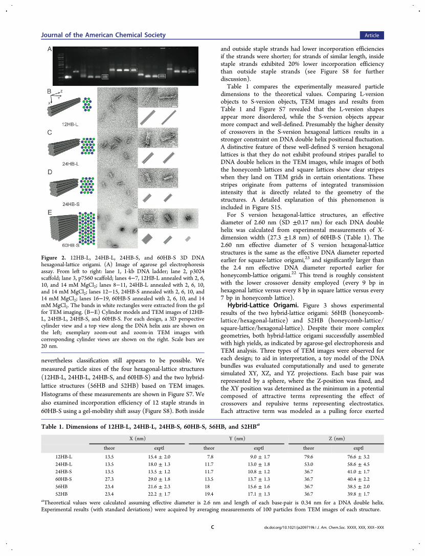
nevertheless classification still appears to be possible. Wemeasured particle sizes of the four hexagonal-lattice structures(12HB-L, 24HB-L, 24HB-S, and 60HB-S) and the two hybrid-lattice structures (56HB and 52HB) based on TEM images.Histograms of these measurements are shown in Figure S7. Wealso examined incorporation efficiency of 12 staple strands in60HB-S using a gel-mobility shift assay (Figure S8). Both inside
and outside staple strands had lower incorporation efficienciesif the strands were shorter; for strands of similar length, insidestaple strands exhibited 20% lower incorporation efficiencythan outside staple strands (see Figure S8 for furtherdiscussion).Table 1 compares the experimentally measured particle
dimensions to the theoretical values. Comparing L-versionobjects to S-version objects, TEM images and results fromTable 1 and Figure S7 revealed that the L-version shapesappear more disordered, while the S-version objects appearmore compact and well-defined. Presumably the higher densityof crossovers in the S-version hexagonal lattices results in astronger constraint on DNA double helix positional fluctuation.A distinctive feature of these well-defined S version hexagonallattices is that they do not exhibit profound stripes parallel toDNA double helices in the TEM images, while images of boththe honeycomb lattices and square lattices show clear stripeswhen they land on TEM grids in certain orientations. Thesestripes originate from patterns of integrated transmissionintensity that is directly related to the geometry of thestructures. A detailed explanation of this phenomenon isincluded in Figure S15.For S version hexagonal-lattice structures, an effective
diameter of 2.60 nm (SD ±0.17 nm) for each DNA doublehelix was calculated from experimental measurements of X-dimension width (27.3 ±1.8 nm) of 60HB-S (Table 1). The2.60 nm effective diameter of S version hexagonal-latticestructures is the same as the effective DNA diameter reportedearlier for square-lattice origami,23 and significantly larger thanthe 2.4 nm effective DNA diameter reported earlier forhoneycomb-lattice origami.22 This trend is roughly consistentwith the lower crossover density employed (every 9 bp inhexagonal lattice versus every 8 bp in square lattice versus every7 bp in honeycomb lattice).
Hybrid-Lattice Origami. Figure 3 shows experimentalresults of the two hybrid-lattice origami: 56HB (honeycomb-lattice/hexagonal-lattice) and 52HB (honeycomb-lattice/square-lattice/hexagonal-lattice). Despite their more complexgeometries, both hybrid-lattice origami successfully assembledwith high yields, as indicated by agarose-gel electrophoresis andTEM analysis. Three types of TEM images were observed foreach design; to aid in interpretation, a toy model of the DNAbundles was evaluated computationally and used to generatesimulated XY, XZ, and YZ projections. Each base pair wasrepresented by a sphere, where the Z-position was fixed, andthe XY position was determined as the minimum in a potentialcomposed of attractive terms representing the effect ofcrossovers and repulsive terms representing electrostatics.Each attractive term was modeled as a pulling force exerted
Figure 2. 12HB-L, 24HB-L, 24HB-S, and 60HB-S 3D DNAhexagonal-lattice origami. (A) Image of agarose gel electrophoresisassay. From left to right: lane 1, 1-kb DNA ladder; lane 2, p3024scaffold; lane 3, p7560 scaffold; lanes 4−7, 12HB-L annealed with 2, 6,10, and 14 mM MgCl2; lanes 8−11, 24HB-L annealed with 2, 6, 10,and 14 mM MgCl2; lanes 12−15, 24HB-S annealed with 2, 6, 10, and14 mM MgCl2; lanes 16−19, 60HB-S annealed with 2, 6, 10, and 14mM MgCl2. The bands in white rectangles were extracted from the gelfor TEM imaging. (B−E) Cylinder models and TEM images of 12HB-L, 24HB-L, 24HB-S, and 60HB-S. For each design, a 3D perspectivecylinder view and a top view along the DNA helix axis are shown onthe left; exemplary zoom-out and zoom-in TEM images withcorresponding cylinder views are shown on the right. Scale bars are20 nm.
Table 1. Dimensions of 12HB-L, 24HB-L, 24HB-S, 60HB-S, 56HB, and 52HBa
X (nm) Y (nm) Z (nm)
theor exptl theor exptl theor exptl
12HB-L 13.5 15.4 ± 2.0 7.8 9.0 ± 1.7 79.6 76.6 ± 3.224HB-L 13.5 18.0 ± 1.3 11.7 13.0 ± 1.8 53.0 58.6 ± 4.524HB-S 13.5 13.5 ± 1.2 11.7 10.8 ± 1.2 36.7 41.0 ± 1.760HB-S 27.3 29.0 ± 1.8 13.5 13.7 ± 1.3 36.7 40.4 ± 2.256HB 23.4 21.6 ± 2.3 18 15.6 ± 1.6 36.7 38.5 ± 2.052HB 23.4 22.2 ± 1.7 19.4 17.1 ± 1.3 36.7 39.8 ± 1.7
aTheoretical values were calculated assuming effective diameter is 2.6 nm and length of each base-pair is 0.34 nm for a DNA double helix.Experimental results (with standard deviations) were acquired by averaging measurements of 100 particles from TEM images of each structure.
Journal of the American Chemical Society Article
dx.doi.org/10.1021/ja209719k | J. Am. Chem.Soc. XXXX, XXX, XXX−XXXC
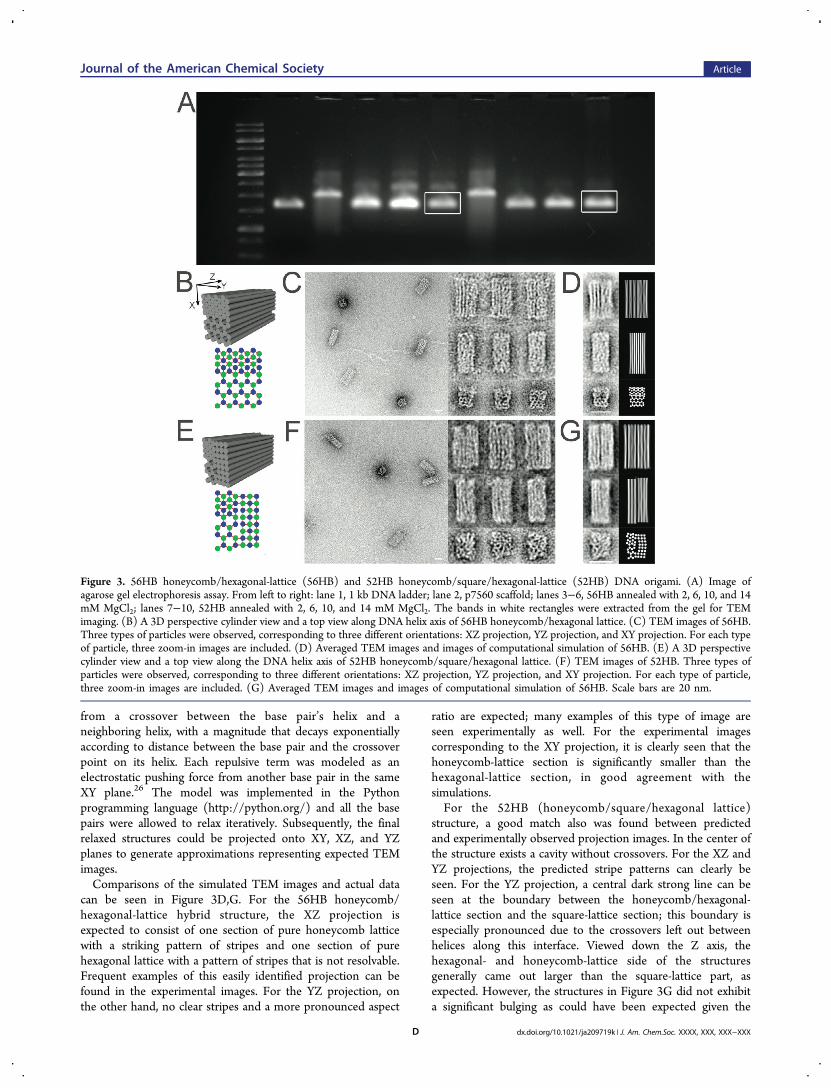
from a crossover between the base pair’s helix and aneighboring helix, with a magnitude that decays exponentiallyaccording to distance between the base pair and the crossoverpoint on its helix. Each repulsive term was modeled as anelectrostatic pushing force from another base pair in the sameXY plane.26 The model was implemented in the Pythonprogramming language (http://python.org/) and all the basepairs were allowed to relax iteratively. Subsequently, the finalrelaxed structures could be projected onto XY, XZ, and YZplanes to generate approximations representing expected TEMimages.Comparisons of the simulated TEM images and actual data
can be seen in Figure 3D,G. For the 56HB honeycomb/hexagonal-lattice hybrid structure, the XZ projection isexpected to consist of one section of pure honeycomb latticewith a striking pattern of stripes and one section of purehexagonal lattice with a pattern of stripes that is not resolvable.Frequent examples of this easily identified projection can befound in the experimental images. For the YZ projection, onthe other hand, no clear stripes and a more pronounced aspect
ratio are expected; many examples of this type of image areseen experimentally as well. For the experimental imagescorresponding to the XY projection, it is clearly seen that thehoneycomb-lattice section is significantly smaller than thehexagonal-lattice section, in good agreement with thesimulations.For the 52HB (honeycomb/square/hexagonal lattice)
structure, a good match also was found between predictedand experimentally observed projection images. In the center ofthe structure exists a cavity without crossovers. For the XZ andYZ projections, the predicted stripe patterns can clearly beseen. For the YZ projection, a central dark strong line can beseen at the boundary between the honeycomb/hexagonal-lattice section and the square-lattice section; this boundary isespecially pronounced due to the crossovers left out betweenhelices along this interface. Viewed down the Z axis, thehexagonal- and honeycomb-lattice side of the structuresgenerally came out larger than the square-lattice part, asexpected. However, the structures in Figure 3G did not exhibita significant bulging as could have been expected given the
Figure 3. 56HB honeycomb/hexagonal-lattice (56HB) and 52HB honeycomb/square/hexagonal-lattice (52HB) DNA origami. (A) Image ofagarose gel electrophoresis assay. From left to right: lane 1, 1 kb DNA ladder; lane 2, p7560 scaffold; lanes 3−6, 56HB annealed with 2, 6, 10, and 14mM MgCl2; lanes 7−10, 52HB annealed with 2, 6, 10, and 14 mM MgCl2. The bands in white rectangles were extracted from the gel for TEMimaging. (B) A 3D perspective cylinder view and a top view along DNA helix axis of 56HB honeycomb/hexagonal lattice. (C) TEM images of 56HB.Three types of particles were observed, corresponding to three different orientations: XZ projection, YZ projection, and XY projection. For each typeof particle, three zoom-in images are included. (D) Averaged TEM images and images of computational simulation of 56HB. (E) A 3D perspectivecylinder view and a top view along the DNA helix axis of 52HB honeycomb/square/hexagonal lattice. (F) TEM images of 52HB. Three types ofparticles were observed, corresponding to three different orientations: XZ projection, YZ projection, and XY projection. For each type of particle,three zoom-in images are included. (G) Averaged TEM images and images of computational simulation of 56HB. Scale bars are 20 nm.
Journal of the American Chemical Society Article
dx.doi.org/10.1021/ja209719k | J. Am. Chem.Soc. XXXX, XXX, XXX−XXXD
large central interface without crossovers in the design. Otherpreparations of the sample did, however, show a strongerbulging, suggesting that it could be dependent on saltconcentration or staining conditions.Given that the toy model assumed a lack of defects, the
strong correspondence between the simulated and experimentaldata indicates that the assembled origami structures are actuallyfolding as expected.
■ DISCUSSIONHere we have designed and realized a new family of 3D DNAorigami where parallel DNA double helices are packed on thehighly compact hexagonal-lattice geometry. Taking advantageof all three available geometries of regular-packing helices inmultilayer DNA origamihoneycomb, square, and hexagonallatticewe also demonstrated that more complicated hybrid3D DNA origami could be constructed. A designer with someknowledge and experience of structural DNA nanotechnologycould design a multilayer hexagonal-lattice origami or hybridorigami with desired geometry in a few hours, using computerprograms such as caDNAno.27 Currently, all crossovers need tobe manually implemented in caDNAno for hexagonal-lattice orhybrid origami. We anticipate an updated version of caDNAnothat can accommodate designs of all three geometries and evenhybrid origami, which will make the process more automatedand less time-consuming (Shawn Douglas, personal communi-cation).2D and 3D DNA-origami structures have been utilized or
proposed for positioning guest macromolecules with preciseorientational and spatial control in 3D space. Such applicationsoften will demand high-level control over overall shape anddensity as well as over the precise positions of each double helixof the DNA-origami structures. The hexagonal-lattice andhybrid-lattice origami design paradigms presented here willexpand our capability to achieve such versatile and detailedcontrol. For instance, since hexagonal-lattice 3D origami havethe highest material density among all three architectures(honeycomb-lattice, square-lattice, and hexagonal-lattice), theyshould exhibit the strongest resistance to twist or compressionper unit volume. Further systematic studies will be needed tobetter understand the mechanical properties of these 3D DNAlattices and thereby bring us closer to realizing the full potentialof DNA-origami-directed self-assembly.
■ ASSOCIATED CONTENT*S Supporting InformationExperimental details, structure design and DNA sequences, andadditional analysis results and discussions. This information isavailable free of charge via the Internet at http://pubs.acs.org.
■ AUTHOR INFORMATIONCorresponding [email protected]
■ ACKNOWLEDGMENTSThis work was supported by Wyss Institute for BiologicallyInspired Engineering, ONR (N000014091118), and NIH NewInnovator (1DP2OD004641-01) grants to W.M.S. and by theDanish National Research Foundation support to K.V.G.
■ REFERENCES(1) Seeman, N. C. Nature 2003, 421, 427−31.
(2) Seeman, N. C. Mol. Biotechnol. 2007, 37, 246−257.(3) Lin, C.; Liu, Y.; Rinker, S.; Yan, H. ChemPhysChem 2006, 7,1641−1647.(4) Yan, H.; Park, S. H.; Finkelstein, G.; Reif, J. H.; LaBean, T. H.Science 2003, 301, 1882−1884.(5) Sharma, J.; Chhabra, R.; Cheng, A.; Brownell, J.; Liu, Y.; Yan, H.Science 2009, 323, 112−116.(6) Sharma, J.; Ke, Y.; Lin, C.; Chhabra, R.; Wang, Q.; Nangreave, J.;Liu, Y.; Yan, H. Angew. Chem., Int. Ed. 2008, 47, 5157−5159.(7) Maune, H.; Han, S.; Barish, R.; Bockrath, M.; Goddard, W.;Rothemund, P.; Winfree, E. Nat. Nanotechnol. 2010, 5, 61−66.(8) Aldaye, F. A.; Palmer, A. L.; Sleiman, H. F. Science 2008, 321,1795−1799.(9) Mao, C.; Sun, W.; Shen, Z.; Seeman, N. C. Nature (London)1999, 397, 144−146.(10) Sherman, W.; Seeman, N. C. Nano Lett. 2004, 4, 1203−1207.(11) Yan, H.; Zhang, X.; Shen, Z.; Seeman, N. C. Nature 2002, 415,62−65.(12) Chakraborty, B.; Sha, R.; Seeman, N. C. Proc. Natl. Acad. Sci.U.S.A. 2008, 105, 17245−17249.(13) Lund, K.; Manzo, A. J.; Dabby, N.; Michelotti, N.; Johnson-Buck, A.; Nangreave, J.; Taylor, S.; Pei, R.; Stojanovic, M. N.; Walter,N. G.; Winfree, E.; Yan, H. Nature 2010, 465, 206−210.(14) Douglas, S. M.; Chou, J.; Shih, W. M. Proc. Natl. Acad. Sci. U.S.A.2007, 104, 6644−6648.(15) Gu, H.; Yang, W.; Seeman, N. C. J. Am. Chem. Soc. 2010, 132,4352−4357.(16) Endo, M.; Katsuda, Y.; Hidaka, K.; Sugiyama, H. J. Am. Chem.Soc. 2010, 132, 1592−1597.(17) Selmi, D. N.; Adamson, R. J.; Attrill, H.; Goddard, A. D.;Gilbert, R. J. C.; Watts, A.; Turberfield, A. J. Nano Lett. 2011, 11, 657−660.(18) Rothemund, P. Nature 2006, 440, 297−302.(19) Andersen, E. S.; Dong, M.; Nielsen, M. M.; Jahn, K.; Subramani,R.; Mamdouh, W.; Golas, M. M.; Sander, B.; Stark, H.; Oliveira, C. L.P.; Pedersen, J. S.; Birkedal, V.; Besenbacher, F.; Gothelf, K. V.; Kjems,J. Nature 2009, 459, 73−76.(20) Kuzuya, A.; Komiyama, M. Chem. Commun. 2009, 28, 4182−4184.(21) Ke, Y.; Sharma, J.; Liu, M.; Jahn, K.; Liu, Y.; Yan, H. Nano Lett.2009, 9, 2445−2447.(22) Douglas, S. M.; Dietz, H.; Liedl, T.; Hogberg, B.; Graf, F.; Shih,W. M. Nature 2009, 459, 414−418.(23) Ke, Y.; Douglas, S. M.; Liu, M.; Sharma, J.; Cheng, A.; Leung,A.; Liu, Y.; Shih, W. M.; Yan, H. J. Am. Chem. Soc 2009, 131, 15903−15908.(24) Wang, R.; Liu, W.; Seeman, N. C. Chem. Biol. 2009, 16, 862−867.(25) Dietz, H.; Douglas, S. M.; Shih, W. M. Science 2009, 325, 725−730.(26) Debye, P.; Huckel, E. Phys. Z. 1923, 24, 185−206.(27) Douglas, S. M.; Marblestone, A. H.; Teerapittayanon, S.;Vazquez, A.; Church, G. M.; Shih, W. M. Nucleic Acids Res. 2009, 37,5001−5006.
Journal of the American Chemical Society Article
dx.doi.org/10.1021/ja209719k | J. Am. Chem.Soc. XXXX, XXX, XXX−XXXE


![253.pptx [Last saved by user] - University of Babylon · HEXAGONAL CLOSE-PACKED STRUCTURE An HCP crystal is a closeAn HCP crystal is a close-packed structure with the stacking sequence](https://img.pdfslide.net/doc/110x75/5af2e01f7f8b9aa91690f0d3/253pptx-last-saved-by-user-university-of-close-packed-structure-an-hcp-crystal.jpg)


![[Akira Yoshizawa] Creative Origami (Sosaku Origami) (Origami Daily)](https://img.pdfslide.net/doc/110x75/577cc0ff1a28aba71191e5ee/akira-yoshizawa-creative-origami-sosaku-origami-origami-daily.jpg)