Embed Size (px)
Citation preview
Multiple Endocrinopathies in an Infant With FatalNeurodegenerative Disease
R. Ravi Shankar,1 Anzar Haider,2 W. Timothy Garvey,3 and Gary R. Freidenberg1*1Department of Pediatrics, Division of Endocrinology, Indiana University, Indianapolis2Akron Children’s Hospital, Akron, Ohio3Department of Medicine, Medical University of South Carolina and Charleston Veterans Affairs Medical Center,Charleston
We report on a male infant with congenitalhypoparathyroidism who developed pri-mary hypothyroidism at 3 months and insu-lin-dependent diabetes mellitus at 25months. He had evidence of widespreadand progressive neurologic dysfunctioncharacterized by severe developmental de-lay, blindness, deafness, seizures, atrophy ofthe cerebellar and frontal lobes, and el-evated spinal fluid protein. Also noted wererenal hypoplasia, hyporeninemic hypoaldo-steronism, chronic anemia, persistent eleva-tion of liver transaminase levels, abnormalintraventricular cardiac conduction, reduc-tion in numbers of helper T-cells, and dis-tinctive facial anomalies. The child died ofmultiorgan failure at 29 months. A mito-chondrial basis for the syndrome was con-sidered but a molecular mechanism has, asyet, not been identified. Am. J. Med. Genet.69:271–279, 1997. © 1997 Wiley-Liss, Inc.
KEY WORDS: hypoparathyroidism; diabe-tes mellitus; renal insuffi-ciency; growth failure; neu-rodegeneration; mitochon-drial
INTRODUCTION
Multiple endocrine failure in infancy is rare. In olderchildren, it is usually the result of autoimmune-me-diated glandular destruction. In infancy, autoimmuneendocrine disease usually involves the pancreatic isletsand results in diabetes mellitus [Neufeld et al., 1980;Arslanian et al., 1985; Vardi et al., 1988]. However,
autoimmune destruction of multiple endocrine glands,resulting in polyendocrinopathy, has not been reportedin infants. We describe an infant with non-autoim-mune failure of the parathyroids, thyroid gland, andpancreatic islets. Additionally, he had progressive neu-rodegenerative disease, chronic anemia, renal insuffi-ciency, and growth failure. He died at 29 months ofmultiorgan failure. We think that he has a previouslyundescribed syndrome. Although the cause of his dis-ease was not established, we suspect a disorder of en-ergy production, perhaps mitochondrial in origin.
Patient Description
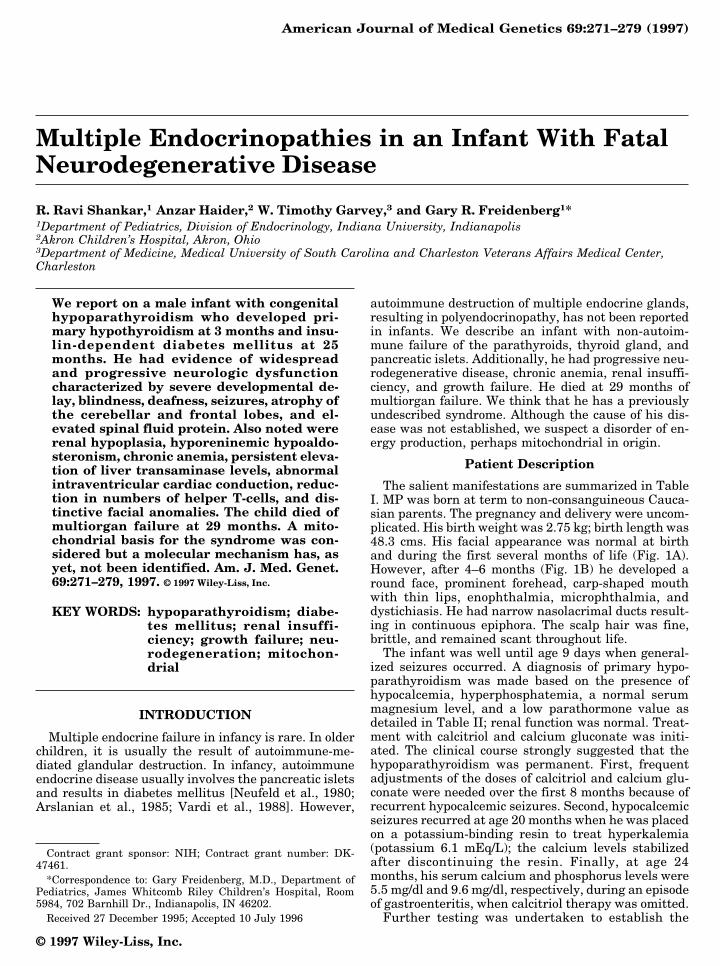
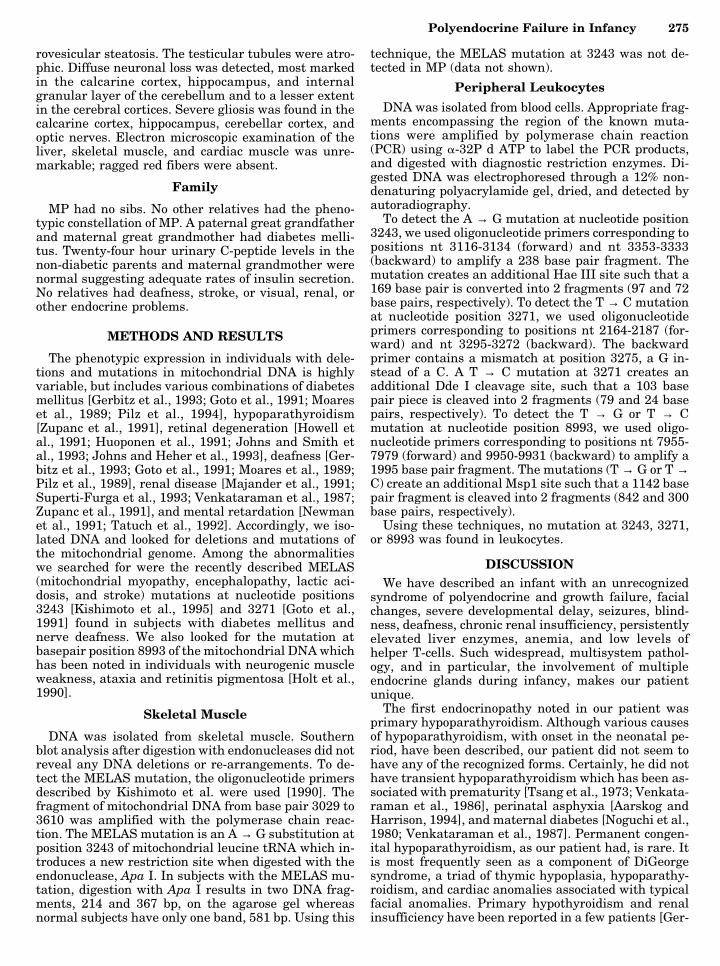
The salient manifestations are summarized in TableI. MP was born at term to non-consanguineous Cauca-sian parents. The pregnancy and delivery were uncom-plicated. His birth weight was 2.75 kg; birth length was48.3 cms. His facial appearance was normal at birthand during the first several months of life (Fig. 1A).However, after 4–6 months (Fig. 1B) he developed around face, prominent forehead, carp-shaped mouthwith thin lips, enophthalmia, microphthalmia, anddystichiasis. He had narrow nasolacrimal ducts result-ing in continuous epiphora. The scalp hair was fine,brittle, and remained scant throughout life.
The infant was well until age 9 days when general-ized seizures occurred. A diagnosis of primary hypo-parathyroidism was made based on the presence ofhypocalcemia, hyperphosphatemia, a normal serummagnesium level, and a low parathormone value asdetailed in Table II; renal function was normal. Treat-ment with calcitriol and calcium gluconate was initi-ated. The clinical course strongly suggested that thehypoparathyroidism was permanent. First, frequentadjustments of the doses of calcitriol and calcium glu-conate were needed over the first 8 months because ofrecurrent hypocalcemic seizures. Second, hypocalcemicseizures recurred at age 20 months when he was placedon a potassium-binding resin to treat hyperkalemia(potassium 6.1 mEq/L); the calcium levels stabilizedafter discontinuing the resin. Finally, at age 24months, his serum calcium and phosphorus levels were5.5 mg/dl and 9.6 mg/dl, respectively, during an episodeof gastroenteritis, when calcitriol therapy was omitted.
Further testing was undertaken to establish the
Contract grant sponsor: NIH; Contract grant number: DK-47461.
*Correspondence to: Gary Freidenberg, M.D., Department ofPediatrics, James Whitcomb Riley Children’s Hospital, Room5984, 702 Barnhill Dr., Indianapolis, IN 46202.
Received 27 December 1995; Accepted 10 July 1996
American Journal of Medical Genetics 69:271–279 (1997)
© 1997 Wiley-Liss, Inc.
cause of the hypoparathyroidism. DiGeorge syndromewas excluded on the basis of a normal thymic shadowon a chest radiograph, absence of anatomic abnormali-ties of the great vessels of the heart, and lack of thecharacteristic deletion on the long arm of chromosome22 observed in most patients with DiGeorge syndrome[de la Chapelle et al., 1981; Greenberg et al., 1986;Kelley et al., 1982]. In addition, the persistent decreasein CD2, CD3, and CD4 T-lymphocytes, as well as thenormal numbers of CD8 cells and decreased levels ofIgG2, were not typical of DiGeorge syndrome (TableIII). Apart from episodic watery diarrhea and an epi-sode of severe bronchiolitis related to an infection withrespiratory syncytial virus, MP had no clinical evi-dence of immune deficiency.
In addition to neonatal hypoparathyroidism, MP alsodeveloped other endocrinopathies. Compensated pri-mary hypothyroidism was evident at 3 months of age(Table III); anti-thyroid antibodies (anti-thyroglobulin,and anti-thyroid peroxidase antibodies) were absent,indicating that an autoimmune cause was unlikely. Heremained clinically and biochemically euthyroid onstandard replacement doses of L-thyroxine. At age25 months, hyperglycemia and relative insulinopeniawere detected (Table III). Islet cell and insulin autoan-tibodies were not present. Good glycemic control wasachieved with twice daily insulin injections (0.4–0.6U/kg/d); the hemoglobin A1c on this regimen was 6.7%(normal: 4.5 ± 0.3; mean ± SD). Neither ketonuria noracidosis ever occurred. At 26 mo, insulin therapy waswithdrawn for 36 hours following a hypoglycemic sei-zure. Hyperglycemia recurred, thus confirming the di-agnosis of diabetes mellitus.
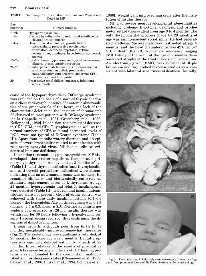
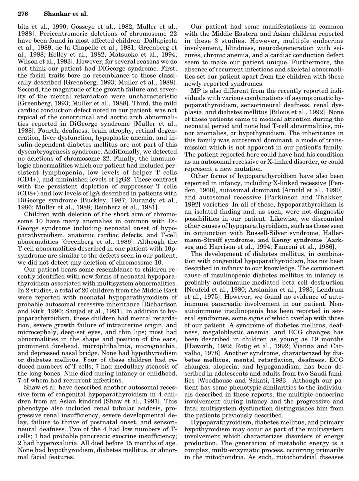
Linear growth, although poor from birth to 18months, inexplicably improved somewhat thereafter(Fig. 2). The skeletal age was significantly retarded; at25 months, the bone age was 3 months. Dental erup-tion was similarly delayed with only 6 teeth at 29months. Interpretation of the results of provocativegrowth hormone testing and levels of IGF binding pro-teins was confounded by the concomitant malnour-ished and insulinopenic states [Clemmons et al., 1989;Salardi et al., 1986; Shisko et al., 1994; Soliman et al.,
1986]. Weight gain improved markedly after the insti-tution of insulin therapy.
MP had severe neurodevelopmental abnormalitiesincluding profound hypotonia, deafness, and psycho-motor retardation evident from age 3 to 4 months. Theonly developmental progress made by 29 months ofage was an inconsistent social smile. He had general-ized areflexia. Microcephaly was first noted at age 5months, and the head circumference was 42.6 cm (−7SD) at death (Fig. 2B). A magnetic resonance imaging(MRI) study of the brain at the age of 7 months dem-onstrated atrophy of the frontal lobes and cerebellum.An electromyogram (EMG) was normal. Multiplebrainstem auditory evoked response studies were con-sistent with bilateral sensorineural deafness. Initially,
Fig. 1. Facial features. A: Relatively normal features at 2 months of ageapart from prominent forehead. B: Facial features at 26 months of age.
TABLE I. Summary of Clinical Manifestations and ProgressionNoted in MP
Age(months) Clinical findings
Birth Hypoparathyroidism1–3 Primary hypothyroidism, mild renal insufficiency,
elevated transaminases4–7 Onset of facial anomalies, growth failure,
microcephaly, progressive psychomotorretardation, deafness, hypotonia, retinaldegeneration, blindness, hypochromic normocyticanemia
19–24 Small kidneys, hyporeninemic hypoaldosteronism,bilateral ptosis, variable esotropia
25–27 Insulinopenic diabetes mellitus, intraventricularcardiac conduction defect, progressiveencephalopathy with seizures, abnormal EEG,increasing spinal fluid protein
29 Progressive renal failure, anasarca, fulminantsepsis, death
272 Shankar et al.
MP appeared to see, and visual evoked responses werenormal at 4 months of age. At age 7 months, examina-tion of his eyes under anesthesia revealed paucity ofretinal vascular arcades, optic atrophy, diffuse pigmen-tary retinopathy, and macular gliosis. An electroret-inogram (ERG) was flat. The retinal degenerative pro-cess was progressive. At age 19 months, bilateral ptosisand variable esotropia were noted. Despite these find-ings, his overall neurologic status was relatively staticuntil age 26 months after which he developed a pro-gressive encephalopathy. He had multiple seizureswhich were not associated with hypocalcemia or hypo-glycemia and required anticonvulsant therapy. Elec-troencephalogram (EEG) recordings showed progres-sive slowing of background activity. Serial lumbarpuncture studies demonstrated progressive increase in
the level of spinal fluid protein (23 mg/dl at age 25months, 81 mg/dl at 27 months, 93 mg/dl at 28 months,and 136 mg/dl pre-terminally).
MP also had chronic renal insufficiency which wasclinically insignificant until the last month of life. El-evated levels of creatinine and BUN were evident fromage 1 month (Table II). At age 5 months, a computer-ized tomogram (CT) scan of the abdomen showed mildrotational abnormalities of both kidneys, which wereotherwise normal. Initially, serial sonographic evalua-tions showed kidney size at the lower end of the normalrange for age. At 19 months, both kidneys were smallfor age and weight, and had not grown over the pre-vious 6 months. A technetium-99 diethelenetriaminepentacetate (DTPA) scan documented decreased renalfunction. On cystoscopy, a web in the mid bulbous ure-
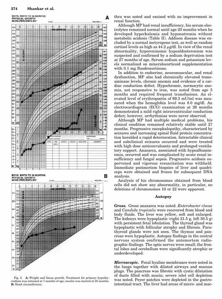
TABLE III. Results of Immunology Studies in the Patient Described
Age (months) 5 9 12 21 29b
Total WBC/mm3
T cell subsets8,400 — 8,500 7,900 5,800
Total lymphocytes/mm3 648 (5,395–7,211) — 2,221 (5,284–6,714) 3,239 (4431–5,508) 4,505 (3,855–5,248)CD2/mm3 303 (3,929–5,275) — 1,465 (3,806–4,881) 2,124 (3,101–3,868) 1,717 (2,649–3,639)CD3/mm3 246 (3,505–5,009) — 1,333 (3,409–4,575) 1,926 (2,766–3,508) 1,975 (2,324–3,295)CD4/mm3 195 (2,780–3,909) — 310 (2,630–3,499) 441 (1,919–2,472) 1,023 (1,538–2,213)CD8/mm3 127 (351–2,479) — 666 (351–2,479) 969 (351–2,479) 1,378 (351–2,479)CD4:CD8 1.5 (1.2–6.2) — 0.46 (1.2–6.2) 0.45 (1.2–6.2) 0.74 (1.2–6.2)
Immunoglobulinsa
IgA — 34 (14–106) 22 (17–123) 72 (17–123) —IgG total — 327 (442–880) 395 (553–971) 1,110 (553–971) —IgG1 — 225 (286–680) 290 (286–680) 1,040 (286–680) —IgG2 — 2 (30–327) 20 (30–327) 5 (30–327) —IgG3 — 30 (13–82) 60 (13–82) 90 (13–82) —IgG4 — 4 (1–65) 3 (1–65) 10 (1–65) —IgM — 91 (34–149) 77 (43–173) 95 (43–173) —
aNormal values for age in parentheses in mg/dl.bNormal values for age in parentheses while receiving 100 mg/m2/d hydrocortisone.
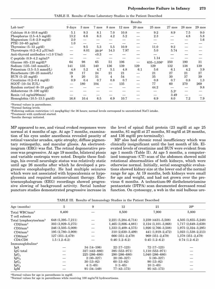
TABLE II. Results of Some Laboratory Studies in the Patient Described
Lab testa
Results
9 days 3 mos 7 mos 8 mos 12 mos 20 mos 25 mos 27 mos 28 mos 29 mos
Calcium (8.4–10.6 mg/dl) 5.1 9.3 8.1 7.0 10.8 — 9.2 8.9 7.5 9.0Phosphorus (2.5–4.5 mg/dl) 13.2 6.6 6.3 4.2 5.2 — 2.3 — 4.8 5.8Magnesium (1.6–2.9 mg/dl) 2.0 — — — — — — — — 1.6PTH (10–65 pg/ml) 1.0 — — — — — — — — —Thyroxine (5–12 mg/dl) — 9.5 5.3 5.5 10.9 — 11.0 9.2 — —Thyrotropin (0.2–6.2 mIU/ml) — 8.01 29.9d 14.3 7.97 — 5.0 5.74 — —Anti-thyroid antibodies (<1.0 U/ml) — — <0.3 — — — — — — —C-peptide (0.9–4.2 ng/ml)b — — — — — — 1.14 — — —Glucose (65–110 mg/dl)b 64 98 65 51 106 — 635–1,036e 250 190 31Sodium (135–145 mmol/L) 141 135 140 136 139 129 129 132 138 139Potassium (3.5–5.5 mmol/L) 4.8 5.2 4.7 5.1 4.8 6.1 5.6 6.1 4.2 3.9Bicarbonate (20–25 mmol/L) 19 17 24 21 21 — 21 27 21 27BUN (5–25 mg/dl) 9 20 31 4 54 — 35 30 37 39Creatinine (0.2–0.4 mg/dl) 0.9 0.4 0.7 0.8 0.7 — 0.8 0.7 0.8 1.3SGOT (10–54 IU/L) — 180 104 — 135 — 77 60 270 269Random cortisol (6–18 mg/dl) — — — — — — 44.2 — — 9.8Aldosterone (8–100 ng/dl) — — — — — — — 5.3c — —Renin (>5 ng/ml/90min) — — — — — — — 3.49 — —Hemoglobin (10.5–13.5 gm/dl) 16.8 10.4 6.5 6.9 10.7 — 6.9 8.0 7.2 7.9
aNormal values in parentheses.bNormal fasting levels.cFollowing sodium restriction (<1 meq/kg/day) for 36 hours; normal levels correspond to unrestricted NaCl intake.dTreatment with synthroid started.eInsulin therapy initiated.
Polyendocrine Failure in Infancy 273
thra was noted and excised with no improvement inrenal function.
Although MP had renal insufficiency, his serum elec-trolytes remained normal until age 20 months when hedeveloped hyperkalemia and hyponatremia withoutmetabolic acidosis (Table II). Addison disease was ex-cluded by a normal metyrapone test, as well as randomcortisol levels as high as 44.2 mg/dl. In view of the renalabnormality, hyporeninemic hypoaldosteronism wassuspected and confirmed by a sodium deprivation testat 27 months of age. Serum sodium and potassium lev-els normalized on mineralocorticoid supplementationwith 0.1 mg fluodrocortisone.
In addition to endocrine, neuromuscular, and renaldysfunction, MP also had chronically elevated trans-aminase levels, chronic anemia and evidence of a car-diac conduction defect. Hypochromic, normocytic ane-mia, not responsive to iron, was noted from age 5months and required frequent transfusions. An el-evated level of erythropoetin of 69.3 mU/ml was mea-sured when the hemoglobin level was 8.0 mg/dl. Anelectrocardiogram (ECG) examination at 26 monthsdemonstrated a mild right intraventricular conductiondefect; however, arrhythmias were never observed.
Although MP had multiple medical problems, hisclinical condition remained relatively stable until 27months. Progressive encephalopathy, characterized byseizures and increasing spinal fluid protein concentra-tion heralded a rapid deterioration. Intractable clinicaland subclinical seizures occurred and were treatedwith high dose anticonvulsants and prolonged ventila-tory support. Anasarca, associated with hypoalbumin-emia, occurred and was complicated by acute renal in-sufficiency and fungal sepsis. Progressive acidosis su-pervened and vigorous resuscitation was withheld.Immediate postmortem biopsies of liver and quadri-ceps were obtained and frozen for subsequent DNAanalysis.
Analysis of his chromosomes obtained from bloodcells did not show any abnormality, in particular, nodeletions of chromosomes 10 or 22 were apparent.
Autopsy
Gross. Gross anasarca was noted. Enterobacter clocaeand Candida tropicalis were recovered from blood andbody fluids. The liver was yellow, soft and enlarged.The kidneys were hypoplastic (right 21.5 g, left 30.5 g)with persistent fetal lobulation. The thyroid gland washypoplastic with follicular atrophy and fibrosis. Para-thyroid glands were not seen. The thymus and pan-creas were hypoplastic. Autopsy findings in the centralnervous system confirmed the antemortem radio-graphic findings. The optic nerves were small; the fron-tal lobes and cerebellum were significantly atrophic orunderdeveloped.
Microscopic. Focal hyaline membranes were noted inthe lungs together with dilated airways and mucousplugs. The pancreas was fibrotic with cystic dilatationof ducts filled with mucin; severe islet cell depletionwas noted. Peyer patches were depleted in the gastro-intestinal tract. The liver had areas of micro- and mac-
Fig. 2. A: Weight and linear growth. Treatment for primary hypothy-roidism was initiated at 7 months of age; insulin was started at 25 months.B: Head circumference.
274 Shankar et al.
rovesicular steatosis. The testicular tubules were atro-phic. Diffuse neuronal loss was detected, most markedin the calcarine cortex, hippocampus, and internalgranular layer of the cerebellum and to a lesser extentin the cerebral cortices. Severe gliosis was found in thecalcarine cortex, hippocampus, cerebellar cortex, andoptic nerves. Electron microscopic examination of theliver, skeletal muscle, and cardiac muscle was unre-markable; ragged red fibers were absent.
Family
MP had no sibs. No other relatives had the pheno-typic constellation of MP. A paternal great grandfatherand maternal great grandmother had diabetes melli-tus. Twenty-four hour urinary C-peptide levels in thenon-diabetic parents and maternal grandmother werenormal suggesting adequate rates of insulin secretion.No relatives had deafness, stroke, or visual, renal, orother endocrine problems.
METHODS AND RESULTS
The phenotypic expression in individuals with dele-tions and mutations in mitochondrial DNA is highlyvariable, but includes various combinations of diabetesmellitus [Gerbitz et al., 1993; Goto et al., 1991; Moareset al., 1989; Pilz et al., 1994], hypoparathyroidism[Zupanc et al., 1991], retinal degeneration [Howell etal., 1991; Huoponen et al., 1991; Johns and Smith etal., 1993; Johns and Heher et al., 1993], deafness [Ger-bitz et al., 1993; Goto et al., 1991; Moares et al., 1989;Pilz et al., 1989], renal disease [Majander et al., 1991;Superti-Furga et al., 1993; Venkataraman et al., 1987;Zupanc et al., 1991], and mental retardation [Newmanet al., 1991; Tatuch et al., 1992]. Accordingly, we iso-lated DNA and looked for deletions and mutations ofthe mitochondrial genome. Among the abnormalitieswe searched for were the recently described MELAS(mitochondrial myopathy, encephalopathy, lactic aci-dosis, and stroke) mutations at nucleotide positions3243 [Kishimoto et al., 1995] and 3271 [Goto et al.,1991] found in subjects with diabetes mellitus andnerve deafness. We also looked for the mutation atbasepair position 8993 of the mitochondrial DNA whichhas been noted in individuals with neurogenic muscleweakness, ataxia and retinitis pigmentosa [Holt et al.,1990].
Skeletal Muscle
DNA was isolated from skeletal muscle. Southernblot analysis after digestion with endonucleases did notreveal any DNA deletions or re-arrangements. To de-tect the MELAS mutation, the oligonucleotide primersdescribed by Kishimoto et al. were used [1990]. Thefragment of mitochondrial DNA from base pair 3029 to3610 was amplified with the polymerase chain reac-tion. The MELAS mutation is an A → G substitution atposition 3243 of mitochondrial leucine tRNA which in-troduces a new restriction site when digested with theendonuclease, Apa I. In subjects with the MELAS mu-tation, digestion with Apa I results in two DNA frag-ments, 214 and 367 bp, on the agarose gel whereasnormal subjects have only one band, 581 bp. Using this
technique, the MELAS mutation at 3243 was not de-tected in MP (data not shown).
Peripheral Leukocytes
DNA was isolated from blood cells. Appropriate frag-ments encompassing the region of the known muta-tions were amplified by polymerase chain reaction(PCR) using a-32P d ATP to label the PCR products,and digested with diagnostic restriction enzymes. Di-gested DNA was electrophoresed through a 12% non-denaturing polyacrylamide gel, dried, and detected byautoradiography.
To detect the A → G mutation at nucleotide position3243, we used oligonucleotide primers corresponding topositions nt 3116-3134 (forward) and nt 3353-3333(backward) to amplify a 238 base pair fragment. Themutation creates an additional Hae III site such that a169 base pair is converted into 2 fragments (97 and 72base pairs, respectively). To detect the T → C mutationat nucleotide position 3271, we used oligonucleotideprimers corresponding to positions nt 2164-2187 (for-ward) and nt 3295-3272 (backward). The backwardprimer contains a mismatch at position 3275, a G in-stead of a C. A T → C mutation at 3271 creates anadditional Dde I cleavage site, such that a 103 basepair piece is cleaved into 2 fragments (79 and 24 basepairs, respectively). To detect the T → G or T → Cmutation at nucleotide position 8993, we used oligo-nucleotide primers corresponding to positions nt 7955-7979 (forward) and 9950-9931 (backward) to amplify a1995 base pair fragment. The mutations (T → G or T →C) create an additional Msp1 site such that a 1142 basepair fragment is cleaved into 2 fragments (842 and 300base pairs, respectively).
Using these techniques, no mutation at 3243, 3271,or 8993 was found in leukocytes.
DISCUSSIONWe have described an infant with an unrecognized
syndrome of polyendocrine and growth failure, facialchanges, severe developmental delay, seizures, blind-ness, deafness, chronic renal insufficiency, persistentlyelevated liver enzymes, anemia, and low levels ofhelper T-cells. Such widespread, multisystem pathol-ogy, and in particular, the involvement of multipleendocrine glands during infancy, makes our patientunique.
The first endocrinopathy noted in our patient wasprimary hypoparathyroidism. Although various causesof hypoparathyroidism, with onset in the neonatal pe-riod, have been described, our patient did not seem tohave any of the recognized forms. Certainly, he did nothave transient hypoparathyroidism which has been as-sociated with prematurity [Tsang et al., 1973; Venkata-raman et al., 1986], perinatal asphyxia [Aarskog andHarrison, 1994], and maternal diabetes [Noguchi et al.,1980; Venkataraman et al., 1987]. Permanent congen-ital hypoparathyroidism, as our patient had, is rare. Itis most frequently seen as a component of DiGeorgesyndrome, a triad of thymic hypoplasia, hypoparathy-roidism, and cardiac anomalies associated with typicalfacial anomalies. Primary hypothyroidism and renalinsufficiency have been reported in a few patients [Ger-
Polyendocrine Failure in Infancy 275
bitz et al., 1990; Gosseye et al., 1982; Muller et al.,1988]. Pericentromeric deletions of chromosome 22have been found in most affected children [Dallapicolaet al., 1989; de la Chapelle et al., 1981; Greenberg etal., 1988; Kelley et al., 1982; Matsuoko et al., 1994;Wilson et al., 1993]. However, for several reasons we donot think our patient had DiGeorge syndrome. First,the facial traits bore no resemblance to those classi-cally described [Greenberg, 1993; Muller et al., 1988].Second, the magnitude of the growth failure and sever-ity of the mental retardation were uncharacteristic[Greenberg, 1993; Muller et al., 1988]. Third, the mildcardiac conduction defect noted in our patient, was nottypical of the conotruncal and aortic arch abnormali-ties reported in DiGeorge syndrome [Muller et al.,1988]. Fourth, deafness, brain atrophy, retinal degen-eration, liver dysfunction, hypoplastic anemia, and in-sulin-dependent diabetes mellitus are not part of thisdysembryogenesis syndrome. Additionally, we detectedno deletions of chromosome 22. Finally, the immuno-logic abnormalities which our patient had included per-sistent lymphopenia, low levels of helper T cells(CD4+), and diminished levels of IgG2. These contrastwith the persistent depletion of suppressor T cells(CD8+) and low levels of IgA described in patients withDiGeorge syndrome [Buckley, 1987; Durandy et al.,1986; Muller et al., 1988; Reinherz et al., 1981].
Children with deletion of the short arm of chromo-some 10 have many anomalies in common with Di-George syndrome including neonatal onset of hypo-parathyroidism, anatomic cardiac defects, and T-cellabnormalities [Greenberg et al., 1986]. Although theT-cell abnormalities described in one patient with 10p-syndrome are similar to the defects seen in our patient,we did not detect any deletion of chromosome 10.
Our patient bears some resemblance to children re-cently identified with new forms of neonatal hypopara-thyroidism associated with multisystem abnormalities.In 2 studies, a total of 20 children from the Middle Eastwere reported with neonatal hypoparathyroidism ofprobable autosomal recessive inheritance [Richardsonand Kirk, 1990; Sanjad et al., 1991]. In addition to hy-poparathyroidism, these children had mental retarda-tion, severe growth failure of intrauterine origin, andmicrocephaly, deep-set eyes, and thin lips; most hadabnormalities in the shape and position of the ears,prominent forehead, microphthalmia, micrognathia,and depressed nasal bridge. None had hypothyroidismor diabetes mellitus. Four of these children had re-duced numbers of T-cells; 7 had medullary stenosis ofthe long bones. Nine died during infancy or childhood,7 of whom had recurrent infections.
Shaw et al. have described another autosomal reces-sive form of congenital hypoparathyroidism in 4 chil-dren from an Asian kindred [Shaw et al., 1991]. Thisphenotype also included renal tubular acidosis, pro-gressive renal insufficiency, severe developmental de-lay, failure to thrive of postnatal onset, and sensori-neural deafness. Two of the 4 had low numbers of T-cells; 1 had probable pancreatic exocrine insufficiency;2 had hyperoxaluria. All died before 15 months of age.None had hypothyroidism, diabetes mellitus, or abnor-mal facial features.
Our patient had some manifestations in commonwith the Middle Eastern and Asian children reportedin these 3 studies. However, multiple endocrineinvolvement, blindness, neurodegeneration with sei-zures, chronic anemia, and a cardiac conduction defectseem to make our patient unique. Furthermore, theabsence of recurrent infections and skeletal abnormali-ties set our patient apart from the children with thesenewly reported syndromes.
MP is also different from the recently reported indi-viduals with various combinations of asymptomatic hy-poparathyroidism, sensorineural deafness, renal dys-plasia, and diabetes mellitus [Bilous et al., 1992]. Noneof these patients came to medical attention during theneonatal period and none had T-cell abnormalities, mi-nor anomalies, or hypothyroidism. The inheritance inthis family was autosomal dominant, a mode of trans-mission which is not apparent in our patient’s family.The patient reported here could have had his conditionas an autosomal recessive or X-linked disorder, or couldrepresent a new mutation.
Other forms of hypoparathyroidism have also beenreported in infancy, including X-linked recessive [Pen-den, 1960], autosomal dominant [Arnold et al., 1990],and autosomal recessive [Parkinson and Thakker,1992] varieties. In all of these, hypoparathyroidism isan isolated finding and, as such, were not diagnosticpossibilities in our patient. Likewise, we discountedother causes of hypoparathyroidism, such as those seenin conjunction with Russell-Silver syndrome, Haller-mann-Streiff syndrome, and Kenny syndrome [Aark-sog and Harrison et al., 1994; Fanconi et al., 1986].
The development of diabetes mellitus, in combina-tion with congenital hypoparathyroidism, has not beendescribed in infancy to our knowledge. The commonestcause of insulinopenic diabetes mellitus in infancy isprobably autoimmune-mediated beta cell destruction[Neufeld et al., 1980; Arslanian et al., 1985; Lendrumet al., 1975]. However, we found no evidence of auto-immune pancreatic involvement in our patient. Non-autoimmune insulinopenia has been reported in sev-eral syndromes, some signs of which overlap with thoseof our patient. A syndrome of diabetes mellitus, deaf-ness, megaloblastic anemia, and ECG changes hasbeen described in children as young as 19 months[Haworth, 1982; Rotig et al., 1992; Vianna and Car-valho, 1978]. Another syndrome, characterized by dia-betes mellitus, mental retardation, deafness, ECGchanges, alopecia, and hypogonadism, has been de-scribed in adolescents and adults from two Saudi fami-lies [Woodhouse and Sakati, 1983]. Although our pa-tient has some phenotypic similarities to the individu-als described in these reports, the multiple endocrineinvolvement during infancy and the progressive andfatal multisystem dysfunction distinguishes him fromthe patients previously described.
Hypoparathyroidism, diabetes mellitus, and primaryhypothyroidism may occur as part of the multisysteminvolvement which characterizes disorders of energyproduction. The generation of metabolic energy is acomplex, multi-enzymatic process, occurring primarilyin the mitochondria. As such, mitochondrial diseases
276 Shankar et al.
may result in multisystem failure. In fact, multiple en-docrinopathies are common in patients with mitochon-drial diseases. Diabetes mellitus is the most frequentlyreported endocrine disease [Dunbar et al., 1993; Ger-bitz et al., 1993; Goto et al., 1991; Johns et al., 1993;Johns et al., 1993; Kelley et al., 1982; Kishimoto et al.,1995; Luder and Barash 1994; Majander et al., 1991;Moares et al., 1989; Munnich et al., 1992; Newman etal., 1991; Pilz et al., 1991; Poulton et al., 1994; Remeset al., 1993; Rogers et al., 1969; Rotig et al., 1992; Su-perti-Furga et al., 1993; Suzuki, 1994; Zupanc et al.,1991] but hypoparathyroidism [Zupanc et al., 1991],growth hormone deficiency [Eviatar et al., 1990; Niau-det et al., 1994], and hypogonadism [Mosewitch et al.,1993; Newman, 1991] have also been described. De-pending on the distribution and numbers of abnormalmitochondria, phenotypic manifestations of mitochon-drial diseases may also include failure to thrive [Evi-atar et al., 1990; Newman et al., 1991; Oldfors et al.,1990; Rogers et al., 1969; Rotig et al., 1993; Rotig et al.,1993; Simonz et al., 1992; Superti-Furga et al., 1993;Suzuki, 1994; Zupanc et al., 1991], encephalopathy [deVriess et al., 1993; Gerbitz et al., 1990; Goto et al.,1991; Holt et al., 1990; Mosewitch et al., 1993; Zevianiet al., 1988; Zupanc et al., 1991], myopathy [de Vreisset al., 1993; Dunbar et al., 1993; Eviatar et al., 1990;Goto et al., 1991; Holt et al., 1990; Moares et al., 1989;Mosewitch et al., 1993; Niaudet et al., 1994; Poulton etal., 1994; Tatuch et al., 1992; Zeviani et al., 1990; Ze-viani et al., 1988; Zupanc et al., 1991], nephropathy[Luder and Barash, 1994; Majander et al., 1991; Niko-skelainen et al., 1987; Rotig et al., 1993; Superti-Furga1993; Zupanc et al., 1991], and pigmentary retinopathy[Eviatar et al., 1990; Howell et al., 1991; Huoponen etal., 1991; Johns et al., 1993; Larsson et al., 1990;Moares et al., 1989; Newman et al., 1991; Nikoskel-ainen et al., 1987; Oldfors et al., 1990; Poulton et al.,1994; Shanske et al., 1990; Wallace et al., 1988; Zupancet al., 1991]. Sensorineural deafness [Eviatar et al.,1990; Gerbitz et al., 1993; Goto et al., 1991; Kishimotoet al., 1995; Oldfors et al., 1990; Poulton et al., 1994;Remes et al., 1993; Rotig et al., 1993; Shanske et al.,1990; Zupanc et al., 1991], blindness [Howell et al.,1991; Huoponen et al., 1991; Johns et al., 1993; New-man et al., 1991; Nikoskelainen et al., 1987; Rotig etal., 1993], anemia [Larsson et al., 1990; Majander etal., 1991; Niaudet et al., 1994; Simonsz et al., 1991;Superti-Furga et al., 1993; Zupanc et al., 1991], lacticacidosis [Gerbitz et al., 1993; Goto et al., 1991; Holt etal., 1990; Luder and Barash, 1994; Remes et al., 1993;Rotig et al., 1992; Superti-Furga et al., 1993; Suzuki,1994; Tatuch et al., 1992; Zupanc et al., 1991], cardiacdysfunction [Gerbitz et al., 1990; Gerbitz et al., 1993;Larsson et al., 1990; Oldfors et al., 1990; Poulton et al.,1994; Shanske et al., 1990; Zeviani et al., 1990], exo-crine pancreatic insufficiency [Majander et al., 1991;Niaudet et al., 1994; Simonsz et al., 1992; Superti-Furga et al., 1993], and cerebellar dysfunction [Larssonet al., 1990; Rotig et al., 1992; Zeviani et al., 1990] havealso been reported.
The constellation of findings, and their inexorableprogression, in our patient is strongly suggestive of amitochondrial cytopathy. If so, our patient is the
youngest reported patient with such widespread endo-crine failure with onset in the neonatal period. Mul-tiple endocrinopathies have been described in olderchildren and adults with known mitochondrial disease[Eviatar et al., 1990; Zupanc et al., 1991]. However,standard textbooks of pediatric endocrinology have,thus far, not included mitochondrial disorders in theetiology of endocrine diseases [Aarskog and Harrison,1994; Mimouni and Tsang, 1990]. In particular, hypo-parathyroidism presenting in the neonatal period, orhypothyroidism and diabetes mellitus manifesting inearly infancy [Aarskog and Harrison, 1994; Mimouniand Tsang, 1990], have not been noted.
We have searched for a mitochondrial basis for ourpatient’s problems. However, we have not demon-strated any large deletion or duplication of mitochon-drial DNA in skeletal muscle or peripheral leuko-cytes. Furthermore, we have not found mutations atbasepairs 3243 [Gerbitz et al., 1993; Kishimoto et al.,1995], 3271 [Goto et al., 1991], and 8993 [de Vriess etal., 1993; Holt et al., 1990] which have been previouslyreported in patients with some mitochondrial diseases.Since mitochondrial proteins are coded by manynuclear as well as mitochondrial genes, it is conceiv-able that defects in nuclear genes could also result indecreased energy production. Thus, defects in either ofthese genomes can result in decreased energy produc-tion at the cellular level, resulting in widespread mani-festations similar to those detailed in our patient. Todate, our attention has focused on the mitochondrialgenome.
In summary, we have described an infant with re-lentlessly progressive multisystem dysfunction withonset of polyendocrinopathy in the immediate neonatalperiod. A diagnosis has not yet been established. It ispossible that MP has a more severe and extensive formof the syndrome described in Saudi, Kuwaiti, andAsian children with hypoparathyroidism [Richardsonand Kirk, 1990; Sanjad et al., 1991; Shaw et al., 1991].On the other hand, his clinical findings are remarkablysimilar to those found in patients with mitochondrialdiseases. In fact, we speculate that the children de-scribed in the Middle Eastern and Asian kindreds mayhave mitochondrial abnormalities. In any case, wethink that our patient has an, as yet, unrecognizedsyndrome. We suggest that disorders of energy produc-tion should be considered in all neonates and childrenwith endocrine failure.
ACKNOWLEDGMENTSWe thank Dr. Sara Shanske and Melissa Brown for
their help in the studies of mitochondrial DNA. A por-tion of the DNA work was supported by NIH grantDK-47461.
REFERENCESAarskog D, Harrison H (1994): Disorders of calcium, phosphate, and vita-
min D. In Kappy MS, Blizzard RM, Migeon CJ (eds): ‘‘Wilkins—TheDiagnosis and Treatment of Endocrine Disorders in Childhood andAdolescence.’’ Illinois: Charles C Thomas, pp 1027–1092.
Arslanian SA, Becker DJ, Rabin B, Atchison D, Eberhardt M, Caender D,et al. (1985): Correlates of insulin antibodies in newly diagnosed chil-dren with insulin-dependent diabetes before insulin therapy. Diabetes34:926–30.
Arnold A, Horst SA, Gardella TJ, Baba H, Levine MA, Kronenberg HM
Polyendocrine Failure in Infancy 277
(1990): Mutation of the signal peptide—encoding region of the pre-proparathyroid hormone gene in familial isolated hypoparathyroidism.J Clin Invest 86:1084–1087.
Bilous RW, Murty G, Parkinson DB, Thakker MG, Burn J, Mathias D,Kendall-Taylor P (1992): Autosomal dominant familial hypoparathy-roidism, sensorineural deafness, and renal dysplasia. New Engl J Med327:1069–1074.
Buckley RH (1987): Immunodeficiency diseases. J Am Med Assoc 258:2841–2850.
Clemmons DR (1989): Insulin-like growth factor binding proteins: Struc-tural analysis and mechanisms of action. Growth and Growth Factors4:3.
Conley ME, Beckwith JB, Mancer JFK, Teuchshoff L (1974): The spectrumof DiGeorge syndrome. J Peds 94:883–900.
Dallapiccola B, Marino B, Giannotti A, Valorani G (1989): DiGeorgeanomaly associated with partial deletion of chromosome 22. Report ofa case with X/22 translocation and a review of the literature. AnnGenet 32:92–96.
de la Chapelle A, Herva R, Koivista M, Aula P (1981): A deletion in chro-mosome 22 can cause DiGeorge syndrome. Hum Genet 57:253–256.
de Vriess DD, van Engelen BG, Gabreels FJ, Ruitenbeek W, van Oost BA(1993): A second missense mutation in the mitochondrial ATPase 6gene in Leigh syndrome. Ann Neurol 34:410–412.
Dunbar DR, Moonie PA, Surirgler RJ, Davidson D, Roberts R, Holt IJ(1993): Maternally transmitted partial direct tandem duplication ofmitochondrial DNA associated with diabetes mellitus. Hum Mol Genet2:1619–1624.
Durandy A, le Deist F, Fischer A, Griscelli C (1986): Impaired T8 lympho-cyte mediated suppressive activity in patients with partial DiGeorgesequence. J Clin Imm 6:265–270.
Eviatar L, Shanske S, Gauthier B, Abrams C, Maytal J, Slavin M, Valder-rama E, DiMauro S (1990): Kearns-Sayre syndrome presenting as re-nal tubular acidosis. Neurol 40:1761–1763.
Fanconi S, Fischer JA, Weiland P, Atares M, Fanconi A, Giedion A, PraderA (1986): Kenny syndrome: evidence for idiopathic hypoparathyroidismin two patients, and for abnormal parathyroid hormone in one. J Peds109:465–475.
Gerbitz KD, Obermaier-Kusser B, Zierz S, Pongratz D, Muller-Hocker J,Lestienne P (1990): Mitochondrial myopathies: divergences of geneticdeletions, biochemical defects, and the clinical syndrome. J Neurol 237:5–10.
Gerbitz KD, Paprotta A, Jaksli M, Zierz S, Dreschel J (1993): Diabetesmellitus is one of the heterogeneous phenotypic features of a mitochon-drial DNA point mutation within the tRNALeu(UUR) gene. FEBS Lett321:194–196.
Gosseye S, Goliare MC, Versllen G, van Lierde M, Claus D (1982): Asso-ciation of bilateral renal agenesis and DiGeorge syndrome in an infantof a diabetic mother. Helv Pediatr Acta 37:471–474.
Goto Y, Nonaka I, Horai S (1991): A new mtDNA mutation associated withmitochondrial myopathy, encephalopathy, lactic acidosis, and stroke-like episodes—MELAS. Biochim Biophys Acta 1097:238–240.
Greenberg F (1993): DiGeorge syndrome: a historical review of clinical andcytogenetic features. J Med Genet 30:803–806.
Greenberg F, Elder FF, Haffner P, Northrup H, Ledbetter DH (1988):Cytogenetic findings in a prospective series of patients with DiGeorgeAnomaly. Am J Hum Genet 43:605–611.
Greenberg F, Valdes C, Rosenblat HM, Kirkland JL, Ledbetter DH (1986):Hypoparathyoridism and T cell immune defect in a patient with 10pdeletion. J Peds 109:489–492.
Hartenstein H, Gardner LI (1966): Tetany of the newborn associated withmaternal parathyroid adenoma. N Engl J Med 274:266–268.
Harrison HE, Harrison HC (1990): Familial hyperparathyroidism. In Lif-shitz F (ed): ‘‘Pediatric Endocrinology: A clinical guide.’’ New York:Marcel Dekker, pp 613–623.
Haworth C, Evans DI, Mitra J, Wickramasinghe SN (1982): Thiamine-responsive megaloblastic anemia: A study of two further cases. Br JHematol 50:549–561.
Holt IJ, Harding AE, Petty RK, Morgan-Hughes JA (1990): A new mito-chondrial disease associated with mitochondrial DNA heteroplasmy.Am J Hum Genet 46:428–433.
Howell N, Bindoff LA, McCullough DA, Kubacka I, Poulton J, Mackey D,Taylor L, Turnbull DM (1991): Leber hereditary optic neuropathy:Identification of the same mitochondrial NDI mutation in six pedi-grees. Am J Hum Genet 49:939–950.
Huoponen K, Vikki J, Pertti A, Nikoskelainen E (1991): A new mtDNAmutation associated with Leber hereditary optic neuro-retinopathy.Am J Hum Genet 48:1147–1153.
Johns DR, Smith KH, Savino PJ, Miller NR (1993a): Leber hereditary opticneuropathy: Clinical manifestations of the 15257 mutation. Ophthal100:981–986.
Johns DR, Heher KL, Miller NR, Smith KH (1993b): Leber hereditary opticneuropathy: Clinical manifestations of the 14484 mutation. Ophthal111:495–498.
Kelley RI, Zackai EH, Emanuel BS, Kistenmacher M, Greenberg F, Pun-nett HH (1982): The association of the DiGeorge anomalad with partialmonosomy of chromosome 22. J Peds 101:197–200.
Kishimoto M, Hashiramoto M, Araki S, Ishida Y, Kazumi T, Kanda F,Kasuga M (1995): Diabetes mellitus carrying a mutation in the mito-chondrial tRNALeu(UUR) gene. Diabetologia 38:193–200.
Larsson NG, Holme E, Kristiansson B, Oldfors A, Tilinius M (1990): Pro-gressive increase of mutated mitochondrial DNA fraction in Kearns-Sayre syndrome. Peds Res 28:131–136.
Luder A, Barash V (1994): Complex I deficiency with diabetes, Fanconisyndrome and mtDNA deletion. J Inher Metab Dis 17:278–300.
Majander A, Suomalainen A, Veltenranta K, Sariola H, Perkkio M, Holm-berg C, Pihko R (1991): Congenital hypoplastic anemia, diabetes andsevere renal tubular dysfunction associated with mitochondrial DNAdeletion. Peds Res 30:327–330.
Matsuoko R, Takao A, Kinura M, Imanura S, Kondo C, Joh-o K, Ikeda K,Nishibatake M, Ando M, Momma K (1994): Confirmation that the cono-truncal anomaly-face syndrome is associated with a deletion within22q11.2. Am J Med Genet 1994; 53:285–289.
Mimouni F, Tsang RC (1990): Parathyroid and vitamin D related disor-ders. In Kaplan SA (ed): ‘‘Clinical Pediatric Endocrinology.’’ Philadel-phia: WB Saunders, pp 427–453.
Moares CT, DiMaoro S, Zeviani M, Lombes A, Shanske S, Miranda AF,Nakase H, Bonilla E, Werneck LC, Servidei S (1989): MitochondrialDNA deletions in progressive external ophthalmoplegia and Kearns-Sayre syndrome. New Engl J Med 320:1293–1299.
Mosewitch RK, Donat JR, Dimauro S, Ciafaloni E, Shanske S, Erasmus M,George D (1993): Syndrome of mitochondrial encephalopathy, lacticacidosis, and stroke-like episodes presenting without strokes. ArchNeurol 50:275–278.
Muller W, Peter HH, Kallfelz HC, Franz A, Reiger CHL (1988): The Di-George syndrome. II. Immunologic findings in partial and completeforms of the disorder. Eur J Peds 149:96–103.
Muller W, Peter HH, Wilken M, Juppner H, Kallfelz HC, Krohn HH, MillerK, Reiger CHL (1988): The DiGeorge syndrome. I. Clinical evaluationand course of partial and complete forms of the syndrome. Eur J Peds147:496–502.
Munnich A, Rustin P, Rotig A, Chretien D, Bonnefont JP, Nuttin C,Cormier V, Vassault A, Parvy P, Bardet J (1992): Clinical aspects ofmitochondrial disorders. J Inherit Metab Dis 15:448–455.
Neufeld M, Maclaren NK, Riley WJ, Lezotte D, McLoaughlin JV, Silver-stein J, Rosenbloom AL (1980): Islet cell and other organ-specific an-tibodies in U.S. Caucasians and Blacks with insulin-dependent diabe-tes mellitus. Diabetes 29:589–592.
Newman N, Lott MT, Wallace DC (1991): The clinical characteristics ofLeber hereditary optic neuropathy with the 11778 mutation. Am JOphthal 111:750–762.
Niaudet P, Heidet L, Munnich A, Schmitz J, Boulssou F, Gubler MC, RotigA (1994): Deletion of mitochondrial DNA in a case of de Toni-Debre-Fanconi syndrome and Pearson syndrome. Pediatr Nephrol 8:164–168.
Nikoskelainen E, Savontaus MJ, Olli PW, Mikko J, Nummelin KU (1987):Leber hereditary optic neuropathy: A maternally inherited disease.Arch Ophthal 105:665–671.
Noguchi A, Eren M, Tsang RC (1980): Parathyroid hormone in hypocalce-mic and normocalcemic infants of diabetic mothers. J Pediatr 97:112–114.
Oka Y, Katagiri H, Yazaki Y, Murase T, Kobayashi T (1993): Mitochondrialgene mutation in islet cell antibody positive patients who were initiallynon-insulin dependent. Lancet 312:527–528.
Oldfors A, Fyhr IM, Holme E, Larsson NG, Tilinius M (1990): Neuropa-thology in Kearns-Sayre syndrome. Neuropathol 80:541–546.
Parkinson DB, Thakker RV (1992): Donor splice mutation in PTH gene isassociated with autosomal recessive hypoparathyroidism. Natur Genet1:149–152.
278 Shankar et al.
Penden V (1960): True idiopathic hypoparathyroidism as a sex-linked re-cessive trait. Am J Hum Genet 12:323–325.
Pilz D, Quarrell OW, Jones EW (1994): Mitochondrial mutation commonlyassociated with Leber’s hereditary optic neuropathy observed in a pa-tient with Wolfram syndrome (DIDMOAD). J Med Genet 31:328–330.
Poulton J, Deadman ME, Bronte-Stewart J, Foulds WS, Gardiner RM(1994): Analysis of mitochondrial DNA in Leber hereditary optic neu-ropathy. J Med Genet 28:765–770.
Reinherz EL, Cooper MD, Schlossman SF, Rosen FS (1984): Abnormalitiesof T cell maturation and regulation in human beings with immunode-ficiency disorders. J Clin Invest 68:699–705.
Remes AM, Majamaa K, Herva R, Hassinen IE (1993): Adult-onset diabe-tes mellitus and neurosensory hearing loss in maternal relatives of aMELAS patient in a family with the tRNA (Leu(UUR)) mutation. Neu-rol 13:1015–1020.
Richardson RJ, Kirk JM (1990): Short stature, mental retardation, andhypoparathyroidism: a new syndrome. Arch Dis Child 65:1113–1117.
Rogers LE, Porter FS, Sidbury JB (1969): Thiamine-responsive megalo-blastic anemia. J Peds 74:494–504.
Rotig A, Bessis JL, Romero N, Cormier V, Saudubray JM, Nancy P, LenoirG, Rustin P, Munnich A (1992): Maternally inherited duplication of themitochondrial genome in a syndrome of proximal tubulopathy, diabetesmellitus, and cerebellar ataxia. Am J Hum Genet 50:361–370.
Rotig A, Cormier V, Chatelain P, Francois R, Saudubray JM, Rustin P,Munnich A (1993): Deletion of mitochondrial DNA in a case of earlyonset diabetes mellitus, optic atrophy and deafness (Wolfram syn-drome, MIM 222300). J Clin Invest 91:1095–1098.
Salardi S, Cacciari E, Ballardini D, Righetti F, Capelli M, Cocognani A,Zuchini S, Natali G, Tassinari D (1986): Relationships between growthfactors (somatomedin-C and growth hormone) and body development,metabolic control, and retinal changes in children and adolescents withIDDM. Diab 35:832–836.
Sanjad SA, Sakati NA, Abu-Osba YK, Kaddoura R, Milner RDG (1991): Anew syndrome of congenital hypoparathyroidism, severe growth fail-ure, and dysmorphic features. Arch Dis Child 66:193–196.
Shanske S, Moraes CT, Lombes A, Miranda AF, Bonilla E, Lewis P,Whelan MA, Ellsworth CA, DiMauro S (1990): Widespread tissue dis-tribution of mitochondrial DNA deletions in Kearns-Sayre syndrome.Neurol 40:24–28.
Shaw NJ, Haigh D, Lealman GT, Karbari G, Brocklebank JT, Dillon MJ(1991): Autosomal recessive hypoparathyroidism with renal insuffi-ciency and developmental delay. Arch Dis Child 66:1191–1194.
Shishko PI, Dreval AV, Abugora IA, Zajarny IU, Goncharov VC (1994):Insulin-like growth factors and binding proteins in patients with recentonset type I (insulin dependent) diabetes mellitus: influence of diabetescontrol and intraportal insulin infusion. Diab Res Clin Prac 25:1–12.
Simonsz HJ, Barlocher K, Rotig A (1992): Kearns-Sayre syndrome devel-oping in a boy who survived Pearson syndrome caused by a mitochon-drial DNA deletion. Doc Ophthal 82:73–79.
Soliman AT, Hassan AE, Aref MK, Hintz RL, Rosenfeld RG, Rogol RG(1986): Serum insulin-like growth factors I and II concentration andgrowth hormone and insulin responses to arginine infusion in children
with protein energy malnutrition before and after nutritional rehabili-tation. Peds Res 20:1122–1130.
Superti-Furga A, Schoenle E, Tuchschmid P, Caduff R, Sabato V, De Mat-tia D, Gitzelmann R, Steinmann B (1993): Pearson bone marrow-pancreas syndrome with insulin-dependent diabetes, progressive renaltubulopathy, organic aciduria and elevated fetal hemoglobin caused bydeletion and duplication of mitochondrial DNA. Eur J Ped 152:11–50.
Suzuki S (1994): Clinical characterization of diabetes mellitus in familieswith mitochondrial encephalomyopathies. Nippon Rinsho—Jap J ClinMed 52:2606–2610.
Tatuch Y, Christodoulou J, Feigenbaum A, Clarke JT, Wherret J, Smith C,Rudd N, Petrova-Benedict R, Robinson BH (1992): HeteroplasmicmtDNA mutation (TØG) at 8993 can cause Leigh disease when thepercentage of abnormal mtDNA is high. Am J Hum Genet 50:852–858.
Tsang RC, Chen I, Freidman MA, Gigger M, Steichen J, Koffor H, FentonL, Brown D, Pramanik A, Keenan W, Strub R, Joyce T (1975): Para-thyroid function in infants of diabetic mothers. J Pediatr 86:399–404.
Tsang RC, Chen IW, Freidman MA, Chen I (1973): Neonatal parathyroidfunction: role of gestational age and postnatal age. J Pediatr 83:728–738.
Vardi P, Zeigler AG, Matthews JH, Dib S, Keller RJ, Ricker AT et al.(1988): Concentration of insulin auto-antibodies at onset of type I dia-betes. Diab Care 11:736–739.
Venkataraman PS, Blick KE, Dasharathy G, Parker MK (1987): Loweredserum calcium, blood ionized calcium, and unresponsive serum para-thyroid hormone with oral glucose ingestion in infants of diabetic moth-ers. J Ped Gastr Nutr 6:931–935.
Venkatamaran PS, Tsang R, Steichen JJ, Grey I, Neylan M, FleischmanAR (1986): Early neonatal hypocalcemia in extremely preterm in-fants—high incidence, early onset, and refractoriness to supraphysi-ologic doses of calcitriol. Am J Dis Child 140:1004–1008.
Vianna MB, Carvalho RI (1978): Thiamine-responsive megaloblastic ane-mia, sensorineural deafness, and diabetes mellitus: a new syndrome? JPeds 93:235–238.
Wallace DC, Singh G, Lott MT, Hodge JA, Scheirr TG, Leeza MSA, ElsasL, Nikoskelainen E (1988): Mitochondrial DNA mutation associatedwith Leber hereditary optic neuropathy. Science 242:1427–1430.
Wilson DI, Burn J, Scambler P, Grodship J (1993): DiGeorge syndrome:part of CATCH 22. J Med Genet 30:852–856.
Woodhouse NJ, Sakati NA (1983): A syndrome of hypogonadism, alopecia,diabetes mellitus, mental retardation, deafness and ECG abnormali-ties. J Med Genet 20:216–219.
Zeviani M, Gallera C, Pannacci M, Uzeil G, Prelle A, Servidei S, DiDonatoS (1990): Tissue distribution and transmission of mitochondrial DNA inmitochondrial myopathies. Ann Neurol 28:94–97.
Zeviani M, Moares CT, DiMauro S, Nakase H, Bonilla E, Schon EA, Row-land LP (1988): Deletion of the mitochondrial DNA in Kearns-Sayresyndrome. Neurol 38:1339–1346.
Zupanc ML, Moraes CT, Shanske S, Langman CB, Ciafaloni E, DiMauro S(1991): Deletion of mitochondrial DNA in a patient with combined fea-tures of Kearns-Sayre and MELAS syndromes. Ann Neurol 29:680–683.
Polyendocrine Failure in Infancy 279