Embed Size (px)
Citation preview
Multisensory maps in parietal cortexMartin I Sereno1,2 and Ruey-Song Huang3
Available online at www.sciencedirect.com
ScienceDirect
Parietal cortex has long been known to be a site of
sensorimotor integration. Recent findings in humans have
shown that it is divided up into a number of small areas
somewhat specialized for eye movements, reaching, and hand
movements, but also face-related movements (avoidance,
eating), lower body movements, and movements coordinating
multiple body parts. The majority of these areas contain rough
sensory (receptotopic) maps, including a substantial
multisensory representation of the lower body and lower visual
field immediately medial to face VIP. There is strong evidence
for retinotopic remapping in LIP and face-centered remapping
in VIP, and weaker evidence for hand-centered remapping. The
larger size of the functionally distinct inferior parietal default
mode network in humans compared to monkeys results in a
superior and medial displacement of middle parietal areas (e.g.,
the saccade-related LIP’s). Multisensory superior parietal areas
located anterior to the angular gyrus such as AIP and VIP are
less medially displaced relative to macaque monkeys, so that
human LIP paradoxically ends up medial to human VIP.
Addresses1 Cognitive Perceptual and Brain Sciences, University College London,
London, UK2 Department of Psychological Sciences, Birkbeck/UCL Centre for
NeuroImaging (BUCNI), Birkbeck College, University of London, London,
UK3 Institute for Neural Computation, University of California San Diego, La
Jolla, CA, United States
Corresponding author: Sereno, Martin I ([email protected],
[email protected]) and
Current Opinion in Neurobiology 2013, 24:39–46
This review comes from a themed issue on Neural maps
Edited by David Fitzpatrick and Nachum Ulanovsky
0959-4388/$ – see front matter, Published by Elsevier Ltd.
http://dx.doi.org/10.1016/j.conb.2013.08.014
Unisensory versus multisensoryThe shortest path between any pair of neurons in the
brain often involves just few intervening synapses. For
example, in mice, primary visual cortex projects directly
to entorhinal cortex [1��]; similarly, in primates, parietal
visual areas project directly, if sparsely, to V1 [2,3��].Thus, in some sense, every brain area is potentially a
‘multisensory’ area [4,5].
But taking primate V1 as an example, single-unit spikes
there are most strongly modulated by the presence of
www.sciencedirect.com
simple visual features (orientation, direction of move-
ment) in the classical excitatory receptive field, or by
large arrays of similar low-level visual features in the non-
classical surround. Simple auditory, vestibular, and soma-
tosensory stimuli have small effects on the spiking of
primate V1 neurons, though they can more strongly
modulate the size or latency of subthreshold membrane
potentials, and consequently EEG/MEG or fMRI signals.
By contrast, spiking activity in neurons in an explicitly
multisensory area, such as primate ventral parietal area
(VIP) and rodent rostrolateral area (RL), is typically
strongly modulated by both visual and somatosensory
stimuli applied to localized regions of the receptor sheets,
either individually or in combination.
Another consideration is that species differ in the overall
depth of their visual cortical area hierarchies. For
example, in small nocturnal mammals that have less well
developed visual capabilities, like mice, V1 neurons are
more strongly modulated by the behavioral context of
stimuli (e.g., see [6]); in primates, there are more inter-
vening synapses from motor cortex to V1 [1��,3��], which
might explain why primate V1 is more strictly visual at the
level of single units. This review concentrates on map-
ping overtly multisensory areas in parietal cortex (for
previous reviews, see [7–9,10��,11]).
Ventral intraparietal area (VIP) — the parietalface areaVIP was originally defined in macaque monkeys as a
visual area containing neurons with large visual receptive
fields that also had aligned somatosensory receptive fields
on the face and shoulders [12]. More recent experiments
have suggested that VIP might instead be thought of as a
somatosensory area focused on operations in face-cen-
tered space that also has visual input. Avoidance and
defensive motor responses from stimulating VIP
[10��,13] and a preference for stereoscopic stimuli near
the face [14] suggest that one primary function is to
protect the face.
In humans, a multisensory area containing somatotopic
maps of air-puff stimuli to the face superimposed and
aligned with retinotopic maps of up-close visual stimuli
was found in the postcentral sulcus, just posterior and
slightly medial to the S-I hand representation [15,16] in
a region originally identified as multisensory by Brem-
mer et al. [17]. This region is also activated during
paradigms as diverse as mental arithmetic [18] and
delayed reaches in complete darkness toward extin-
guished visual targets [19], and so it is likely to be
involved in many cognitive functions involving actions
Current Opinion in Neurobiology 2014, 24:39–46
40 Neural maps
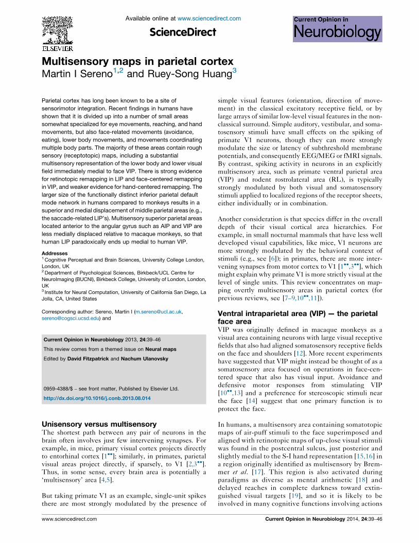
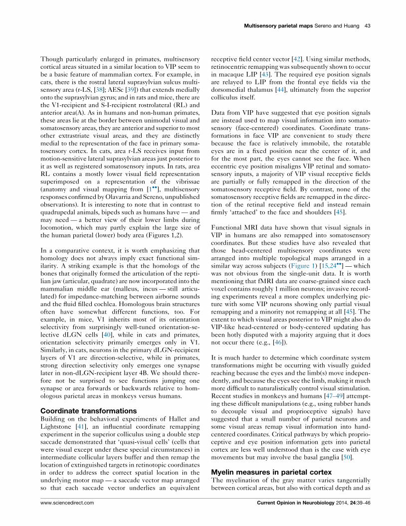
Figure 1
Current Opinion in Neurobiology
LH RH
Fingers Lips Face Shoulders Legs Toes
Overlapping retinotopic (upper panel) and somatotopic (lower panel) maps in human anterior parietal cortex. The upper row close-ups show 24-subject
average polar angle maps from wide-field direct-view fMRI mapping using a moving video wedge (complex-valued surface-based spherical coordinate
system averaging method described in [56]). Four lower ! middle ! upper field traverses are visible in VIP+ in each hemisphere (yellow arrows). The
color contours in the lower row show spherically aligned somatotopic whole body mapping data from [24��] (face data [15]) over the grayed visual data
(body part key is at bottom). Top insets show the location of the magnified views on the unfolded, dorsolaterally tilted cortex.
or events in real or metaphorical peripersonal space. For
example, when we say ‘the holidays are approaching’,
we treat the holidays as if they were looming objects
(compare the syntactically equivalent ‘the children are
approaching’) [20]. One of the overlaid functions of
multisensory parietal areas in humans may be to gen-
erate or interpret the meaning of such utterances.
Recent fMRI evidence in macaque monkeys has demon-
strated that face somatosensory inputs and visual inputs
overlap in one or more localized regions of the fundus of the
intraparietal sulcus rather than extending along its entire
length [21]. This result is compatible with human mapping
studies, which have uncovered multiple, somewhat
Current Opinion in Neurobiology 2014, 24:39–46
variable, overlapped representations of the face and retina
[15]. Surface-based cross-subject average retinotopic maps
suggest that the population average pattern in the anterior
most part of visual parietal cortex consists of two separated
(anterior and posterior) upper field representations and two
separated (medial and lateral) lower field representations
(see Figure 1, upper). This results in four lower-to-upper
visual field progressions. Multiple aligned representations
of the face overlay a portion of these visual maps (see red
contours in Figure 1, lower).
The parietal body area (greater VIP)Electrical stimulation studies in parietal cortex of several
different non-human primates using the ‘extended
www.sciencedirect.com
Multisensory parietal maps Sereno and Huang 41
stimulation trains’ method [22] had shown that parietal
cortex is involved in generating movements well beyond
facial defensive movements [23]. Subsequent bimodal
(somatosensory and visual) fMRI mapping experiments
in humans [24��] then revealed that the multisensory
zone in superior parietal cortex is larger than was
initially suspected (see cyan/pink/purple/black con-
tours, Figure 1, lower). The rough body homunculus
in human parietal cortex is arranged in a different order
than the ones in MI and SI (where the face is lateral, the
hand is intermediate, and the leg is medial). In human
superior parietal cortex, moving lateral to medial, the
face and lips in VIP proper are adjoined medially by the
shoulders, and then further medially by the lower parts
of the body (leg and toes), skipping the hand. The hand,
by contrast, is represented ‘out of order’, lateral to the
VIP face, in area AIP in the lateral part of the post-
central sulcus [16,24��], which is situated just posterior
to the S-I face representation (Figure 1, bottom, green
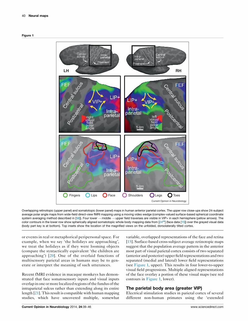
Figure 2
LeftHemisphere
(human)
Multisensoryactions
D: Defense
F: Feed
G: Grasp
P: Point
R: Reach
S: Saccade
T: Touch
W: Walk
Lower visual field
PreCent.
PreCent.
IFS
IFS
SFS
SFS
Lip
Tongue
Throat T
Human parietal eye, hand, face, and body areas. A rough structural (top) and
of the areas is likely to be involved in multiple additional functions beyond t
www.sciencedirect.com
contour). The visual field map overlying the lower body
representation in superior parietal cortex is primarily
lower field, as would be expected if part of its function
was to defend and coordinate the lower part of the
body with respect to visual and somatosensory objects
in the lower part of near peripersonal space; for example,
when watching your step. Several of these results were
prefigured in the excellent review by Rizzolatti et al.[11].
Multisensory areas for visually guidedreachingThere is a separate representation of hand and arm-
related multisensory areas more posteriorly on the medial
bank of the intraparietal sulcus in macaque monkeys and
extending onto the medial wall in the precuneus. This
general region has been divided into a number of differ-
ent areas, some of which overlap each other, including
MIP on the lateral surface, PEc near the dorsal convexity,
LS
LS
S-IM
-I
IPS
IPS
Eyes
Central sulcus
Central sulcus
PostCent.
PostCent.
ArmLegTrunk
TrunkTrunk
Leg/ToesArm
Leg
ArmHandHand
HandHand/Arm
FaceFace
Faces
Lips
Lips
Tonguehroat
R
D
W
F
GG/T
/R P/S
Current Opinion in Neurobiology
functional (bottom) parcellation of human parietal cortex is shown. Each
hose listed here.
Current Opinion in Neurobiology 2014, 24:39–46
42 Neural maps
and V6A (itself subdivided), and the greater ‘parietal
reach region’(see[8,25,26,27��,28])onthemidline.Recently
reach-related and grasp-related areas have been more pre-
cisely localized, subdivided, and renamed in humans
[29,30,31��,32,33,34��]. Figure 2 shows a summary of the
overall location of body parts (top) and a rough guide to
functional localization (bottom) drawn from references [7–9,
10��,11–16,18,19,21–23,24��,25,26,28–30,31��,32–33,34��].
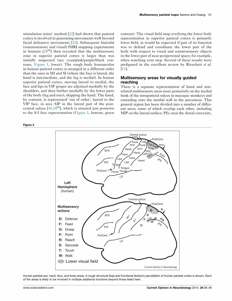
Comparative anatomy of parietal areasParietal cortex has long been known to be a site of
multisensory interactions on the basis of the effects of
brain lesions there in humans and in particular hemi-
neglect. Subsequent anatomical and physiological
investigations especially in macaque monkeys provided
support for this idea. However, some confusion has
persisted in correlating invasive non-human primate
data with human brain imaging data. In particular, the
relatively larger size of the angular gyrus component of
the ‘default mode network’ in the inferior parietal
lobule of humans compared to macaque monkeys results
in a substantial superior and medial displacement of
some — but not other — parietal visual areas in humans,
compared to their location in non-human primates (see
Figure 3). Thus, the initial report of a retinotopically
organized human homologue of macaque area LIP was
Figure 3
LIP+
LIP
LIP
VIP
VIP
AIP
AIPfolded
inflated
Def
Def
gyrussulcus
(not curvature)
VIPMacaqueMonkey
The large relative expansion of the inferior parietal component of the default m
monkeys results in the medial displacement of LIP+ (nominally, the lateral intr
macaque and human hemispheres. The monkey parietal default mode netwo
network was defined as the zone bounded by retinotopic, tonotopic, and som
scale.
Current Opinion in Neurobiology 2014, 24:39–46
controversial given how close to the midline the
putative human LIP was situated [35]. Subsequent
studies further subdivided parietal areas on the medial
bank of the human IPS [36,37]. The initial unease
with such a medial LIP derived from the fact that
macaque VIP, in the fundus of the intraparietal sulcus,
was conventionally thought of as being medial to LIP,
which as LIP’s name suggests, sits on the lateral bank
of the intraparietal sulcus. But on the unfolded cortex,
VIP can also be thought of as anterior to LIP in the
sense of being closer to somatosensory cortex. In
humans, VIP is mostly anterior to the relatively
enlarged angular gyrus areas; this results in VIP being
less medially displaced by them than LIP is. This
paradoxically results in human VIP+ sitting lateral to
human LIP+ (IPS1-4) (see Figure 3).
The superior and medial displacement of human LIP out
of the lateral bank of the intraparietal sulcus recalls a
related relative displacement of human MT+, which
moves laterally and inferiorly out of the superior temporal
sulcus, so that like LIP, its position no longer exactly
matches its name (the ‘middle temporal area’). That
downward displacement is also largely the result of the
increased relative size of the lateral parietal default mode
network in humans.
+ Human
Current Opinion in Neurobiology
ode network (Def, transparent purple) in humans compared to macaque
aparietal area) to a position medial to VIP+, shown on folded and inflated
rk component is taken from [57��]; the human angular gyrus default mode
atotopic maps in this subject. All cortical surfaces are shown at the same
www.sciencedirect.com
Multisensory parietal maps Sereno and Huang 43
Though particularly enlarged in primates, multisensory
cortical areas situated in a similar location to VIP seem to
be a basic feature of mammalian cortex. For example, in
cats, there is the rostral lateral suprasylvian sulcus multi-
sensory area (r-LS, [38]; AESc [39]) that extends medially
onto the suprasylvian gyrus; and in rats and mice, there are
the V1-recipient and S-I-recipient rostrolateral (RL) and
anterior area(A). As in humans and non-human primates,
these areas lie at the border between unimodal visual and
somatosensory areas, they are anterior and superior to most
other extrastriate visual areas, and they are distinctly
medial to the representation of the face in primary soma-
tosensory cortex. In cats, area r-LS receives input from
motion-sensitive lateral suprasylvian areas just posterior to
it as well as registered somatosensory inputs. In rats, area
RL contains a mostly lower visual field representation
superimposed on a representation of the vibrissae
(anatomy and visual mapping from [1��], multisensory
responses confirmed by Olavarria and Sereno, unpublished
observations). It is interesting to note that in contrast to
quadrupedal animals, bipeds such as humans have — and
may need — a better view of their lower limbs during
locomotion, which may partly explain the large size of
the human parietal (lower) body area (Figures 1,2).
In a comparative context, it is worth emphasizing that
homology does not always imply exact functional sim-
ilarity. A striking example is that the homologs of the
bones that originally formed the articulation of the repti-
lian jaw (articular, quadrate) are now incorporated into the
mammalian middle ear (malleus, incus — still articu-
lated) for impedance-matching between airborne sounds
and the fluid filled cochlea. Homologous brain structures
often have somewhat different functions, too. For
example, in mice, V1 inherits most of its orientation
selectivity from surprisingly well-tuned orientation-se-
lective dLGN cells [40], while in cats and primates,
orientation selectivity primarily emerges only in V1.
Similarly, in cats, neurons in the primary dLGN-recipient
layers of V1 are direction-selective, while in primates,
strong direction selectivity only emerges one synapse
later in non-dLGN-recipient layer 4B. We should there-
fore not be surprised to see functions jumping one
synapse or area forwards or backwards relative to hom-
ologous parietal areas in monkeys versus humans.
Coordinate transformationsBuilding on the behavioral experiments of Hallet and
Lightstone [41], an influential coordinate remapping
experiment in the superior colliculus using a double step
saccade demonstrated that ‘quasi-visual cells’ (cells that
were visual except under these special circumstances) in
intermediate collicular layers buffer and then remap the
location of extinguished targets in retinotopic coordinates
in order to address the correct spatial location in the
underlying motor map — a saccade vector map arranged
so that each saccade vector underlies an equivalent
www.sciencedirect.com
receptive field center vector [42]. Using similar methods,
retinocentric remapping was subsequently shown to occur
in macaque LIP [43]. The required eye position signals
are relayed to LIP from the frontal eye fields via the
dorsomedial thalamus [44], ultimately from the superior
colliculus itself.
Data from VIP have suggested that eye position signals
are instead used to map visual information into somato-
sensory (face-centered) coordinates. Coordinate trans-
formations in face VIP are convenient to study there
because the face is relatively immobile, the rotatable
eyes are in a fixed position near the center of it, and
for the most part, the eyes cannot see the face. When
eccentric eye position misaligns VIP retinal and somato-
sensory inputs, a majority of VIP visual receptive fields
are partially or fully remapped in the direction of the
somatosensory receptive field. By contrast, none of the
somatosensory receptive fields are remapped in the direc-
tion of the retinal receptive field and instead remain
firmly ‘attached’ to the face and shoulders [45].
Functional MRI data have shown that visual signals in
VIP in humans are also remapped into somatosensory
coordinates. But these studies have also revealed that
those head-centered multisensory coordinates were
arranged into multiple topological maps arranged in a
similar way across subjects (Figure 1) [15,24��] — which
was not obvious from the single-unit data. It is worth
mentioning that fMRI data are coarse-grained since each
voxel contains roughly 1 million neurons; invasive record-
ing experiments reveal a more complex underlying pic-
ture with some VIP neurons showing only partial visual
remapping and a minority not remapping at all [45]. The
extent to which visual areas posterior to VIP might also do
VIP-like head-centered or body-centered updating has
been hotly disputed with a majority arguing that it does
not occur there (e.g., [46]).
It is much harder to determine which coordinate system
transformations might be occurring with visually guided
reaching because the eyes and the limb(s) move indepen-
dently, and because the eyes see the limb, making it much
more difficult to naturalistically control visual stimulation.
Recent studies in monkeys and humans [47–49] attempt-
ing these difficult manipulations (e.g., using rubber hands
to decouple visual and proprioceptive signals) have
suggested that a small number of parietal neurons and
some visual areas remap visual information into hand-
centered coordinates. Critical pathways by which proprio-
ceptive and eye position information gets into parietal
cortex are less well understood than is the case with eye
movements but may involve the basal ganglia [50].
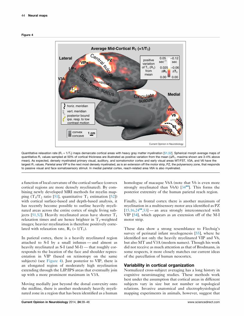
Myelin measures in parietal cortexThe myelination of the gray matter varies tangentially
between cortical areas, but also with cortical depth and as
Current Opinion in Neurobiology 2014, 24:39–46
44 Neural maps
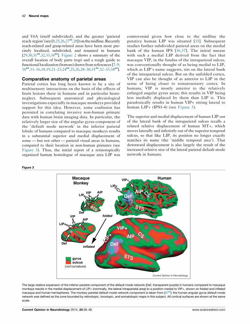
Figure 4
0.05sec–1
0.00
horiz. meridian
vert. meridian
concave1 cmconvex
posterior boundipsi. resp. to lowcontrast motion
0.00
Medial
Lateral
Average Mid-Cortical R1 (=1/T 1)
–0.12sec
0.025ΔR1
–0.06ΔT1
positivevariationof T1 (R 1)
frommean
Current Opinion in Neurobiology
Quantitative relaxation rate (R1 = 1/T1) maps demarcate cortical areas with heavy gray matter myelination [51,52]. Spherical morph average maps of
quantitative R1 values sampled at 50% of cortical thickness are illustrated as positive variation from the mean (DR1, maxima shown are 3-4% above
mean). As expected, densely myelinated primary visual, auditory, and somatomotor cortex and early visual areas MT/FST, V3A, and V6 have the
largest R1 values. Parietal area VIP is the next most densely myelinated, as is an extension off the motor strip, PZ, the polysensory zone, that responds
to passive visual and face somatosensory stimuli. In medial parietal cortex, reach-related area V6A is also myelinated.
a function of local curvature of the cortical surface (convex
cortical regions are more densely myelinated). By com-
bining newly developed MRI methods for myelin map-
ping (T1/T2 ratio [51], quantitative T1 estimation [52])
with cortical surface-based and depth-based analysis, it
has recently become possible to outline heavily myeli-
nated areas across the entire cortex of single living sub-
jects [51,52]. Heavily myelinated areas have shorter T1
relaxation times and are hence brighter in T1-weighted
images; heavier myelination is therefore positively corre-
lated with relaxation rate, R1 (= 1/T1).
In parietal cortex, there is a heavily myelinated region
attached to S-I by a small isthmus — and almost as
heavily myelinated as S-I (and M-I) — that roughly cor-
responds to the location of the face and shoulder repres-
entation in VIP (based on retinotopy on the same
subjects) (see Figure 4). Just posterior to VIP, there is
an elongated region of moderately high myelination
extending through the LIP/IPS areas that eventually join
up with a more prominent maximum in V3A.
Moving medially just beyond the dorsal convexity onto
the midline, there is another moderately heavily myeli-
nated zone in a region that has been identified as a human
Current Opinion in Neurobiology 2014, 24:39–46
homologue of macaque V6A (note that V6 is even more
strongly myelinated than V6A) [34��]. This forms the
posterior extremity of the human parietal reach region.
Finally, in frontal cortex there is another maximum of
myelination in a multisensory motor area identified as PZ
[15,16,24��,53] — an area strongly interconnected with
VIP [54], which appears as an extension off of the M-I
motor strip.
These data show a strong resemblance to Flechsig’s
survey of perinatal infant myelogenesis [55], where he
identified not only the heavily myelinated VIP and V6,
but also MT and V3A (modern names). Though his work
did not receive as much attention as that of Brodmann, in
some respects, it more closely matches our current ideas
of the parcellation of human neocortex.
Variability in cortical organizationNormalized cross-subject averaging has a long history in
cognitive neuroimaging studies. These methods work
best under the assumption that cortical areas in different
subjects vary in size but not number or topological
relations. Invasive anatomical and electrophysiological
mapping experiments in animals, however, suggest that
www.sciencedirect.com
Multisensory parietal maps Sereno and Huang 45
areas vary not only in size but sometimes also in neighbor
relations. The same may occur in humans. For example,
the number of discrete upper field representations found
in individual subjects between the upper field repres-
entation of V3A and the more posterior multisensory
upper-face-plus-upper-visual-field representation in
VIP varies from 1 to 3 in different humans (e.g., see
[36]). Given the large differences in individual area size
and in neighbor relations among visual areas among
closely related primates species, within-species variations
are perhaps not surprising.
ConclusionParietal multisensory maps are present in all mammals
and are especially well developed in primates and
humans. They seem to be specialized for coordinating
eye and limb movements in near peripersonal space for
the defense of the entire body, but also for acquisitive
movements such as hand-to-mouth and biting. In
humans, parietal multisensory areas are also active in a
variety of cognitive acts, some of which may involve
fictive or metaphoric acquisition, object manipulation,
or body defense.
Much work remains to be done in the field of active
sensory-guided limb movements, which involve complex
coordination of sensory inputs (visual, auditory, vestibu-
lar, somatosensory) as well as multiple sources of effer-
ence copy signals (saccades, smooth eye movements, face
and lip movements, neck movements, limb movements,
finger and toe movements). This area is particularly
challenging because of the difficulty of controlling these
multisensory stimuli, and in the case of human neuroima-
ging, maintaining data quality while making movements.
AcknowledgementsSupported by NIH R01 MH 081990 (Sereno, Huang), Royal SocietyWolfson Research Merit Award (Sereno).
References and recommended readingPapers of particular interest, published within the period of review,have been highlighted as:
� of special interest
�� of outstanding interest
1.��
Wang Q, Sporns O, Burkhalter A:: Network analysis ofcorticocortical connections reveals ventral and dorsalprocessing streams in mouse visual cortex. J Neurosci 2012,32:4386-4399.
Contains a comprehensive catalog of visual area connections fromflatmounted cortex including multisensory areas RL and A in mice andshows that visual areas are closer to being fully interconnected in micethan in monkeys.
2. Borra E, Rockland KS:: Projections to early visual areas V1 andV2 in the calcarine fissure from parietal association areas inthe macaque. Front Neuroanat 2011, 5:35.
3.��
Markov NT, Ercsey-Ravasz MM, Ribeiro Gomes AR, Lamy C,Magrou L, Vezoli J, Misery P, Falchier A, Quilodran R, Gariel MAet al.: A weighted and directed interareal connectivity matrixfor macaque cerebral cortex. Cereb Cortex 2012. [September25, Epub ahead of print].
www.sciencedirect.com
Contains a comprehensive catalog of visual area connections in macaquemonkeys including of which 30% have not been previously reported. Thegreat majority of connections come from areas within 12 mm of theinjection site. Compare with [1].
4. Ghazanfar AA, Schroeder CE:: Is neocortex essentiallymultisensory? Trends Cogn Sci 2006, 10:278-285.
5. Shams L, Kim R:: Crossmodal influences on visual perception.Phys Life Rev 2010, 7:269-284.
6. Niell CM, Stryker MP:: Modulation of visual responses bybehavioral state in mouse visual cortex. Neuron 2010, 65:472-479.
7. Culham JC, Valyear KF:: Human parietal cortex in action. CurrOpin Neurobiol 2006, 16:205-212.
8. Filimon F:: Human cortical control of hand movements:parietofrontal networks for reaching, grasping, and pointing.Neuroscientist 2010, 16:388-407.
9. Bremmer F:: Multisensory space: from eye-movements to self-motion. J Physiol 2011, 589:815-823.
10.��
Kaas JH, Gharbawie OA, Stepniewska I:: The organization andevolution of dorsal stream multisensory motor pathways inprimates. Front Neuroanat 2011, 5:34.
Parietal cortex stimulation in non-human primates results in coordinatedfacial and limb movements including reaching, grasping, hand-to-mouth,and defensive and aggressive movements with a region-specific orga-nization.
11. Rizzolatti G, Luppino G, Matelli M:: The organization of thecortical motor system: new concepts. Electroenceph ClinNeurophysiol 1998, 106:283-296.
12. Colby CL, Duhamel JR, Goldberg ME:: Ventral intraparietal areaof the macaque: anatomic location and visual responseproperties. J Neurophysiol 1993, 69:902-914.
13. Graziano MS, Cooke DF:: Parieto-frontal interactions, personalspace, and defensive behavior. Neuropsychologia 2006,44:2621-2635.
14. Bremmer F, Schlack A, Kaminiarz A, Hoffmann KP:: Encoding ofmovement in near extrapersonal space in primate area VIP.Front Behav Neurosci 2013, 7:8.
15. Sereno MI, Huang RS:: A human parietal face area containsaligned head-centered visual and tactile maps. Nat Neurosci2006, 9:1337-1343.
16. Huang RS, Sereno MI:: Dodecapus: an MR-compatible systemfor somatosensory stimulation. Neuroimage 2007, 34:1060-1073.
17. Bremmer F, Schlack A, Shah NJ, Zafiris O, Kubischik M,Hoffmann K, Zilles K, Fink GR:: Polymodal motion processing inposterior parietal and premotor cortex: a human fMRI studystrongly implies equivalencies between humans andmonkeys. Neuron 2001, 29:287-296.
18. Knops A, Thirion B, Hubbard EM, Michel V, Dehaene S::Recruitment of an area involved in eye movements duringmental arithmetic. Science 2009, 324:1583-1585.
19. Filimon F, Nelson JD, Huang RS, Sereno MI:: Multiple parietalreach regions in humans: cortical representations for visualand proprioceptive feedback during online reaching. JNeurosci 2009, 29:2961-2971.
20. Nunez R, Motz B, Teuscher U:: Time after time: thepsychological reality of the Ego- and time-reference-pointdistinction in metaphorical construals of time. MetaphorSymbol 2006, 21:133-146.
21. Guipponi O, Wardak C, Ibarrola D, Comte JC, Sappey-Marinier D,Pinede S, Ben Hamed S:: Multimodal convergence within theintraparietal sulcus of the macaque monkey. J Neurosci 2013,33:4128-4139.
22. Cooke DF, Taylor CS, Moore T, Graziano MS:: Complexmovements evoked by microstimulation of the ventralintraparietal area. Proc Natl Acad Sci U S A 2003, 100:6163-6168.
Current Opinion in Neurobiology 2014, 24:39–46
46 Neural maps
23. Stepniewska I, Fang PC, Kaas JH:: Organization of the posteriorparietal cortex in galagos: I, Functional zones identified bymicrostimulation. J Comp Neurol 2009, 517:765-782.
24.��
Huang RS, Chen CF, Tran AT, Holstein KL, Sereno MI:: Mappingmultisensory parietal face and body areas in humans. Proc NatlAcad Sci U S A 2012, 109:18114-18119.
By combining full body air-puff mapping with wide field visual stimulation, itwas shown that the region of visual somatosensory/visual overlap in parietalcortex extends beyond VIP as traditionally defined to include a medialregion concentrating on the lower body and extreme lower visual fields.
25. Gamberini M, Galletti C, Bosco A, Breveglieri R, Fattori P:: Is themedial posterior parietal area V6A a single functional area? JNeurosci 2011, 31:5145-5157.
26. Bakola S, Gamberini M, Passarelli L, Fattori P, Galletti C:: Corticalconnections of parietal field PEc in the macaque: linking visionand somatic sensation for the control of limb action. CerebCortex 2010, 20:2592-2604.
27.��
Seelke AM, Padberg JJ, Disbrow E, Purnell SM, Recanzone G,Krubitzer L:: Topographic maps within Brodmann’s Area 5 ofmacaque monkeys. Cereb Cortex 2012, 22:1834-1850.
Presents detailed microelectrode mapping of area 5 (superior parietalcortex) showing multiple maps of body parts from a region partiallyoverlapping the region electrically stimulated by Kaas et al. [10].
28. Kaas JH, Stepniewska I, Gharbawie O:: Cortical networkssubserving upper limb movements in primates. Eur J PhysRehabil Med 2012, 48:299-306.
29. Hinkley LB, Krubitzer LA, Padberg J, Disbrow EA:: Visual-manualexploration and posterior parietal cortex in humans. JNeurophysiol 2009, 102:3433-3446.
30. Cavina-Pratesi C, Monaco S, Fattori P, Galletti C, McAdam TD,Quinlan DJ, Goodale MA, Culham JC:: Functional magneticresonance imaging reveals the neural substrates of armtransport and grip formation in reach-to-grasp actions inhumans. J Neurosci 2010, 30:10306-10323.
31.��
Konen CS, Mruczek RE, Montoya JL, Kastner S:: Functionalorganization of human posterior parietal cortex: grasping- andreaching-related activations relative to topographicallyorganized cortex. J Neurophysiol 2013, 109:2897-2908.
Parietal cortex retinotopic maps are used as a basemap for examiningreaching and grasping movements, showing that much of reaching andgrasping activity takes place in areas containing sensory maps.
32. Mruczek RE, von Loga IS, Kastner S:: The representation of tooland non-tool object information in the human intraparietalsulcus. J Neurophysiol 2013, 109:2883-2896.
33. Rossit S, McAdam T, McLean DA, Goodale MA, Culham JC:: fMRIreveals a lower visual field preference for hand actions inhuman superior parieto-occipital cortex (SPOC) andprecuneus. Cortex 2013, January [Epub ahead of print].
34.��
Pitzalis S, Sereno MI, Committeri G, Fattori P, Galati G, Tosoni A,Galletti C:: The human homologue of macaque area V6A.Neuroimage 2013, 82C:517-530.
By combining retinotopy with reaching, the posterior portion of the medialparietal reach region in humans was shown to contain a representation ofthe lower visual field. See also [33].
35. Sereno MI, Pitzalis S, Martinez A:: Mapping of contralateralspace in retinotopic coordinates by a parietal cortical area inhumans. Science 2001, 294:1350-1354.
36. Swisher JD, Halko MA, Merabet LB, McMains SA, Somers DC::Visual topography of human intraparietal sulcus. J Neurosci2007, 27:5326-5337.
37. Silver MA, Kastner S:: Topographic maps in human frontal andparietal cortex. Trends Cogn Sci 2009, 13:488-495.
38. Palmer LA, Rosenquist AC, Tusa RJ:: The retinotopicorganization of lateral suprasylvian visual areas in the cat. JComp Neurol 1978, 177:237-256.
Current Opinion in Neurobiology 2014, 24:39–46
39. Monteiro GA, Clemo HR, Meredith MA:: Anterior ectosylviancortical projections to the rostral suprasylvian multisensoryzone in cat. Neuroreport 2003, 14:2139-2145.
40. Scholl B, Tan AY, Corey J, Priebe NJ:: Emergence of orientationselectivity in the mammalian visual pathway. J Neurosci 2013,33:10616-10624.
41. Hallett PE, Lightstone AD:: Saccadic eye movements towardsstimuli triggered by prior saccades. Vision Res 1976, 16:99-106.
42. Mays LE, Sparks DL:: Dissociation of visual and saccade-related responses in superior colliculus. J Neurophysiol 1980,43:207-232.
43. Goldberg ME, Colby CL, Duhamel JR:: Representation ofvisuomotor space in the parietal lobe of the monkey. ColdSpring Harb Symp Quant Biol 1990, 55:729-739.
44. Sommer MA, Wurtz RH:: A pathway in primate brain for internalmonitoring of movements. Science 2002, 296:1480-1482.
45. Avillac M, Deneve S, Olivier E, Pouget A, Duhamel JR:: Referenceframes for representing visual and tactile locations in parietalcortex. Nat Neurosci 2005, 8:941-949.
46. Golomb JD, Kanwisher N:: Higher level visual cortex representsretinotopic, not spatiotopic, object location. Cereb Cortex2012, 22:2794-2810.
47. Buneo CA, Andersen RA:: Integration of target and handposition signals in the posterior parietal cortex: effects ofworkspace and hand vision. J Neurophysiol 2012, 108:187-199.
48. Makin TR, Holmes NP, Zohary E:: Is that near my hand?Multisensory representation of peripersonal space in humanintraparietal sulcus. J Neurosci 2007, 27:731-740.
49. Brozzoli C, Gentile G, Petkova VI, Ehrsson HH:: FMRI adaptationreveals a cortical mechanism for the coding of space near thehand. J Neurosci 2011, 31:9023-9031.
50. Clower DM, Dum RP, Strick PL:: Basal ganglia and cerebellarinputs to ‘AIP’. Cereb Cortex 2005, 15:913-920.
51. Glasser MF, Van Essen DC:: Mapping human cortical areas invivo based on myelin content as revealed by T1- and T2-weighted MRI. J Neurosci 2011, 31:11597-11616.
52. Sereno MI, Lutti A, Weiskopf N, Dick F:: Mapping the humancortical surface by combining quantitative T1 with retinotopy.Cereb Cortex 2012, 23(July) http://dx.doi.org/10.1093/cercor/bhs213.
53. Graziano MS, Gandhi S:: Location of the polysensory zone inthe precentral gyrus of anesthetized monkeys. Exp Brain Res2000, 135:259-266.
54. Lewis JW, Van Essen DC:: Corticocortical connections ofvisual, sensorimotor, and multimodal processing areas in theparietal lobe of the macaque monkey. J Comp Neurol 2000,428:112-137.
55. Flechsig P:: Antomie des menschlichen Gehirns und Ruckenmarksauf myelogenetischer Grundlage. Leipzig: Georg Thieme; 1920, .
56. Hagler DJ, Riecke L, Sereno MI:: Parietal and superior frontalvisuospatial maps activated by pointing and saccades.NeuroImage 2007, 35:1562-1577.
57.��
Mantini D, Gerits A, Nelissen K, Durand JB, Joly O, Simone L,Sawamura H, Wardak C, Orban GA, Buckner RL, Vanduffel W::Behavioral/systems/cognitive default mode of brain functionin monkeys. J Neurosci 2011, 31:12954-12962.
This meta-analysis of task-related deactivations combined with analysisof resting state has identified components of the default-mode network inmacaque monkeys.
www.sciencedirect.com