Embed Size (px)
Citation preview
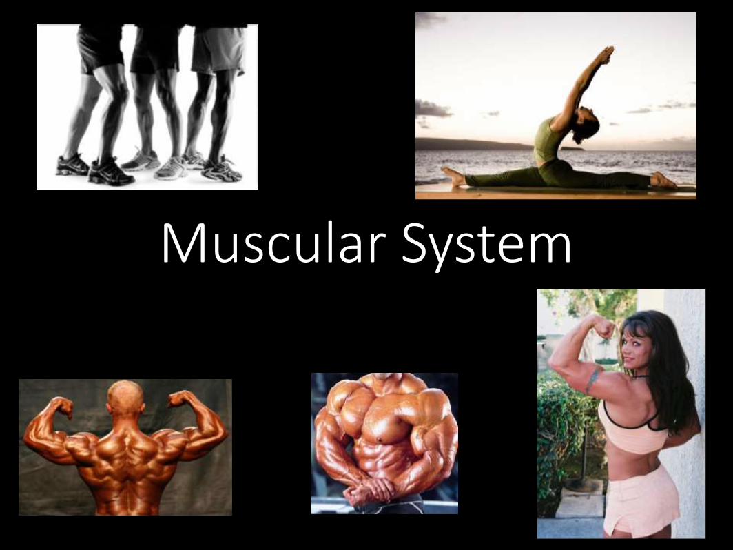
Muscular System
Written Response #1
1. Based on what you know about Latin root words, what do you think these terms refer to?• Sarcomere
• Sarcoplasm
• Myofibril
• Epimysium
• Perimysium
• Endomysium
2. What structure connects muscle to bone?
Muscles
• “muscle” = myo- or mys-
• sarco- = “flesh” - also refers to muscles
Main Functions of Muscles
1. Produce movement
2. Maintain posture & body position
3. Stabilize joints
4. Generate heat
• Additional functions: protect organs, valves, dilate pupils, raise hairs
Written Response #2
1. Describe how connective tissue is part of a skeletal muscle.
2. Describe the general structure of a skeletal muscle fiber.
3. Explain why skeletal muscle fibers appear striated.
4. Explain the relationship between the sarcoplasmic reticulum and the transverse tubules.
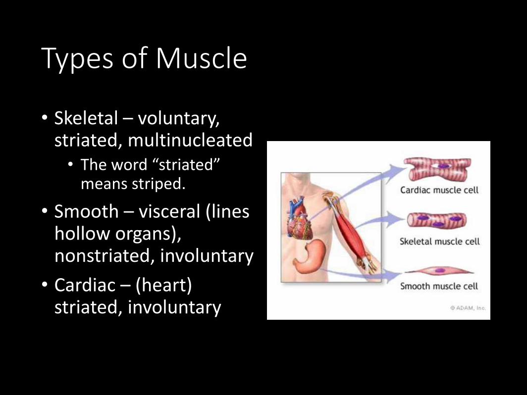
Types of Muscle
• Skeletal – voluntary, striated, multinucleated• The word “striated”
means striped.
• Smooth – visceral (lines hollow organs), nonstriated, involuntary
• Cardiac – (heart) striated, involuntary
Special Characteristics of Muscle
• Excitability – can receive and respond to stimuli
• Contractility – can shorten forcibly
• Extensibility – can be stretched or extended
• Elasticity – can recoil and resume resting length after being stretched
Gross Anatomy of Skeletal Muscle
• 1 muscle = 1 organ
• Each muscle served by a nerve, artery and vein
• Rich blood supply: need energy & oxygen
• Connective tissue sheaths: wraps each cell and reinforce whole muscle
• Attachment is either1. directly to bone
2. by tendons or aponeuroses to bone, cartilage, or other muscles
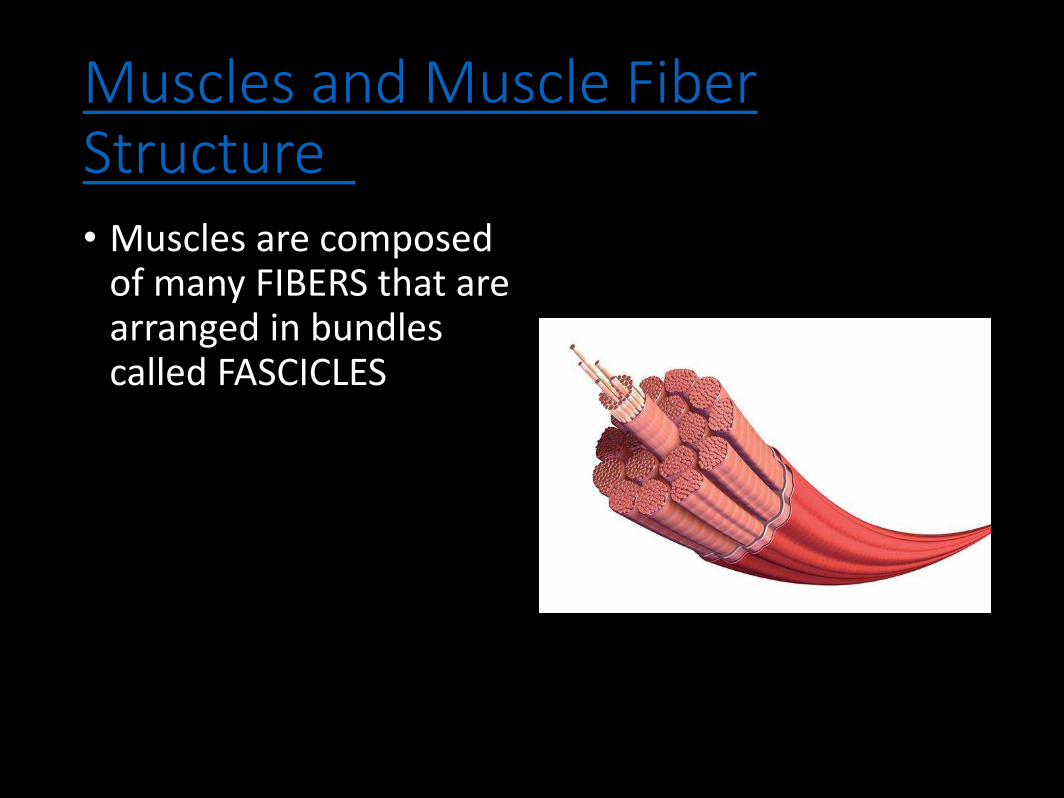
Muscles and Muscle FiberStructure• Muscles are composed
of many FIBERS that are arranged in bundles called FASCICLES
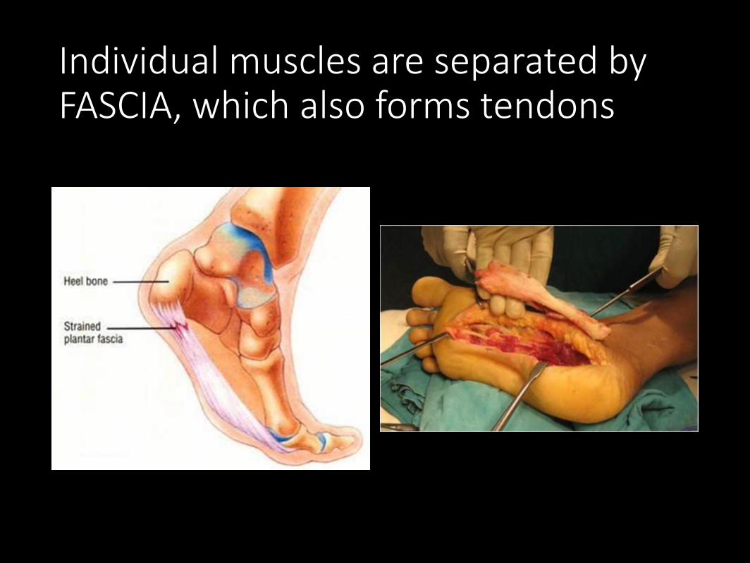
Individual muscles are separated by FASCIA, which also forms tendons
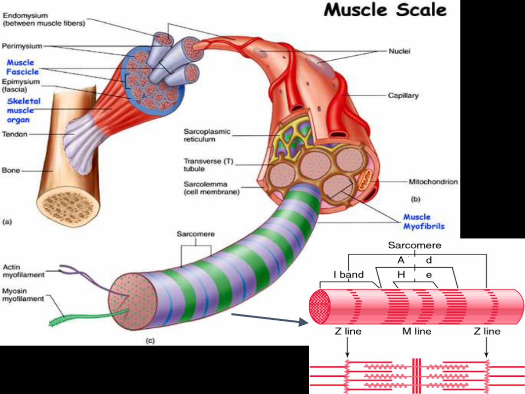
Muscle Structure
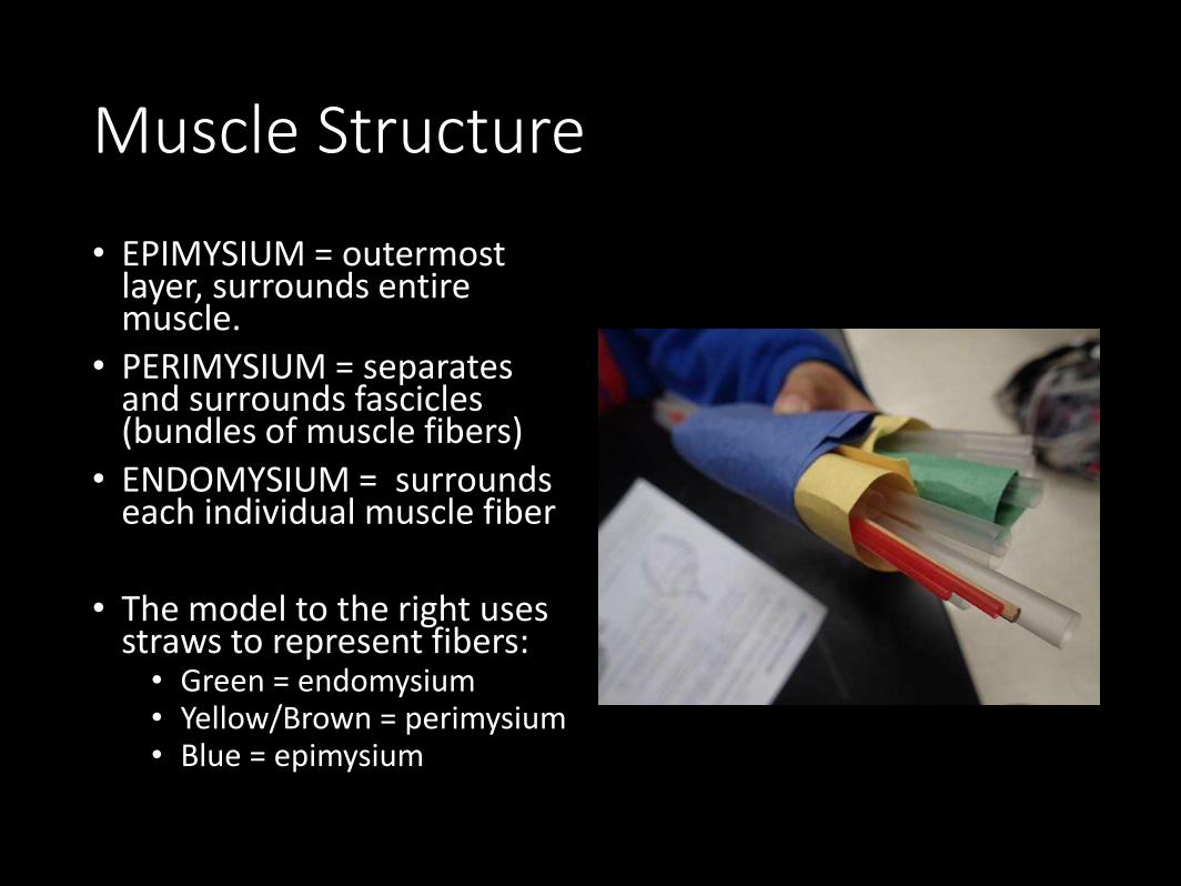
• EPIMYSIUM = outermost layer, surrounds entire muscle.
• PERIMYSIUM = separates and surrounds fascicles (bundles of muscle fibers)
• ENDOMYSIUM = surrounds each individual muscle fiber
• The model to the right uses straws to represent fibers:• Green = endomysium• Yellow/Brown = perimysium• Blue = epimysium
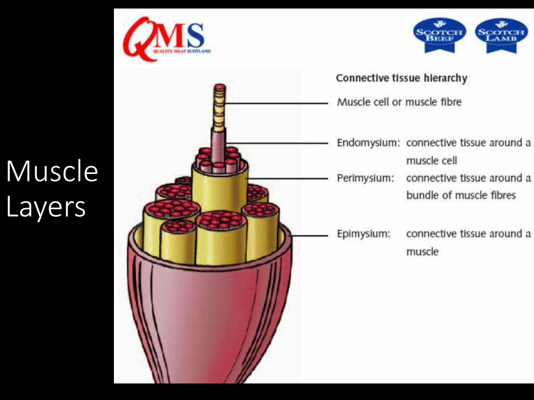
Muscle Layers
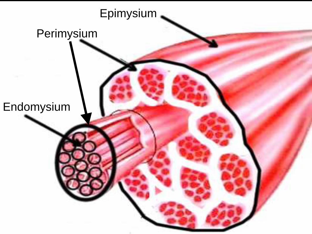
Epimysium
Perimysium
Endomysium

Muscles / Cells
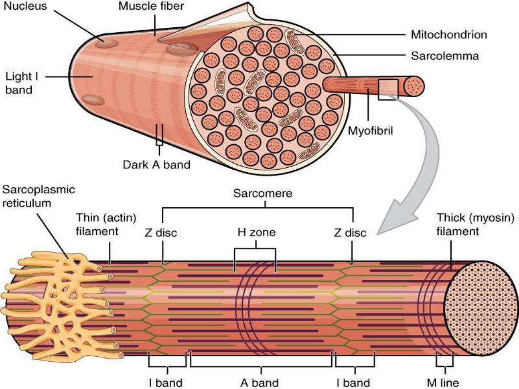
• Sarcolemma = muscle fiber membrane
• Sarcoplasm = inner material surrounding fibers (like cytoplasm)
• Myofibrils = individual parallel muscle fibers, within sarcoplasm
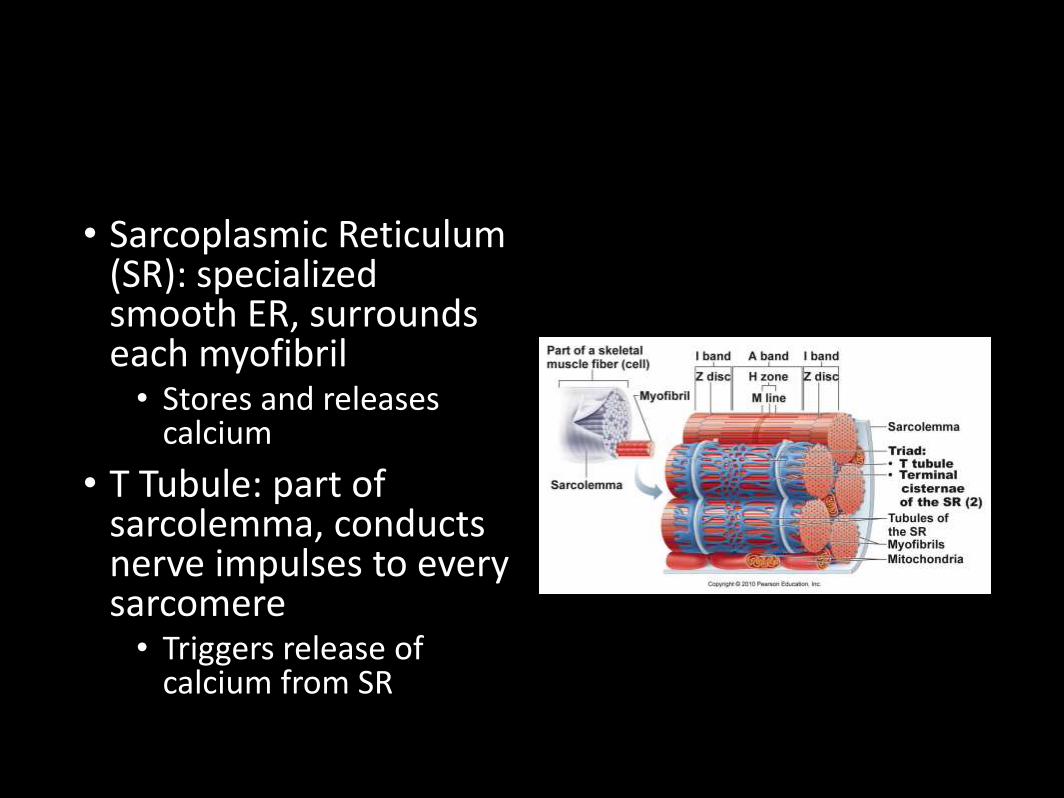
• Sarcoplasmic Reticulum (SR): specialized smooth ER, surrounds each myofibril• Stores and releases
calcium
• T Tubule: part of sarcolemma, conducts nerve impulses to every sarcomere• Triggers release of
calcium from SR
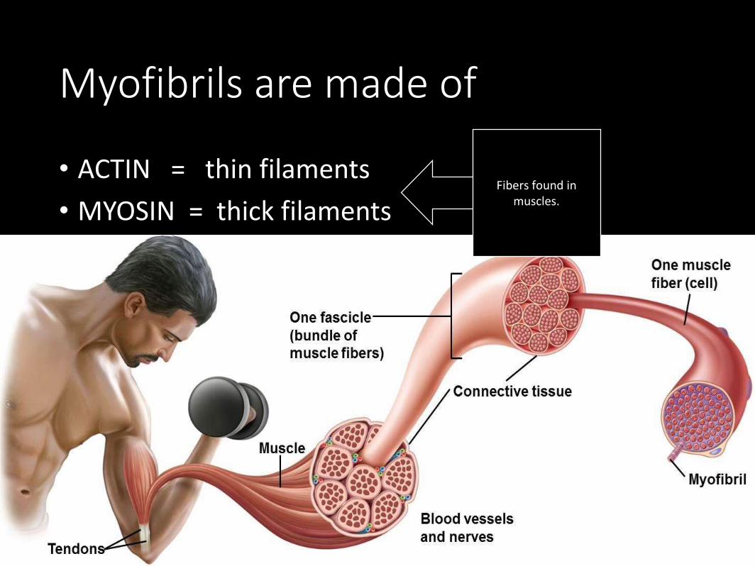
Myofibrils are made of
• ACTIN = thin filaments
• MYOSIN = thick filamentsFibers found in
muscles.
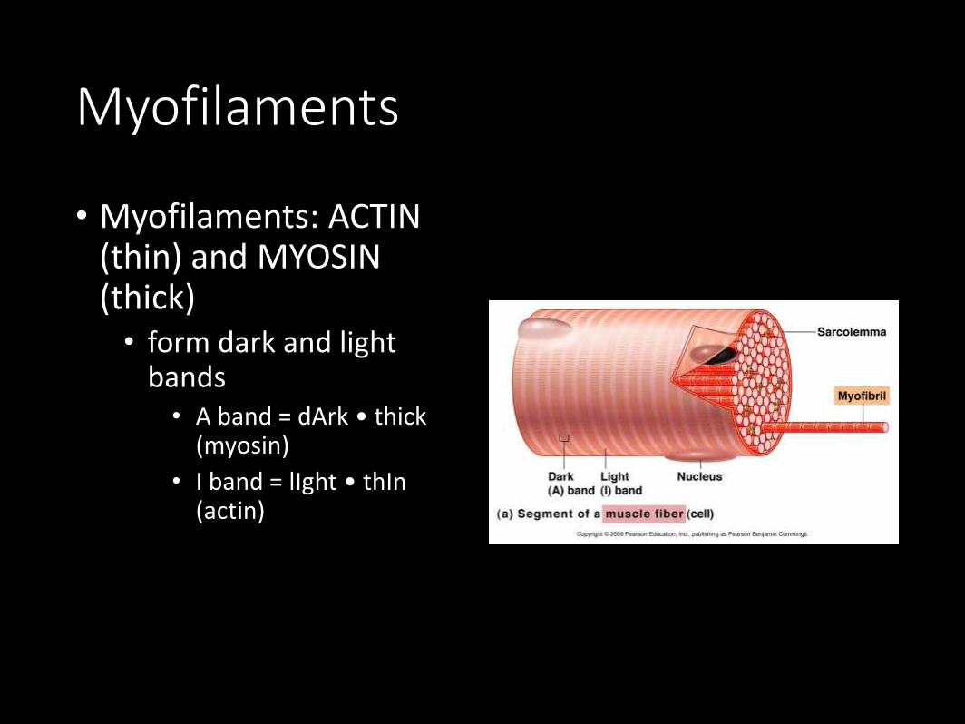
Myofilaments
• Myofilaments: ACTIN (thin) and MYOSIN (thick)• form dark and light
bands • A band = dArk • thick
(myosin)
• I band = lIght • thIn(actin)
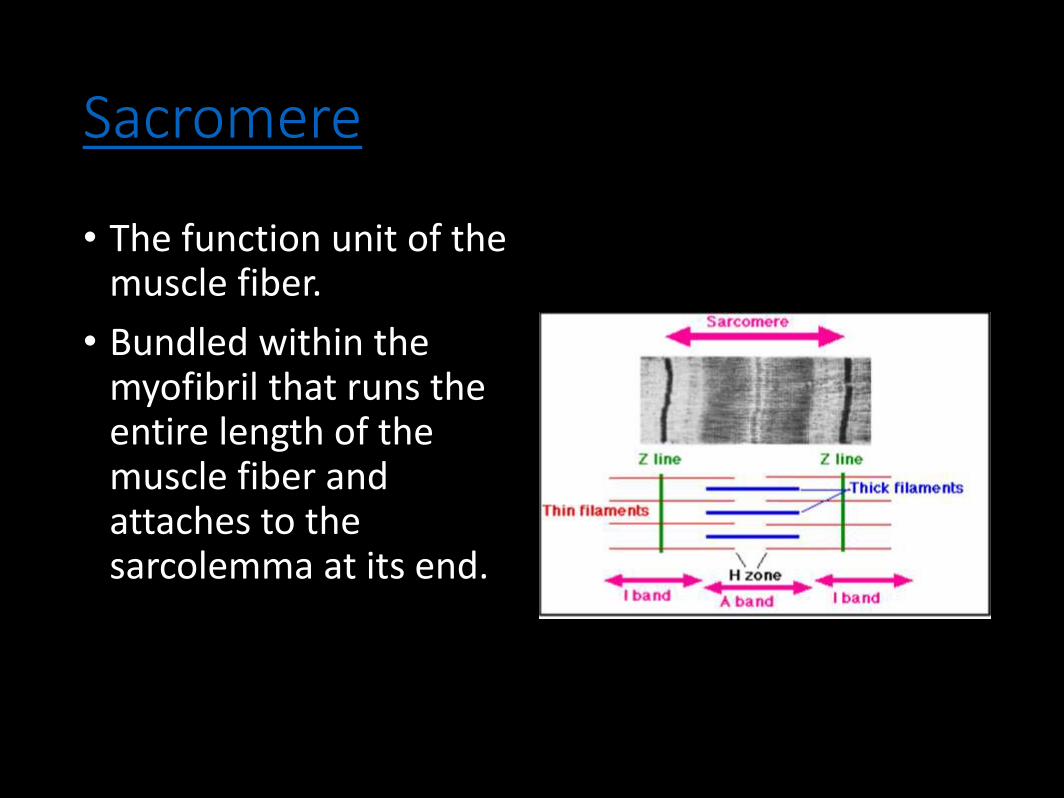
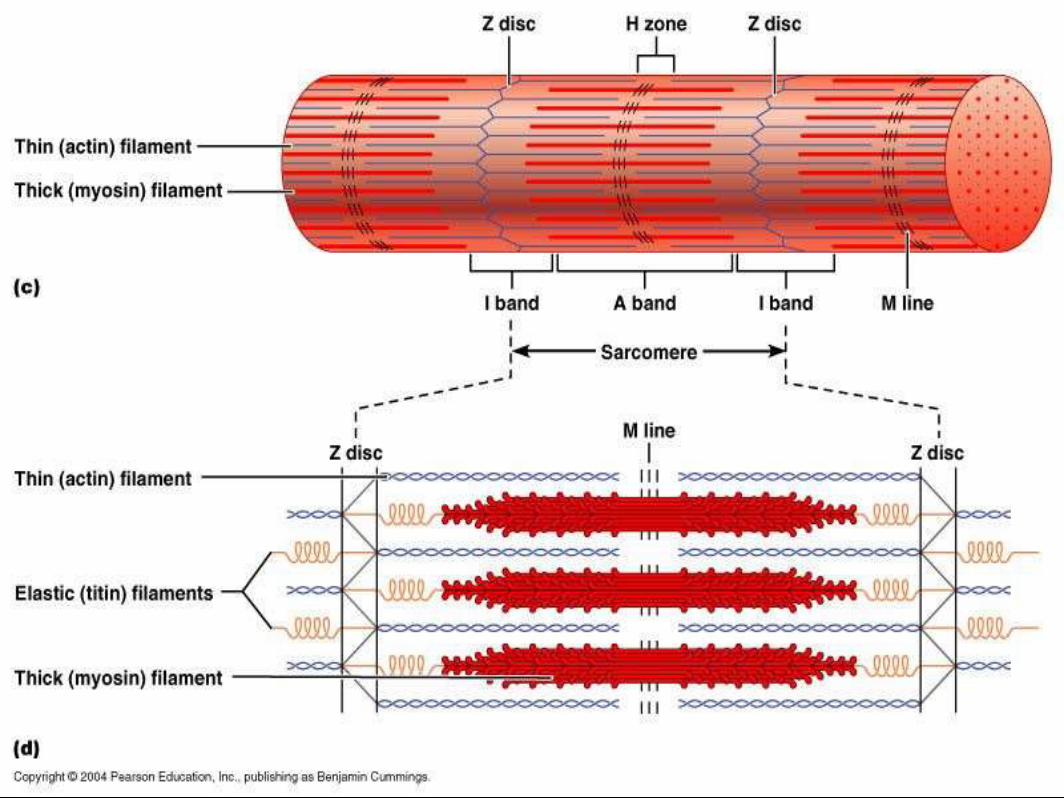
Sacromere
• The function unit of the muscle fiber.
• Bundled within the myofibril that runs the entire length of the muscle fiber and attaches to the sarcolemma at its end.
Skeletal Muscle Structures
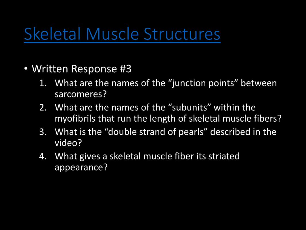
• Written Response #31. What are the names of the “junction points” between
sarcomeres?
2. What are the names of the “subunits” within the myofibrils that run the length of skeletal muscle fibers?
3. What is the “double strand of pearls” described in the video?
4. What gives a skeletal muscle fiber its striated appearance?
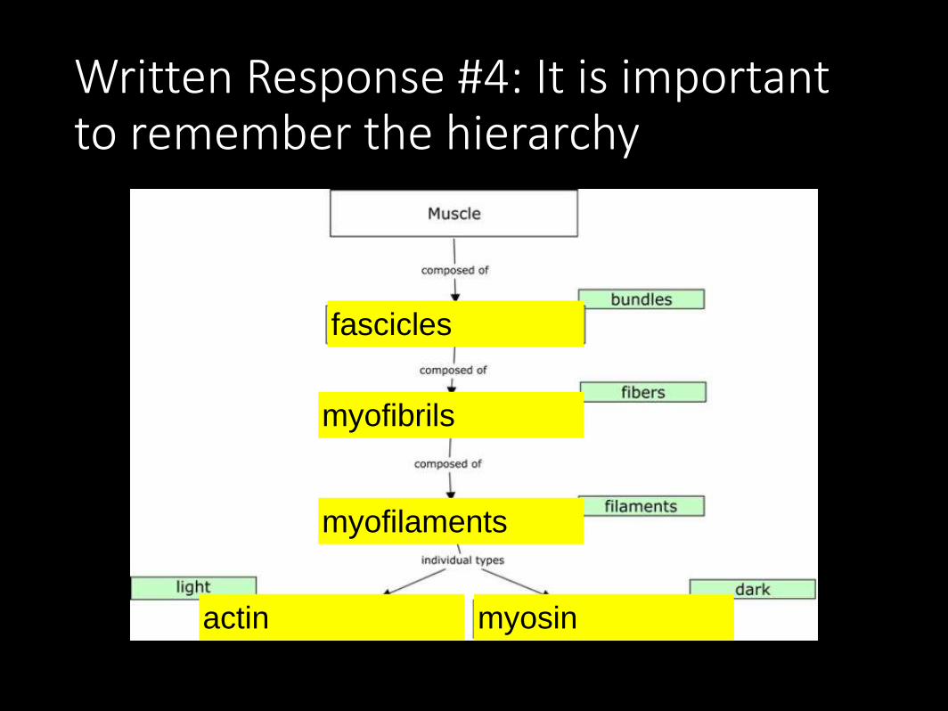
Written Response #4: It is important to remember the hierarchy
fascicles
myofibrils
myofilaments
actin myosin

Sarcomere Filament Coloring -Handout

Written Response #5
1. What is the definition of a motor unit?
2. What is the structural and functional difference between a large motor unit and a small motor unit?
3. Give an example of a large motor unit and a small motor unit.
4. Why is the neurotransmitter acetylcholine degraded after binding to its receptor?
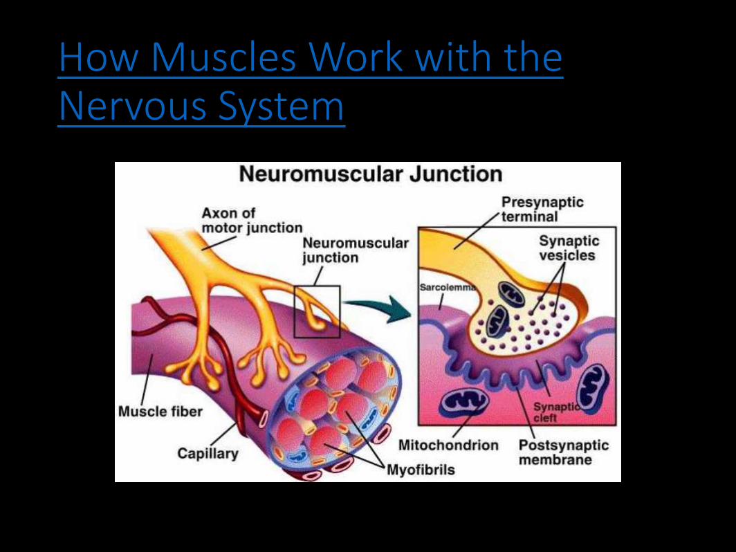
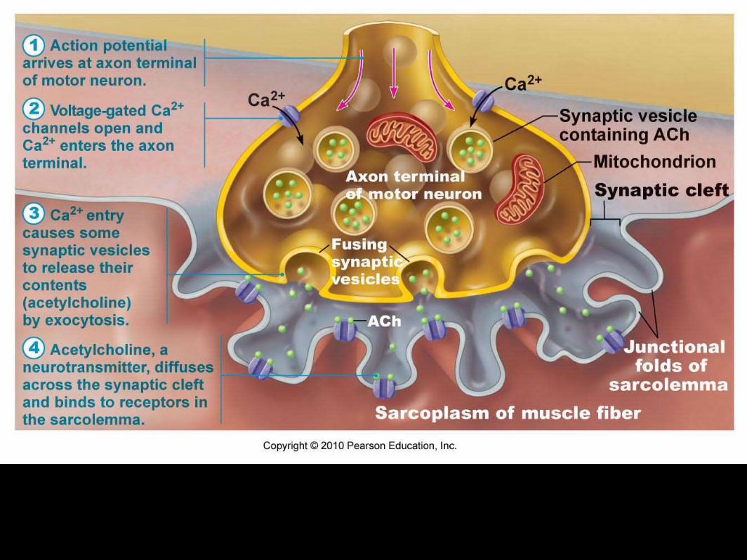
• NEUROMUSCULAR JUNCTION - where a nerve and muscle fiber come together
• MOTOR END PLATE - folded area where muscle and neuron communicate
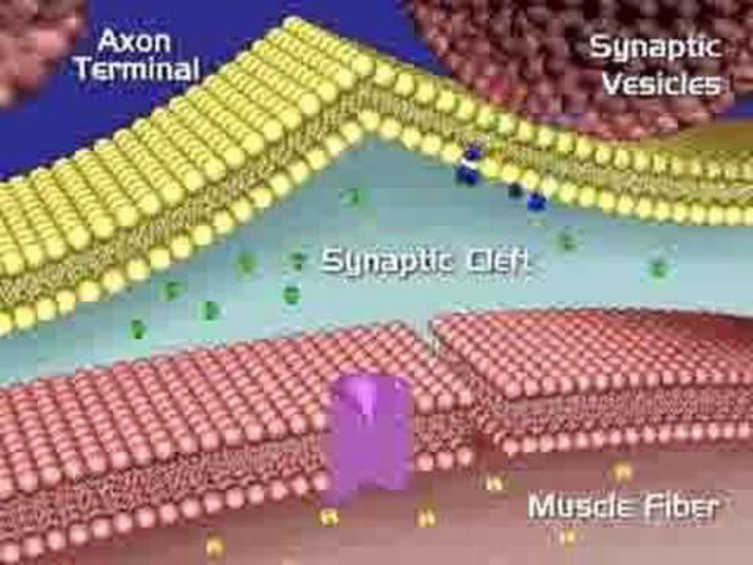
• SYNAPTIC CLEFT - gap between the neuron and motor end plate
• SYNAPTIC VESICLES - where neurotransmitters are stored• these are released into the cleft and tell the muscle to
contract
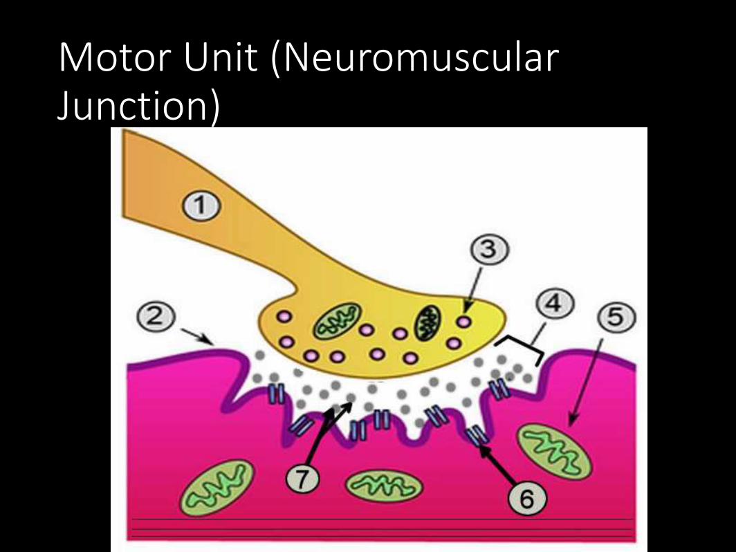
1. Neuron 2. Sarcolemma (or motor end plate)
3. Vesicle 4. Synapse 5. Mitochondria
6. Receptors 7. Acetylcholine
Motor Unit or Neuromuscular Junction
Motor Unit (Neuromuscular Junction)
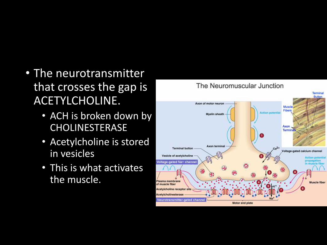
• The neurotransmitter that crosses the gap is ACETYLCHOLINE.• ACH is broken down by
CHOLINESTERASE
• Acetylcholine is stored in vesicles
• This is what activates the muscle.
Written Response #6
1. Which biochemical provides the energy to regenerate ATP?
2. What are the sources of oxygen for aerobic respiration?
3. How are lactic acid, oxygen debt, and muscle fatigue related?
4. What is the relationship between cellular respiration and heat production?
Sliding Filament Theory (Model)
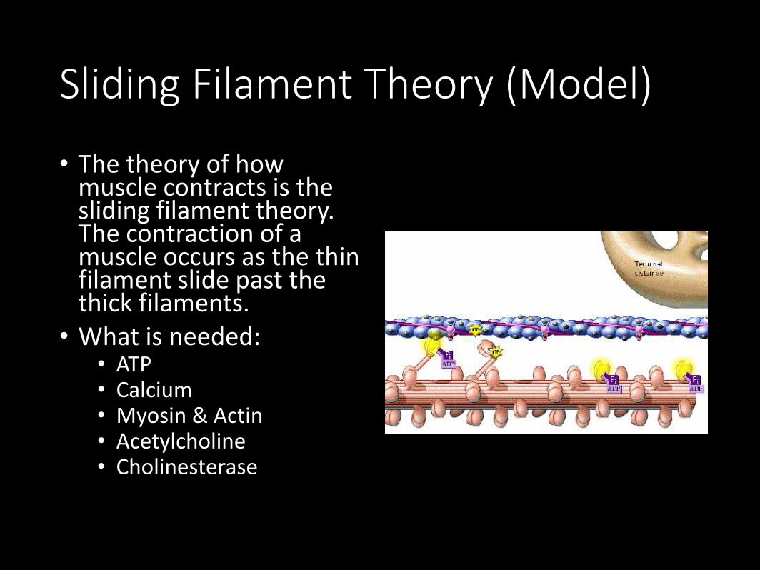
• The theory of how muscle contracts is the sliding filament theory. The contraction of a muscle occurs as the thin filament slide past the thick filaments.
• What is needed:• ATP• Calcium• Myosin & Actin• Acetylcholine• Cholinesterase
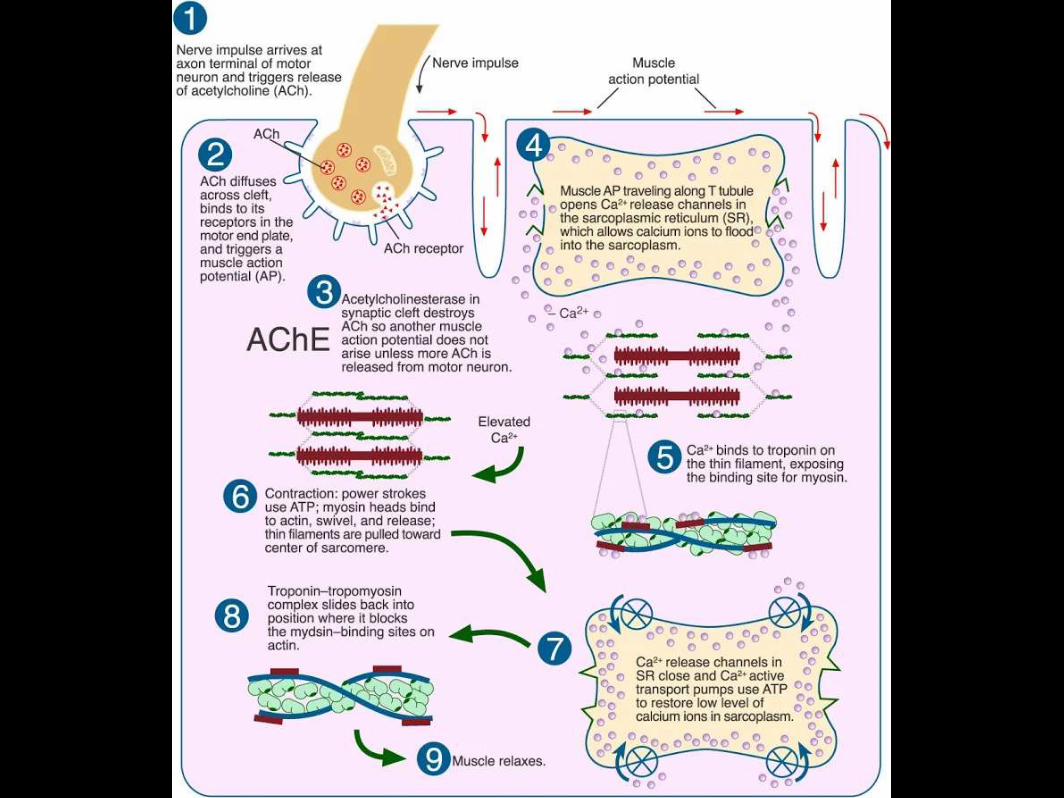
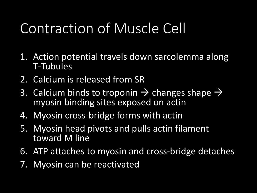
Contraction of Muscle Cell
1. Action potential travels down sarcolemma along T-Tubules
2. Calcium is released from SR
3. Calcium binds to troponin changes shape myosin binding sites exposed on actin
4. Myosin cross-bridge forms with actin
5. Myosin head pivots and pulls actin filament toward M line
6. ATP attaches to myosin and cross-bridge detaches
7. Myosin can be reactivated
Lab: Muscle Fatigue
Hank explains muscles and the sliding filament model.
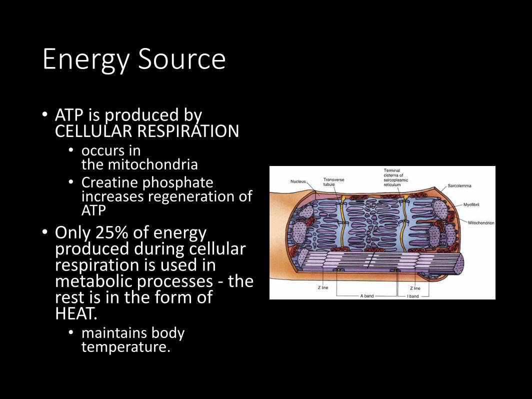
Energy Source
• ATP is produced by CELLULAR RESPIRATION• occurs in
the mitochondria• Creatine phosphate
increases regeneration of ATP
• Only 25% of energy produced during cellular respiration is used in metabolic processes - the rest is in the form of HEAT. • maintains body
temperature.
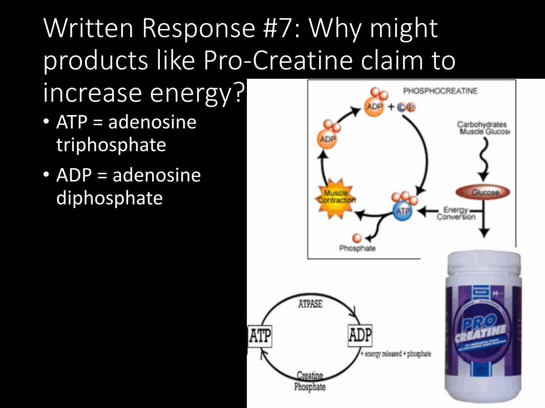
Written Response #7: Why might products like Pro-Creatine claim to increase energy?• ATP = adenosine
triphosphate
• ADP = adenosine diphosphate
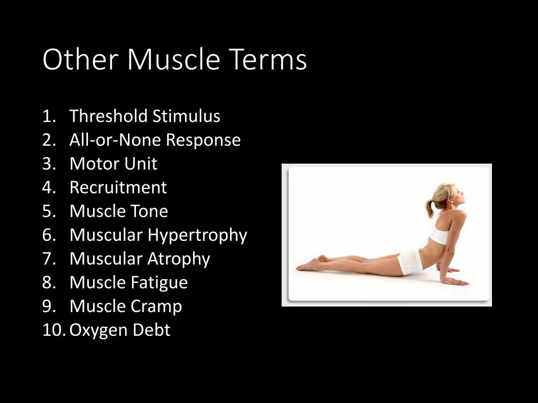
Other Muscle Terms
1. Threshold Stimulus2. All-or-None Response3. Motor Unit4. Recruitment5. Muscle Tone6. Muscular Hypertrophy7. Muscular Atrophy8. Muscle Fatigue9. Muscle Cramp10. Oxygen Debt
• Threshold Stimulus:• Minimal strength required to cause a contraction
• Motor neuron releases enough acetylcholine to reach threshold
• All-or-None Response• Fibers do not contract partially, they either do or don't
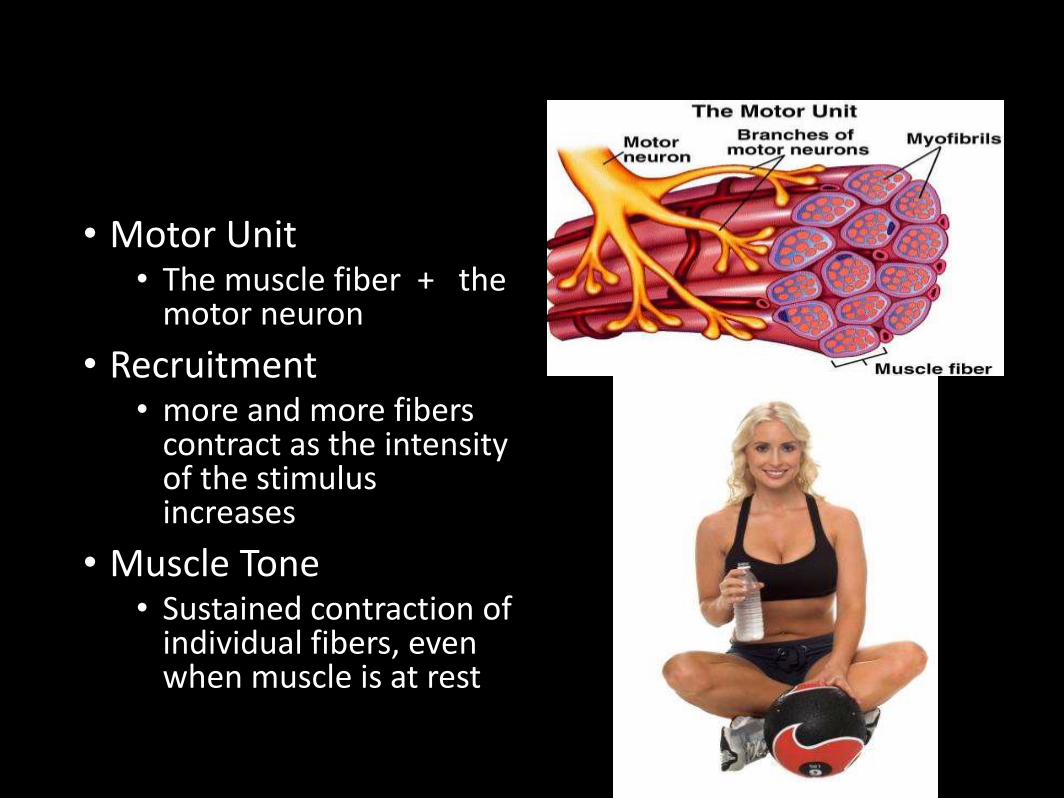
• Motor Unit• The muscle fiber + the
motor neuron
• Recruitment• more and more fibers
contract as the intensity of the stimulus increases
• Muscle Tone• Sustained contraction of
individual fibers, even when muscle is at rest
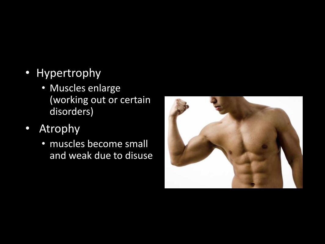
• Hypertrophy• Muscles enlarge
(working out or certain disorders)
• Atrophy• muscles become small
and weak due to disuse
• Muscle Fatigue• muscle loses ability to contract after prolonged exercise
or strain
• Muscle Cramp• a sustained involuntary contraction
• Oxygen Debt• oxygen is used to create ATP, -- not having enough
oxygen causes Lactic Acid to accumulate in the muscles → Soreness
• Magic School Bus
Case Study: The Tired Swimmer –Handout• Pick up a case study from the front counter.
• Read and complete independently.
• Turn in when you have completed.
Other Handouts to Complete (Notebook):• Muscle Groups Coloring
• Naming of Muscles
• Crosswords – Muscle Physiology and
Written Response #8
1. What is rigor mortis?
2. Explain why rigor mortis occurs after death at the physiological level.• Think about what we have learned with the sliding
filament theory and respiration to answer this question.
Crime Scene Investigation
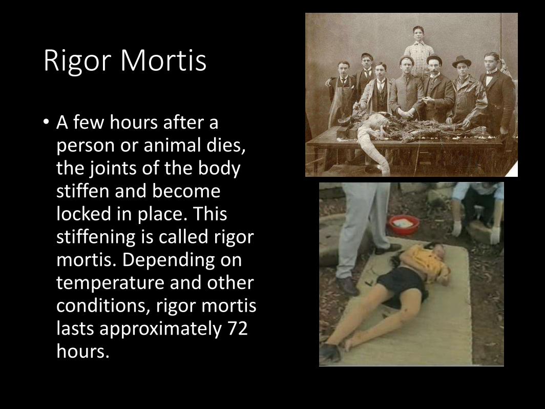
Rigor Mortis
• A few hours after a person or animal dies, the joints of the body stiffen and become locked in place. This stiffening is called rigor mortis. Depending on temperature and other conditions, rigor mortis lasts approximately 72 hours.
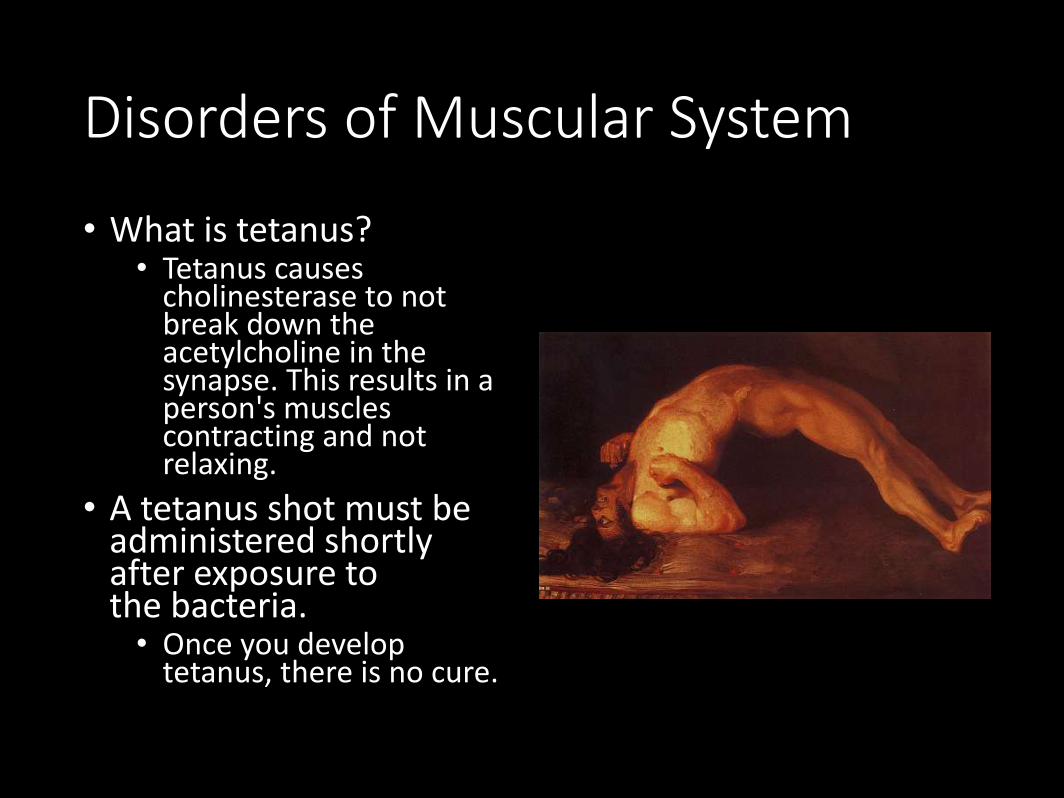
Disorders of Muscular System
• What is tetanus?• Tetanus causes
cholinesterase to not break down the acetylcholine in the synapse. This results in a person's muscles contracting and not relaxing.
• A tetanus shot must be administered shortly after exposure to the bacteria.• Once you develop
tetanus, there is no cure.
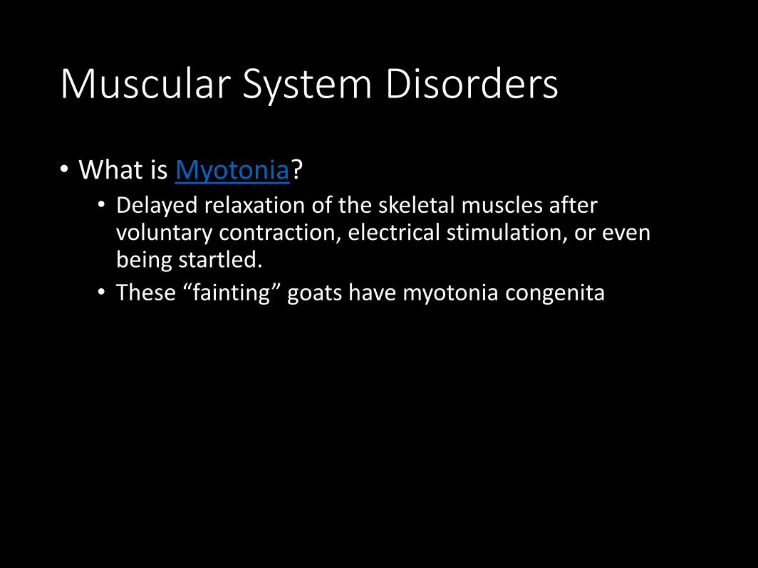
Muscular System Disorders
• What is Myotonia?• Delayed relaxation of the skeletal muscles after
voluntary contraction, electrical stimulation, or even being startled.
• These “fainting” goats have myotonia congenita
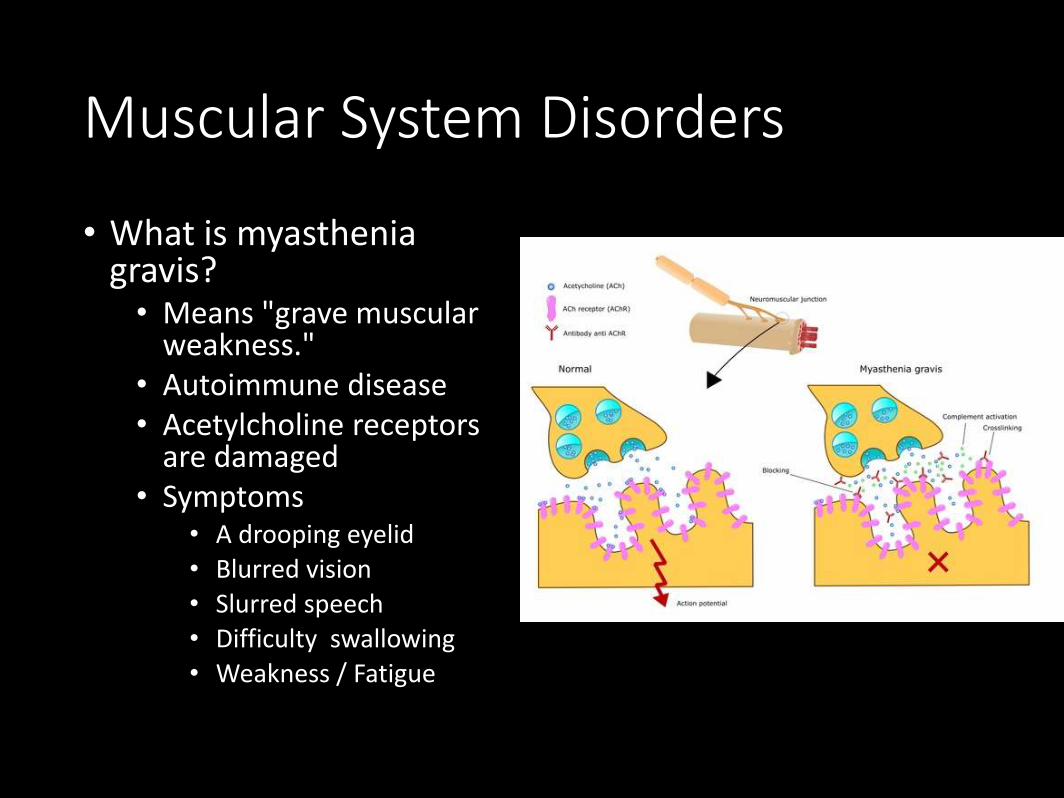
Muscular System Disorders
• What is myasthenia gravis?• Means "grave muscular
weakness." • Autoimmune disease• Acetylcholine receptors
are damaged• Symptoms
• A drooping eyelid• Blurred vision• Slurred speech• Difficulty swallowing• Weakness / Fatigue
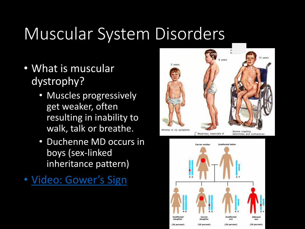
Muscular System Disorders
• What is muscular dystrophy?• Muscles progressively
get weaker, often resulting in inability to walk, talk or breathe.
• Duchenne MD occurs in boys (sex-linked inheritance pattern)
• Video: Gower’s Sign
Muscular System Disorders
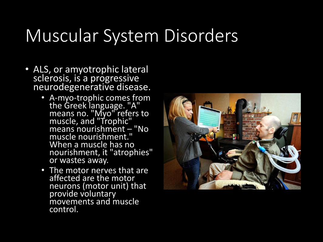
• ALS, or amyotrophic lateral sclerosis, is a progressive neurodegenerative disease. • A-myo-trophic comes from
the Greek language. "A" means no. "Myo" refers to muscle, and "Trophic" means nourishment – "No muscle nourishment." When a muscle has no nourishment, it "atrophies" or wastes away.
• The motor nerves that are affected are the motor neurons (motor unit) that provide voluntary movements and muscle control.
Poisons that Affect the Neuromuscular Junction – Botulism
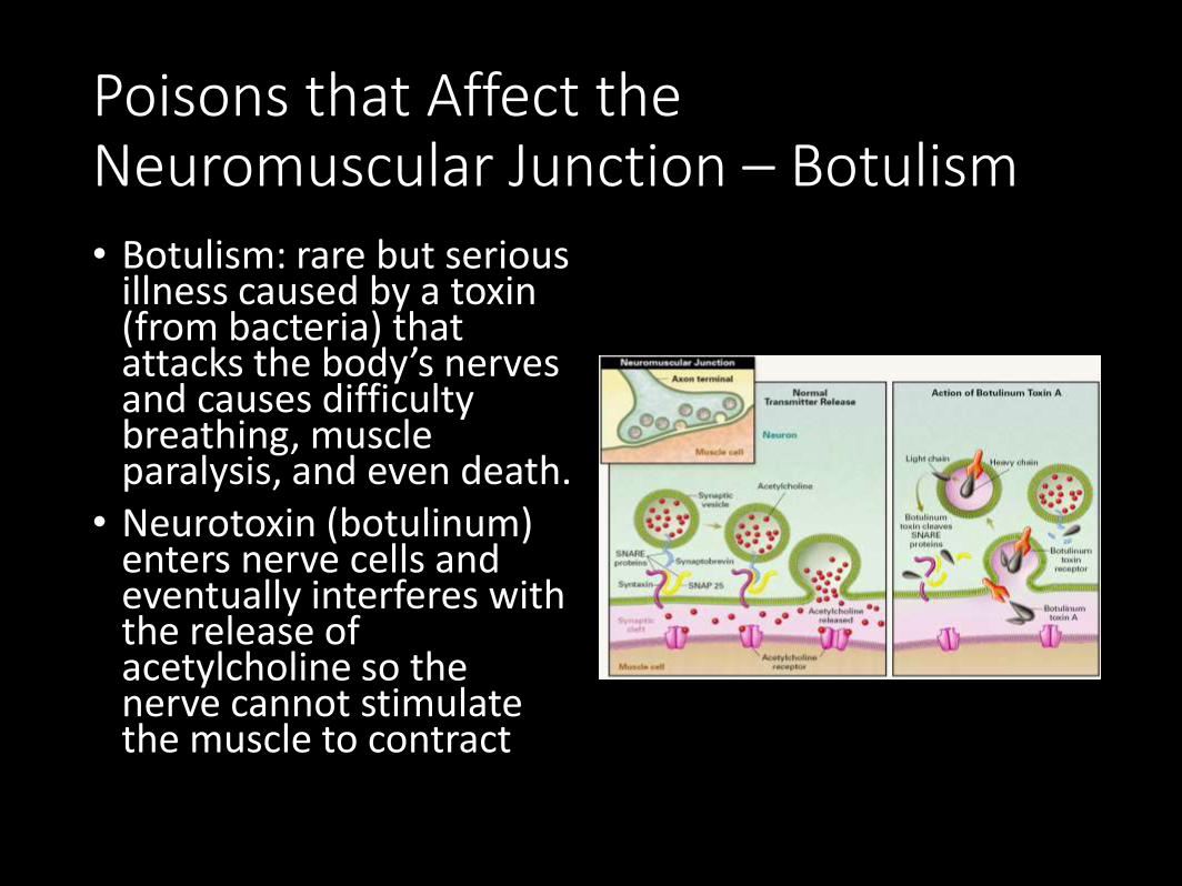
• Botulism: rare but serious illness caused by a toxin (from bacteria) that attacks the body’s nerves and causes difficulty breathing, muscle paralysis, and even death.
• Neurotoxin (botulinum) enters nerve cells and eventually interferes with the release of acetylcholine so the nerve cannot stimulate the muscle to contract
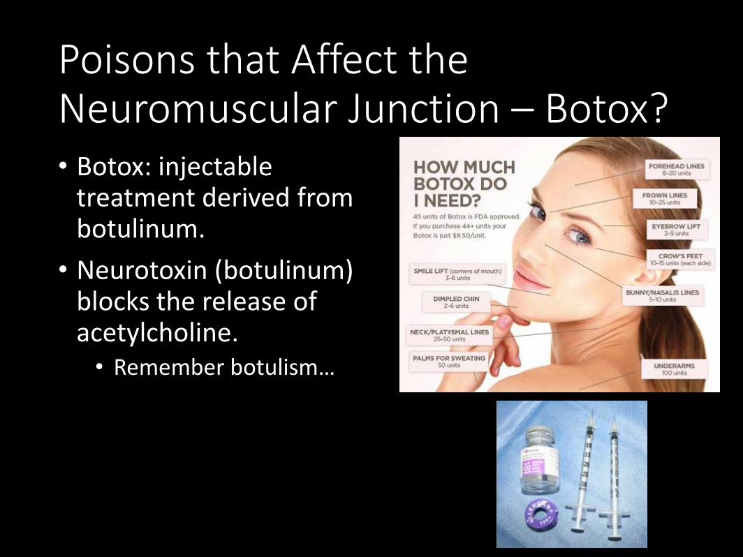
Poisons that Affect the Neuromuscular Junction – Botox?• Botox: injectable
treatment derived from botulinum.
• Neurotoxin (botulinum) blocks the release of acetylcholine.• Remember botulism…
Article: If Looks Could Kill –Handout• Read the article.
• Complete a summary about the article in 1-2 paragraphs.
• In another paragraph, choose a stance on whether botox is safe for humans or not and explain your reasoning.
• This means you should have a total of 2-3 paragraphs.
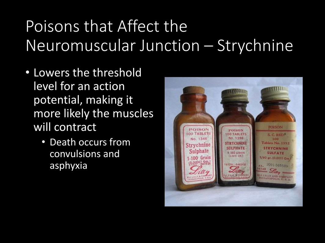
Poisons that Affect the Neuromuscular Junction – Strychnine
• Lowers the threshold level for an action potential, making it more likely the muscles will contract• Death occurs from
convulsions and asphyxia
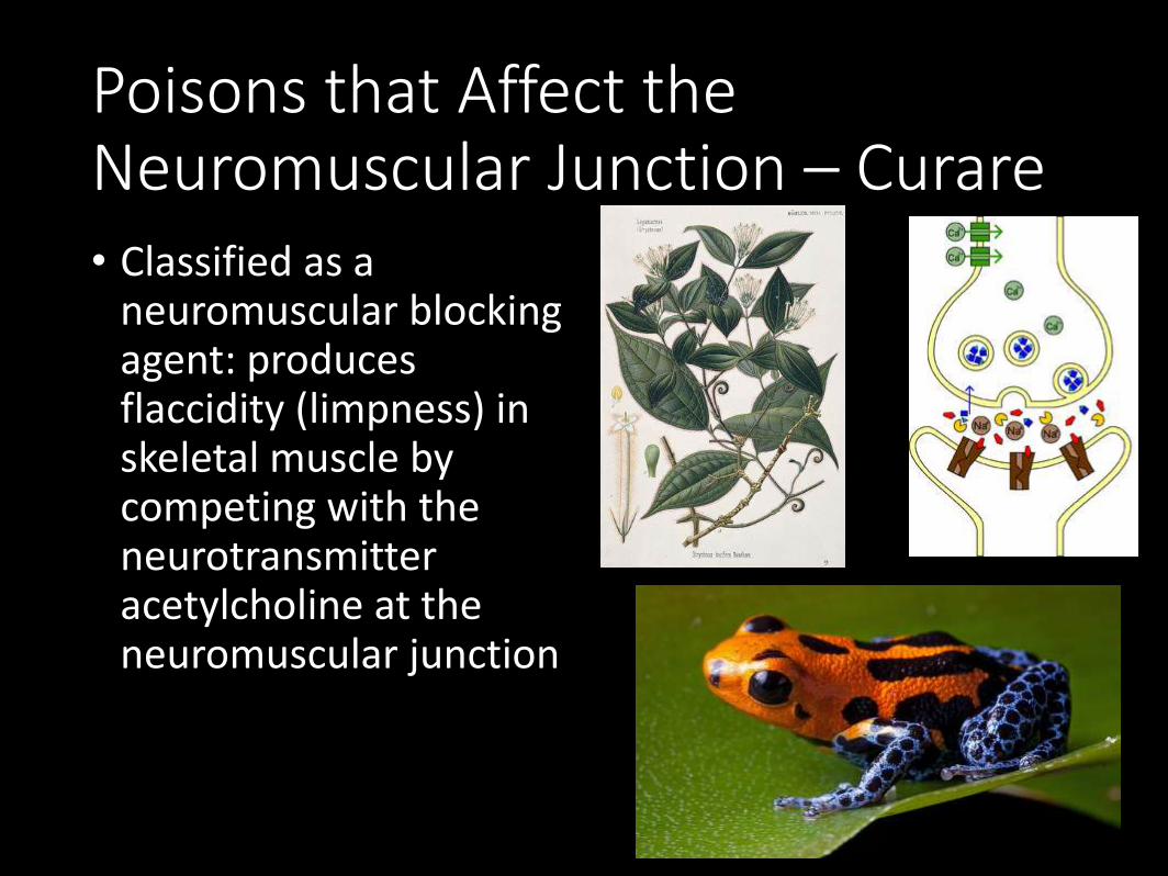
Poisons that Affect the Neuromuscular Junction – Curare• Classified as a
neuromuscular blocking agent: produces flaccidity (limpness) in skeletal muscle by competing with the neurotransmitter acetylcholine at the neuromuscular junction
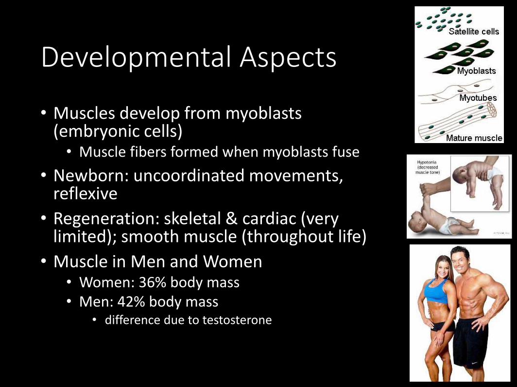
Developmental Aspects
• Muscles develop from myoblasts (embryonic cells)• Muscle fibers formed when myoblasts fuse
• Newborn: uncoordinated movements, reflexive
• Regeneration: skeletal & cardiac (very limited); smooth muscle (throughout life)
• Muscle in Men and Women• Women: 36% body mass • Men: 42% body mass
• difference due to testosterone
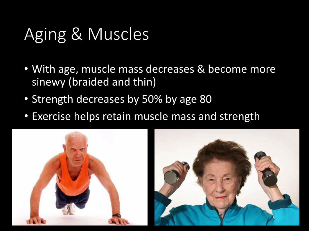
Aging & Muscles
• With age, muscle mass decreases & become more sinewy (braided and thin)
• Strength decreases by 50% by age 80
• Exercise helps retain muscle mass and strength
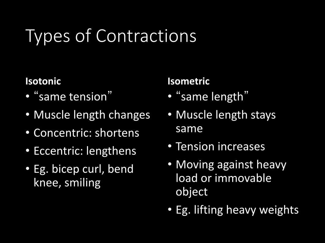
Types of Contractions
Isotonic
• “same tension”
• Muscle length changes
• Concentric: shortens
• Eccentric: lengthens
• Eg. bicep curl, bend knee, smiling
Isometric
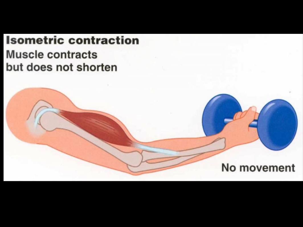
• “same length”
• Muscle length stays same
• Tension increases
• Moving against heavy load or immovable object
• Eg. lifting heavy weights
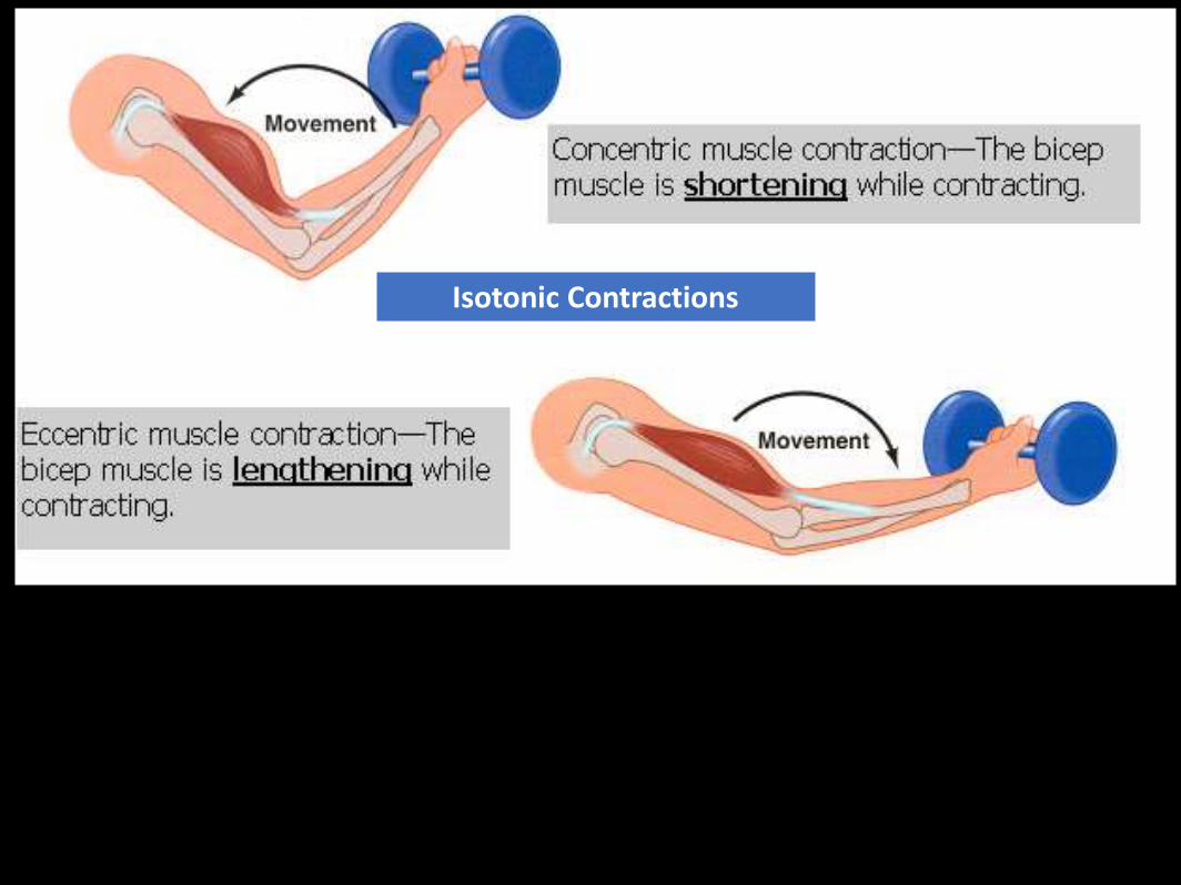
Isotonic Contractions
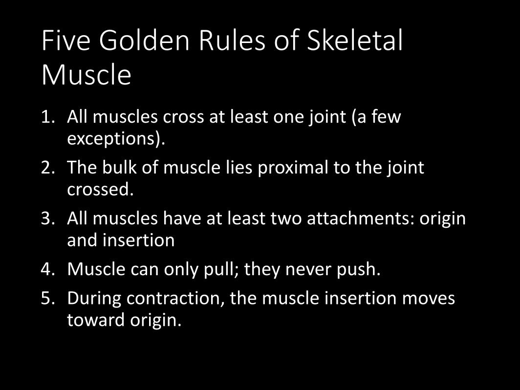
Five Golden Rules of Skeletal Muscle1. All muscles cross at least one joint (a few
exceptions).
2. The bulk of muscle lies proximal to the joint crossed.
3. All muscles have at least two attachments: origin and insertion
4. Muscle can only pull; they never push.
5. During contraction, the muscle insertion moves toward origin.
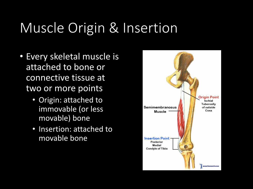
Muscle Origin & Insertion
• Every skeletal muscle is attached to bone or connective tissue at two or more points• Origin: attached to
immovable (or less movable) bone
• Insertion: attached to movable bone
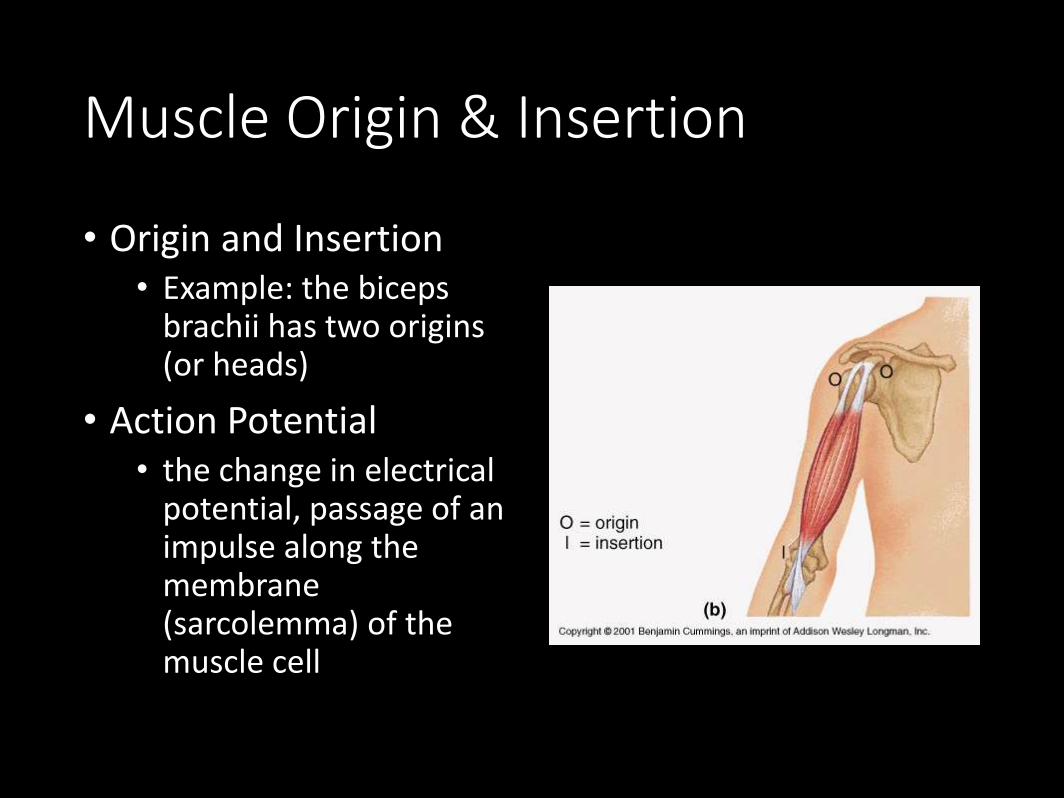
Muscle Origin & Insertion
• Origin and Insertion• Example: the biceps
brachii has two origins (or heads)
• Action Potential • the change in electrical
potential, passage of an impulse along the membrane (sarcolemma) of the muscle cell
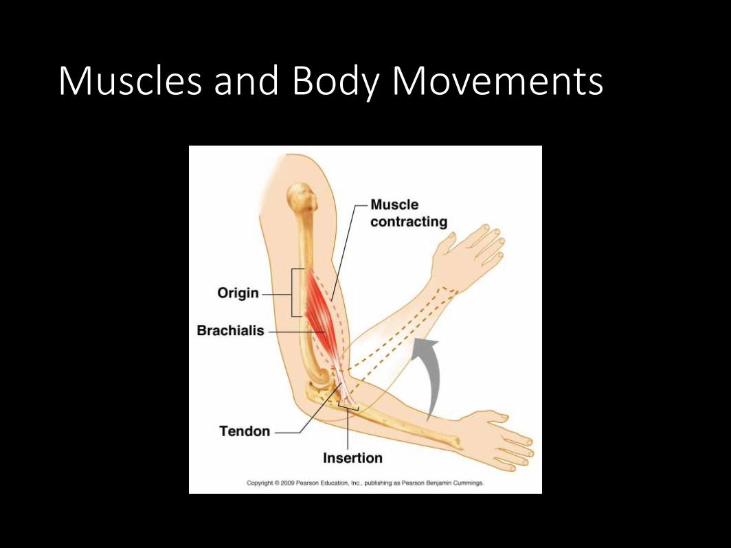
Muscles and Body Movements
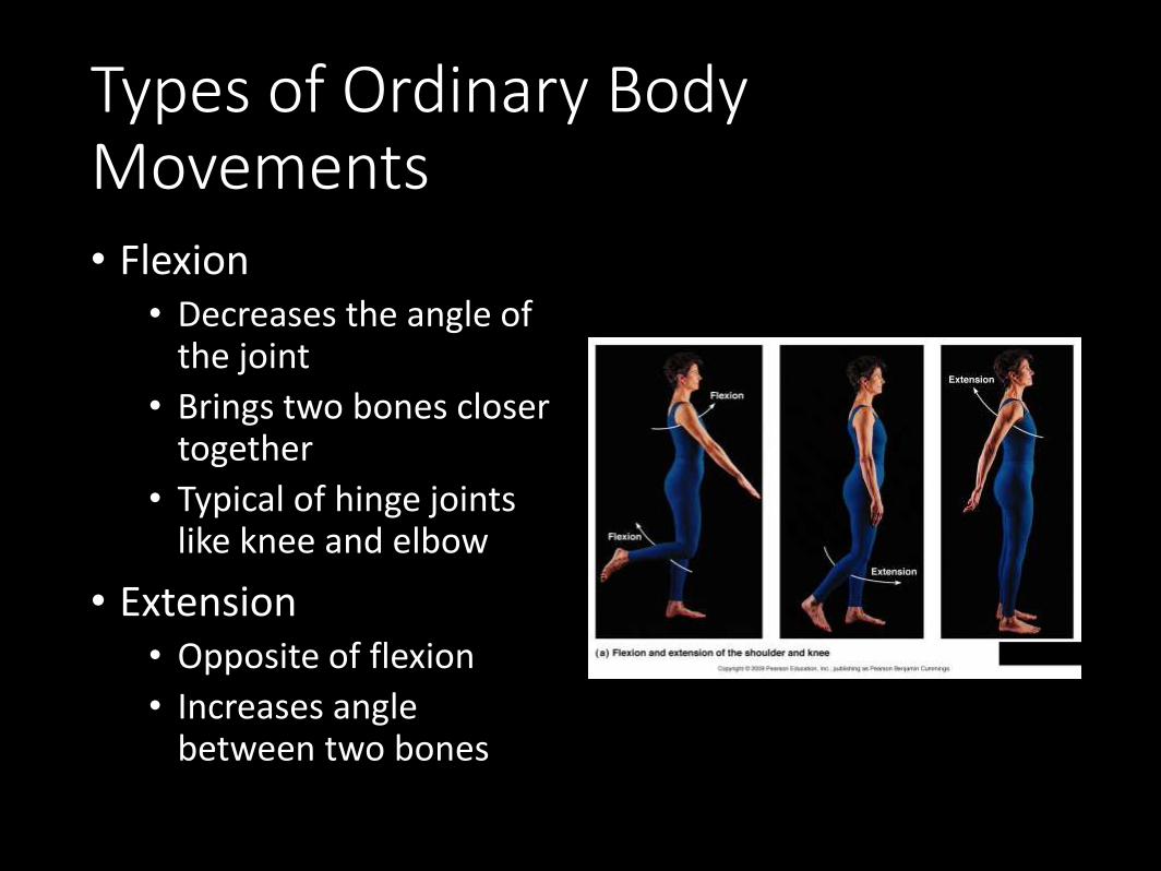
Types of Ordinary Body Movements• Flexion
• Decreases the angle of the joint
• Brings two bones closer together
• Typical of hinge joints like knee and elbow
• Extension• Opposite of flexion
• Increases angle between two bones
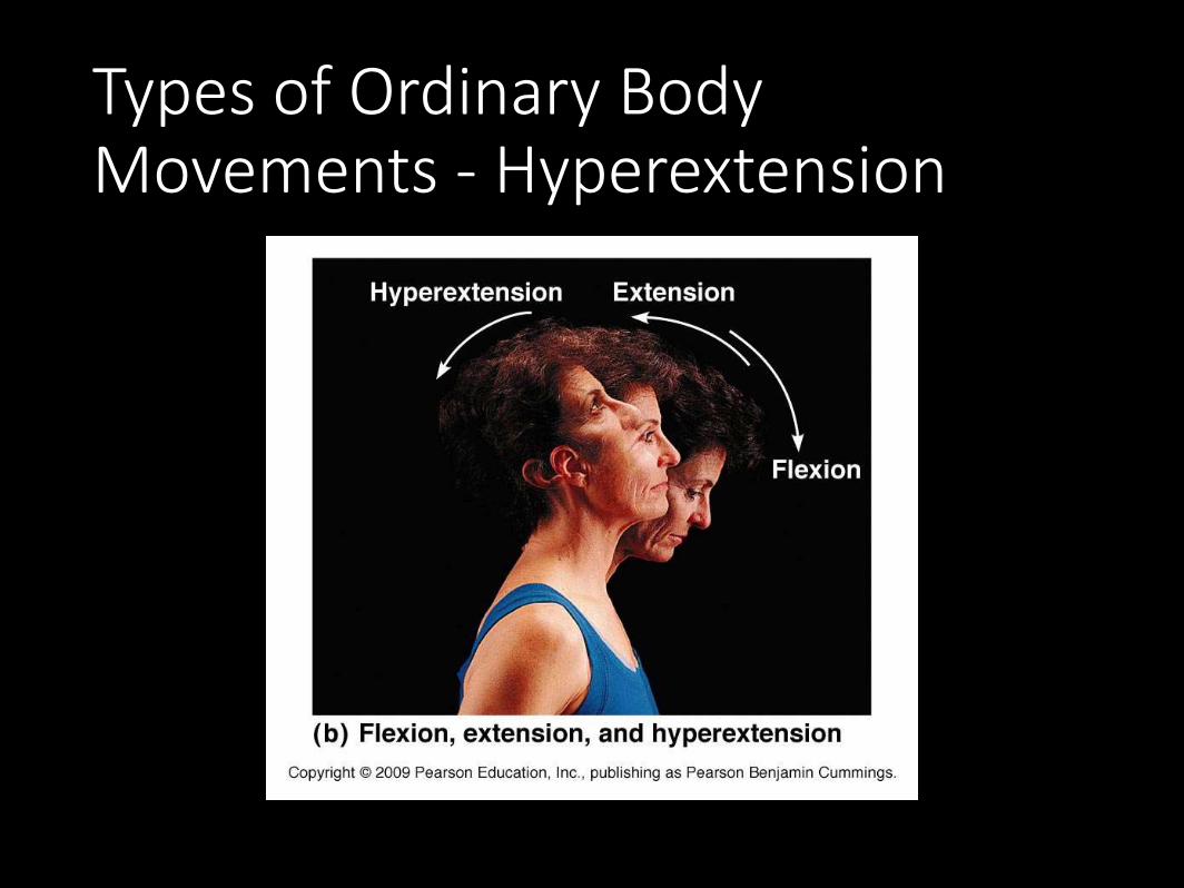
Types of Ordinary Body Movements - Hyperextension
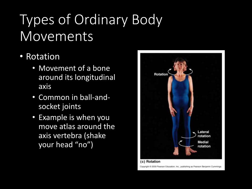
Types of Ordinary Body Movements• Rotation
• Movement of a bone around its longitudinal axis
• Common in ball-and-socket joints
• Example is when you move atlas around the axis vertebra (shake your head “no”)
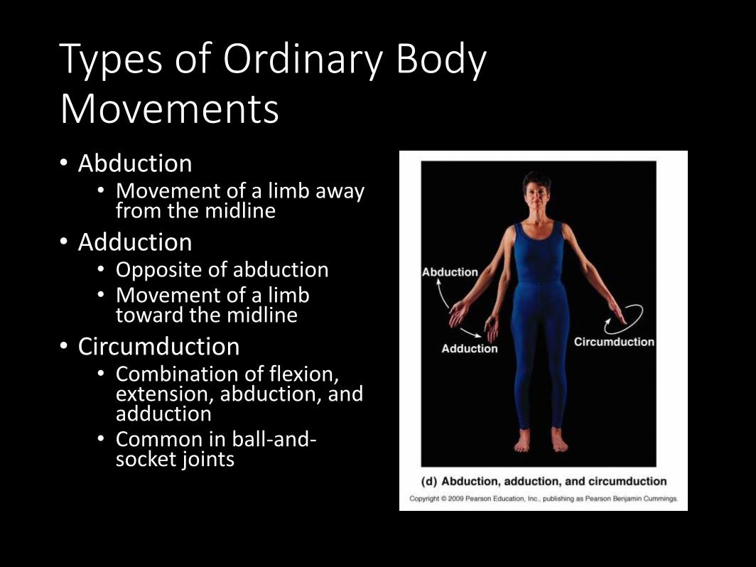
Types of Ordinary Body Movements• Abduction
• Movement of a limb away from the midline
• Adduction• Opposite of abduction• Movement of a limb
toward the midline
• Circumduction• Combination of flexion,
extension, abduction, and adduction
• Common in ball-and-socket joints
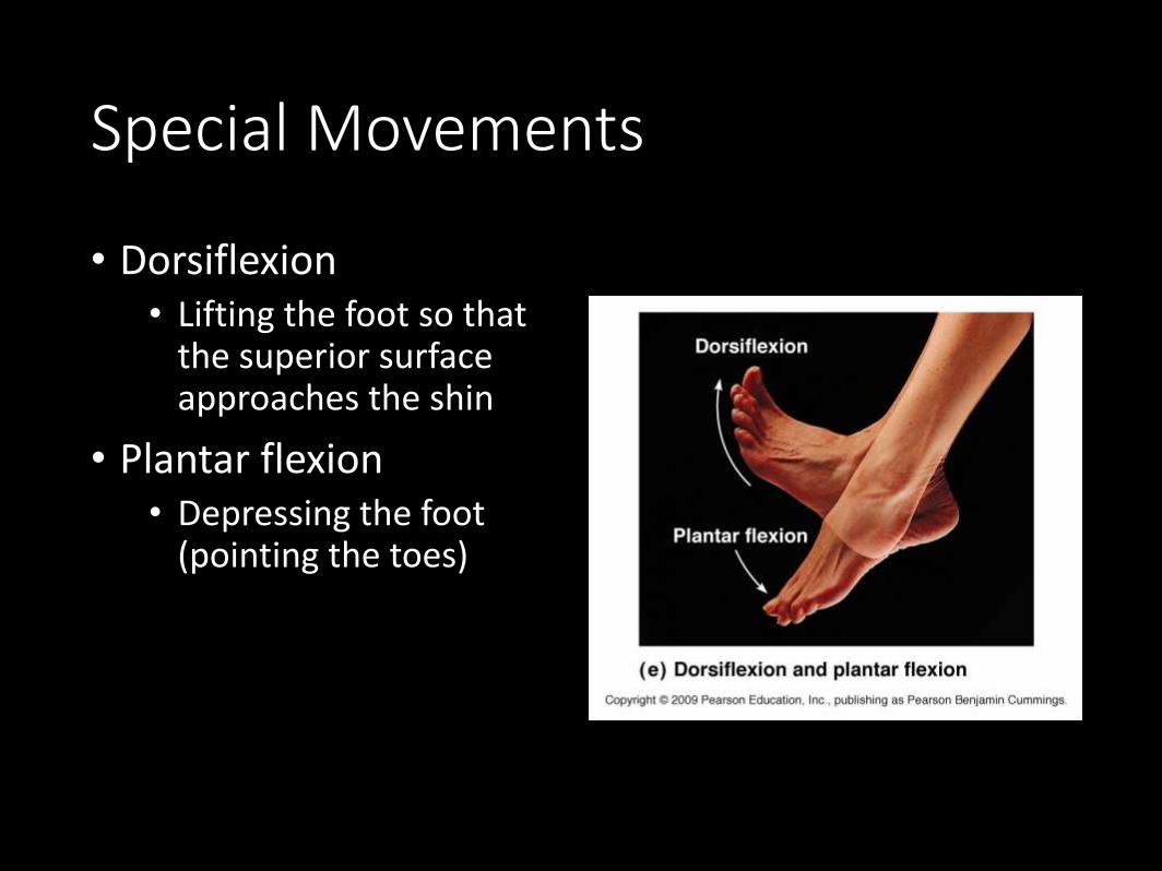
Special Movements
• Dorsiflexion• Lifting the foot so that
the superior surface approaches the shin
• Plantar flexion• Depressing the foot
(pointing the toes)
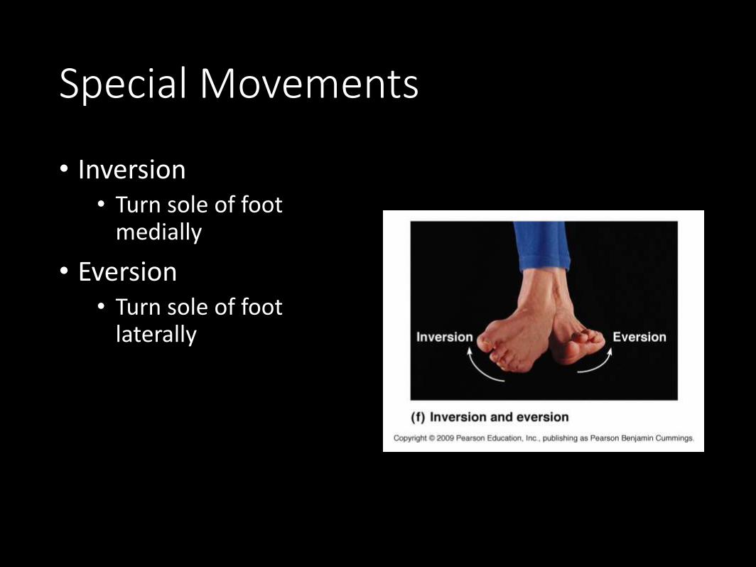
Special Movements
• Inversion• Turn sole of foot
medially
• Eversion• Turn sole of foot
laterally
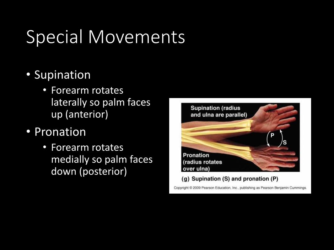
Special Movements
• Supination• Forearm rotates
laterally so palm faces up (anterior)
• Pronation• Forearm rotates
medially so palm faces down (posterior)
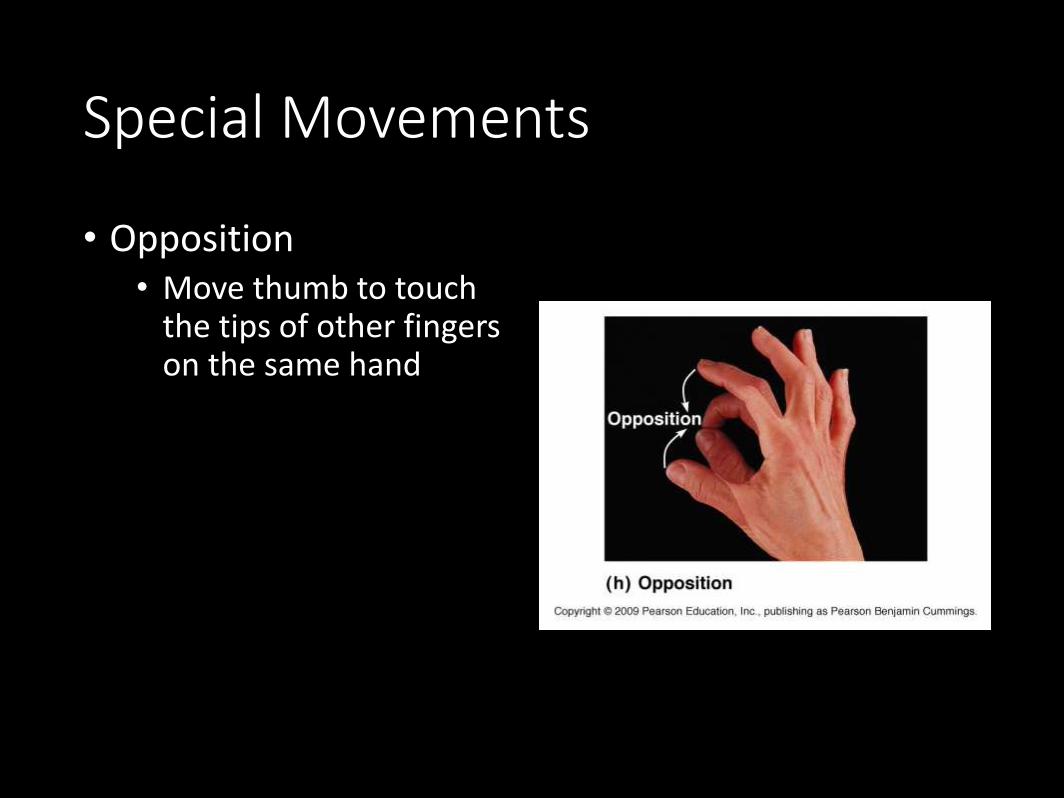
Special Movements
• Opposition• Move thumb to touch
the tips of other fingers on the same hand
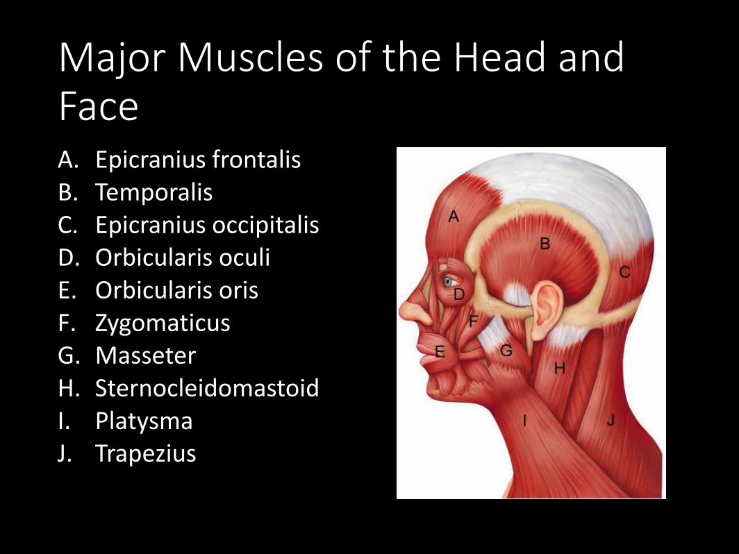
Major Muscles of the Head and FaceA. Epicranius frontalisB. TemporalisC. Epicranius occipitalisD. Orbicularis oculiE. Orbicularis orisF. ZygomaticusG. MasseterH. SternocleidomastoidI. PlatysmaJ. Trapezius
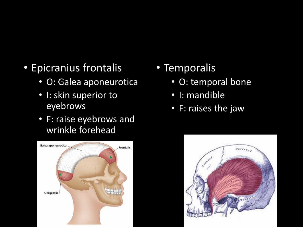
• Epicranius frontalis• O: Galea aponeurotica
• I: skin superior to eyebrows
• F: raise eyebrows and wrinkle forehead
• Temporalis• O: temporal bone
• I: mandible
• F: raises the jaw
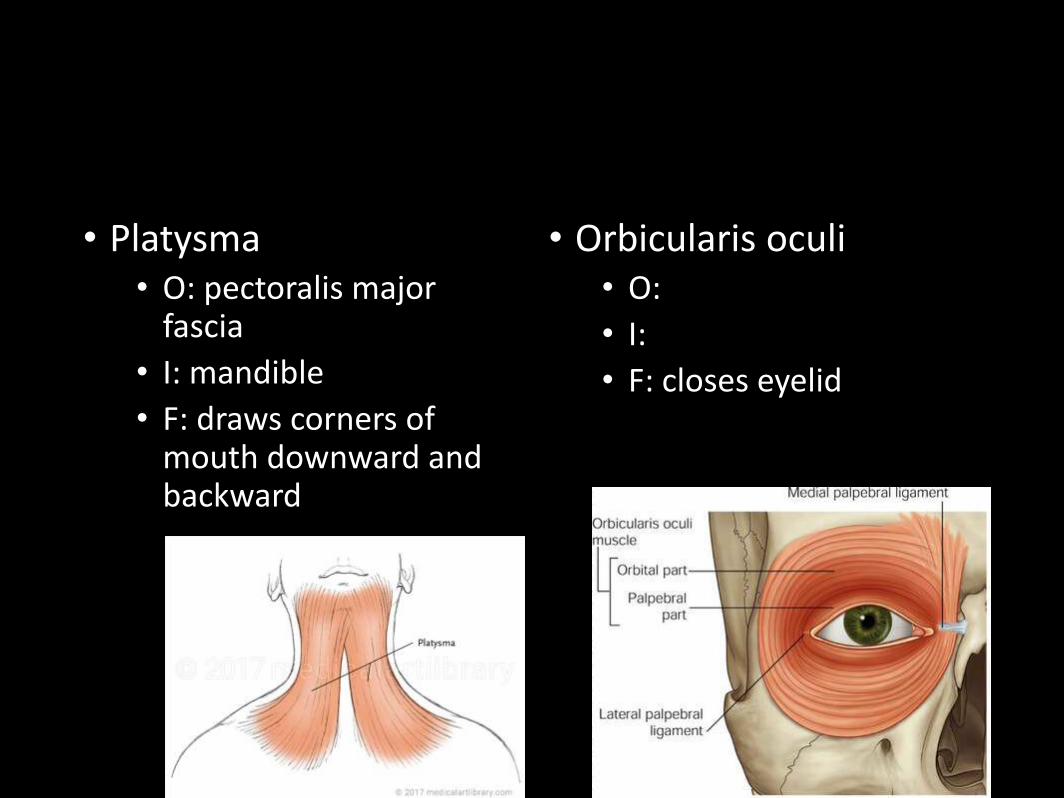
• Platysma• O: pectoralis major
fascia
• I: mandible
• F: draws corners of mouth downward and backward
• Orbicularis oculi• O:
• I:
• F: closes eyelid
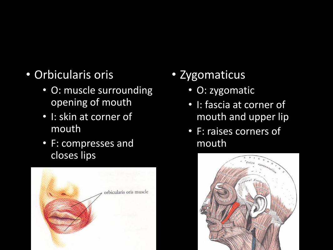
• Orbicularis oris• O: muscle surrounding
opening of mouth
• I: skin at corner of mouth
• F: compresses and closes lips
• Zygomaticus• O: zygomatic
• I: fascia at corner of mouth and upper lip
• F: raises corners of mouth
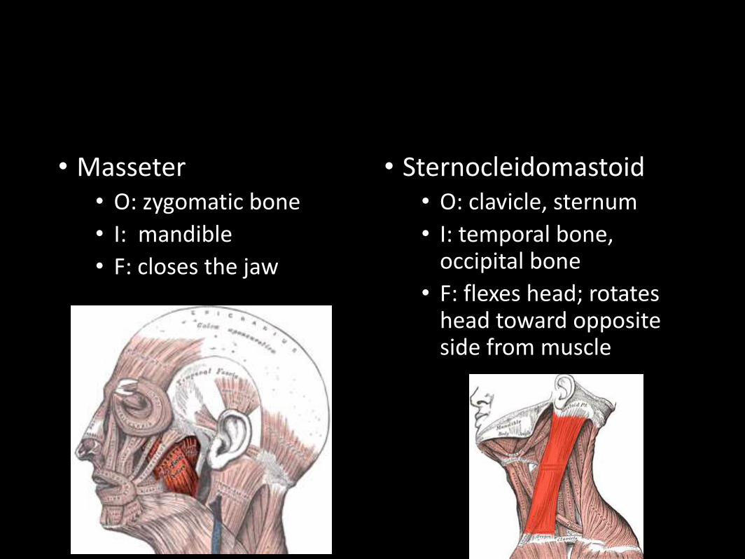
• Masseter• O: zygomatic bone
• I: mandible
• F: closes the jaw
• Sternocleidomastoid• O: clavicle, sternum
• I: temporal bone, occipital bone
• F: flexes head; rotates head toward opposite side from muscle
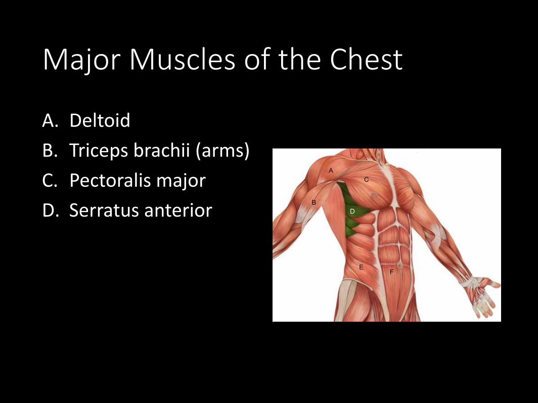
Major Muscles of the Chest
A. Deltoid
B. Triceps brachii (arms)
C. Pectoralis major
D. Serratus anterior
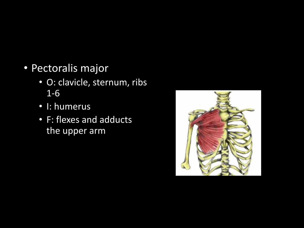
• Pectoralis major• O: clavicle, sternum, ribs
1-6
• I: humerus
• F: flexes and adducts the upper arm
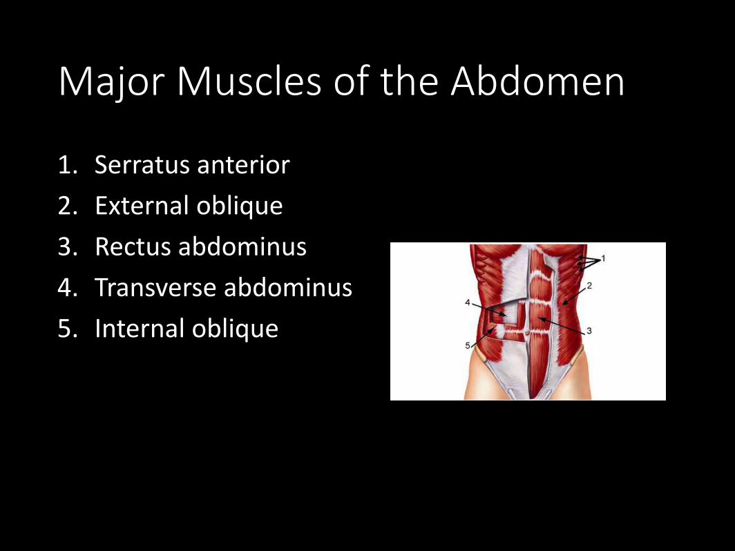
Major Muscles of the Abdomen
1. Serratus anterior
2. External oblique
3. Rectus abdominus
4. Transverse abdominus
5. Internal oblique
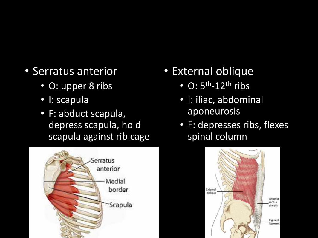
• Serratus anterior• O: upper 8 ribs
• I: scapula
• F: abduct scapula, depress scapula, hold scapula against rib cage
• External oblique• O: 5th-12th ribs
• I: iliac, abdominal aponeurosis
• F: depresses ribs, flexes spinal column
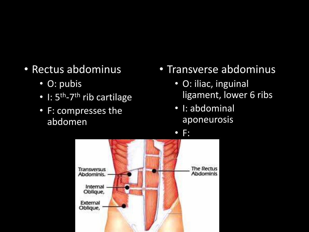
• Rectus abdominus• O: pubis
• I: 5th-7th rib cartilage
• F: compresses the abdomen
• Transverse abdominus• O: iliac, inguinal
ligament, lower 6 ribs
• I: abdominal aponeurosis
• F:
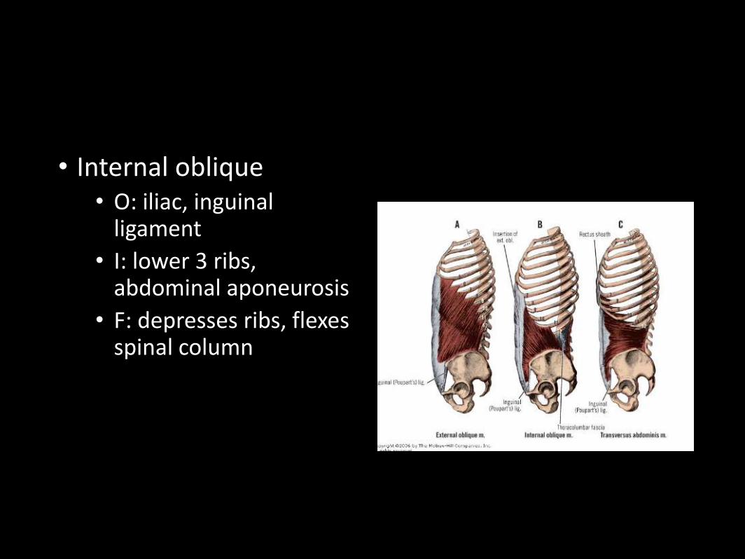
• Internal oblique• O: iliac, inguinal
ligament
• I: lower 3 ribs, abdominal aponeurosis
• F: depresses ribs, flexes spinal column
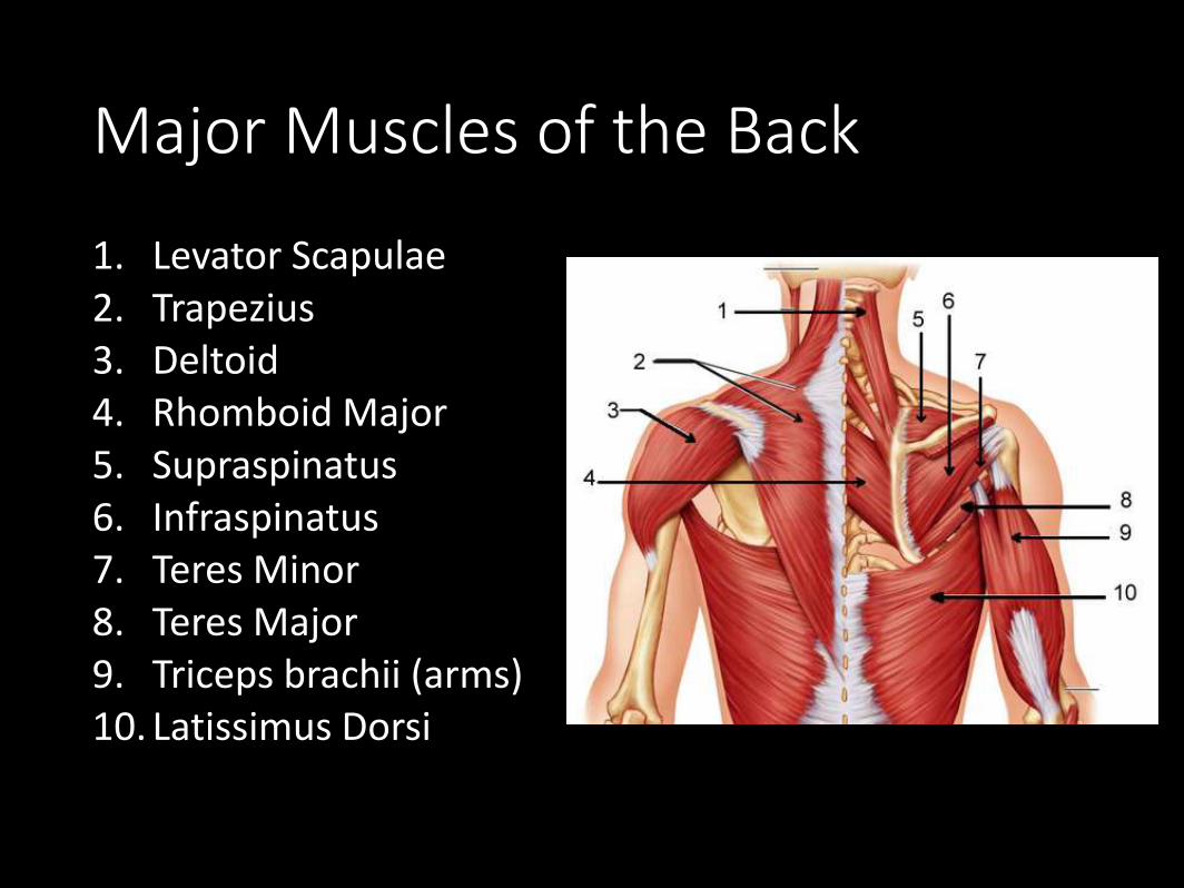
Major Muscles of the Back
1. Levator Scapulae2. Trapezius3. Deltoid4. Rhomboid Major5. Supraspinatus6. Infraspinatus7. Teres Minor8. Teres Major9. Triceps brachii (arms)10. Latissimus Dorsi
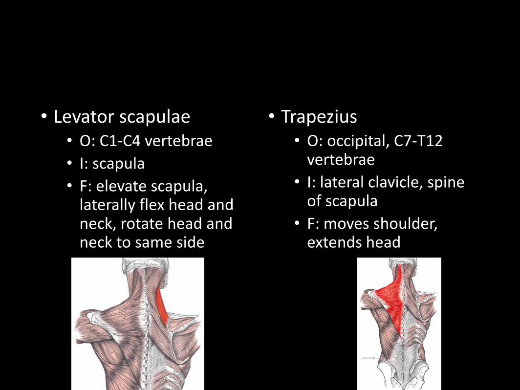
• Levator scapulae• O: C1-C4 vertebrae
• I: scapula
• F: elevate scapula, laterally flex head and neck, rotate head and neck to same side
• Trapezius• O: occipital, C7-T12
vertebrae
• I: lateral clavicle, spine of scapula
• F: moves shoulder, extends head
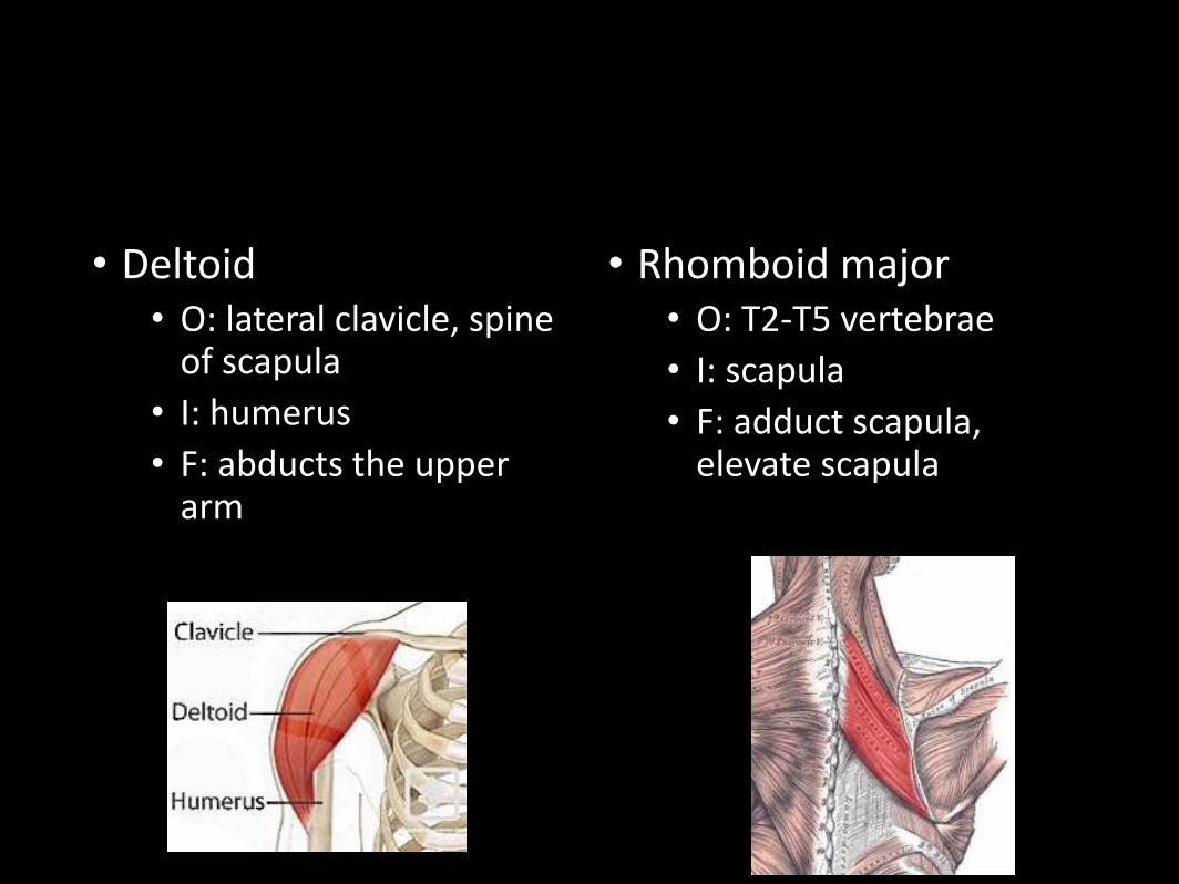
• Deltoid• O: lateral clavicle, spine
of scapula
• I: humerus
• F: abducts the upper arm
• Rhomboid major• O: T2-T5 vertebrae
• I: scapula
• F: adduct scapula, elevate scapula
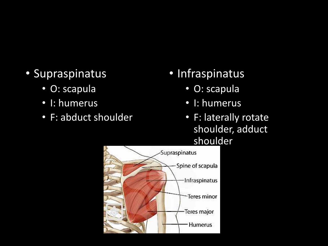
• Supraspinatus• O: scapula
• I: humerus
• F: abduct shoulder
• Infraspinatus• O: scapula
• I: humerus
• F: laterally rotate shoulder, adduct shoulder
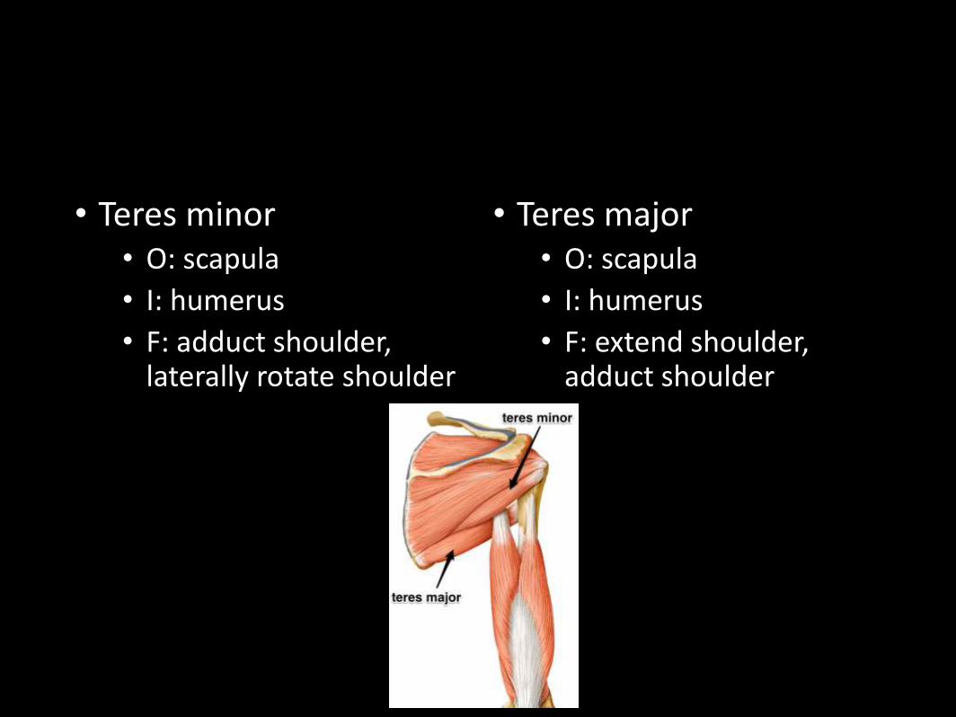
• Teres minor• O: scapula
• I: humerus
• F: adduct shoulder, laterally rotate shoulder
• Teres major• O: scapula
• I: humerus
• F: extend shoulder, adduct shoulder
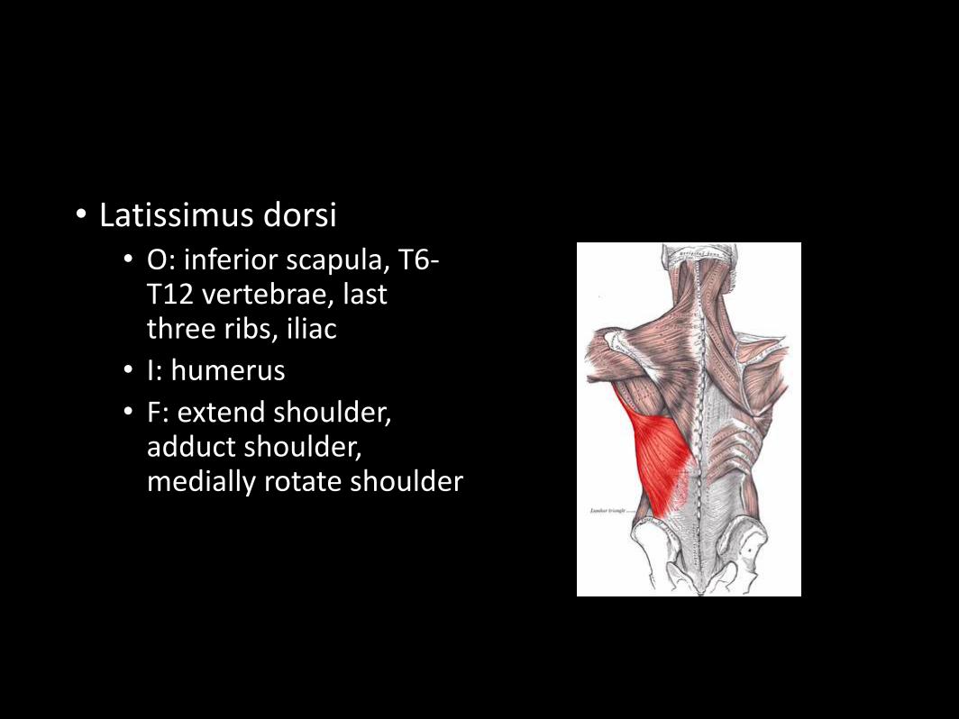
• Latissimus dorsi• O: inferior scapula, T6-
T12 vertebrae, last three ribs, iliac
• I: humerus
• F: extend shoulder, adduct shoulder, medially rotate shoulder
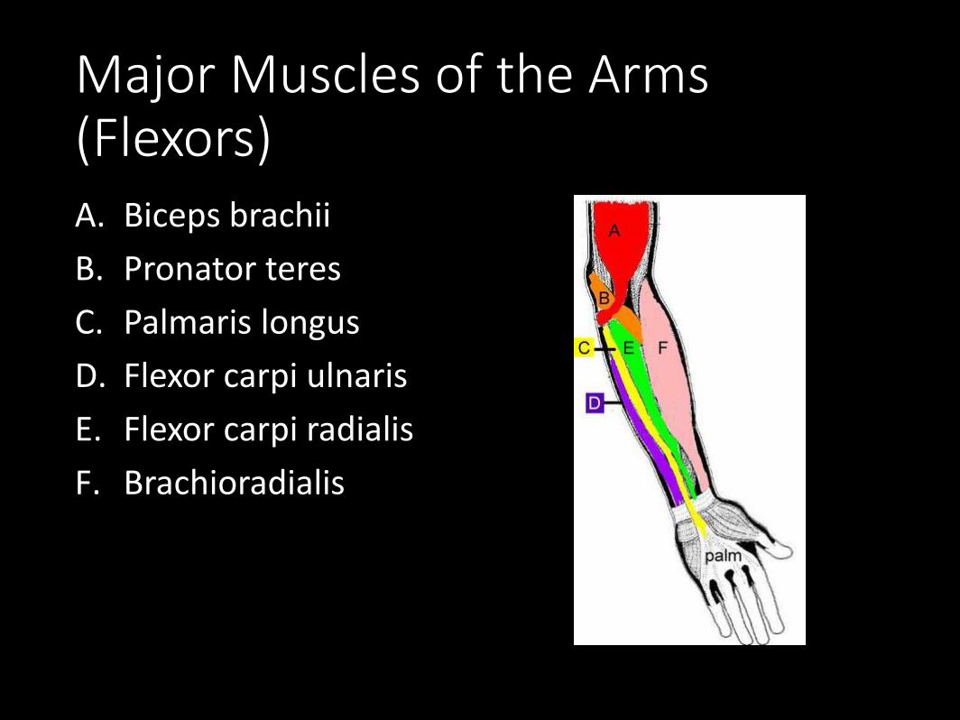
Major Muscles of the Arms (Flexors)A. Biceps brachii
B. Pronator teres
C. Palmaris longus
D. Flexor carpi ulnaris
E. Flexor carpi radialis
F. Brachioradialis
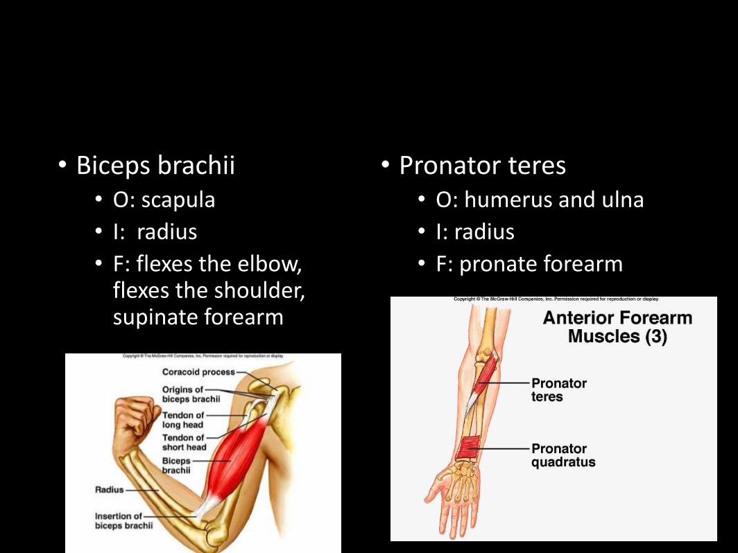
• Biceps brachii• O: scapula
• I: radius
• F: flexes the elbow, flexes the shoulder, supinate forearm
• Pronator teres• O: humerus and ulna
• I: radius
• F: pronate forearm
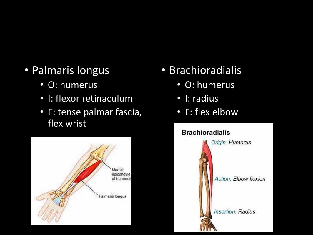
• Palmaris longus• O: humerus
• I: flexor retinaculum
• F: tense palmar fascia, flex wrist
• Brachioradialis• O: humerus
• I: radius
• F: flex elbow
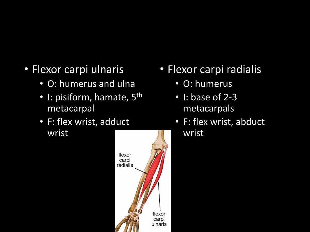
• Flexor carpi ulnaris• O: humerus and ulna
• I: pisiform, hamate, 5th
metacarpal
• F: flex wrist, adduct wrist
• Flexor carpi radialis• O: humerus
• I: base of 2-3 metacarpals
• F: flex wrist, abduct wrist
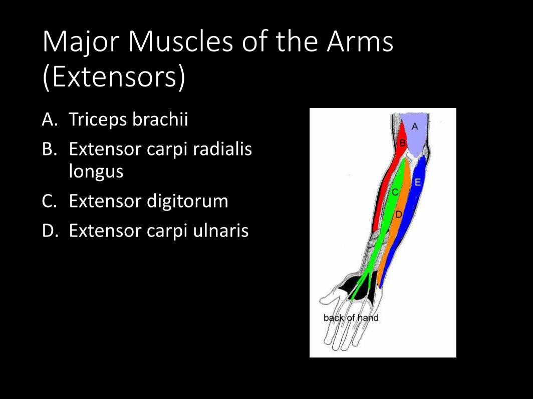
Major Muscles of the Arms (Extensors)A. Triceps brachii
B. Extensor carpi radialislongus
C. Extensor digitorum
D. Extensor carpi ulnaris
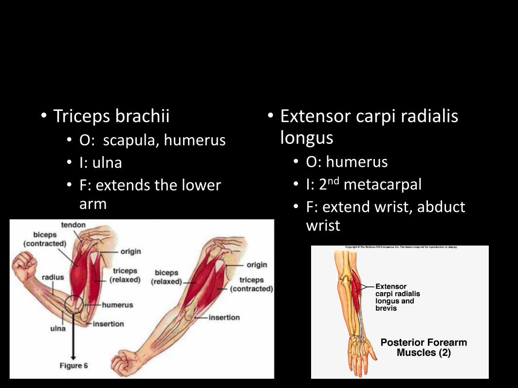
• Triceps brachii• O: scapula, humerus
• I: ulna
• F: extends the lower arm
• Extensor carpi radialislongus• O: humerus
• I: 2nd metacarpal
• F: extend wrist, abduct wrist
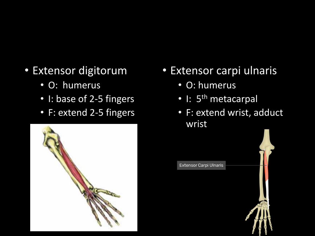
• Extensor digitorum• O: humerus
• I: base of 2-5 fingers
• F: extend 2-5 fingers
• Extensor carpi ulnaris• O: humerus
• I: 5th metacarpal
• F: extend wrist, adduct wrist
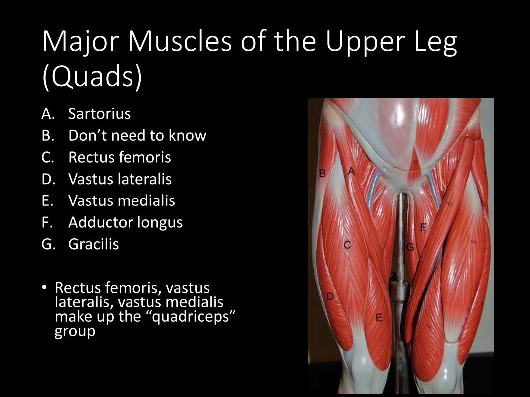
Major Muscles of the Upper Leg (Quads)A. Sartorius
B. Don’t need to know
C. Rectus femoris
D. Vastus lateralis
E. Vastus medialis
F. Adductor longus
G. Gracilis
• Rectus femoris, vastuslateralis, vastus medialismake up the “quadriceps” group
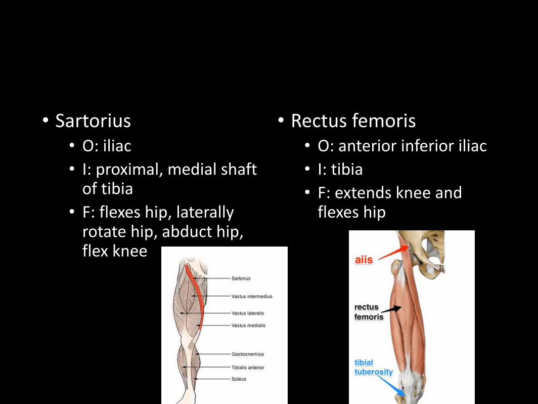
• Sartorius• O: iliac
• I: proximal, medial shaft of tibia
• F: flexes hip, laterally rotate hip, abduct hip, flex knee
• Rectus femoris• O: anterior inferior iliac
• I: tibia
• F: extends knee and flexes hip
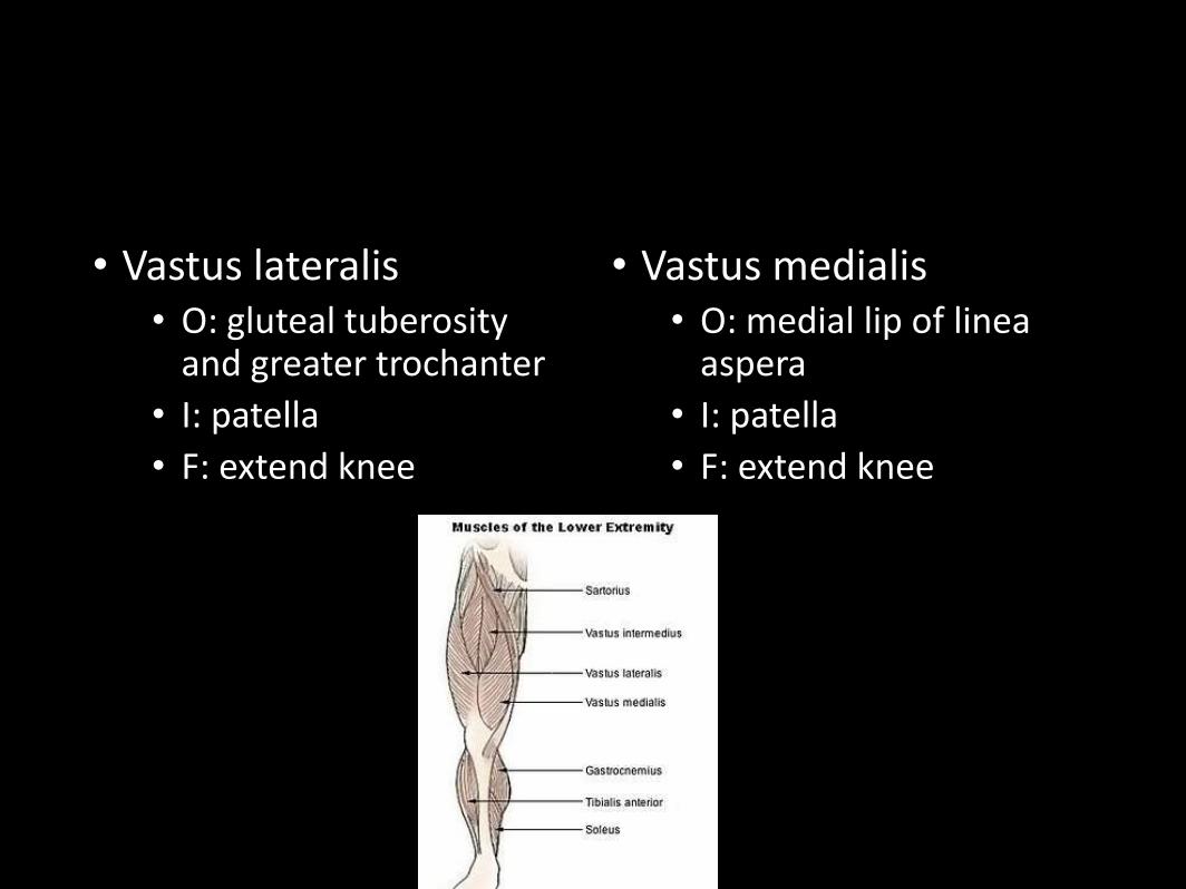
• Vastus lateralis• O: gluteal tuberosity
and greater trochanter
• I: patella
• F: extend knee
• Vastus medialis• O: medial lip of linea
aspera
• I: patella
• F: extend knee
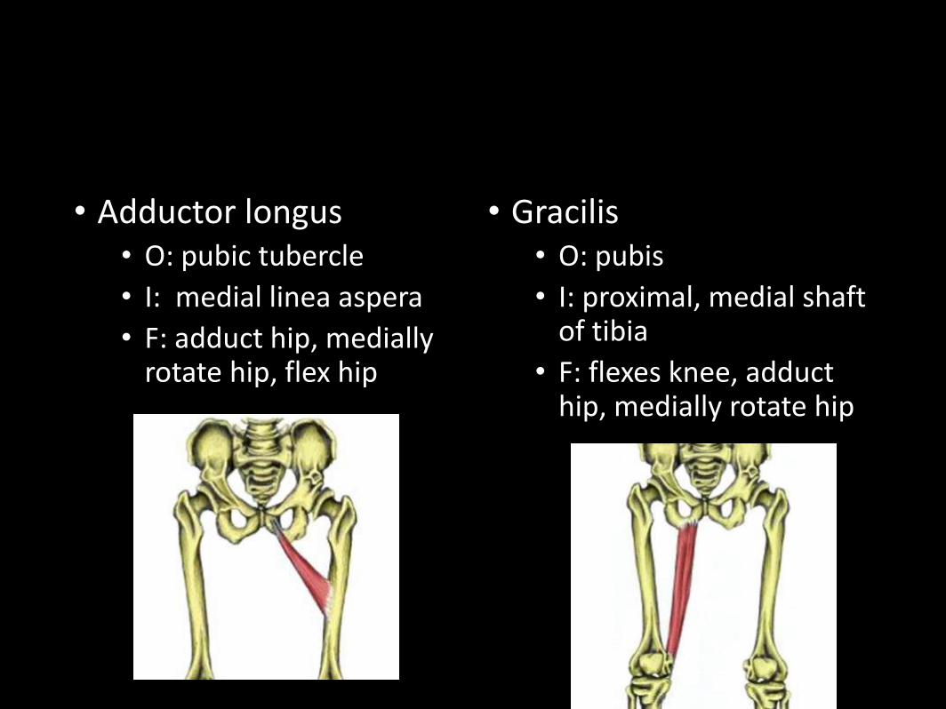
• Adductor longus• O: pubic tubercle
• I: medial linea aspera
• F: adduct hip, medially rotate hip, flex hip
• Gracilis• O: pubis
• I: proximal, medial shaft of tibia
• F: flexes knee, adduct hip, medially rotate hip
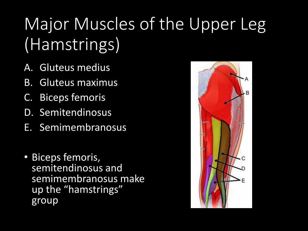
Major Muscles of the Upper Leg (Hamstrings)A. Gluteus medius
B. Gluteus maximus
C. Biceps femoris
D. Semitendinosus
E. Semimembranosus
• Biceps femoris, semitendinosus and semimembranosus make up the “hamstrings” group
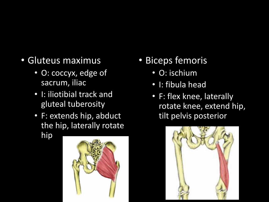
• Gluteus maximus• O: coccyx, edge of
sacrum, iliac
• I: iliotibial track and gluteal tuberosity
• F: extends hip, abduct the hip, laterally rotate hip
• Biceps femoris• O: ischium
• I: fibula head
• F: flex knee, laterally rotate knee, extend hip, tilt pelvis posterior
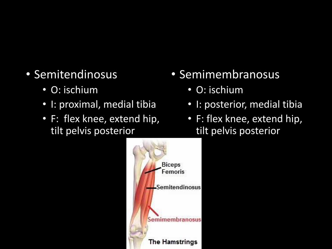
• Semitendinosus• O: ischium
• I: proximal, medial tibia
• F: flex knee, extend hip, tilt pelvis posterior
• Semimembranosus• O: ischium
• I: posterior, medial tibia
• F: flex knee, extend hip, tilt pelvis posterior
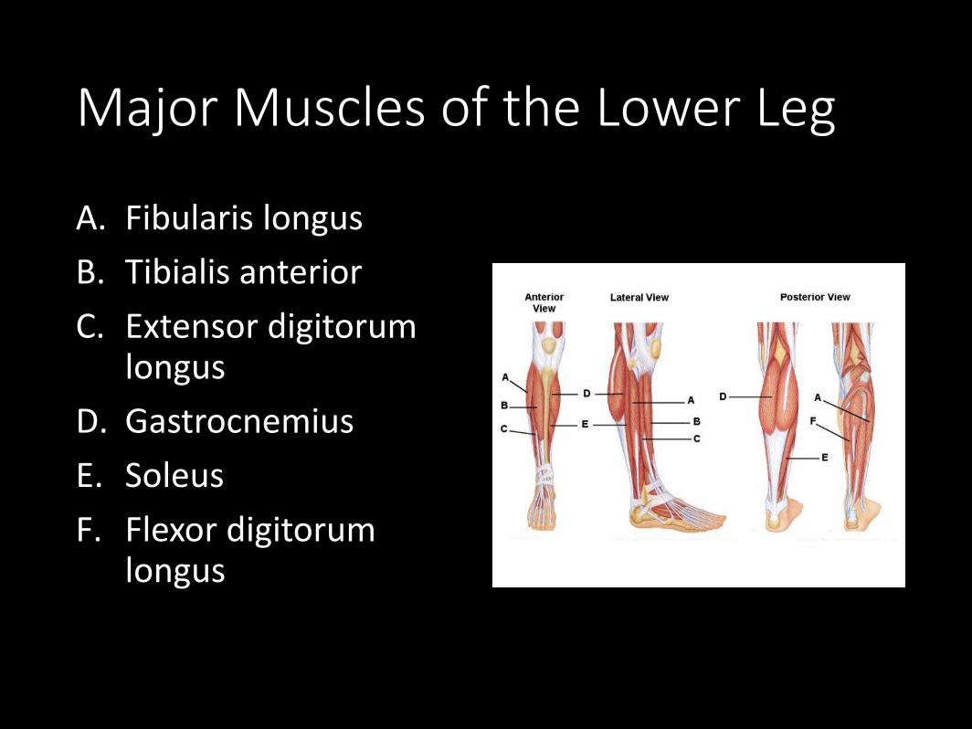
Major Muscles of the Lower Leg
A. Fibularis longus
B. Tibialis anterior
C. Extensor digitorumlongus
D. Gastrocnemius
E. Soleus
F. Flexor digitorumlongus
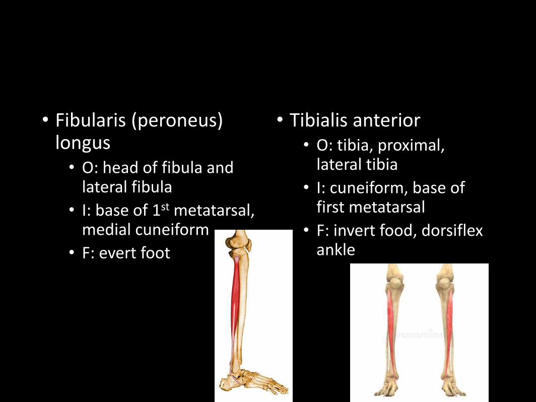
• Fibularis (peroneus) longus• O: head of fibula and
lateral fibula
• I: base of 1st metatarsal, medial cuneiform
• F: evert foot
• Tibialis anterior• O: tibia, proximal,
lateral tibia
• I: cuneiform, base of first metatarsal
• F: invert food, dorsiflexankle
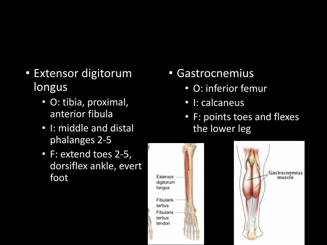
• Extensor digitorumlongus• O: tibia, proximal,
anterior fibula
• I: middle and distal phalanges 2-5
• F: extend toes 2-5, dorsiflex ankle, evert foot
• Gastrocnemius• O: inferior femur
• I: calcaneus
• F: points toes and flexes the lower leg
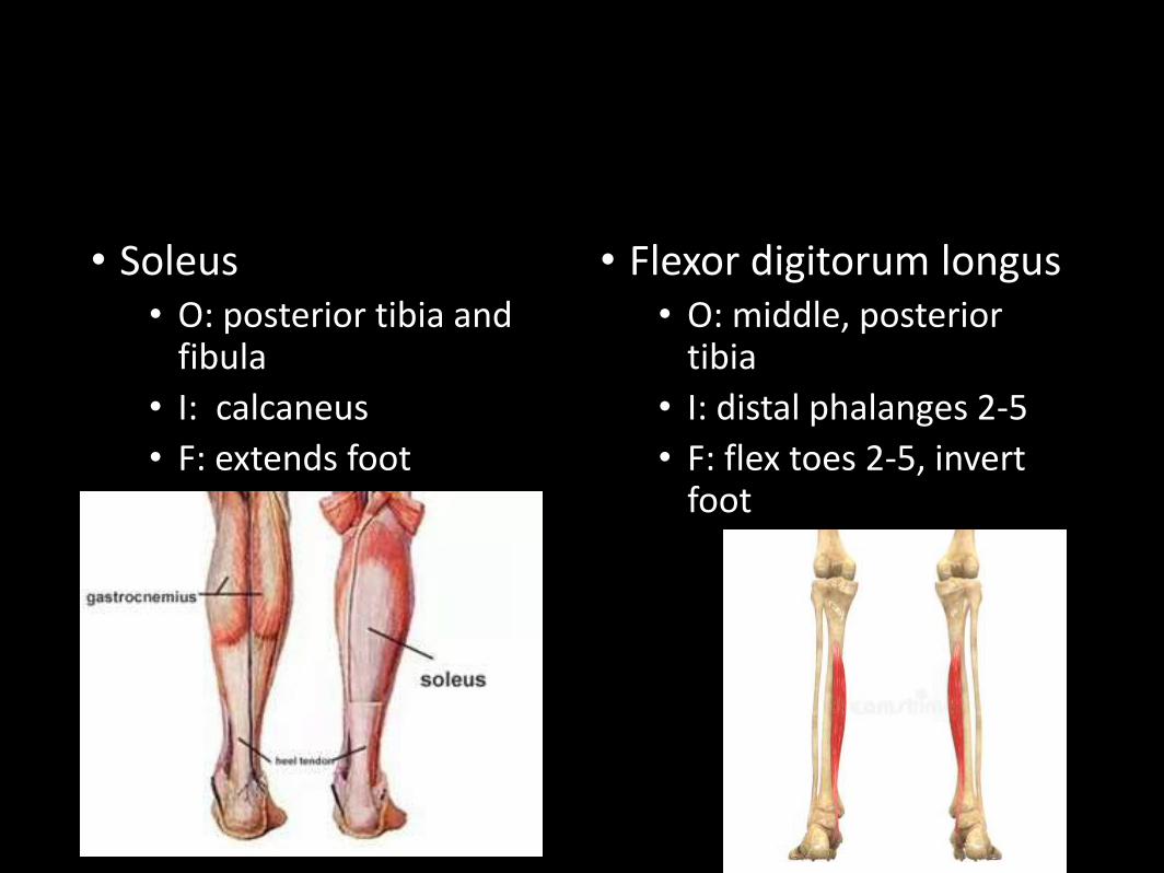
• Soleus• O: posterior tibia and
fibula
• I: calcaneus
• F: extends foot
• Flexor digitorum longus• O: middle, posterior
tibia
• I: distal phalanges 2-5
• F: flex toes 2-5, invert foot
Frawg Dissections
• Labs to Complete:• 4.1 Observing Skeletal
Muscle Through the Microscope
• 4.2 Identifying The Frog’s Muscles
• 4.3 The Muscles of the Human Body –Introduction
• 4.5 Naming Muscle Movements