Embed Size (px)
Citation preview
ORIGINAL ARTICLEnpg
Mutation of kri1l causes definitive hematopoiesis failure via PERK-dependent excessive autophagy inductionXiao-E Jia1, 4, *, Ke Ma1, *, Tao Xu1, *, Lei Gao1, Shuang Wu1, Cong Fu1, Wenjuan Zhang1, Zhizhang Wang1, Kaiyu Liu6, Mei Dong1, Changbin Jing1, Chunguang Ren1, Zhiwei Dong1, Yi Chen2, Yi Jin2, Qiuhua Huang2, Xing Chang1, 9, Min Deng1, Li Li8, Lingfei Luo8, Jun Zhu7, 9, Yongjun Dang6, Hung-Chun Chang5, Leonard I Zon3, Yi Zhou3, Saijuan Chen2, 9, Weijun Pan1, 9
1Key Laboratory of Stem Cell Biology, Institute of Health Sciences, Shanghai Institutes for Biological Sciences, Chinese Academy of Sciences & Shanghai Jiao Tong University School of Medicine, Shanghai, China; 2State Key Laboratory for Medical Genom-ics, Shanghai Institute of Hematology, RuiJin Hospital, Shanghai Jiao Tong University School of Medicine, Shanghai, China; 3Stem Cell Program, Hematology/Oncology Program at Children’s Hospital Boston, Harvard Medical School, Boston, MA 02114, USA; 4Biomedical Research Center, Baotou Medical College, Baotou, Inner Mongolia Autonomous Region, China; 5Institute of Neuroscience, Shanghai Institutes for Biological Sciences, Chinese Academy of Sciences, Shanghai, China; 6Key Laboratory of Metabolism and Molecular Medicine, Ministry of Education, Department of Biochemistry and Molecular Biology, School of Basic Medical Sciences, Fudan University, Shanghai, China; 7CNRS LIA, Shanghai Institute of Hematology, RuiJin Hospital, 197 RuiJin Road II, Shanghai, China; 8Key Laboratory of Freshwater Fish Reproduction and Development, Ministry of Education, Laborato-ry of Molecular Developmental Biology, School of Life Sciences, Southwest University, Beibei, Chongqing, China; 9Collaborative Innovation Center of Systems Biomedicine, Shanghai Jiao Tong University School of Medicine, Shanghai, China
Dysregulation of ribosome biogenesis causes human diseases, such as Diamond-Blackfan anemia, del (5q-) syn-drome and bone marrow failure. However, the mechanisms of blood disorders in these diseases remain elusive. Through genetic mapping, molecular cloning and mechanism characterization of the zebrafish mutant cas002, we reveal a novel connection between ribosomal dysfunction and excessive autophagy in the regulation of hematopoi-etic stem/progenitor cells (HSPCs). cas002 carries a recessive lethal mutation in kri1l gene that encodes an essential component of rRNA small subunit processome. We show that Kri1l is required for normal ribosome biogenesis, ex-pansion of definitive HSPCs and subsequent lineage differentiation. Through live imaging and biochemical studies, we find that loss of Kri1l causes the accumulation of misfolded proteins and excessive PERK activation-dependent autophagy in HSPCs. Blocking autophagy but not inhibiting apoptosis by Bcl2 overexpression can fully rescue hema-topoietic defects, but not the lethality of kri1lcas002 embryos. Treatment with autophagy inhibitors (3-MA and Baf A1) or PERK inhibitor (GSK2656157), or knockdown of beclin1 or perk can markedly restore HSPC proliferation and definitive hematopoietic cell differentiation. These results may provide leads for effective therapeutics that benefit patients with anemia or bone marrow failure caused by ribosome disorders.Keywords: Hematopoietic stem cells; zebrafish; kri1l; ribosome biogenesis; autophagy; PERK; misfolded/unfolded proteinCell Research (2015) 25:946-962. doi:10.1038/cr.2015.81; published online 3 July 2015
*These three authors contributed equally to this work.Correspondence: Weijun Pana, Saijuan Chenb, Yi Zhouc
aE-mail: [email protected]: [email protected] cE-mail: [email protected] 12 January 2015; revised 3 May 2015; accepted 28 May 2015; published online 3 July 2015
Introduction
Vertebrate hematopoiesis is an evolutionarily con-served and highly regulated process involving the pro-duction of differentiated blood cell lineages from hema-topoietic stem cells (HSCs) [1-3]. The zebrafish (Danio rerio) is a powerful genetic and developmental model to study the development of vertebrate circulatory system,
Cell Research (2015) 25:946-962.www.nature.com/cr
Xiao-E Jia et al.947
npg
www.cell-research.com | Cell Research
especially hematopoiesis [4, 5]. Definitive hematopoie-sis in zebrafish is highly conserved with mammals [6]. It occurs at the ventral wall of dorsal aorta in a structure called aorta-gonad-mesonephros (AGM) around 28 hour post-fertilization (hpf) [7]. By 2 days post-fertilization (dpf), the AGM-derived HSCs migrate to caudal hemato-poietic tissue (CHT), an embryonic structure analogous to the mammalian fetal liver [8-10], for rapid pool ex-pansion and lineage differentiation.
Normal ribosome biogenesis is critical for cell sur-vival, proliferation and function. In eukaryotic cells the small subunit (SSU) processome, a ribonucleoprotein complex, plays essential roles in 18S rRNA maturation, small ribosomal subunit assembly and subsequent ribo-some biogenesis [11]. The SSU processome is composed of pre-rRNA, small nucleolar RNAs and more than 70 different associated proteins. Dysfunction of ribosome biogenesis is associated with human diseases, including Diamond–Blackfan anemia (DBA), 5q minus (del (5q−)) syndrome, dyskeratosis congenita and Bowen-Conradi syndrome [12-14]. In addition to carrying genetic mu-tations affecting ribosome biogenesis, these human dis-eases share same clinical features, and are thus termed as “ribosomopathies” [13].
Previous works have shown that upregulation of p53 by ribosome dysfunction contributes to bone mar-row failure syndromes [15] that have been genetically mapped to loss-of-function mutations in rps14 [16], rps19 [17, 18], rpl11 [19] and rps29 [15]. The apoptotic phenotype of hematopoietic cells in these diseases can be partially restored by p53 downregulation [16, 17]. How-ever, some studies have reported that p53 pathway is not involved in some ribosome biogenesis disorders [20-22]. Furthermore, not all bone marrow samples from patients with del (5q−) syndrome or DBA show p53 activation, suggesting that other mechanisms may contribute to ri-bosomopathies [23]. For example, loss of pwhp2h causes gut degeneration and hyperactivated autophagy in a p53- and mTOR-independent manner [21]. Knockdown of Rpl22 in zebrafish embryos blocks T-lineage progenitor development, while knockdown of the Rpl22 paralog Rpl22l impairs the emergence of HSC in AGM by abro-gating Smad1 expression and Runx1 induction [24].
Autophagy and apoptosis are two major stress-re-sponse pathways. Dysregulation of autophagy has been linked to many human diseases such as neurodegenera-tion [25, 26], autoimmunity and cancer [27, 28]. Multiple upstream signaling mechanisms, including mTOR path-way, unfolded protein response (UPR), ER stress and nutrition stress regulate autophagy, with Beclin1-VPS34 complex playing an important role in autophagy initia-tion [29, 30]. Autophagy is a critical mechanism that pro-
tects HSCs from stress damages [31]. In mice, a condi-tional deletion of atg7 in HSCs renders the loss of HSC’s self-renewal property and severe myeloproliferation due to a failure of HSPCs to respond normally to stress from reactive oxygen species (ROS) [32]. Appropriate autoph-agy level is also important for lymphocyte survival [33, 34] and erythroid cell maturation [35-37]. Patients with certain ribosomopathies have elevated levels of autoph-agy in peripheral blood cells resulted from S6K-induced inhibition on insulin pathway activation [38]. However, the potential cures for these disorders have not been found.
In the present study, we report that kri1l gene is essen-tial for definitive hematopoiesis. Loss of Kri1l, a critical component of SSU complex, causes ribosomal biogen-esis defects, accumulation of misfolded proteins and activation of PERK-eif2a signaling. These deficiencies subsequently hyperactivate autophagy and ultimately lead to the inhibition of HSPC proliferation. Treatment with autophagy or PERK inhibitors, or knockdown of beclin1 or perk by morpholino (MO), can successfully rescue HSPC proliferation and lineage differentiation in kri1l mutant.
Results
cas002 mutant displays a hematopoietic failure pheno-type
In a large-scale ENU mutagenesis screen for definitive hematopoietic mutations, we obtained cas002, a novel mutant with severe hematopoietic defects and recessive lethality. cas002 embryos are morphologically indis-tinguishable from wild-type siblings before 3 dpf, with normal blood flow and heart beats (Figure 1A-1B). How-ever, whole-mount in situ hybridization (WISH) of cmyb reveals a markedly reduced HSPC population in caudal hematopoietic tissue (CHT) of mutant embryos at 3 dpf (Figure 1C-1D), and in CHT, thymus and kidney at 5 dpf (Figure 1E-1F). cas002 mutant embryos eventually die at 6-10 dpf with abnormal head shape, cardiac edema and smaller eyes.
To examine hematopoiesis phenotype in detail, we performed WISH analysis of different cell lineage mark-ers: gata1 (erythrocyte progenitors), αe1-globin (em-bryonic erythrocytes), l-plastin (pan-myeloid cells), lyz and mpo (neutrophils). The expression of these markers is the same as wild-type siblings at 3 dpf (Supplemen-tary information, Figure S1), but becomes significantly reduced in cas002 mutant embryos at 5 dpf (Supplemen-tary information, Figure S2A-S2J). The expression of rag1 (T lymphocyte), is substantially reduced at 4 dpf in cas002 (Supplementary information, Figure S2K-S2L),

948Excessive autophagy limits HSPCs proliferationnpg
Cell Research | Vol 25 No 8 | August 2015
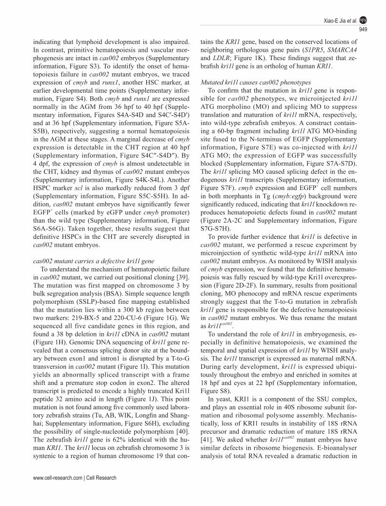
Figure 1 Hematopoietic defects and positional cloning of cas002 mutant. (A-B) Light microscope images of zebrafish wild-type (WT) and cas002 embryos at 3 dpf. (C-F) WISH analysis of cmyb expression in WT and cas002 embryos at indicated development stages. Black arrows indicate thymus, kidney marrow and CHT. (C′-D′) enlarged CHT regions in C and D. (G) Genetic mapping of the cas002 region on chromosome 3. Bulk segregation analysis locates cas002 mutation to Chr. 3. Fine mapping using SSLPs narrows down to a region between markers 219-BX-5 and 220-CU-6, containing kri1l and four other genes as indicated. (H) The sequencing results of kri1l cDNA from mutant embryos show a 38 bp deletion (MU) compared with kri1l cDNA from WT embryos. (I, J) The sequencing result of kri1l genomic DNA shows a T-G transversion at the kri1l exon 1-intron 1 consensus splicing donor site (I), which causes a frame shift (H) and a premature stop codon leading to the production of a truncated Kri1l protein (J). (K) Synteny between zebrafish kri1l I and human KRI1 loci. (Left) Six genes, in-cluding KRI1, are located within a genomic region on human chromosome 19. (Right) Six zebrafish homologs are listed ac-cording to their map positions on chromosome 3 (Ensembl website). CHT, caudal hematopoietic tissue; KM, kidney marrow; Mb, mega base; T, thymus; het, heterozygote.
Xiao-E Jia et al.949
npg
www.cell-research.com | Cell Research
indicating that lymphoid development is also impaired. In contrast, primitive hematopoiesis and vascular mor-phogenesis are intact in cas002 embryos (Supplementary information, Figure S3). To identify the onset of hema-topoiesis failure in cas002 mutant embryos, we traced expression of cmyb and runx1, another HSC marker, at earlier developmental time points (Supplementary infor-mation, Figure S4). Both cmyb and runx1 are expressed normally in the AGM from 36 hpf to 40 hpf (Supple-mentary information, Figures S4A-S4D and S4C′-S4D′) and at 36 hpf (Supplementary information, Figure S5A-S5B), respectively, suggesting a normal hematopoiesis in the AGM at these stages. A marginal decrease of cmyb expression is detectable in the CHT region at 40 hpf (Supplementary information, Figure S4C″-S4D″). By 4 dpf, the expression of cmyb is almost undetectable in the CHT, kidney and thymus of cas002 mutant embryos (Supplementary information, Figure S4K-S4L). Another HSPC marker scl is also markedly reduced from 3 dpf (Supplementary information, Figure S5C-S5H). In ad-dition, cas002 mutant embryos have significantly fewer EGFP+ cells (marked by eGFP under cmyb promoter) than the wild type (Supplementary information, Figure S6A-S6G). Taken together, these results suggest that definitive HSPCs in the CHT are severely disrupted in cas002 mutant embryos.
cas002 mutant carries a defective kri1l geneTo understand the mechanism of hematopoietic failure
in cas002 mutant, we carried out positional cloning [39]. The mutation was first mapped on chromosome 3 by bulk segregation analysis (BSA). Simple sequence length polymorphism (SSLP)-based fine mapping established that the mutation lies within a 300 kb region between two markers: 219-BX-5 and 220-CU-6 (Figure 1G). We sequenced all five candidate genes in this region, and found a 38 bp deletion in kri1l cDNA in cas002 mutant (Figure 1H). Genomic DNA sequencing of kri1l gene re-vealed that a consensus splicing donor site at the bound-ary between exon1 and intron1 is disrupted by a T-to-G transversion in cas002 mutant (Figure 1I). This mutation yields an abnormally spliced transcript with a frame shift and a premature stop codon in exon2. The altered transcript is predicted to encode a highly truncated Kri1l peptide 32 amino acid in length (Figure 1J). This point mutation is not found among five commonly used labora-tory zebrafish strains (Tu, AB, WIK, Longfin and Shang-hai; Supplementary information, Figure S6H), excluding the possibility of single-nucleotide polymorphism [40]. The zebrafish kri1l gene is 62% identical with the hu-man KRI1. The kri1l locus on zebrafish chromosome 3 is syntenic to a region of human chromosome 19 that con-
tains the KRI1 gene, based on the conserved locations of neighboring orthologous gene pairs (S1PR5, SMARCA4 and LDLR; Figure 1K). These findings suggest that ze-brafish kri1l gene is an ortholog of human KRI1.
Mutated kri1l causes cas002 phenotypesTo confirm that the mutation in kri1l gene is respon-
sible for cas002 phenotypes, we microinjected kri1l ATG morpholino (MO) and splicing MO to suppress translation and maturation of kri1l mRNA, respectively, into wild-type zebrafish embryos. A construct contain-ing a 60-bp fragment including kri1l ATG MO-binding site fused to the N-terminus of EGFP (Supplementary information, Figure S7E) was co-injected with kri1l ATG MO; the expression of EGFP was successfully blocked (Supplementary information, Figure S7A-S7D). The kri1l splicing MO caused splicing defect in the en-dogenous kri1l transcripts (Supplementary information, Figure S7F). cmyb expression and EGFP+ cell numbers in both morphants in Tg (cmyb:egfp) background were significantly reduced, indicating that kri1l knockdown re-produces hematopoietic defects found in cas002 mutant (Figure 2A-2C and Supplementary information, Figure S7G-S7H).
To provide further evidence that kri1l is defective in cas002 mutant, we performed a rescue experiment by microinjection of synthetic wild-type kri1l mRNA into cas002 mutant embryos. As monitored by WISH analysis of cmyb expression, we found that the definitive hemato-poiesis was fully rescued by wild-type Kri1l overexpres-sion (Figure 2D-2F). In summary, results from positional cloning, MO phenocopy and mRNA rescue experiments strongly suggest that the T-to-G mutation in zebrafish kri1l gene is responsible for the defective hematopoiesis in cas002 mutant embryos. We thus rename the mutant as kri1lcas002.
To understand the role of kri1l in embryogenesis, es-pecially in definitive hematopoiesis, we examined the temporal and spatial expression of kri1l by WISH analy-sis. The kri1l transcript is expressed as maternal mRNA. During early development, kri1l is expressed ubiqui-tously throughout the embryo and enriched in somites at 18 hpf and eyes at 22 hpf (Supplementary information, Figure S8).
In yeast, KRI1 is a component of the SSU complex, and plays an essential role in 40S ribosome subunit for-mation and ribosomal polysome assembly. Mechanis-tically, loss of KRI1 results in instability of 18S rRNA precursor and dramatic reduction of mature 18S rRNA [41]. We asked whether kri1lcas002 mutant embryos have similar defects in ribosome biogenesis. E-bioanalyser analysis of total RNA revealed a dramatic reduction in
950Excessive autophagy limits HSPCs proliferationnpg
Cell Research | Vol 25 No 8 | August 2015
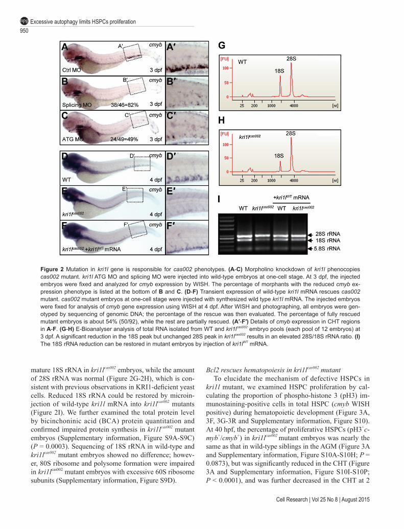
Figure 2 Mutation in kri1l gene is responsible for cas002 phenotypes. (A-C) Morpholino knockdown of kri1l phenocopies cas002 mutant. kri1l ATG MO and splicing MO were injected into wild-type embryos at one-cell stage. At 3 dpf, the injected embryos were fixed and analyzed for cmyb expression by WISH. The percentage of morphants with the reduced cmyb ex-pression phenotype is listed at the bottom of B and C. (D-F) Transient expression of wild-type kri1l mRNA rescues cas002 mutant. cas002 mutant embryos at one-cell stage were injected with synthesized wild type kri1l mRNA. The injected embryos were fixed for analysis of cmyb gene expression using WISH at 4 dpf. After WISH and photographing, all embryos were gen-otyped by sequencing of genomic DNA; the percentage of the rescue was then evaluated. The percentage of fully rescued mutant embryos is about 54% (50/92), while the rest are partially rescued. (A′-F′) Details of cmyb expression in CHT regions in A-F. (G-H) E-Bioanalyser analysis of total RNA isolated from WT and kri1lcas002 embryo pools (each pool of 12 embryos) at 3 dpf. A significant reduction in the 18S peak but unchanged 28S peak in kri1lcas002 results in an elevated 28S/18S rRNA ratio. (I) The 18S rRNA reduction can be restored in mutant embryos by injection of kri1lWT mRNA.
mature 18S rRNA in kri1lcas002 embryos, while the amount of 28S rRNA was normal (Figure 2G-2H), which is con-sistent with previous observations in KRI1-deficient yeast cells. Reduced 18S rRNA could be restored by microin-jection of wild-type kri1l mRNA into kri1lcas002 mutants (Figure 2I). We further examined the total protein level by bicinchoninic acid (BCA) protein quantitation and confirmed impaired protein synthesis in kri1lcas002 mutant embryos (Supplementary information, Figure S9A-S9C) (P = 0.0003). Sequencing of 18S rRNA in wild-type and kri1lcas002 mutant embryos showed no difference; howev-er, 80S ribosome and polysome formation were impaired in kri1lcas002 mutant embryos with excessive 60S ribosome subunits (Supplementary information, Figure S9D).
Bcl2 rescues hematopoiesis in kri1lcas002 mutantTo elucidate the mechanism of defective HSPCs in
kri1l mutant, we examined HSPC proliferation by cal-culating the proportion of phospho-histone 3 (pH3) im-munostaining-positive cells in total HSPC (cmyb WISH positive) during hematopoietic development (Figure 3A, 3F, 3G-3R and Supplementary information, Figure S10). At 40 hpf, the percentage of proliferative HSPCs (pH3+c-myb+/cmyb+) in kri1lcas002 mutant embryos was nearly the same as that in wild-type siblings in the AGM (Figure 3A and Supplementary information, Figure S10A-S10H; P = 0.0873), but was significantly reduced in the CHT (Figure 3A and Supplementary information, Figure S10I-S10P; P < 0.0001), and was further decreased in the CHT at 2

Xiao-E Jia et al.951
npg
www.cell-research.com | Cell Research
Figure 3 Transient expression of Bcl2 rescues the defective hematopoiesis in kri1lcas002 mutant embryos. (A) Statistic analysis of the proportion of proliferative HSPCs (cmyb and PH3 double positive) in total HSPCs in kri1lcas002 mutant embryos and wild-type siblings during hematopoietic development (40 hpf-2 dpf). N ≥ 10 for each group. 40 hpf AGM, p = 0.0873; 40 hpf CHT, p < 0.0001; 2 dpf CHT, p<0.0001. Error bars represent SEM. **P ≤ 0.01; ***P ≤ 0.001 (Student’s t-test). (B) Proportion of kri1lcas002 mutant embryos with classified hematopoiesis phenotype (indicated by cmyb staining) with or without microinjection of p53 MO, treatment with caspase inhibitor (Z-VAD-FMK) or Bcl2 expression, respectively. (C-E) The CHT hematopoiesis is recovered by Bcl2 overexpression in kri1lcas002 mutant embryos. Embryos from two heterozygous parents were injected with bcl2 mRNA at one-cell stage and harvested for WISH analysis. After WISH and photographing, all embryos were genotyped by genomic DNA sequencing; percentage of rescue was then calculated. 26 out of 33 mutant embryos injected at one-cell stage with bcl2 mRNA are fully rescued, while the rest of embryos are partially rescued. (C′-E′) The enlarged CHT regions. (F) Percentage of the proliferative HSPCs in the CHT of kri1lcas002 mutant embryos and wild-type siblings with or without Bcl2 overexpression at 3 dpf. N = 12 for each group. Without Bcl2 overexpression, p < 0.0001; With Bcl2 overexpression, p = 0.3136. Error bars represent SEM. **P ≤ 0.01; ***P ≤ 0.001 (Student’s t-test). (G-R) Representative confocal images of double stain-ing of cmyb RNA (red) and pH3 protein (green) in the CHT at 3 dpf. G, K and O are bright-field images overlaid with fluores-cent staining. Scale bars, 50 µm.
952Excessive autophagy limits HSPCs proliferationnpg
Cell Research | Vol 25 No 8 | August 2015
dpf (Figure 3A and Supplementary information, Figure S10Q-S10X; P < 0.0001) and 3 dpf (Figure 3F-3R) (P = 0.0003).
Previous reports have demonstrated that p53-depen-dent apoptosis pathway is activated and responsible for hematopoietic defects in both ribosomopathy-related mice and zebrafish models [15-18]. We carried out termi-nal transferase dUTP nick end labeling (TUNEL) assays in wild-type siblings and kri1lcas002 mutant embryos from 40 hpf to 3 dpf; however, there was no significant differ-ence in apoptotic signals in the CHT region between two genotypes (Supplementary information, Figure S11).
Furthermore, we microinjected p53 MO [42] into kri1lcas002 mutant embryos (Figure 3B and Supplementa-ry information, Figure S12A-S12D), and kri1l MO into p53M214K mutant embryos (Supplementary information, Figure S13A-S13D). The defective hematopoiesis re-mained in these morphants, suggesting that p53-depen-dent apoptosis is not responsible for the hematopoietic failure in kri1lcas002 mutant. We further tested whether the activation of caspase pathway contributes to HSPC defects and found treatment of pan-caspase inhibitor, Z-VAD-FMK peptide, could not rescue kri1lcas002 pheno-type (Figure 3B and Supplementary information, Figure S12E-S12F). Quantitative PCR and immunoblotting analysis also showed that ∆113-p53, an inhibitor of full-length p53 [43], but not full-length p53, was dramatically upregulated in kri1lcas002 mutant embryos (Supplementary information, Figure S14).
In parallel, we overexpressed an EGFP-Bcl2 fusion protein in kri1lcas002 mutant embryos. To our surprise, microinjection of egfp-bcl2 mRNA fully restored cmyb expression in the CHT of kri1lcas002 mutant embryos at 3 dpf (Figure 3B-3E). We performed WISH analysis of αe1-globin and lyz expression at 4 dpf, and found Bcl2 overexpression also successfully restored the erythro-cytes (74%; Supplementary information, Figure S15A-S15D) and neutrophils (61%; Supplementary informa-tion, Figure S15E-S15H). Very importantly, ectopic expression of Bcl2 almost fully restored the percentage of proliferating HSPCs (Figure 3F-3R; P = 0.3136) in kri1lcas002 mutant embryos.
Autophagy is hyperactivated in kri1lcas002 mutantRecent reports have shown that Bcl2 functions not
only as an anti-apoptosis protein, but also as an anti-auto-phagy protein by interacting with Beclin1 and disrupting VPS34-Beclin1 complex [44, 45]. To check whether the regulatory role of Bcl2 in autophagy is responsible for the rescue of HSPC in kri1lcas002 mutant, we carried out immunoblotting on the whole embryo lysates using an antibody against an autophagy marker, microtubule-as-sociated protein light chain 3 (Lc3) [46, 47]. We found
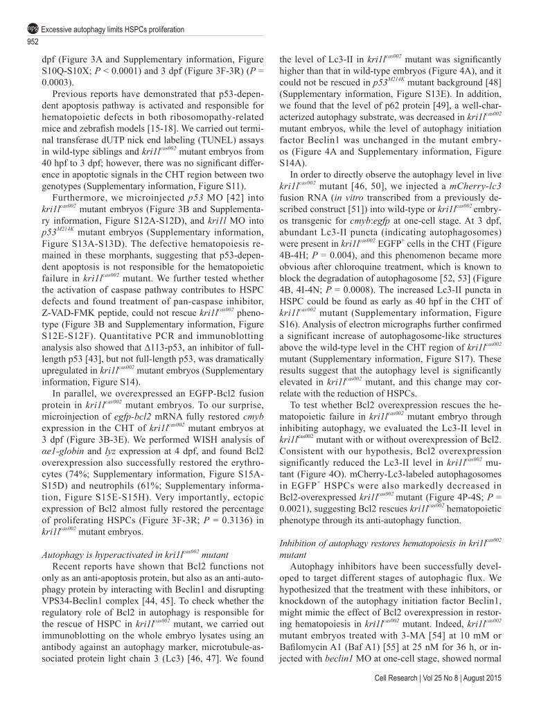
the level of Lc3-II in kri1lcas002 mutant was significantly higher than that in wild-type embryos (Figure 4A), and it could not be rescued in p53M214K mutant background [48] (Supplementary information, Figure S13E). In addition, we found that the level of p62 protein [49], a well-char-acterized autophagy substrate, was decreased in kri1lcas002 mutant embryos, while the level of autophagy initiation factor Beclin1 was unchanged in the mutant embry-os (Figure 4A and Supplementary information, Figure S14A).
In order to directly observe the autophagy level in live kri1lcas002 mutant [46, 50], we injected a mCherry-lc3 fusion RNA (in vitro transcribed from a previously de-scribed construct [51]) into wild-type or kri1lcas002 embry-os transgenic for cmyb:egfp at one-cell stage. At 3 dpf, abundant Lc3-II puncta (indicating autophagosomes) were present in kri1lcas002 EGFP+ cells in the CHT (Figure 4B-4H; P = 0.004), and this phenomenon became more obvious after chloroquine treatment, which is known to block the degradation of autophagosome [52, 53] (Figure 4B, 4I-4N; P = 0.0008). The increased Lc3-II puncta in HSPC could be found as early as 40 hpf in the CHT of kri1lcas002 mutant (Supplementary information, Figure S16). Analysis of electron micrographs further confirmed a significant increase of autophagosome-like structures above the wild-type level in the CHT region of kri1lcas002 mutant (Supplementary information, Figure S17). These results suggest that the autophagy level is significantly elevated in kri1lcas002 mutant, and this change may cor-relate with the reduction of HSPCs.
To test whether Bcl2 overexpression rescues the he-matopoietic failure in kri1lcas002 mutant embryo through inhibiting autophagy, we evaluated the Lc3-II level in kri1lcas002 mutant with or without overexpression of Bcl2. Consistent with our hypothesis, Bcl2 overexpression significantly reduced the Lc3-II level in kri1lcas002 mu-tant (Figure 4O). mCherry-Lc3-labeled autophagosomes in EGFP+ HSPCs were also markedly decreased in Bcl2-overexpressed kri1lcas002 mutant (Figure 4P-4S; P = 0.0021), suggesting Bcl2 rescues kri1lcas002 hematopoietic phenotype through its anti-autophagy function.
Inhibition of autophagy restores hematopoiesis in kri1lcas002 mutant
Autophagy inhibitors have been successfully devel-oped to target different stages of autophagic flux. We hypothesized that the treatment with these inhibitors, or knockdown of the autophagy initiation factor Beclin1, might mimic the effect of Bcl2 overexpression in restor-ing hematopoiesis in kri1lcas002 mutant. Indeed, kri1lcas002 mutant embryos treated with 3-MA [54] at 10 mM or Bafilomycin A1 (Baf A1) [55] at 25 nM for 36 h, or in-jected with beclin1 MO at one-cell stage, showed normal
Xiao-E Jia et al.953
npg
www.cell-research.com | Cell Research
Figure 4 Ectopic expression of Bcl2 inhibits excessive autophagic flux in kri1lcas002 HSPCs. (A) Representative immuno-blotting images of Lc3-I, Lc3-II, Beclin1 and p62 in whole embryo lysates of wild-type (WT) and kri1lcas002 embryos at 3 dpf, treated with or without chloroquine (5 µM) for 6 h before harvest. Tubulin serves as the loading control. (B) Statistics of the average number of autophagosome (indicated by mCherry-Lc3 puncta) in kri1lcas002 mutant embryos and WT siblings with or without chloroquine treatment at 3 dpf. mCherry-Lc3 puncta are counted in over 20 EGFP+ cells (in cmyb:egfp background) per embryo, total 6-8 embryos under each condition. Without chloroquine treatment, P = 0.0040; with chloroquine treatment, P = 0.0008. Error bars represent SEM. **P ≤ 0.01; ***P ≤ 0.001 (Student’s t-test). (C-N) Representative confocal images of mCherry-Lc3 puncta (autophagosomes) in HSPCs of live zebrafish embryos. Wild type siblings or kri1lcas002 mutant embryos in the Tg (cmyb: egfp) transgenic background, were injected with mcherry-lc3 mRNA at one-cell stage and imaged at 3 dpf with or without pre-treatment with chloroquine (5 µM) for 6 h. Scale bars: 5 µm. (O) Immunoblotting analysis of whole embryo lysates from WT and kri1lcas002 mutant embryos with or without bcl2 mRNA injection. (P) Statistics of the average number of autophagosome for kri1lcas002 mutant embryos and wild type siblings with Bcl2 overexpression at 3 dpf. mCherry-Lc3 puncta are counted in over 20 EGFP+ (cmyb: egfp) cells per embryo, total 8-11 individual embryos. P = 0.0021. Error bars represent SEM. **P ≤ 0.01; ***P ≤ 0.001 (Student’s t-test). (Q-S) Representative confocal images of mCherry-Lc3 puncta (autophago-somes) in HSPCs of live zebrafish embryos with Bcl2 overexpression. Scale bars, 5 µm.
954Excessive autophagy limits HSPCs proliferationnpg
Cell Research | Vol 25 No 8 | August 2015
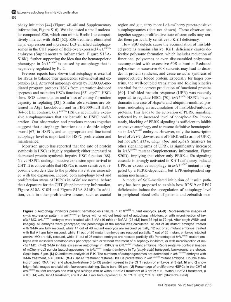
cmyb expression in the CHT (Figure 5A-5E). 3-MA sig-nificantly reduced autophagy level in kri1lcas002 HSPCs, which was visualized by live imaging and quantitation of Lc3-II puncta (Figure 5F-5L; P = 0.0037). Furthermore, both Baf A1 (Figure 5M-5U) and 3-MA (data no shown) treatment could restore kri1lcas002 HSPC proliferation. At 3 dpf, the proportion of pH3+cmyb+ cells in kri1lcas002 mutant embryos treated with Baf A1 was almost the same as that in wild type (Figure 5U; P = 0.2344). Therefore, inhibi-tion of autophagic flux at different stages is able to rescue HSPC proliferation defects in kri1lcas002 mutant.
Accumulation of misfolded proteins trigger PERK-de-pendent autophagy
To understand how Kri1l deficiency-caused ribosome defects trigger excessive autophagy via a p53-indepen-dent manner, we hypothesized that impaired ribosome biogenesis might cause dysfunction in protein synthe-sis to trigger misfolded protein accumulation, which is known to activate autophagy [56, 57].
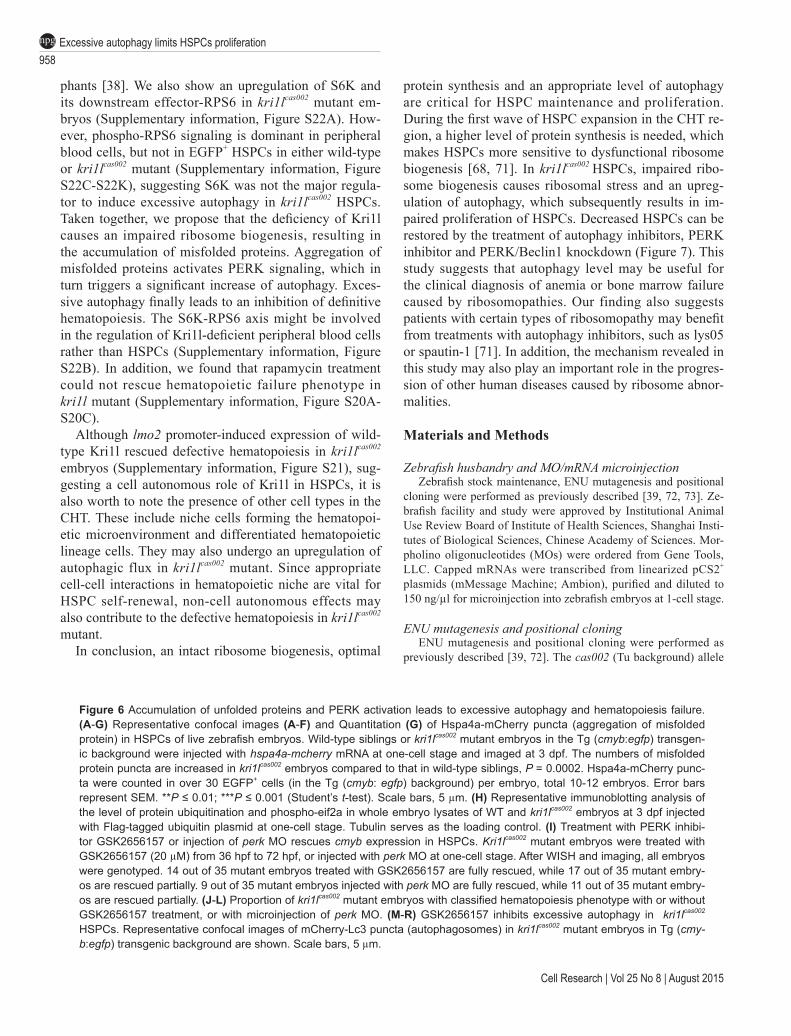
Protein remodeling factor Hsp110 is known to cooper-ate with Hsp70 and Hsp40 to dissolve and reactivate ag-gregated proteins. It is used as a molecular probe to detect puncta foci where misfolded proteins accumulate [58]. We injected an in vitro transcribed mRNA coding for Hspa4a (zebrafish homolog of human Hsp110) [59] fused to mCherry into one-cell stage wild-type or kri1lcas002 embryos stably transgenic for cmyb:egfp. Abundant Hspa4a-mCherry puncta were observed in kri1lcas002 EGFP+ cells in the CHT region (Figure 6A-6G; P = 0.0002), indicating the accumulation of misfolded pro-teins in Kri1l deficient HSPCs.
Ubiquitination is an important protein post-translation-al modification, which governs protein quality control. Misfolded or unfolded proteins contain more lysine resi-dues on their surface, and are prone to be recognized and subjected to ubiquitination and degradation [60]. Micro-injection of Flag-tagged ubiquitin-expressing construct was performed with or without kri1l ATG MO. We found a dramatic accumulation of ubiquitin-modified proteins in kri1l ATG morphant (Figure 6H).
Accumulation of misfolded/unfolded proteins is known to trigger activation of PERK, which subsequent-ly phosphorylates eif2a to inhibit protein synthesis [61]. Immunoblotting analysis showed that the phosphory-lation level of eif2a protein was indeed upregulated in kri1lcas002 mutant (Figure 6H). To assess whether PERK signaling plays an important role in the induction of excessive autophagy after the accumulation of un-folded proteins in Kri1l-deficient HSPCs, we treated kri1lcas002 mutant embryos with PERK-specific inhibitor GSK2656157. GSK2656157 treatment indeed success-fully restored the cmyb expression in kri1lcas002 embryos
(Figure 6I-6K). Live imaging and immunoblotting analy-sis further revealed that GSK2656157 treatment inhibited the formation of autophagosomes indicated by LC3-II puncta (Figure 6M-6R and Supplementary information, Figure S18L; P = 0.0091). In addition, we performed microinjection of perk/eif2ak3 MO in kri1lcas002 mutants. Knockdown of PERK reduced the upregulated level of phosphorylated eif2a (Supplementary information, Fig-ure S19A) and LC3-II (Supplementary information, Fig-ure S19B), meanwhile WISH analysis showed a recovery of cmyb expression in kri1lcas002 embryos (Figure 6I and 6L). These data together support the notion that PERK activation-mediated excessive autophagy in the HSPCs causes hematopoiesis failure in kri1lcas002.
Discussion
Through the characterization of a recessive zebrafish mutant kri1lcas002 with hematopoietic defects we have uncovered a novel connection between ribosome bio-genesis and autophagy in HSPCs. Loss of kri1l blocks HSPC proliferation in the CHT region and depletes most downstream hematopoietic lineages during definitive hematopoiesis. Due to the dysfunction of SSU complex, in which Kri1l functions as a critical component [41], the level of 18S rRNA is dramatically reduced, although 28S rRNA is spared (Figure 2G-2I). The ribosomal biogenesis defects reduce the rate of protein synthesis (Supplemen-tary information, Figure S9A-S9C) and cause misfolded proteins to aggregate (Figure 6A-6G). Accumulation of misfolded proteins and inefficient protein synthesis trig-ger PERK activation (Figure 6H), which subsequently upregulates autophagy [56, 57] (Figure 4A, 4C-4N and Supplementary information, Figure S16). Inhibition of autophagy or PERK signaling using drug treatments (Figure 5, 3-MA and Baf A1 targeting autophagy; Figure 6J-6R, GSK2656157 targeting PERK signaling) or MOs (Figure 5D and 5E, targeting beclin1; Figure 6I and 6L, targeting perk) successfully rescue hematopoiesis defects in kri1lcas002 mutant embryos.
This finding is distinct from the known regulatory role of apoptosis in bone marrow failure or anemia caused by ribosome disorders [15-18]. Apoptotic signals are normal in HSPCs in the CHT of kri1lcas002 embryos (Sup-plementary information, Figure S11). Neither p53 MO, nor caspase3 inhibitor, nor p53 null allele, rescued the HSPC defects (Supplementary information, Figure S12, S13A-S13D). Results from our biochemical analyses of Lc3-II level and live imaging of Lc3 puncta in HSPCs, however, attribute hematopoiesis defects in kri1lcas002
embryos to a hyperactivation of autophagy, which can be inhibited by Bcl2 overexpression that is known to disrupt the formation of VPS34-Beclin1complex during auto-

Xiao-E Jia et al.955
npg
www.cell-research.com | Cell Research
956Excessive autophagy limits HSPCs proliferationnpg
Cell Research | Vol 25 No 8 | August 2015
phagy initiation [44] (Figure 4B-4N and Supplementary information, Figure S16). We also tested a small molecu-lar compound Z36, which can mimic Beclin1 to compet-itively interact with Bcl2 [62]. Z36 treatment eliminated cmyb expression and increased Lc3-enriched autophago-somes in the CHT region of Bcl2-overexpressed kri1lcas002 embryos (Supplementary information, Figure S18A-S18K), further supporting the idea that the hematopoietic phenotype in kri1lcas002 is caused by autophagy that is negatively regulated by Bcl2.
Previous reports have shown that autophagy is essential for HSCs to balance their quiescence, self-renewal and ex-pansion [31]. Activated autophagy driven by FOXO3A-me-diated program protects HSCs from starvation-induced apoptosis and maintains HSCs functions [63]. atg7−/− HSCs show ROS accumulation and a loss of colony formation capacity in replating [32]. Similar observations are ob-tained in Atg5 knockdown and in FIP2000-null HSCs [64-66]. In contrast, kri1l cas002 HSPCs accumulate exces-sive autophagosomes that are harmful to HSPC prolif-eration. Our observation and previous reports together suggest that autophagy functions as a double-edged sword [67] in HSPCs, and an appropriate and fine-tuned autophagy level is important for HSPC proliferation and maintenance.
Morrison group has reported that the rate of protein synthesis in HSCs is highly regulated; either increased or decreased protein synthesis impairs HSC function [68]. Naive HSPCs undergo massive expansion upon arrival in CHT. It is conceivable that HSPCs is more sensitive to ri-bosome disorders due to the proliferative stress associat-ed with the expansion. Indeed, both autophagy level and proliferation status of HSPCs in AGM are normal before their departure for the CHT (Supplementary information, Figure S10A-S10H and Figure S16A-S16F). In addi-tion, cells in other proliferative tissues, such as cranial
region and gut, carry more Lc3-mCherry puncta-positive autophagosomes (data not shown). These observations together suggest proliferative state of stem cells may ren-der them particularly sensitive to Kri1l deficiency.
How SSU defects cause the accumulation of misfold-ed proteins remains elusive. Kri1l deficiency causes de-fective polysome formation, which includes reduction of functional polysomes or even disassembled polysomes accompanied with excessive 60S subunits. Reduced polysomes or excessive 60S subunits may lead to disor-der in protein synthesis, and cause de novo synthesis of unproductively folded protein. Especially for larger pro-teins, the well-coupled translation and folding kinetics are vital for the correct production of functional proteins [69]. Unfolded protein response (UPR) was recently reported to regulate HSCs [70]. kri1lcas002 HSPCs have a dramatic increase of Hspa4a and ubiquitin-modified pro-teins, indicating an accumulation of misfolded/unfolded proteins. This leads to the activation of PERK signaling, reflected by an increased level of phospho-eif2a. Impor-tantly, blocking of PERK signaling is sufficient to inhibit excessive autophagy and to rescue definitive hematopoie-sis in kri1lcas002 embryos. However, only the transcription level of ATF4 (downstream of PERK-eif2a arm of UPR), but not BIP, ATF6, chop, xbp1 and xpb1ls (markers for other signaling arms of UPR), is significantly increased in kri1lcas002 mutant (Supplementary information, Figure S20D), implying that either only PERK-eif2a signaling cascade is strongly activated in Kri1l deficiency-induced UPR, or excessive autophagy in kri1lcas002 mutant is trig-gered by a PERK-dependent, but UPR-independent sig-naling mechanism.
A model of S6K-mediated inhibition of insulin path-way has been proposed to explain how RPS19 or RPS7 deficiencies induce the upregulation of autophagy level in peripheral blood cells of patients and zebrafish mor-
Figure 5 Autophagy inhibitors prevent hematopoiesis failure in kri1lcas002 mutant embryos. (A-D) Representative images of cmyb expression pattern in kri1lcas002 embryos with or without treatment of autophagy inhibitors, or with microinjection of be-clin1 MO. kri1lcas002 embryos were treated with 3-MA (10 mM) or Baf A1 (25 nM) from 36 hpf to 72 hpf. After cmyb WISH and imaging, all embryos were genotyped; the percentage of the rescue was calculated. 18 out of 40 mutant embryos treated with 3-MA are fully rescued, while 17 out of 40 mutant embryos are rescued partially. 12 out of 26 mutant embryos treated with Baf A1 are fully rescued, while 11 out of 26 mutant embryos are rescued partially. 7 out of 26 mutant embryos injected beclin1 MO are fully rescued, while 11 out of 26 mutant embryos are rescued partially. (E) Percentage of kri1lcas002 mutant em-bryos with classified hematopoiesis phenotype with or without treatment of autophagy inhibitors, or with microinjection of be-clin1 MO. (F-K) 3-MA inhibits excessive autophagy in HSPCs in kri1lcas002 mutant embryos. Representative confocal images of mCherry-Lc3 puncta (autophagosomes) in kri1lcas002 mutant embryos in Tg (cmyb:egfp) transgenic background are shown. Scale bars, 5 µm. (L) Quantitative analysis of F-K. The numbers of autophagosomes are decreased in kri1lcas002 embryos with 3-MA treatment, p = 0.0037. (M-T) Baf A1 treatment restores HSPCs proliferation in kri1lcas002 mutant embryos. Double stain-ing of cmyb RNA (red) and phospho-histone 3 (pH3) protein (green) in the CHT region of embryos at 3 dpf. M and Q show bright-field images overlaid with fluorescent staining. Scale bars, 50 µm. (U) Percentage of proliferative HSPCs in the CHT of kri1lcas002 mutant embryos and wild type siblings with or without Baf A1 treatment at 3 dpf N = 10. Without Baf A1 treatment, p = 0.0014; with Baf A1 treatment, P = 0.2344. Error bars represent SEM. **P ≤ 0.01; ***P ≤ 0.001 (Student’s t-test).

Xiao-E Jia et al.957
npg
www.cell-research.com | Cell Research
958Excessive autophagy limits HSPCs proliferationnpg
Cell Research | Vol 25 No 8 | August 2015
phants [38]. We also show an upregulation of S6K and its downstream effector-RPS6 in kri1lcas002 mutant em-bryos (Supplementary information, Figure S22A). How-ever, phospho-RPS6 signaling is dominant in peripheral blood cells, but not in EGFP+ HSPCs in either wild-type or kri1lcas002 mutant (Supplementary information, Figure S22C-S22K), suggesting S6K was not the major regula-tor to induce excessive autophagy in kri1lcas002 HSPCs. Taken together, we propose that the deficiency of Kri1l causes an impaired ribosome biogenesis, resulting in the accumulation of misfolded proteins. Aggregation of misfolded proteins activates PERK signaling, which in turn triggers a significant increase of autophagy. Exces-sive autophagy finally leads to an inhibition of definitive hematopoiesis. The S6K-RPS6 axis might be involved in the regulation of Kri1l-deficient peripheral blood cells rather than HSPCs (Supplementary information, Figure S22B). In addition, we found that rapamycin treatment could not rescue hematopoietic failure phenotype in kri1l mutant (Supplementary information, Figure S20A-S20C).
Although lmo2 promoter-induced expression of wild-type Kri1l rescued defective hematopoiesis in kri1lcas002 embryos (Supplementary information, Figure S21), sug-gesting a cell autonomous role of Kri1l in HSPCs, it is also worth to note the presence of other cell types in the CHT. These include niche cells forming the hematopoi-etic microenvironment and differentiated hematopoietic lineage cells. They may also undergo an upregulation of autophagic flux in kri1lcas002 mutant. Since appropriate cell-cell interactions in hematopoietic niche are vital for HSPC self-renewal, non-cell autonomous effects may also contribute to the defective hematopoiesis in kri1lcas002 mutant.
In conclusion, an intact ribosome biogenesis, optimal
protein synthesis and an appropriate level of autophagy are critical for HSPC maintenance and proliferation. During the first wave of HSPC expansion in the CHT re-gion, a higher level of protein synthesis is needed, which makes HSPCs more sensitive to dysfunctional ribosome biogenesis [68, 71]. In kri1lcas002 HSPCs, impaired ribo-some biogenesis causes ribosomal stress and an upreg-ulation of autophagy, which subsequently results in im-paired proliferation of HSPCs. Decreased HSPCs can be restored by the treatment of autophagy inhibitors, PERK inhibitor and PERK/Beclin1 knockdown (Figure 7). This study suggests that autophagy level may be useful for the clinical diagnosis of anemia or bone marrow failure caused by ribosomopathies. Our finding also suggests patients with certain types of ribosomopathy may benefit from treatments with autophagy inhibitors, such as lys05 or spautin-1 [71]. In addition, the mechanism revealed in this study may also play an important role in the progres-sion of other human diseases caused by ribosome abnor-malities.
Materials and Methods
Zebrafish husbandry and MO/mRNA microinjectionZebrafish stock maintenance, ENU mutagenesis and positional
cloning were performed as previously described [39, 72, 73]. Ze-brafish facility and study were approved by Institutional Animal Use Review Board of Institute of Health Sciences, Shanghai Insti-tutes of Biological Sciences, Chinese Academy of Sciences. Mor-pholino oligonucleotides (MOs) were ordered from Gene Tools, LLC. Capped mRNAs were transcribed from linearized pCS2+ plasmids (mMessage Machine; Ambion), purified and diluted to 150 ng/µl for microinjection into zebrafish embryos at 1-cell stage.
ENU mutagenesis and positional cloningENU mutagenesis and positional cloning were performed as
previously described [39, 72]. The cas002 (Tu background) allele
Figure 6 Accumulation of unfolded proteins and PERK activation leads to excessive autophagy and hematopoiesis failure. (A-G) Representative confocal images (A-F) and Quantitation (G) of Hspa4a-mCherry puncta (aggregation of misfolded protein) in HSPCs of live zebrafish embryos. Wild-type siblings or kri1lcas002 mutant embryos in the Tg (cmyb:egfp) transgen-ic background were injected with hspa4a-mcherry mRNA at one-cell stage and imaged at 3 dpf. The numbers of misfolded protein puncta are increased in kri1lcas002 embryos compared to that in wild-type siblings, P = 0.0002. Hspa4a-mCherry punc-ta were counted in over 30 EGFP+ cells (in the Tg (cmyb: egfp) background) per embryo, total 10-12 embryos. Error bars represent SEM. **P ≤ 0.01; ***P ≤ 0.001 (Student’s t-test). Scale bars, 5 µm. (H) Representative immunoblotting analysis of the level of protein ubiquitination and phospho-eif2a in whole embryo lysates of WT and kri1lcas002 embryos at 3 dpf injected with Flag-tagged ubiquitin plasmid at one-cell stage. Tubulin serves as the loading control. (I) Treatment with PERK inhibi-tor GSK2656157 or injection of perk MO rescues cmyb expression in HSPCs. Kri1lcas002 mutant embryos were treated with GSK2656157 (20 µM) from 36 hpf to 72 hpf, or injected with perk MO at one-cell stage. After WISH and imaging, all embryos were genotyped. 14 out of 35 mutant embryos treated with GSK2656157 are fully rescued, while 17 out of 35 mutant embry-os are rescued partially. 9 out of 35 mutant embryos injected with perk MO are fully rescued, while 11 out of 35 mutant embry-os are rescued partially. (J-L) Proportion of kri1lcas002 mutant embryos with classified hematopoiesis phenotype with or without GSK2656157 treatment, or with microinjection of perk MO. (M-R) GSK2656157 inhibits excessive autophagy in kri1lcas002 HSPCs. Representative confocal images of mCherry-Lc3 puncta (autophagosomes) in kri1lcas002 mutant embryos in Tg (cmy-b:egfp) transgenic background are shown. Scale bars, 5 µm.

Xiao-E Jia et al.959
npg
www.cell-research.com | Cell Research
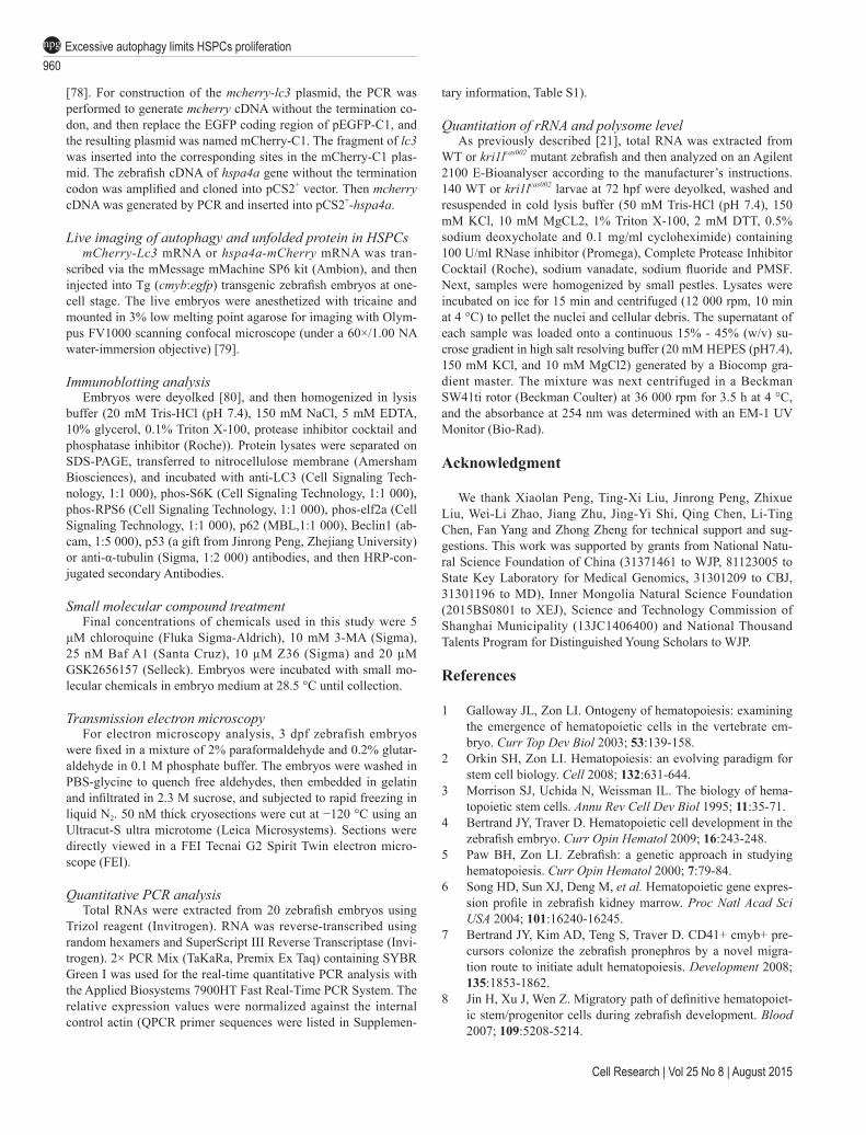
Figure 7 Working model for Kri1l dysfunction-induced hematopoietic defects and its treatment. (A) In normal HSPCs, ribo-some biogenesis is tightly regulated; autophagy occurs at an appropriate level. HSPCs expand massively in the CHT region and differentiate into multiple hematopoietic lineages, including erythrocytes, myeloid cells (neutrophils and monocytes) and lymphocytes. (B) In kri1lcas002 HSPCs, abnormal ribosome biogenesis leads to the accumulation of misfolded proteins, which triggers PERK activation and excessive autophagy. The excessive autophagy leads to inhibition of HPSC proliferation, deple-tion of HSPC pool, and a profound hematopoiesis failure. (C) Blocking of PERK signaling with specific inhibitor (GSK2656157), Bcl2 overexpression or treatment with autophagy inhibitors (3-MA, Baf A1) reduces autophagy level and restores HSPC pro-liferation defects and hematopoietic lineage differentiation in kri1lcas002 mutant embryo.
was mapped by out-crossing Tu background heterozygous fish into polymorphic WIK background wild-type strain. We scanned the genome for linked SSLP markers by BSA. SSLP markers [74, 75] used for BSA were selected from the Massachusetts General Hospital Zebrafish Server website (http://zebrafish.mgh.harvard.edu). Fine mapping using mainly SSLP markers was carried out to narrow down the genetic interval. The cDNAs of candidate genes in the interval were cloned and sequenced from pooled mutants, and candidate mutation was confirmed by sequencing genomic DNA of individual mutant embryo. All primers used for this study are provided in Supplementary Table 1.
WISH, TUNEL assay and immunostainingAntisense RNA probes were transcribed using linearized
constructs with T3 or T7 polymerase (Ambion) in the presence of digoxigenin (DIG, Roche)-labeled UTP using the DIG-RNA Labeling Kit (Roche). WISH was performed as described previ-ously using NBT/BCIP (Sigma) as substrates [76, 77]. TUNEL was performed with In Situ Cell Death Detection Kit and TMR
Red Kit (Roche) following manufacturer’s instruction. To detect both cmyb RNA and mitosis marker pH3 simultaneously, embryos were first hybridized with the DIG-labeled antisense cmyb RNA probe, incubated at 4 °C overnight with a peroxidase-conjugated anti-DIG antibody (1:500; Roche), and stained with Alexa Fluor cy3-conjugated tyramide as substrate (PerkinElmer). The embryos were then incubated with primary anti-pH3 (ser10) antibody (1:500; Santa Cruz), and finally incubated with Alexa Fluor 488-labeled goat anti-rabbit IgG antibody (1:500; Invitrogen).
Plasmid constructionThe zebrafish cDNA of kri1l gene was amplified from reverse
transcription products and cloned into pCS2+ vector. For construc-tion of the kri1l (1-60)-gfp reporter plasmid, the zebrafish cDNA (1-60 aa) of kri1l gene was amplified and cloned into pCS2+-egfp vector. For construction of Tg (lmo2:kri1l) transient transgenesis plasmid, the zebrafish lmo2 promoter was obtained by PCR ampli-fication from lmo2-Cre-PBSK-I-SceI plasmid, and then cloned into the Tol2 transposon backbone together with full-length kri1l gene
960Excessive autophagy limits HSPCs proliferationnpg
Cell Research | Vol 25 No 8 | August 2015
[78]. For construction of the mcherry-lc3 plasmid, the PCR was performed to generate mcherry cDNA without the termination co-don, and then replace the EGFP coding region of pEGFP-C1, and the resulting plasmid was named mCherry-C1. The fragment of lc3 was inserted into the corresponding sites in the mCherry-C1 plas-mid. The zebrafish cDNA of hspa4a gene without the termination codon was amplified and cloned into pCS2+ vector. Then mcherry cDNA was generated by PCR and inserted into pCS2+-hspa4a.
Live imaging of autophagy and unfolded protein in HSPCs mCherry-Lc3 mRNA or hspa4a-mCherry mRNA was tran-
scribed via the mMessage mMachine SP6 kit (Ambion), and then injected into Tg (cmyb:egfp) transgenic zebrafish embryos at one-cell stage. The live embryos were anesthetized with tricaine and mounted in 3% low melting point agarose for imaging with Olym-pus FV1000 scanning confocal microscope (under a 60×/1.00 NA water-immersion objective) [79].
Immunoblotting analysisEmbryos were deyolked [80], and then homogenized in lysis
buffer (20 mM Tris-HCl (pH 7.4), 150 mM NaCl, 5 mM EDTA, 10% glycerol, 0.1% Triton X-100, protease inhibitor cocktail and phosphatase inhibitor (Roche)). Protein lysates were separated on SDS-PAGE, transferred to nitrocellulose membrane (Amersham Biosciences), and incubated with anti-LC3 (Cell Signaling Tech-nology, 1:1 000), phos-S6K (Cell Signaling Technology, 1:1 000), phos-RPS6 (Cell Signaling Technology, 1:1 000), phos-elf2a (Cell Signaling Technology, 1:1 000), p62 (MBL,1:1 000), Beclin1 (ab-cam, 1:5 000), p53 (a gift from Jinrong Peng, Zhejiang University) or anti-α-tubulin (Sigma, 1:2 000) antibodies, and then HRP-con-jugated secondary Antibodies.
Small molecular compound treatmentFinal concentrations of chemicals used in this study were 5
µM chloroquine (Fluka Sigma-Aldrich), 10 mM 3-MA (Sigma), 25 nM Baf A1 (Santa Cruz), 10 µM Z36 (Sigma) and 20 µM GSK2656157 (Selleck). Embryos were incubated with small mo-lecular chemicals in embryo medium at 28.5 °C until collection.
Transmission electron microscopyFor electron microscopy analysis, 3 dpf zebrafish embryos
were fixed in a mixture of 2% paraformaldehyde and 0.2% glutar-aldehyde in 0.1 M phosphate buffer. The embryos were washed in PBS-glycine to quench free aldehydes, then embedded in gelatin and infiltrated in 2.3 M sucrose, and subjected to rapid freezing in liquid N2. 50 nM thick cryosections were cut at −120 °C using an Ultracut-S ultra microtome (Leica Microsystems). Sections were directly viewed in a FEI Tecnai G2 Spirit Twin electron micro-scope (FEI).
Quantitative PCR analysisTotal RNAs were extracted from 20 zebrafish embryos using
Trizol reagent (Invitrogen). RNA was reverse-transcribed using random hexamers and SuperScript III Reverse Transcriptase (Invi-trogen). 2× PCR Mix (TaKaRa, Premix Ex Taq) containing SYBR Green I was used for the real-time quantitative PCR analysis with the Applied Biosystems 7900HT Fast Real-Time PCR System. The relative expression values were normalized against the internal control actin (QPCR primer sequences were listed in Supplemen-
tary information, Table S1).
Quantitation of rRNA and polysome levelAs previously described [21], total RNA was extracted from
WT or kri1lcas002 mutant zebrafish and then analyzed on an Agilent 2100 E-Bioanalyser according to the manufacturer’s instructions. 140 WT or kri1lcas002 larvae at 72 hpf were deyolked, washed and resuspended in cold lysis buffer (50 mM Tris-HCl (pH 7.4), 150 mM KCl, 10 mM MgCL2, 1% Triton X-100, 2 mM DTT, 0.5% sodium deoxycholate and 0.1 mg/ml cycloheximide) containing 100 U/ml RNase inhibitor (Promega), Complete Protease Inhibitor Cocktail (Roche), sodium vanadate, sodium fluoride and PMSF. Next, samples were homogenized by small pestles. Lysates were incubated on ice for 15 min and centrifuged (12 000 rpm, 10 min at 4 °C) to pellet the nuclei and cellular debris. The supernatant of each sample was loaded onto a continuous 15% - 45% (w/v) su-crose gradient in high salt resolving buffer (20 mM HEPES (pH7.4), 150 mM KCl, and 10 mM MgCl2) generated by a Biocomp gra-dient master. The mixture was next centrifuged in a Beckman SW41ti rotor (Beckman Coulter) at 36 000 rpm for 3.5 h at 4 °C, and the absorbance at 254 nm was determined with an EM-1 UV Monitor (Bio-Rad).
Acknowledgment
We thank Xiaolan Peng, Ting-Xi Liu, Jinrong Peng, Zhixue Liu, Wei-Li Zhao, Jiang Zhu, Jing-Yi Shi, Qing Chen, Li-Ting Chen, Fan Yang and Zhong Zheng for technical support and sug-gestions. This work was supported by grants from National Natu-ral Science Foundation of China (31371461 to WJP, 81123005 to State Key Laboratory for Medical Genomics, 31301209 to CBJ, 31301196 to MD), Inner Mongolia Natural Science Foundation (2015BS0801 to XEJ), Science and Technology Commission of Shanghai Municipality (13JC1406400) and National Thousand Talents Program for Distinguished Young Scholars to WJP.
References
1 Galloway JL, Zon LI. Ontogeny of hematopoiesis: examining the emergence of hematopoietic cells in the vertebrate em-bryo. Curr Top Dev Biol 2003; 53:139-158.
2 Orkin SH, Zon LI. Hematopoiesis: an evolving paradigm for stem cell biology. Cell 2008; 132:631-644.
3 Morrison SJ, Uchida N, Weissman IL. The biology of hema-topoietic stem cells. Annu Rev Cell Dev Biol 1995; 11:35-71.
4 Bertrand JY, Traver D. Hematopoietic cell development in the zebrafish embryo. Curr Opin Hematol 2009; 16:243-248.
5 Paw BH, Zon LI. Zebrafish: a genetic approach in studying hematopoiesis. Curr Opin Hematol 2000; 7:79-84.
6 Song HD, Sun XJ, Deng M, et al. Hematopoietic gene expres-sion profile in zebrafish kidney marrow. Proc Natl Acad Sci USA 2004; 101:16240-16245.
7 Bertrand JY, Kim AD, Teng S, Traver D. CD41+ cmyb+ pre-cursors colonize the zebrafish pronephros by a novel migra-tion route to initiate adult hematopoiesis. Development 2008; 135:1853-1862.
8 Jin H, Xu J, Wen Z. Migratory path of definitive hematopoiet-ic stem/progenitor cells during zebrafish development. Blood 2007; 109:5208-5214.
Xiao-E Jia et al.961
npg
www.cell-research.com | Cell Research
9 Kissa K, Murayama E, Zapata A, et al. Live imaging of emerging hematopoietic stem cells and early thymus coloni-zation. Blood 2008; 111:1147-1156.
10 Murayama E, Kissa K, Zapata A, et al. Tracing hematopoi-etic precursor migration to successive hematopoietic organs during zebrafish development. Immunity 2006; 25:963-975.
11 Sondalle SB, Baserga SJ. Human diseases of the SSU proces-some. Biochim Biophys Acta 2014; 1842:758-764.
12 Liu JM, Ellis SR. Ribosomes and marrow failure: coincidental association or molecular paradigm? Blood 2006; 107:4583-4588.
13 Narla A, Ebert BL. Ribosomopathies: human disorders of ri-bosome dysfunction. Blood 2010; 115:3196-3205.
14 Aguissa-Toure AH, Da Costa L, Leblanc T, Tchernia G, Fribourg S, Gleizes PE. Diamond-Blackfan anemia reveals the dark side of ribosome biogenesis. Med Sci (Paris) 2009; 25:69-76.
15 Taylor AM, Humphries JM, White RM, Murphey RD, Burns CE, Zon LI. Hematopoietic defects in rps29 mutant zebrafish depend upon p53 activation. Exp Hematol 2012; 40:228-237.
16 Barlow JL, Drynan LF, Hewett DR, et al. A p53-dependent mechanism underlies macrocytic anemia in a mouse model of human 5q- syndrome. Nat Med 2010; 16:59-66.
17 McGowan KA, Li JZ, Park CY, et al. Ribosomal mutations cause p53-mediated dark skin and pleiotropic effects. Nat Genet 2008; 40:963-970.
18 Danilova N, Sakamoto KM, Lin S. Ribosomal protein S19 deficiency in zebrafish leads to developmental abnormalities and defective erythropoiesis through activation of p53 protein family. Blood 2008; 112:5228-5237.
19 Chakraborty A, Uechi T, Higa S, Torihara H, Kenmochi N. Loss of ribosomal protein L11 affects zebrafish embryonic de-velopment through a p53-dependent apoptotic response. PLoS One 2009; 4:e4152.
20 Provost E, Wehner KA, Zhong X, et al. Ribosomal biogenesis genes play an essential and p53-independent role in zebrafish pancreas development. Development 2012; 139:3232-3241.
21 Boglev Y, Badrock AP, Trotter AJ, et al. Autophagy induction is a Tor- and Tp53-independent cell survival response in a ze-brafish model of disrupted ribosome biogenesis. PLoS Genet 2013; 9:e1003279.
22 Torihara H, Uechi T, Chakraborty A, Shinya M, Sakai N, Kenmochi N. Erythropoiesis failure due to RPS19 deficiency is independent of an activated Tp53 response in a zebrafish model of Diamond-Blackfan anaemia. Br J Haematol 2011; 152:648-654.
23 Dutt S, Narla A, Lin K, et al. Haploinsufficiency for ribosom-al protein genes causes selective activation of p53 in human erythroid progenitor cells. Blood 2011; 117:2567-2576.
24 Zhang Y, Duc AC, Rao S, et al. Control of hematopoietic stem cell emergence by antagonistic functions of ribosomal protein paralogs. Dev Cell 2013; 24:411-425.
25 Hara T, Nakamura K, Matsui M, et al. Suppression of basal autophagy in neural cells causes neurodegenerative disease in mice. Nature 2006; 441:885-889.
26 Komatsu M, Waguri S, Chiba T, et al. Loss of autophagy in the central nervous system causes neurodegeneration in mice. Nature 2006; 441:880-884.
27 Espina V, Liotta LA. What is the malignant nature of human
ductal carcinoma in situ? Nat Rev Cancer 2011; 11:68-75.28 Degenhardt K, Mathew R, Beaudoin B, et al. Autophagy pro-
motes tumor cell survival and restricts necrosis, inflammation, and tumorigenesis. Cancer Cell 2006; 10:51-64.
29 He C, Klionsky DJ. Regulation mechanisms and signaling pathways of autophagy. Annu Rev Genet 2009; 43:67-93.
30 Funderburk SF, Wang QJ, Yue Z. The Beclin 1-VPS34 com-plex--at the crossroads of autophagy and beyond. Trends Cell Biol 2010; 20:355-362.
31 Guan JL, Simon AK, Prescott M, et al. Autophagy in stem cells. Autophagy 2013; 9:830-849.
32 Mortensen M, Soilleux EJ, Djordjevic G, et al. The autophagy protein Atg7 is essential for hematopoietic stem cell mainte-nance. J Exp Med 2011; 208:455-467.
33 Miller BC, Zhao Z, Stephenson LM, et al. The autophagy gene ATG5 plays an essential role in B lymphocyte develop-ment. Autophagy 2008; 4:309-314.
34 Pua HH, Dzhagalov I, Chuck M, Mizushima N, He YW. A critical role for the autophagy gene Atg5 in T cell survival and proliferation. J Exp Med 2007; 204:25-31.
35 Kundu M, Lindsten T, Yang CY, et al. Ulk1 plays a critical role in the autophagic clearance of mitochondria and ribo-somes during reticulocyte maturation. Blood 2008; 112:1493-1502.
36 Mortensen M, Ferguson DJ, Edelmann M, et al. Loss of au-tophagy in erythroid cells leads to defective removal of mito-chondria and severe anemia in vivo. Proc Natl Acad Sci USA 2010; 107:832-837.
37 Sandoval H, Thiagarajan P, Dasgupta SK, et al. Essential role for Nix in autophagic maturation of erythroid cells. Nature 2008; 454:232-235.
38 Heijnen HF, van Wijk R, Pereboom TC, et al. Ribosomal pro-tein mutations induce autophagy through S6 kinase inhibition of the insulin pathway. PLoS Genet 2014; 10:e1004371.
39 Bahary N, Davidson A, Ransom D, et al. The Zon laboratory guide to positional cloning in zebrafish. Methods Cell Biol 2004; 77:305-329.
40 Whitehead GG, Makino S, Lien CL, Keating MT. fgf20 is es-sential for initiating zebrafish fin regeneration. Science 2005; 310:1957-1960.
41 Sasaki T, Toh EA, Kikuchi Y. Yeast Krr1p physically and functionally interacts with a novel essential Kri1p, and both proteins are required for 40S ribosome biogenesis in the nu-cleolus. Mol Cell Biol 2000; 20:7971-7979.
42 Yuan H, Zhou J, Deng M, et al. Small ubiquitin-related mod-ifier paralogs are indispensable but functionally redundant during early development of zebrafish. Cell Res 2010; 20:185-196.
43 Chen J, Ng SM, Chang C, et al. p53 isoform delta113p53 is a p53 target gene that antagonizes p53 apoptotic activity via BclxL activation in zebrafish. Genes Dev 2009; 23:278-290.
44 Pattingre S, Tassa A, Qu X, et al. Bcl-2 antiapoptotic proteins inhibit Beclin 1-dependent autophagy. Cell 2005; 122:927-939.
45 Maiuri MC, Le Toumelin G, Criollo A, et al. Functional and physical interaction between Bcl-X(L) and a BH3-like do-main in Beclin-1. EMBO J 2007; 26:2527-2539.
46 Klionsky DJ, Abdalla FC, Abeliovich H, et al. Guidelines for the use and interpretation of assays for monitoring autophagy.
962Excessive autophagy limits HSPCs proliferationnpg
Cell Research | Vol 25 No 8 | August 2015
Autophagy 2012; 8:445-544.47 Klionsky DJ, Abeliovich H, Agostinis P, et al. Guidelines for
the use and interpretation of assays for monitoring autophagy in higher eukaryotes. Autophagy 2008; 4:151-175.
48 Berghmans S, Murphey RD, Wienholds E, et al. tp53 mutant zebrafish develop malignant peripheral nerve sheath tumors. Proc Natl Acad Sci USA 2005; 102:407-412.
49 Galavotti S, Bartesaghi S, Faccenda D, et al. The autopha-gy-associated factors DRAM1 and p62 regulate cell migration and invasion in glioblastoma stem cells. Oncogene 2013; 32:699-712.
50 He C, Klionsky DJ. Analyzing autophagy in zebrafish. Auto-phagy 2010; 6:642-644.
51 He C, Bartholomew CR, Zhou W, Klionsky DJ. Assaying autophagic activity in transgenic GFP-Lc3 and GFP-Gabarap zebrafish embryos. Autophagy 2009; 5:520-526.
52 Kimmelman AC. The dynamic nature of autophagy in cancer. Genes Dev 2011; 25:1999-2010.
53 Schonewolf CA, Mehta M, Schiff D, et al. Autophagy inhibi-tion by chloroquine sensitizes HT-29 colorectal cancer cells to concurrent chemoradiation. World J Gastrointest Oncol 2014; 6:74-82.
54 Wu YT, Tan HL, Shui G, et al. Dual role of 3-methyladenine in modulation of autophagy via different temporal patterns of inhibition on class I and III phosphoinositide 3-kinase. J Biol Chem 2010; 285:10850-10861.
55 Yamamoto A, Tagawa Y, Yoshimori T, Moriyama Y, Masaki R, Tashiro Y. Bafilomycin A1 prevents maturation of autophagic vacuoles by inhibiting fusion between autophagosomes and lysosomes in rat hepatoma cell line, H-4-II-E cells. Cell Struct Funct 1998; 23:33-42.
56 Hoyer-Hansen M, Jaattela M. Connecting endoplasmic retic-ulum stress to autophagy by unfolded protein response and calcium. Cell Death Differ 2007; 14:1576-1582.
57 Kouroku Y, Fujita E, Tanida I, et al. ER stress (PERK/eIF2al-pha phosphorylation) mediates the polyglutamine-induced LC3 conversion, an essential step for autophagy formation. Cell Death Differ 2007; 14:230-239.
58 Buchberger A, Bukau B, Sommer T. Protein quality control in the cytosol and the endoplasmic reticulum: brothers in arms. Mol Cell 2010; 40:238-252.
59 Crawford KC, Vega Flores M, Oehlers SH, Hall CJ, Crosier KE, Crosier PS. Zebrafish heat shock protein a4 genes in the intestinal epithelium are up-regulated during inflammation. Genesis 2011; 49:905-911.
60 Amm I, Sommer T, Wolf DH. Protein quality control and elimination of protein waste: the role of the ubiquitin-protea-some system. Biochim Biophys Acta 2014; 1843:182-196.
61 Wek RC, Cavener DR. Translational control and the unfolded protein response. Antioxid Redox Signal 2007; 9:2357-2371.
62 Lin J, Zheng Z, Li Y, et al. A novel Bcl-XL inhibitor Z36 that induces autophagic cell death in Hela cells. Autophagy 2009; 5:314-320.
63 Warr MR, Binnewies M, Flach J, et al. FOXO3A directs a protective autophagy program in haematopoietic stem cells. Nature 2013; 494:323-327.
64 Salemi S, Yousefi S, Constantinescu MA, Fey MF, Simon HU. Autophagy is required for self-renewal and differentia-
tion of adult human stem cells. Cell Res 2012; 22:432-435.65 Liu F, Guan JL. FIP200, an essential component of mamma-
lian autophagy is indispensible for fetal hematopoiesis. Auto-phagy 2011; 7:229-230.
66 Liu F, Lee JY, Wei H, et al. FIP200 is required for the cell-au-tonomous maintenance of fetal hematopoietic stem cells. Blood 2010; 116:4806-4814.
67 Shintani T, Klionsky DJ. Autophagy in health and disease: a double-edged sword. Science 2004; 306:990-995.
68 Signer RA, Magee JA, Salic A, Morrison SJ. Haematopoietic stem cells require a highly regulated protein synthesis rate. Nature 2014; 509:49-54.
69 Kim YE, Hipp MS, Bracher A, Hayer-Hartl M, Hartl FU. Molecular chaperone functions in protein folding and proteo-stasis. Annu Rev Biochem 2013; 82:323-355.
70 van Galen P, Kreso A, Mbong N, et al. The unfolded protein response governs integrity of the haematopoietic stem-cell pool during stress. Nature 2014; 510:268-272.
71 McAfee Q, Zhang Z, Samanta A, et al. Autophagy inhibitor Lys05 has single-agent antitumor activity and reproduces the phenotype of a genetic autophagy deficiency. Proc Natl Acad Sci USA 2012; 109:8253-8258.
72 Mullins MC, Nusslein-Volhard C. Mutational approaches to studying embryonic pattern formation in the zebrafish. Curr Opin Genet Dev 1993; 3:648-654.
73 Kimmel CB, Ballard WW, Kimmel SR, Ullmann B, Schilling TF. Stages of embryonic development of the zebrafish. Dev Dyn 1995; 203:253-310.
74 Knapik EW, Goodman A, Ekker M, et al. A microsatellite genetic linkage map for zebrafish (Danio rerio). Nat Genet 1998; 18:338-343.
75 Shimoda N, Knapik EW, Ziniti J, et al. Zebrafish genetic map with 2000 microsatellite markers. Genomics 1999; 58:219-232.
76 Thisse C, Thisse B. High-resolution in situ hybridization to whole-mount zebrafish embryos. Nat Protoc 2008; 3:59-69.
77 Zhang Y, Bai XT, Zhu KY, et al. In vivo interstitial migration of primitive macrophages mediated by JNK-matrix metallo-proteinase 13 signaling in response to acute injury. J Immunol 2008; 181:2155-2164.
78 Wang L, Zhang Y, Zhou T, et al. Functional characterization of Lmo2-Cre transgenic zebrafish. Dev Dyn 2008; 237:2139-2146.
79 Le Guyader D, Redd MJ, Colucci-Guyon E, et al. Origins and unconventional behavior of neutrophils in developing zebraf-ish. Blood 2008; 111:132-141.
80 Link V, Shevchenko A, Heisenberg CP. Proteomics of early zebrafish embryos. BMC Dev Biol 2006; 6:1.
(Supplementary information is linked to the online version of the paper on the Cell Research website.)
This work is licensed under the Creative Commons Attribution-NonCommercial-No Derivative Works
3.0 Unported License. To view a copy of this license, visit http://creativecommons.org/licenses/by-nc-nd/3.0