Embed Size (px)
Citation preview

HAL Id: inserm-00126175https://www.hal.inserm.fr/inserm-00126175v1
Submitted on 15 Jun 2007 (v1), last revised 13 Aug 2007 (v2)
HAL is a multi-disciplinary open accessarchive for the deposit and dissemination of sci-entific research documents, whether they are pub-lished or not. The documents may come fromteaching and research institutions in France orabroad, or from public or private research centers.
L’archive ouverte pluridisciplinaire HAL, estdestinée au dépôt et à la diffusion de documentsscientifiques de niveau recherche, publiés ou non,émanant des établissements d’enseignement et derecherche français ou étrangers, des laboratoirespublics ou privés.
Mutations in the gene encoding the synaptic scaffoldingprotein SHANK3 are associated with autism spectrum
disorders.Christelle Durand, Catalina Betancur, Tobias Boeckers, Juergen Bockmann,
Pauline Chaste, Fabien Fauchereau, Gudrun Nygren, Maria Rastam, I CarinaGillberg, Henrik Anckarsäter, et al.
To cite this version:Christelle Durand, Catalina Betancur, Tobias Boeckers, Juergen Bockmann, Pauline Chaste, etal.. Mutations in the gene encoding the synaptic scaffolding protein SHANK3 are associated withautism spectrum disorders.. Nature Genetics, Nature Publishing Group, 2007, 39 (1), pp.25-7.<10.1038/ng1933>. <inserm-00126175v1>

Durand et al. 1
Mutations of the gene encoding the synaptic scaffoldingprotein SHANK3 are associated with autism spectrum
disorders
Christelle M. Durand1, Catalina Betancur2, Tobias M Boeckers3, Juergen Bockmann3, Pauline Chaste1,
Fabien Fauchereau1,4, Gudrun Nygren5, Maria Rastam5, I. Carina Gillberg5, Henrik Anckarsäter5, Eili
Sponheim6, Hany Goubran-Botros1, Richard Delorme1, Nadia Chabane7, Marie-Christine Mouren-
Simeoni7, Philippe de Mas8, Eric Bieth8, Bernadette Rogé9, Delphine Héron10, Lydie Burglen11,
Christopher Gillberg5,12, Marion Leboyer2,13, Thomas Bourgeron1,4*
1 Human Genetics and Cognitive Functions, Institut Pasteur, Paris, France2 INSERM U513, Université Paris XII, Créteil, France3 Institute for Anatomy and Cell Biology, Ulm University, Ulm, Germany4 University Denis Diderot Paris 7, Paris, France5 Department of Child and Adolescent Psychiatry, Göteborg University, Göteborg, Sweden6 Centre for Child and Adolescent Psychiatry, University of Oslo, Oslo, Norway7 Service de Psychopathologie de l'Enfant et de l'Adolescent, Hôpital Robert Debré, Assistance
Publique-Hôpitaux de Paris, Paris, France8 Purpan Hospital, Department of Medical Genetics, Toulouse, France9 Centre d’Etudes et de Recherches en PsychoPathologie, Université de Toulouse le Mirail, Toulouse,
France10 Département de Génétique, Groupe Hospitalier Pitié-Salpêtrière, Paris, France11Service de Génétique, Hôpital Trousseau, Assistance Publique-Hôpitaux de Paris, Paris, France12 Saint George’s Hospital Medical School, London, United Kingdom13 Département de Psychiatrie, Groupe Hospitalier Henri Mondor et Albert Chenevier, Assistance
Publique-Hôpitaux de Paris, Créteil, France
* Correspondence should be addressed to Thomas Bourgeron, PhD, Human Genetics and Cognitive
Functions, Institut Pasteur, 25 rue du Docteur Roux, 75015 Paris, France. Tel: 33-1-40-61-32-16. Fax:
33-1-40-61-39-53. E-mail: [email protected]
HA
L author manuscript inserm
-00126175, version 1
HAL author manuscriptNat Genet 01/2007; 39(1): 25-7

Durand et al. 2
Abstract
SHANK3/ProSAP2 regulates the structural organization of dendritic spines and is a binding
partner of neuroligins, previously found to be mutated in autism and Asperger syndrome. Here,
we report that a mutation of a single copy of SHANK3 on chromosome 22q13 is sufficient to
develop language and/or social communication disorders. These mutations concern only a small
number of individuals, but shed light on one synaptic pathway, sensitive to gene dosage, involved
in autism spectrum disorders.
Autism spectrum disorder (ASD) affects about 6/1000 children and is characterized by impairments in
reciprocal social interaction and communication as well as restricted and stereotyped patterns of
interests and activities1. ASD ranges from severe (autistic disorder with moderate or severe cognitive
impairment) to a milder variant (Asperger syndrome [AS] with higher cognitive ability). Although the
causative genes remain largely unknown2, familial and twin studies indicate that ASD is one of the
most genetic neuropsychiatric disorders. Standard karyotype analyses reveal chromosomal
rearrangements in 3%-6% of cases, the most common being deletions/duplications on chromosomes
15q, 22q and 7q3. Among the most frequent rearrangements associated with cognitive deficits, the
22q13.3 microdeletion syndrome is characterized by neonatal hypotonia, global developmental delay,
normal to accelerated growth, absent to severely delayed speech, autistic behavior, and minor
dysmorphic features4. The loss of terminal 22q13.3 can be subtle and go undetected by routine
chromosome analysis; fluorescence in situ hybridization (FISH) is often required to confirm the
presence of this deletion.
Among the three genes (ACR, RABL2B, SHANK3) located in the minimal telomeric region5,
SHANK3 (also known as ProSAP2) is the strongest candidate for the neurobehavioral symptoms
observed in patients with 22q13 deletions. SHANK3 is a scaffolding protein found in excitatory
synapses directly opposite to the presynaptic active zone. Shank proteins are believed to function as
master organizers of the postsynaptic density (PSD), due to their ability to form multimeric complexes
with postsynaptic receptors, signaling molecules and cytoskeletal proteins present in dendritic
spines/PSDs6,7. SHANK3 can bind to the cell adhesion proteins neuroligins (NLGN)8, which we
previously found to be mutated in autism and Asperger syndrome9. SHANK3 was disrupted by a de
novo balanced translocation in a child with all the features of the 22q13.3 deletion syndrome10. In this
paper, we report evidence showing that abnormal gene dosage of SHANK3 is associated with severe
cognitive deficits, including language/speech disorder and ASD.
We investigated chromosome 22q13 and SHANK3 in patients with ASD by FISH analysis (n=97)
and/or direct sequencing (n=227). We also sequenced all SHANK3 exons in a minimum of 190
controls to ascertain the diversity of SHANK3 non-synonymous variations in the general population.
SHANK3 spans 57 kb and contains 24 exons. Seven exons are alternatively spliced, including exon 18,
which is mostly detected in the brain (Supplementary Fig. 1). During our screening, three families
with ASD showed unambiguous alteration of 22q13/SHANK3. In family ASD 1, the proband with
HA
L author manuscript inserm
-00126175, version 1

Durand et al. 3
autism, no language and moderate mental retardation, carried a de novo deletion of 22q13 (the clinical
description of all patients is provided in Supplementary note 1). The deletion breakpoint was located
in intron 8 of SHANK3 and removed 142 kb of the terminal 22q13 (Fig. 1a). This deletion had been
"repaired" by addition of telomeric repeats and was similar to the minimum deleted region described
previously5. The occurrence of recurrent deletions in this region may be due to the quadruplex forming
G-rich sequence (QGRS) surrounding the breakpoint (Supplementary Fig. 2), which provides a
structural substrate for inappropriate telomere formation.
In family ASD 2, two brothers with autism were heterozygous for an insertion of a guanine
nucleotide in exon 21 (Fig. 1b). Both brothers had severely impaired speech and severe mental
retardation. The mutation was absent in an unaffected brother and the unaffected parents. Using
fourteen informative single nucleotide polymorphisms (SNPs), we found that the mutation was located
on the same maternal haplotype in the two affected brothers and that the unaffected brother did not
have this haplotype (Supplementary Fig. 3). The mutation was absent in the DNA isolated from blood
leukocytes and mouth cells of the mother. These results strongly suggest a germinal mosaicism in the
mother. The guanine insertion creates a frame-shift at nucleotide 3680, modifying the C–terminal
sequence of the protein (Fig1b). This putative truncated protein lacks several crucial domains involved
in mGluR- and actin binding (Homer, AbP1, cortactin) and in the synaptic targeting and postsynaptic
assembly of SHANK3 multimers11,12. Consistent with the loss of these domains, the mutant protein
over-expressed in rat hippocampus neuronal cells showed no synaptic localization compared to the
wild-type sequence (Supplementary Fig. 4).
In family ASD 3, we identified a terminal 22q deletion in a girl with autism and severe language
delay and a 22qter partial trisomy in her brother with Asperger syndrome, who exhibited precocious
language development and fluent speech (Fig. 1c). We demonstrated that these unbalanced cytogenetic
abnormalities were inherited from a paternal translocation t(14;22)(p11.2;q13.33). The chromosome
14p11.2 breakpoint falls within the heterochromatic DNA sequence characteristic of acrocentric
chromosomes and contains no putative transcripts or genes. On chromosome 22q13.33, using
informative SNPs and quantitative PCR, we mapped the breakpoint between ALG12 and MLC1 (Fig.
1d). The deletion/duplication rearrangement observed in both siblings involved 25 genes, including
SHANK3 , located in the 800 kb terminal sequence of 22q13. No other SHANK3 deletions or
duplications were observed after screening by quantitative PCR 155 individuals (58 with autism, 38
with Asperger syndrome and 59 controls).
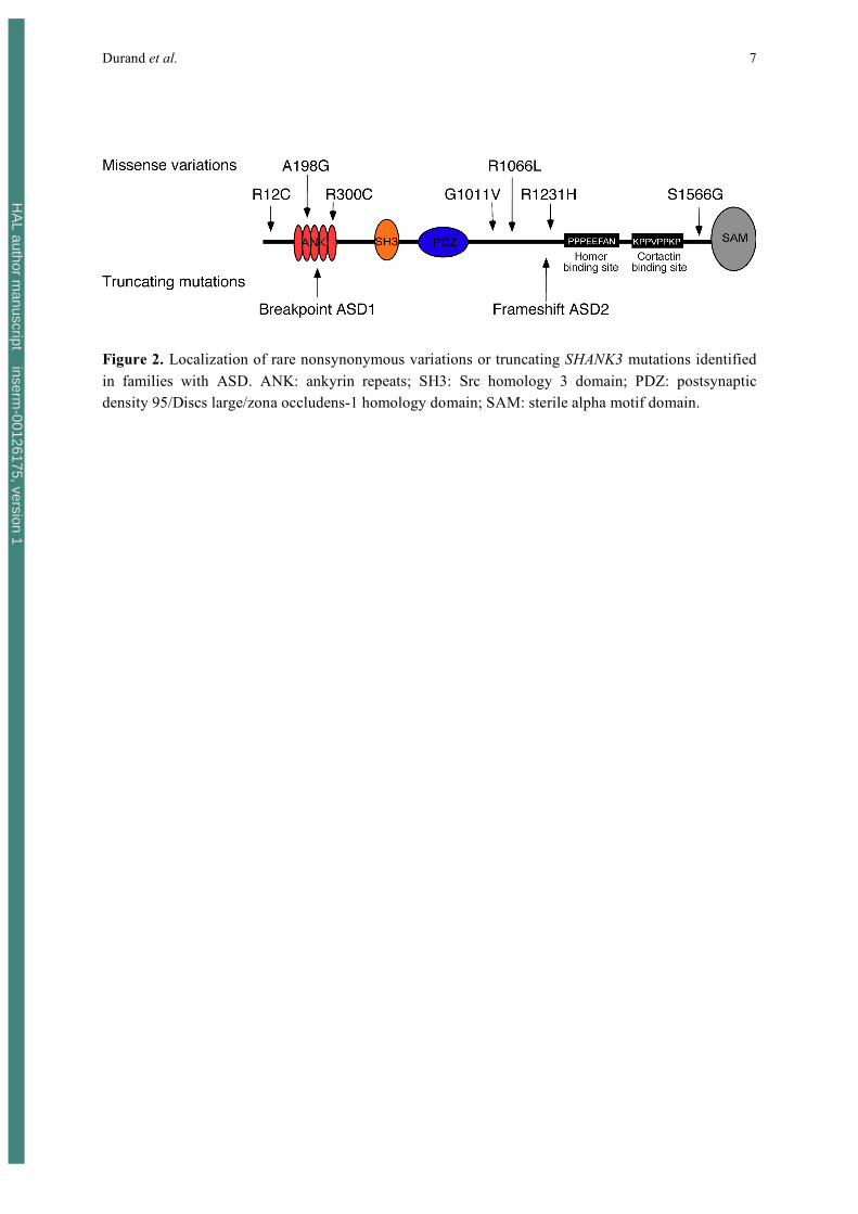
In the remaining individuals with ASD, we identified seven who had rare non-synonymous
variations, which were not observed in controls (n=270-333; Fig. 2; Supplementary Table 1).
However, all these variations were inherited from healthy parents, ruling out their direct involvement
as dominant mutations in the disorder. Interestingly, for two substitutions modifying highly conserved
amino acids (R12C and R300C; Supplementary Fig. 3), we observed that the over-expressed mutated
GFP Shank3 fusion proteins clustered but showed significantly less co-localization with the
presynaptic marker protein Bassoon, suggesting nonsynaptic clustering (Supplementary Fig. 4). These
HA
L author manuscript inserm
-00126175, version 1

Durand et al. 4
observations might reflect posttranslational modifications or abnormal folding of the protein. Thus,
although these genetic variations cannot be considered as causal mutations, they might nevertheless
modify the synaptic scaffolding and represent risk factors for ASD in interaction with other
susceptibility genes.
In this study, we show that a SHANK3 heterozygous mutation can cause ASD. Interestingly, in
the ASD 3 boy with Asperger syndrome, the presence of an additional copy of 22q13/SHANK3 did not
impair his language ability, but appears to have led to a severe impairment in social communication.
These results, together with previous reports13,14, highlight the importance of a fine gene dosage for the
development of speech-language and/or social communication in humans.
The mutations identified in these patients are believed to have affected the function/localization
of SHANK3 at PSD/dendritic spines. These results are consistent with the alterations of dendrites and
the spine/PSD compartment in individuals with learning disabilities15. In mice, Shank-3 promotes the
maturation and the enlargement of dendritic spine heads and is even able to induce spine formation in
aspiny neurons11. In ASD, an abnormality of synapse formation/maintenance was first suggested by
the identification of mutations in X-linked NLGN3 and NLGN49, and next confirmed by functional
studies of the causative mutations. Therefore, we hypothesize that the protein complex including
NLGN/SHANK participates in the assembly of specialized postsynaptic structures required for the
development of language and social communication.
References1. Folstein, S.E. & Rosen-Sheidley, B. Nat Rev Genet 2, 943-955 (2001).2. Persico, A.M. & Bourgeron, T. Trends Neurosci 29, 349-358 (2006).3. Vorstman, J.A. et al. Mol Psychiatry 11, 1, 18-28 (2006).4. Manning, M.A. et al. Pediatrics 114, 451-7 (2004).5. Bonaglia, M.C. et al. J Med Genet 43, 822-8 (2006).6. Naisbitt, S. et al. Neuron 23, 569-82 (1999).7. Boeckers, T.M., Bockmann, J., Kreutz, M.R. & Gundelfinger, E.D. J Neurochem 81, 903-10
(2002).8. Meyer, G., Varoqueaux, F., Neeb, A., Oschlies, M. & Brose, N. Neuropharmacology 47, 724-33
(2004).9. Jamain, S. et al. Nat. Genet. 34, 27-29 (2003).10. Bonaglia, M.C. et al. Am J Hum Genet 69, 261-8 (2001).11. Roussignol, G. et al. J Neurosci 25, 3560-70 (2005).12. Baron, M.K. et al. Science 311, 531-5 (2006).13. Lai, C.S., Fisher, S.E., Hurst, J.A., Vargha-Khadem, F. & Monaco, A.P. Nature 413, 519-23.
(2001).14. Somerville, M.J. et al. N Engl J Med 353, 1694-701 (2005).15. Carlisle, H.J. & Kennedy, M.B. Trends Neurosci 28, 182-7 (2005).
HA
L author manuscript inserm
-00126175, version 1

Durand et al. 5
Acknowledgements
We thank the patients and their families for participating in this study and all the collaborators of the
Paris Autism Research International Sibpair Study (PARIS): Sweden: Department of Child and
Adolescent Psychiatry, Göteborg University, Göteborg: Christopher Gillberg, Maria Råstam, Carina
Gillberg, Gudrun Nygren, Henrik Anckarsäter, Ola Ståhlberg; France: Department of Psychiatry,
Groupe Hospitalier Albert Chenevier et Henri Mondor, Créteil: Marion Leboyer; INSERM U513,
Université Paris XII, Créteil: Catalina Betancur; Service de Psychopathologie de l’Enfant et
l’Adolescent, Hôpital Robert Debré, Paris: Catherine Colineaux, Deborah Cohen, Nadia Chabane,
Marie-Christine Mouren-Siméoni; INSERM U679, Hôpital Pitié-Salpêtrière, Paris: Alexis Brice;
Norway: Centre for Child and Adolescent Psychiatry, University of Oslo, Oslo: Eili Sponheim;
Department of Pediatrics, Rikshospitalet, University of Oslo, Oslo: Ola H. Skjeldal; USA: Department
of Pediatrics, Georgetown University School of Medicine, Washington D.C.: Mary Coleman;
Children's National Medical Center, George Washington University School of Medicine, Washington,
D.C.: Philip L. Pearl; New York State Institute for Basic Research in Developmental Disabilities,
Staten Island, New York: Ira L. Cohen, John Tsiouris; Italy: Divisione di Neuropsichiatria Infantile,
Azienda Ospedaliera Senese, Siena: Michele Zappella; Austria: Department of General Psychiatry,
University Hospital, Vienna: Harald Aschauer ; Belgium: Centre de Génétique Humaine, Institut de
Pathologie et de Génétique, Gerpinnes, Loverval: Lionel Van Maldergem
We also thank the DNA and cell bank of INSERM U679 (IFR des Neurosciences, Hôpital Pitié-
Salpêtrière), the Centre d'Investigations Cliniques of the Hôpital Robert Debré, C. Bouchier and S.
Duthoy for the use of sequencing facilities at the Génopole Pasteur, and A. Hchikat, L. Margarit, and
G. Rouffet for technical assistance. This work was supported by the Pasteur Institute, INSERM,
Assistance Publique-Hôpitaux de Paris, Fondation France Telecom, Cure Autism Now, Fondation de
France, Fondation Biomédicale de la Mairie de Paris, Fondation pour la Recherche Médicale,
EUSynapse European Commission FP6, AUTISM MOLGEN European Commission FP6, Fondation
NRJ, the Swedish Science Council and the Deutsche Forschungsgemeinschaft DFG, SFB 497.
HA
L author manuscript inserm
-00126175, version 1

Durand et al. 6
Figure 1. Genetic analyses of three families with ASD and SHANK3 mutations. (a) In family ASD 1, the probandcarries a de novo terminal deletion of the paternal chromosome 22q13. The deletion breakpoint is located in intron 8 ofSHANK3. The breakpoint was sequenced after amplification of the proband DNA using primer 1 in SHANK3 andprimer 2 in the telomeric repeats. The heterogenous smear in the proband is likely due to the difference in telomerelength from chromosome to chromosome and/or priming at different locations by the telomeric primer. (b) In familyASD 2, the two probands carry the same de novo SHANK3 frame-shift mutation on the maternal chromosome 22q13.The mutation is absent from the mother blood and buccal cells, suggesting a germinal mosaicism. The guanine insertionis located in exon 21 of SHANK3, leading to a premature truncated protein. (c) In family ASD 3, the father carries abalanced translocation t(14,22)(p11.2;q13.33), proband A (Asperger syndrome) presents a partial 22qter trisomy andproband B (autism) has a 22qter deletion. (d) Using quantitative fluorescent PCR, we mapped the breakpoint betweenthe genes ALG12 and MLC1. The dosage quotient has a theoretical value of 0.5 for a deletion and 1.5 for a duplication.
HA
L author manuscript inserm
-00126175, version 1
Durand et al. 7
Figure 2. Localization of rare nonsynonymous variations or truncating SHANK3 mutations identified in families with ASD. ANK: ankyrin repeats; SH3: Src homology 3 domain; PDZ: postsynaptic density 95/Discs large/zona occludens-1 homology domain; SAM: sterile alpha motif domain.
HA
L author manuscript inserm
-00126175, version 1

Durand et al. 8
SUPPLEMENTARY MATERIAL
Materials and Methods
FamiliesFamilies (n=226) with at least one child with ASD were recruited by the PARIS (Paris AutismResearch International Sibpair) study at specialized clinical centers in seven countries (France,Sweden, Norway, Italy, Belgium, Austria, and the United States); 163 families had one child withASD and 63 families had at least two children with ASD. Diagnosis was based on clinical evaluationby experienced clinicians, DSM-IV criteria1, and the Autism Diagnostic Interview-Revised (ADI-R)2.In Sweden, the Diagnostic Interview for Social and Communication Disorders (DISCO-10)3 was usedinstead of the ADI-R in some cases. Like the ADI-R, the DISCO-10 utilizes algorithms based on ICD-10 and DSM-IV research criteria to diagnose autism. However, it can also be used to diagnose otherpervasive developmental disorders, such as pervasive developmental disorder not otherwise specified(PDD-NOS) and Asperger syndrome. The diagnosis of Asperger syndrome was confirmed using theAsperger Syndrome Diagnostic Interview (ASDI)4. The sample consisted of 177 males and 50females; there were 194 patients with autistic disorder and 29 with Asperger syndrome; 4 individualsnarrowly missed the criteria for autistic disorder and were considered to have atypical autism (PDD-NOS). Laboratory tests to rule out medical causes of autism included standard karyotyping, fragile-Xtesting, and metabolic screening; brain imaging and EEG were performed when possible. Patientsdiagnosed with medical disorders, such as fragile X syndrome or chromosomal anomalies, wereexcluded from the study. The local research ethics boards reviewed and approved the study. Informedconsent was obtained from all families. There were 206 Caucasian, 7 Black, 1 Asian and 13 familiesof mixed ethnicity.
The control sample (n=270) comprised 120 Caucasians from France and 150 Caucasians living inSweden. An additional control group of 63 individuals from China was screened for rare variantsbecause one proband carrying the R300C mutation had parents originating from this country. Forexons with no mutation in ASD (2, 3, 9, 11, 13-17), 190 controls (95 from France and 95 fromSweden) were sequenced. Using this minimum sample size (n = 380 chromosomes), we can detect apolymorphism with a frequency of 1% with 95% power5. The controls from Sweden were part of twopopulation-based cohorts living in Göteborg, originally recruited for a study of obesity and body fatdistribution6,7, who volunteered to provide a blood sample for genetic studies. Women (n=30) wereborn in 1956 and men (n=65) were born in 1944; subjects with one or two parents probably orcertainly being non-Caucasian were excluded.
The controls from France (40 females and 55 males) were healthy volunteers, between 19 and 65years old, interviewed with the Diagnostic Interview for Genetic Studies (DIGS) and the FamilyInterview for Genetic Studies (FIGS) to confirm the absence of both personal and family history ofpsychiatric disorders in first- and second-degree relatives.
Genomic structure and RT-PCR analyses of SHANK3The genomic structure of SHANK3 was deduced using the published data from Wilson et al. 8, theExpressed Sequence Tags (ESTs), and the rat cDNA (AJ133120) published in databases. SHANK3transcripts were detected in human brain regions from three independent controls (two females andone male) and in human tissues using the Clontech cDNA panel (Clontech Laboratories Inc). Total
HA
L author manuscript inserm
-00126175, version 1

Durand et al. 9
RNA was isolated from brain tissues by the acid-guanidium thiocyanate phenol chlorophorm methodand reverse transcribed by oligodT priming using SuperScript™ II Reverse Transcriptase (Invitrogen).Primer sequences for RT-PCR are indicated in Supplementary Table 2. Before sequencing thealternatively spliced exons, the breakpoint in family ASD 1 and the frame-shift mutation in familyASD 2, the PCR products were cloned with TOPO–TA cloning kit (Invitrogen). Prediction of theQuadruplex forming G-Rich Sequences (QGRS) at chromosome 22q13.3 was performed using theGQRS mapper software (http://bioinformatics.ramapo.edu/QGRS/index.php)9.
Mutation analysisDNA was extracted from blood leukocytes or B lymphoblastoid cell lines with the phenolchlorophorm method10. In the mother of family ASD 2, DNA from buccal cells was extracted usingthe BuccalAmp DNA extraction kit (Tebu-bio). For mutation analysis, the 24 coding exons ofSHANK3 were amplified from genomic DNA with specific primers (Supplementary Table 2).Amplification was performed on 20 ng of DNA template with HotStar Taq polymerase (Qiagen) forall exons except for exons 1, 11, 21, 22, 22b, and 22c, for which amplification was performed withTaq polymerase from Eurobio and 10% GC melt (Clontech GC rich kit). Two PCR protocols wereused: (i) Standard protocol: 95° for 15 min, followed by 35 cycles at 95°C for 30 sec, 55 to 65°C for20 sec, 72°C for 30 sec to 1 min, with a final cycle at 72°C for 10 min; and (ii) Touchdown protocol:95°C for 15 min followed by 20 cycles at 95°C for 30 sec, 70°-60° or 65-55°C for 30 sec, and 72°Cfor 30 sec, followed by 20 cycles at 95°C for 30 sec, 60° or 55°C for 10 sec, and 72°C for 30 sec, witha final cycle at 72°C for 10 min. Sequence analysis was performed by direct sequencing of the PCRproducts, using a 373A automated DNA sequencer (Applied Biosystems).
Detection of 22q13/SHANK3 deletions and duplicationsFor quantitative analysis, the forward primer was labeled with fluorescent 6-carboxyfluorescein (6-FAM). PCR amplification (25 cycles) was as described above for standard protocol. Fluorescence-labeled PCR products were run on a 373A automated DNA sequencer (Applied Biosystems) withGENEFLO 625 DNA Ladder, ROX l (EurX). Following data collection, samples were analyzed withGenescan 3.7 software program. Two independent SHANK3 PCRs (exon 9 and exon 17) werecompared to two autosomal control genes NLGN1 and ANKRD15, located on chromosome 3q26.31and 9p24.3, respectively. The peak ratio of each PCR was used to calculate the dosage quotient (DQ)value. DQ= (PCR SHANK3/PCR in control region) in tested individual / (PCR SHANK3/PCR incontrol region) in control individual. Thus, DQ gives a theoretical value of 0.5 for a deletion and 1.5for a duplication. A total of 155 individuals (58 with autism, 38 with Asperger, and 59 controls) werescreened for deletion/duplication of SHANK3. All primers are indicated in supplementary table 2.
In vitro mutagenesis and transfection studies in hippocampal neuronsFull-length rat Shank3 cDNA (sequence AJ133120) was cloned into a pEGFP-C2 vector (Clontech).Mutagenesis was made using the QuickChange II XL site directed mutagenesis kit (Invitrogen) on 100ng of wild type plasmid. Each clone was purified with Endofree Plasmid Maxi kit (Qiagen) andentirely sequenced to rule out additional mutations in the Shank3 cDNA. Moreover, the GFPconstructs were transfected into Cos cells and analyzed by Western Blot using a GFP antibody. Thepreparation of rat hippocampal cultures and the Shank3 localization experiments were performed
HA
L author manuscript inserm
-00126175, version 1

Durand et al. 10
essentially as described previously11,12. Neurons were transfected after 14 days in culture. For co-localization of transfected Shank3 full length construct or the mutated constructs with the pre- andpostsynaptic marker proteins SAP-90/PSD95, synaptophysin or Bassoon13, cells were fixed on day 17in 4% paraformaldehyde for 20 min at room temperature (20°C). Secondary antibodies conjugated toCyanine 3 (Cy3) fluorophore were used (anti mouse Cy3 , anti-rabbit Cy3, Chemicon, Temecula, CA,USA), and the cells were visualized by fluorescence microscopy. All animal experiments wereperformed in compliance with the guidelines for the welfare of experimental animals issued by theFederal Government of Germany, the National Institutes of Health and The Max Planck Society.
Quantitative and qualitative measurement of GFP expressionQuantitative and qualitative measurement of GFP expression was performed essentially as describedby Craven and Bredt14. In brief, transfected neurons were chosen at random from three independenttransfections of each construct (4-5 cells per construct). The perimeter of the dendrites was traced(excluding spine heads/clusters), and the average pixel intensity was calculated. Similarly, the spineheads/clusters were traced, and the average pixel intensity was obtained. The ratio of the average pixelintensity in dendrites vs. spine heads/clusters was defined as the synaptic clustering ratio (SCR): aratio of zero indicates complete synaptic clustering whereas a ratio of ≥ 0.7 indicates diffuse dendriticfluorescence with no clusters. Consecutively, we determined the synaptic localization of the clusteringconstructs by immunostaining with an antibody directed against the presynaptic marker proteinBassoon13. Between 250 and 300 synapses per construct were analyzed and the percentage of co-localization between Bassoon and the different GFP-constructs was determined (P/B in percentage).
References1. American Psychiatric Association. Diagnostic and Statistical Manual of Mental Disorders, 4th
Ed., (American Psychiatric Press, Washington D.C., 1994).2. Lord, C., Rutter, M. & Le Couteur, A. J Autism Dev Disord 24, 659-685 (1994).3. Wing, L., Leekam, S.R., Libby, S.J., Gould, J. & Larcombe, M. J. Child Psychol. Psychiatry 43,
307-25 (2002).4. Gillberg, C., Rastam, M. & Wentz, E. Autism 5, 57-66 (2001).5. Collins, J.S. & Schwartz, C.E. Am J Hum Genet 71, 1251-2 (2002).6. Rosmond, R., Lapidus, L., Marin, P. & Bjorntorp, P. Obes Res 4, 245-52 (1996).7. Rosmond, R. & Bjorntorp, P. Obes Res 6, 338-45 (1998).8. Wilson, H.L. et al. J Med Genet 40, 575-84 (2003).9. Kikin, O., D'Antonio, L. & Bagga, P.S. Nucleic Acids Res 34, W676-82 (2006).10. Sambrook, J. & Russell, D.W. Molecular Cloning, a Laboratory Manual, (Cold Spring Harbor
Laboratory Press, Cold Spring Harbor, New York, 2001).11. Boeckers, T.M. et al. J Neurochem 92, 519-24 (2005).12. Dresbach, T. et al. Mol Cell Neurosci 23, 279-91 (2003).13. tom Dieck, S. et al. J Cell Biol 142, 499-509 (1998).14. Craven, S.E. & Bredt, D.S. J Biol Chem 275, 20045-51 (2000).
HA
L author manuscript inserm
-00126175, version 1

Durand et al. 11
Supplementary note 1. Clinical cases
In family ASD 1, the proband is the only child of healthy unrelated parents. Family history is negativefor mental retardation and ASD. He was born at 41 weeks after a pregnancy marked by bleedings.Birth growth parameters were large (weight 4850 g, length 54 cm, head circumference [HC] 36 cm).He exhibited neonatal hypotonia, poor suction and cried a lot as a baby. Early motor milestones werenormal. He said a few words such as mama and dada but stopped using them around 30-36 months,when his parents noted a regression, both for language and play behavior. Recurrent otitis media wasobserved during early infancy and he had sleep difficulties. There was no history of seizures.Psychiatric evaluation at the age of 9 years showed total absence of language and moderate mentalretardation. Non verbal communication was impaired, as well as social interaction, and he had motorstereotypies. The clinical diagnosis of autism was confirmed with the ADI-R. Physical examination atthe age of 12 years showed no minor dysmorphic features except large ears and elbow extensionlimitation. Weight was 44 kg (+1.5 SD), height was 143 cm (–0.5 SD), and HC was 54.5 cm(average). Neurological examination was normal. A standard karyotype and fragile X testing werenormal. Routine screening of the 22q13 region by FISH revealed a de novo terminal deletion. An MRIand EEG were performed and were normal.
In family ASD 2, the probands are the first and the third children of three. The unaffected brothermeets DSM-IV criteria for ADHD but has no autism spectrum symptoms and no language delay. Themother meets DSM-IV criteria for social phobia. Family history is otherwise negative for mentalretardation, language disorders, ASD and other psychiatric disorders. Both children were born fromfull term and uneventful pregnancies. Birth parameters were average. Neonatal history was limited toneonatal jaundice and no hypotonia was recorded. The eldest boy had a normal development duringthe first year but was very agitated. He started walking at 16 months. He spoke his first words at 5years and first sentences at 6 years, but never acquired functional language. At the age of 20 years,after he was transferred to a new institution, a regression with severe outbursts was noted, he lost skillsincluding cleanliness, and he developed anorexia and marked weight loss. At that time he had aseizure-induced aspiration, he was hospitalized and died a few days later. The second affected brotherhad normal measures at birth (weight 3420 g, length 50 cm, HC 35.5 cm) but he showed progressivemacrocephaly until 9 months of age (+2 SD), followed by slowed growth (HC + 1 SD). He had limitedeye contact since birth. He started walking at 18 months. He never acquired words, except mama anddada. At the age of 16 years he had an episode of aspiration with loss of consciousness, whichnecessitated hospitalization for a few days, and a second episode at the age of 20. Since then, he is feda special diet with no liquids and soft food. When he was 16, after being transferred to the sameinstitution where his brother had been, a regression was also noted, with loss of autonomy andcleanliness, and marked weight loss. He developed epilepsy at the age of 17 and was started onclonazepam. Physical examination at the age of 20 years showed growth parameters in the average(height 175 cm, HC 55.5 cm), strabismus and no dysmorphic features. Both brothers had severelanguage impairment, severe mental retardation and marked deficits in social interaction. The clinicaldiagnosis of autism was confirmed for both of them with the ADI-R.
In family ASD 3, the probands are the first and the third born of three children. The unaffectedbrother has no history of psychiatric disorder. The mother fulfills DSM-IV criteria for agoraphobia;the father used to have symptoms of ADHD as a child but doesn’t fulfill the criteria as an adult. There
HA
L author manuscript inserm
-00126175, version 1
Durand et al. 12
is no family history of mental retardation or ASD, but the paternal grandfather has numerous ritualsand routines and is socially isolated. The eldest boy was born full term (41 weeks of gestation) after anuneventful pregnancy, with large birth parameters (weight: 4040 g, length: 53 cm, HC: 38 cm). He hadnormal early motor milestones, said his first words at 12 months, first sentences at 18 months andalready knew the letters at that time. He learned to read at 5 years, before attending primary school. Hehas impairment in social interactions, is often awkward in social situations and is quite isolated atschool. He doesn’t like to be observed by teachers and has marked difficulties to speak in class. Healso has difficulties to identify social cues and to understand others people’s reactions. His interestsare focused on mechanical sports and he has great knowledge about this. He has excellent memory forcar plates and is very rapid for mental calculation. He also has routines, especially when taking ashower. He has impairments in the interpretation of implied meanings. He has a small repertoire offacial expressions and limited use of gestures. Direct eye gaze is rare. His voice is often too loud,lacking modulation. He is quite clumsy and awkward. He has mood swings, hypersensitivity to noise,and phobia of chimney fire. Clinical evaluation at the age of 14.5 years confirmed the diagnosis ofAsperger syndrome and showed a normal IQ. Physical examination at this age showed increasedheight (184 cm) and marfanoid habitus, without significant dysmorphic features. The youngest sisterwas born at 38 weeks of gestation, after an uneventful pregnancy. Birth parameters were low (weight:2050 g, height: 47.5 cm, HC: 33 cm). She had discrete neonatal hypotonia but had normal early motormilestones. Language development was severely delayed with few words around 4 years anddevelopment of very short sentences before regression. Around 5 years old, she presented echolalia.She had motor stereotypies and exhibited some routines and need for sameness. She is now 12 yearsold and uses few words, with pronunciation errors. She communicates with pictograms. She went toschool until 7 years old and now attends a day hospital. An IQ evaluation at 12.9 years showed aperformance IQ of 44 (developmental age 5.8 years), and a verbal IQ of 15 (developmental age 24months). Her non verbal communication is impaired, as well as her social interaction. At present sheinitiates activities by herself. She especially enjoys puzzles, she is very fast at them and can spend thewhole day on this activity. She also plays with construction games and has a tendency to align thepieces. She still shows insistence on sameness. The diagnosis of autism was confirmed with the ADI-R. She has some self-aggressive behaviors, hitting her head. She has no hypersensitivity to noise andher reaction to pain is normal. She presents nocturnal enuresis. Fine and global motor skills areimpaired. Physical examination at 10 years showed average growth parameters, without dysmorphicfeatures. A brain MRI at 5 years of age was normal. The terminal 22q deletion in the girl wasdiagnosed by FISH when she was 8 years old. Study of other family members revealed a translocationt(14;22)(p11.2;q13.33) in the father and a 22qter partial trisomy in her brother.
The proband of family ASD 4 carries the R12C SHANK3 mutation. He is the third child ofhealthy unrelated parents. His two brothers are healthy. Medical and psychiatric history of thematernal family includes Parkinson disease in both maternal grandparents, depression in thegrandmother and thyroid disease in the grandmother, mother and several aunts. The mother had amajor episode of depression during pregnancy and after the birth of the proband. In the father’s family,two brothers of the grandmother committed suicide and two paternal uncles have a history of alcoholabuse. There is no history of mental retardation, language disorder or ASD. The proband was bornafter a full term and uneventful pregnancy. Delivery was unremarkable and birth parameters were inthe average (weight 3750 g, length 51 cm, HC 35 cm). He didn't establish eye contact and early
HA
L author manuscript inserm
-00126175, version 1

Durand et al. 13
milestones were delayed: he sat at 18 months and started walking at 4 years. EEG and brain imagingby tomodensitometry (TDM) were performed during his first year and were both normal. Vision waschecked and was not impaired. Physical examination at the age of 23 years showed a narrow face,large ears, long philtrum, saddle nose, flattened midface, thin superior lip, and prominent supraorbitalridge. Height was 160 cm (-2 SD), weight 60 kg, and HC 55.3 cm. Neurological examination wasnormal and there was no history of seizures. There was a high pain threshold and low noise threshold.Psychiatric examination showed total absence of language and severe mental retardation. Non verbalcommunication was impaired, as well as social interaction, and he had motor stereotypies. There wasno sleep disorder or hyperactivity. The clinical diagnosis of autism was confirmed with the ADI-R.
The proband of family ASD 5 carries the R300C SHANK3 mutation, which he inherited from hismother. He is the first of two children from non-consanguineous healthy parents of Chinese origin. Hisfamily history is non-contributory. The patient was born at term, after an uncomplicated pregnancy.The first symptoms were noticed at 2 years, when he lost some words (he said mama, dada, bye-bye,and could name certain things he wanted) and exhibited unusual eye contact. From this time, heshowed no interest in other children and preferred to play on his own. He had sleeping problemsduring childhood. He showed repetitive behaviors and circumscribed interests, opening and closingdoors, lining up objects, finding similar objects, leafing through certain books, or letting sand siftthrough his fingers. When evaluated at 42 months, he had no expressive verbal language, but useddifferent sounds and song melodies. He had some receptive language; he could point out some familiarobjects and was able understand some familiar sentences. He fulfilled the criteria for autistic disorderaccording to the ADI-R and had a Childhood Autism Rating Scale (CARS) score of 37.5, indicatingsevere autism. His developmental age at that time, evaluated with the Psycho-Educational Profile-Revised (PEP-R) was 18-20 months. No somatic pathology was detected at that time. A standardkaryotype, fragile X testing and metabolic screening were normal. A brain CT scan performed at thistime was also normal. Three EEGs, performed at 3, 7 and 10 years, did not show significantpathology. He started to speak at 5-6 years, but his language problems persisted. At the age of 10years, his language was stereotyped and repetitive; his adaptive language function was below mentalage of 3 years according to the Vineland Adaptive Behavior Scales. A Wechsler Intelligence Scale forChildren (WISC) performed at 10 years showed a total IQ of 67, a verbal IQ of 63, and a performanceIQ of 81. On examination at 11 years 4 months, his weight was 38 kg, his height 144.5 cm, and HC 55cm. Neurological exam showed generalized hypotonia and brisk reflexes throughout all extremities.He had long arms, with narrow hands and long fingers. He had a supernumerary upper incisor(mesiodens) and he still had his milk teeth in his upper jaw except for the two front teeth. No obviousdysmorphic features were observed. His parents noted that the patient often took off his clothes andcomplained about being warm (patients with 22 deletion syndrome have a tendency to becomeoverheated due to reduced perspiration1).
Reference1. Phelan, M.C. et al. Am J Med Genet 101, 91-9 (2001).
HA
L author manuscript inserm
-00126175, version 1

Durand et al. 14
Supplementary Table 1. SHANK3 variations identified in families with ASD and controls.Variations Sex of
probandTransmission and ethnicity Additional family information Occurrence
Exon Changea Parents Siblings Ethnicity ASD(n=227)
Controlsb
(n=190-333)ASD
E1 R12C Male Mother Present in one unaffected brotherand absent in one unaffectedbrother
European The mother has social phobia traits; thefather and the two brothers are healthy
1 0
E5 A198G Female Mother Absent in the affected brother European No history of psychiatric disorders ineither parent
1 0
E8 R300C Male Mother Absent in the unaffected sister Asian Language delay in the father; the mother ishealthy
1 0c
E21a G1011V Female Mother (3 brothers, DNA not available) European No history of psychiatric disorders in anyfamily member
2 0
E21a G1011V Male Father Present in the affectedmonozygotic twin
European No history of psychiatric disorders ineither parent
E21a R1066L Male Mother Present in the unaffected sister NorthAfrican
The father has a depressive syndrome; themother had depression during pregnancy
1 0
E21a ins(G)3680at aminoacid 1227
Male De novo(germinalmosaicism inthe mother)
Present in the two affectedbrothers and absent in theunaffected brother
European No history of psychiatric disorders ineither parent
1 0
E21a R1231H Female Mother Absent in the affected brother European No history of psychiatric disorders ineither parent
1 0
E22b S1566G Male Father (2 unaffected siblings, DNA notavailable)
European No history of psychiatric disorders in anyfamily member
1 0
ASD andControls
E21a A963G Female Mother (1 healthy brother, DNA notavailable)
European No history of psychiatric disorders in anyfamily member
2 1
E21a A963G Male Father (1 brother, DNA not available) European The father has broad autism phenotype(very shy, some autistic traits); theyounger brother has mental retardationand autistic traits (not autism)
E22c P1654T Male Father Absent in the affected brother European The father talked late and has stereotypedbody movements; the mother also was lateto talk.
2 1
E22c P1654T Male Nd (1 sister, DNA not available) European No history of psychiatric disorders ineither parent or the sister
ControlsE6 A224T Female — — European 0 1E21a V1333G Male — — European 0 2
a SHANK3 cDNA sequence: NM_033517.1. b Exon 8 was sequenced in 333 controls; exons 1, 4-7, 10, 12, 18-20, 21a, and 22abc were sequenced in 270 controls; andexons 2, 3, 9, 11, and 13-17 were sequenced in 190 controls. Nd = not determined.
HA
L author manuscript inserm
-00126175, version 1

Durand et al. 15
Supplementary Table 2. Primers used in this study
Primers for mutation screening
Forward primer (5’-3’) Reverse Primer (5’-3’) Size(bp) Condition
SHANK3E1F GCGCTCCGTTCCCCGGCGCGA SHANK3E1R CCTCCGCGAACCGCGGCCGAA 325 70°C + GC melt 10%
SHANK3E2F GACCTGAGCTCACGAGCCCGCT SHANK3E2R CTGCCGTGCCCTTCACTGGTC 322 55°C
SHANK3E3F TCCACTGTGGTAGTATGACTG SHANK3E3R TGGAACACATCACTGTACCAC 435 57°C
SHANK3E4F GAGGAAGGCGGGTGATGTTCA SHANK3E7R AGTATATCCACACTCGGTGCA 1047 TD 66-56°C
SHANK3E8F GTGTGCATTCCTGTGTGCGCA SHANK3E8R GGCTTCTGCACCCCTGCTGGT 485 TD 66-56°C
SHANK3E9F ATCCCAGTTACAGACAAGAGT SHANK3E9R CAATGTTCACTCAACACAGGC 450 55°C
SHANK3E10F GCCTGGGCAAACTGGACAAGT SHANKE10R TCCCCAACCAGGAAGCCCTAG 500 55°C
SHANK3E11F GGCATCGCGTCCGTCACCTACGT SHANK3E11R AAGGTTCCTGCCGTGCGGGT 415 55°C + GC melt 10%
SHANK3E12F AGCTGGGAGAAAGTGGGAAGG SHANK3E12R GTCACACACGTCCTATGTGTC 500 TD 65-55°C
SHANK3E13F ACTGGTGACCAGCATGGGTGA SHANK3E14R GGCAGAAGCAAGAAGCTGAAG 560 TD 65-55°C
SHANK3E15F CGCAGTCATCTCTTTCCTGTG SHANK3E16R GGCAGAAGCAAGAAGCTGAAG 589 55°C
SHANK3E17F ACCTGAACAAGATCCTGGCAC SHANK3E17R CACCCATTCACCTCTGACCTG 390 55°C
SHANK3E18F GCATGTACCAACTGACTCCAG SHANK3E18R CCATCACAGTCTCAGAGGGTC 500 55°C
SHANK3E19F CTCTGTCAGCATCACGGGTG SHANK3E20R CTTCAAACCCAAGTCCACCCT 1010 61°C
SHANK3E21AF1 AAGGCTGGCCTCTGTGGGAGG SHANK3E21AR1 ACGGACAGGAACACAGTGGAG 825 TD 70-60°C + DMSO 5%
SHANK3E21AF2 CAAGAGCCCCCTGGTGAAGCA SHANK3E21AR2 GCTCTCGGGCAGCCAGGGCAA 300 TD 70-60°C + DMSO 5%
SHANK3E21AF3 TGCCCTGAAGCCGTTGGTCAG SHANK3E21AR3 ACCTTCTCTGCCTCCCTGCGA 300 58°C
SHANK3E21AF4 CCTGTTTGTGGATGTACAGGCC SHANK3E21AR4 ACCGTGGAGATGGTGCTTGTG 716 55°C + GC melt
SHANK3E21AF5 CTCAGGGAAGCCCAGCAGTGA SHANK3E21AR5 AGAACAGACAAGAGGAATGAC 668 TD 70-60°C
SHANK3E21BF GAGGAGCCCTTCGGGCCCGTG SHANK3E21BR GGCAGAGAAGAGCGGAGGGAG 641 TD 70-60°C
SHANK3E21CF GTCTCGAAGGGAAACATGAAC SHANKE21CR CAGTGTCCATGTCTGACTTCC 431 55°C
SHANK3E22F1 CCGTAGGATCCCACCCTTTA SHANK3E22R1 GCCTAGGTGGATGCTCTCCAG 500 60°C
SHANK3E22F2 CTTCGTGGTGCGCAGCGTGAG SHANK3E22R2 ACAGCAAACAGGACGATTCA 485 TD 65-55°C + DMSO 5%
Primers for expression
Forward primer (5’-3’) Reverse Primer (5’-3’) Size(bp) Condition
SHANKEX18F CTGCGCTCCAAGTCCATGACA SHANK3EX18R GGCCCTGGCGTTCAAACAATG219,198 55°C
SHANK3EX21F GCCTGAAGACGACAAACCAA SHANK3EX21R GAGCTGCAGCGGCTTCTGCTG571,403,321
TD 70-60°C + DMSO 5%
Primers for in vitro mutagenesisForward primer (5’-3’) Reverse Primer (5’-3’)SHANK3R12CF GGGCCAGCGCCGTGGTCGTGTGCGTCGGCATCCCGGAC SHANK3R12CR TCCGGGATGCCGACGCACACGACCACGGCGCTGGCCC
SHANK3R300CF ACCAGGAGAGCTGTGCCTGCGTCCTGCTTTTCCGTGG SHANK3R300CR CCACGGAAAAGCAGGACGCAGGCACAGCTCTCCTGGT
SHANK3INSF GAGCCCAACAGGCTGGGGGGCTGAAGAGGAGCGCC SHANK3INSR GGCGCTCCTCTTCAGCCCCCCAGCCTGTTGGGCTC
Primers for telomere-SHANK3 PCRForward primer (5’-3’) Reverse Primer (5’-3’)BP22QF GTGACTTGACTTCTCTGAACCTTGG BP22QR TATGGATCCCTAACCCTAACCCTAACCC
Primers for quantitative PCRForward primer (5’-3’) Reverse Primer (5’-3’)SHANK3E9F-FAM mATCCCAGTTACAGACAAGAGT SHANK3E9R CAATGTTCACTCAACACAGGC
SHANK3E17F-FAM mACCTGAACAAGATCCTGGCAC SHANK3E17R CACCCATTCACCTCTGACCTG
NLGN1F-FAM mCCTTGATTAATACAGGCTTCA NLGN1R ATACAGAGCATCACATACTAC
ANKRD15F-FAM mGAAGAACTAACGACCACTTG ANKRD15R TCTCTGAGATGAGTCACAACC
HA
L author manuscript inserm
-00126175, version 1

Durand et al. 16
Supplementary Figure 1. Genomic structure and mRNA expression of the human SHANK3 gene. (a) Genomic structure of the SHANK3 gene, which is located on chromosome 22q13.3, spans 57 kb and contains 24 exons. The mouse gene on chromosome 15qE3, the rat gene on chromosome 7q34, and the human gene share the same genomic structure. (b) Specific RT-PCRs were performed on total RNA from different brain regions and cDNA libraries (Clontech) to detect SHANK3 mRNA and the alternative splice of exon 18, exon 22a, b and c. The age of the male and the two females studied were 74, 55, and 36 years, with a post-mortem delay of 10, 24 and 2 h, respectively. FC, frontal cortex; TC, temporal cortex; OC, occipital cortex; H, hippocampus; T, thalamus; C, cerebellum. Normal control human brains were obtained at autopsy under guidelines approved by the ethics committee. BLCL: B lymphoblastoid cell lines.
HA
L author manuscript inserm
-00126175, version 1
Durand et al. 17
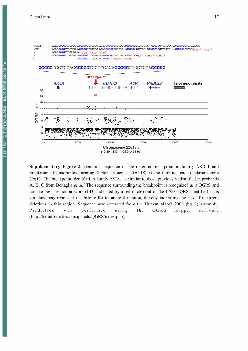
Supplementary Figure 2. Genomic sequence of the deletion breakpoint in family ASD 1 andprediction of quadruplex forming G-rich sequences (QGRS) at the terminal end of chromosome22q13. The breakpoint identified in family ASD 1 is similar to those previously identified in probandsA, B, C from Bonaglia et al.5 The sequence surrounding the breakpoint is recognized as a QGRS andhas the best prediction score (143, indicated by a red circle) out of the 1700 GQRS identified. Thisstructure may represent a substrate for telomere formation, thereby increasing the risk of recurrentdeletions in this region. Sequence was extracted from the Human March 2006 (hg18) assembly.P r e d i c t i o n w a s p e r f o r m e d u s i n g t h e Q G R S m a p p e r s o f t w a r e(http://bioinformatics.ramapo.edu/QGRS/index.php).
HA
L author manuscript inserm
-00126175, version 1

Durand et al. 18
Supplementary Figure 3. Pedigree structure, haplotype analyses and conservation of the SHANK3mutations and variants identified in individuals with autism. In family ASD 1, the proband carries a denovo 22q13 deletion on the paternal chromosome. In family ASD 2, the two affected siblings carry aG insertion on the maternal chromosome, originating from a germinal mosaicism. The insertion inexon 21 of SHANK3 leads to a premature truncated protein. The proband of family ASD 4 carries theR12C SHANK3 mutation, transmitted by the mother and shared with his healthy brother. The study of10 SNPs revealed that the two brothers carrying the R12C variation don’t share the same paternalallele of SHANK3. The proband of family ASD 5 carries the R300C SHANK3 mutation, transmitted bythe mother, located in the ankyrin domain. The promoter region, the 5’UTR and the 3’UTR ofSHANK3 were sequenced in the patients ASD 4 and ASD 5, but no additional variations wereidentified.
HA
L author manuscript inserm
-00126175, version 1

Durand et al. 19
Supplementary Figure 4. Analyses of SHANK3 mutations in rat hippocampal neuronal cultures. (a)Western Blot analysis of the GFP constructs revealed similar sizes of Shank3 WT and fusion proteinscarrying point mutations (R12C, R300C). The frame-shift mutation (InsG3680) results in a truncation ofthe protein. (b, c) Compared to the WT protein, the truncated Shank3 molecule (missing the C-terminalSAM domain), is evenly distributed in dendrites and does not cluster at synapses (synaptic clusteringratio, SCR, 0.8) as revealed by immunostaining (IH) against the presynaptic marker molecule Bassoon. Incontrast, the two other mutated Shank3 proteins have the potential to cluster (SCR ≤ 0.35). However, onlyabout one third of these clusters are Bassoon positive (S/B = Shank3/Bassoon ratio, arrows, ***≤ 0.001).
HA
L author manuscript inserm
-00126175, version 1


![Evolving Synaptic Plasticity with an Evolutionary …...been suggested for such systems, including Artificial Ontogeny [8], Computational Embryogeny [9], Cellular Encoding [10,11],](https://img.pdfslide.net/doc/110x75/5f3f8ae8693e0a7d4e5ec431/evolving-synaptic-plasticity-with-an-evolutionary-been-suggested-for-such-systems.jpg)