Embed Size (px)
Citation preview

© Copyright WJEC CBAC Ltd. All rights reserved Page 1 of 20
My Question Paper

© Copyright WJEC CBAC Ltd. All rights reserved Page 2 of 20
• Doxorubicin (DOX) and idarubicin (IDA) are antibiotics.
• They are widely used in human cancer treatment.
• DOX causes rapid changes in red blood cell membranes following injection.
• These changes are- decreased fluidity of the hydrophobic parts of the lipid bilayer- the membrane proteins change shape.
• IDA is considered to be less toxic to cancer patients than DOX.
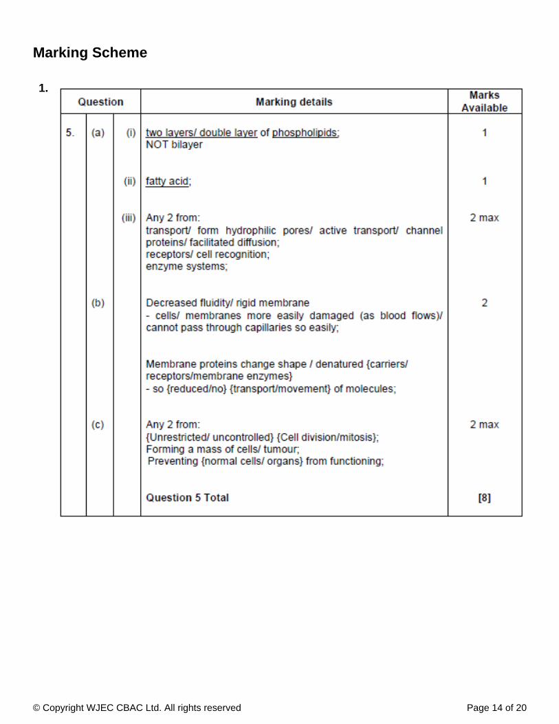
(a) (i) Explain what is meant by the term 'lipid bilayer'.
[1]
(ii) Name the 'hydrophobic parts' referred to in the information above.
[1]
(iii) State functions of membrane proteins.two
[2]
(b) Use the information above to suggest why the changes in red blood cell membranes caused by makeDOXit more toxic than .IDA
[2]
1.

© Copyright WJEC CBAC Ltd. All rights reserved Page 3 of 20
(c) These drugs are used in cancer treatment. Explain briefly what is meant by the term cancer.
[2]
Total
[8]
(a) Samples of epithelial tissue were examined using a light microscope. Drawings of cells from these tissuesare shown below. Identify the type of epithelial tissue shown, and suggest from where in the body the sampleswere taken.
2.

© Copyright WJEC CBAC Ltd. All rights reserved Page 4 of 20
The electron micrograph below shows part of a typical animal cell.
© Copyright WJEC CBAC Ltd. All rights reserved Page 5 of 20
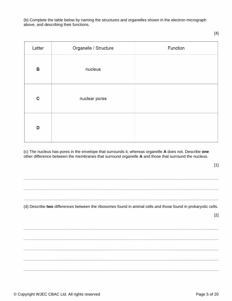
(b) Complete the table below by naming the structures and organelles shown in the electron micrographabove, and describing their functions.
[4]
(c) The nucleus has pores in the envelope that surrounds it, whereas organelle does not. Describe A oneother difference between the membranes that surround organelle and those that surround the nucleus.A
[1]
(d) Describe differences between the ribosomes found in animal cells and those found in prokaryotic cells.two
[2]

© Copyright WJEC CBAC Ltd. All rights reserved Page 6 of 20
The diagram below shows two molecules which are sub-units of proteins.3.

© Copyright WJEC CBAC Ltd. All rights reserved Page 7 of 20
[3]
(ii) Name the type of reaction involved.
[1]
. . . . . . . . . . . . . . . . . . . . . . . . . . . . . . . . . . . . . . . . . . . . . . . . . . . . . . . . . . . . . . . . . . . . .
(iii) Name the type of bond formed.
[1]
. . . . . . . . . . . . . . . . . . . . . . . . . . . . . . . . . . . . . . . . . . . . . . . . . . . . . . . . . . . . . . . . . . . . . .
(b) (i) Why is the model of the structure of biological membranes described as 'fluid mosaic'?
[2]
The diagrams below represent two glycoprotein molecules found in the plasma membranes of mammaliancells.
(i) Complete the diagram above to show how a reaction takes place to join the twomolecules.
(a)

© Copyright WJEC CBAC Ltd. All rights reserved Page 8 of 20
[1]
Molecule . . . . . . . . . . . . . . . . . . . . . . . . . . . . . . . . . . . . . . . . . . . .
(iii) Draw a labelled diagram of the plasma membrane using the diagrams above to show the correctpositioning of glycoproteins A and B.
[2]
(iv) Give one function of the carbohydrate chains on the glycoproteins.
[1]
Which of the molecules A or B will form an intrinsic protein in the plasmamembrane?
(ii)

© Copyright WJEC CBAC Ltd. All rights reserved Page 9 of 20
[1]
. . . . . . . . . . . . . . . . . . . . . . . . . . . . . . . . . . . . . . . . . . . . . . . . . . . . . . . . . . . . . .
(ii) Which organelles are involved in synthesising proteins?
[1]
. . . . . . . . . . . . . . . . . . . . . . . . . . . . . . . . . . . . . . . . . . . . . . . . . . . . . . . . . . . . . .
(d) The following diagram shows one way that prions may pass into cells.
Some diseases are caused by abnormal proteins called prions. Some prions have a higherproportion of β pleated sheet in place of the normal helix structure.(i) What level of protein structure is described by the terms helix and β pleatedsheet?
(c)
[1]
. . . . . . . . . . . . . . . . . . . . . . . . . . . . . . . . . . . . . . . . . . . . . . . . . .
(ii) Name two other ways in which substances might pass into the cell.
[2]
Name the process shown in the diagram above.(i)
Answer one of the following questions.Any diagrams included in your answers must be fully annotated.
, (a) Using examples to illustrate your answer, describe how the structures of polysaccharides areEitherrelated to their functions.
[10]
Or (b) Globular proteins are an important component of plasma membranes. Describe the structure andfunction of membrane proteins.
[10]
4.

© Copyright WJEC CBAC Ltd. All rights reserved Page 10 of 20
© Copyright WJEC CBAC Ltd. All rights reserved Page 11 of 20
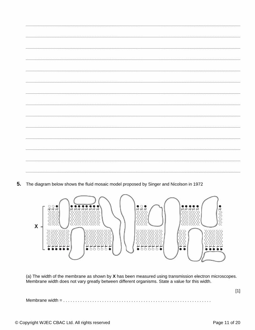
The diagram below shows the fluid mosaic model proposed by Singer and Nicolson in 1972
(a) The width of the membrane as shown by has been measured using transmission electron microscopes.XMembrane width does not vary greatly between different organisms. State a value for this width.
[1]
Membrane width = . . . . . . . . . . . . . . . . . . . . . . . . . . . . . . . . . . . . . . . . . . . . . . . . . . . . . . . . . . . . .
5.

© Copyright WJEC CBAC Ltd. All rights reserved Page 12 of 20
(b) Glucose is water soluble. Vitamin A is lipid soluble. Describe and explain how each molecule crosses themembrane shown above.
[4]
Vitamin A
Glucose
(c) Beetroot vacuoles contain a red pigment called betacyanin. When beetroot discs are cut with a borer andimmersed in a solution of 70% ethanol (an organic solvent) at 15°C, the red pigment begins to leak out of thecells into the ethanol turning it red.
(i) Using your knowledge of the structure of cell membranes, explain why this leakage of pigment occurs.
[2]

© Copyright WJEC CBAC Ltd. All rights reserved Page 13 of 20
(ii) When the experiment was repeated at 30°C, the time taken for the ethanol to turn red decreased. Explainwhy.
[2]
© Copyright WJEC CBAC Ltd. All rights reserved Page 14 of 20
Marking Scheme
1.

© Copyright WJEC CBAC Ltd. All rights reserved Page 15 of 20
2.

© Copyright WJEC CBAC Ltd. All rights reserved Page 16 of 20
(a)(i)OH and H removal shown on diagram;formation of water (H2O) shown;dipeptide correctly drawn with C joined to N; [3]
(ii)Condensation; [1]
(iii)Peptide; NOT dipeptide; [1]
(b)(i)Mosaic: Proteins are scattered (in lipid layer);Fluid: molecules / components / (phospho)lipids / proteins are free to move around; [2]
(ii)B; [1]
(iii)Drawing shows a lipid bilayer with A and B in the correct places, B intrinsic (through the middle) A extrinsic (ontop or bottom, outside phosphate heads);Need not use N and P, but must be clear which is A and Bany 1 correct label from phospholipid / hydrophobic / hydrophilic / cholesterol / phosphate (head) / lipid or fattyacid (tails);11(iv)Cell {recognition / interaction / identification / cell to cell recognition / adhesion / signalling} / receptor qualifiede.g. {hormone receptor / antigens}; [1]
(c)(i)Secondary; [1]
(ii)Ribosomes / rough endoplasmic reticulum;Accept nucleus;NOT golgi body / nucleolus. [1]
(d)(i)Endocytosis (accept phagocytosis / pinocytosis);NOT exocytosis. [1]
(ii)Any 2:Diffusion / osmosis;Facilitated diffusion;Active transport; [2]
3.

© Copyright WJEC CBAC Ltd. All rights reserved Page 17 of 20
4.

© Copyright WJEC CBAC Ltd. All rights reserved Page 18 of 20
5.
© Copyright WJEC CBAC Ltd. All rights reserved Page 19 of 20
Examiner's Comments
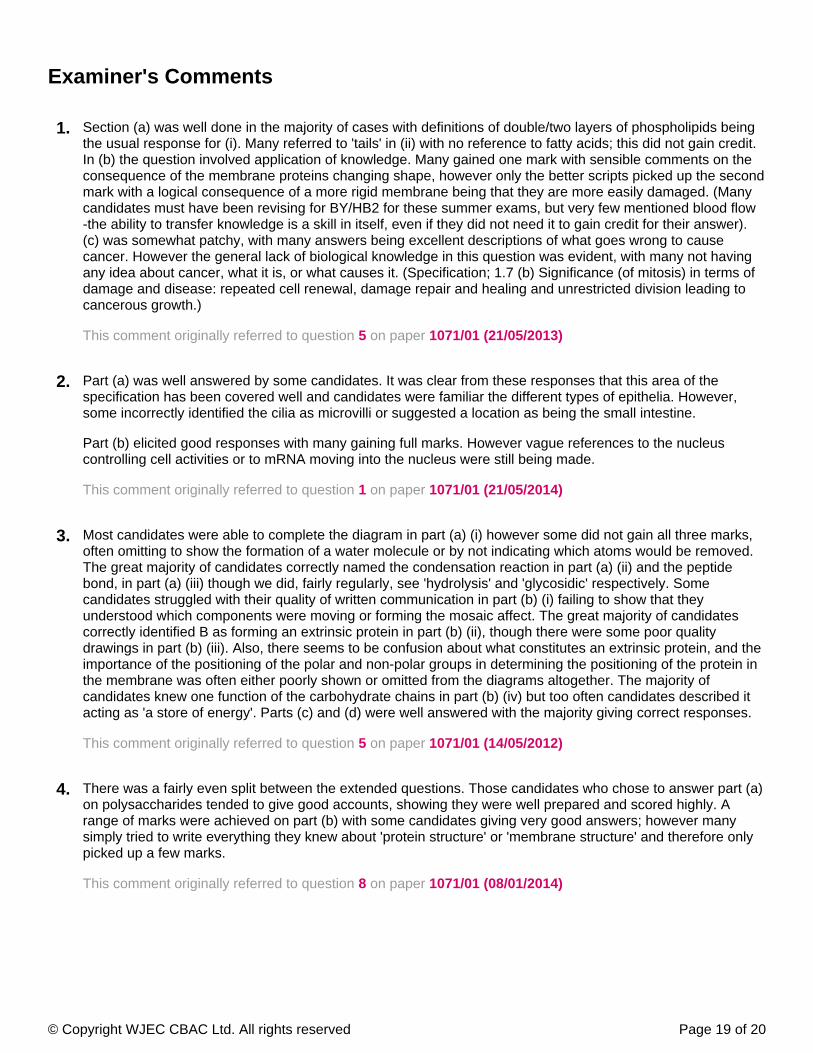
Section (a) was well done in the majority of cases with definitions of double/two layers of phospholipids beingthe usual response for (i). Many referred to 'tails' in (ii) with no reference to fatty acids; this did not gain credit.In (b) the question involved application of knowledge. Many gained one mark with sensible comments on theconsequence of the membrane proteins changing shape, however only the better scripts picked up the secondmark with a logical consequence of a more rigid membrane being that they are more easily damaged. (Manycandidates must have been revising for BY/HB2 for these summer exams, but very few mentioned blood flow-the ability to transfer knowledge is a skill in itself, even if they did not need it to gain credit for their answer).(c) was somewhat patchy, with many answers being excellent descriptions of what goes wrong to causecancer. However the general lack of biological knowledge in this question was evident, with many not havingany idea about cancer, what it is, or what causes it. (Specification; 1.7 (b) Significance (of mitosis) in terms ofdamage and disease: repeated cell renewal, damage repair and healing and unrestricted division leading tocancerous growth.)
This comment originally referred to question on paper 5 1071/01 (21/05/2013)
1.
Part (a) was well answered by some candidates. It was clear from these responses that this area of thespecification has been covered well and candidates were familiar the different types of epithelia. However,some incorrectly identified the cilia as microvilli or suggested a location as being the small intestine.
Part (b) elicited good responses with many gaining full marks. However vague references to the nucleuscontrolling cell activities or to mRNA moving into the nucleus were still being made.
This comment originally referred to question on paper 1 1071/01 (21/05/2014)
2.
Most candidates were able to complete the diagram in part (a) (i) however some did not gain all three marks,often omitting to show the formation of a water molecule or by not indicating which atoms would be removed.The great majority of candidates correctly named the condensation reaction in part (a) (ii) and the peptidebond, in part (a) (iii) though we did, fairly regularly, see 'hydrolysis' and 'glycosidic' respectively. Somecandidates struggled with their quality of written communication in part (b) (i) failing to show that theyunderstood which components were moving or forming the mosaic affect. The great majority of candidatescorrectly identified B as forming an extrinsic protein in part (b) (ii), though there were some poor qualitydrawings in part (b) (iii). Also, there seems to be confusion about what constitutes an extrinsic protein, and theimportance of the positioning of the polar and non-polar groups in determining the positioning of the protein inthe membrane was often either poorly shown or omitted from the diagrams altogether. The majority ofcandidates knew one function of the carbohydrate chains in part (b) (iv) but too often candidates described itacting as 'a store of energy'. Parts (c) and (d) were well answered with the majority giving correct responses.
This comment originally referred to question on paper 5 1071/01 (14/05/2012)
3.
There was a fairly even split between the extended questions. Those candidates who chose to answer part (a)on polysaccharides tended to give good accounts, showing they were well prepared and scored highly. Arange of marks were achieved on part (b) with some candidates giving very good answers; however manysimply tried to write everything they knew about 'protein structure' or 'membrane structure' and therefore onlypicked up a few marks.
This comment originally referred to question on paper 8 1071/01 (08/01/2014)
4.

© Copyright WJEC CBAC Ltd. All rights reserved Page 20 of 20
Very few candidates were able to state the correct range of membrane width asked for in part (a). TheTeachers' guide publishes this as 7/8nm but we accepted any in the range of 6-10nm to allow for otherpublications. However, many candidates were quoting figures in cm which meant they lacked any concept ofthe thinness of the cell membrane.
Most candidates answered part (b) well.
Part (c) often elicited references to osmosis and water potential gradient or vague references to ethanol(i)dissolving the membrane. Part (c) often elicited vague references to “molecules” rather than(ii)specifying dye or membrane molecules. Few candidates mentioned diffusion.
This comment originally referred to question on paper 6 1071/01 (21/05/2014)
5.


![GCE MARKING SCHEME - Weeblycahsbiology.weebly.com/.../jan_11_markscheme.pdf · GCE MARKING SCHEME BIOLOGY/HUMAN BIOLOGY ... surface area:volume [1] (c) ... K. Diffusion of gases in](https://img.pdfslide.net/doc/110x75/5aa2cd0d7f8b9a46238d865e/gce-marking-scheme-marking-scheme-biologyhuman-biology-surface-areavolume.jpg)