Embed Size (px)
Citation preview

Myelination in Pediatric Neurology
Robert Carson MD PhD
Pediatric Neurology Resident Lecture SeriesDOT 815509.13.2013

GoalsReview development of MyelinDiscuss Normal Myelin ImagingOutline an approach to
Leukodystrophies

What we will not discuss todayADEMMSPrimary inflammatory disorders
of CNSMyelin basic science (2 weeks)My groundbreaking research in
exquisite detail :^(

Abnormal connectivity may contribute to neurocognitive deficits in:◦Autism◦TSC◦Angelman’s◦Periventricular leukomalacia
White matter in neurodevelopment.
(Nave, K. 2010)
(spike-timing dependent plasticity)

Timing of myelination mirrors human development

Cortical thickness reaches a developmental nadir while myelin continues to increase

Normal myelination/general MRI patternsNeed to know what is normal to know what
is not normal. ◦ Neonate: T1 hypo T2 hyper◦ Fully myelinated: T1 hyper T2 hypo
T1 signal increases with increasing cholesterol and galactocerebroside
T2 signal decreases with decreasing amount of brain water◦ displaced by myelin◦ Increased length hydrocarbons and double bonds
T2 changes lag behind T1 changes
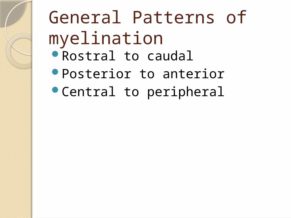
General Patterns of myelinationRostral to caudalPosterior to anteriorCentral to peripheral


T1 T1
T2 FLAIR

T1 FLAIR
T2-FSE T2-FSE

T1 FLAIR
T2-FSE T2-FSE

T1 FLAIR
T2-FSE T2-FSE

FLAIR FLAIR
T2-FSE T2-FSE

FLAIR FLAIR
T2-FSE T2-FSE
Normal Variant

FLAIR Signal evolution

9m 15m
2y 3y

Terminal Zones of myelination

Components of myelin:Sheath: protein-lipid-protein-lipid-
proteinGlycolipids: glalctocerebroside,
sulfatide, cholesterol◦Outer layer of membrane
Long chain fatty acids (middle)Phospholipids:
◦Hydrophobic, on inner membraneOthers: MAG, MOG, PLP, MBP,
CNPase

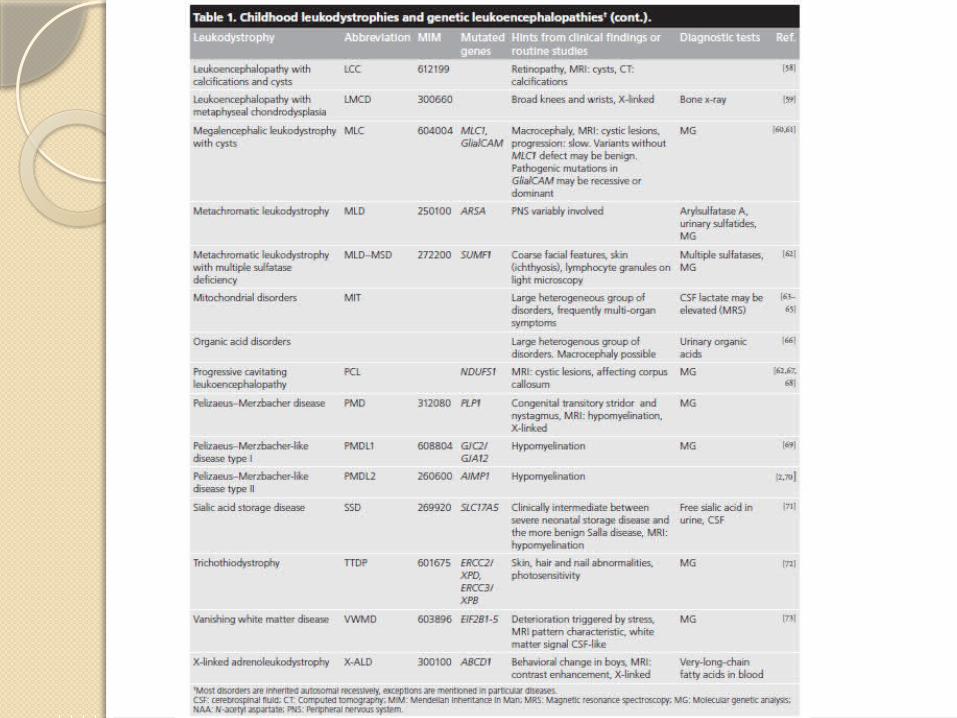
LeukodystrophiesGenetic, with degeneration of
myelin sheaths in CNS (+/-) PNS◦Related to synthesis and maintenance
of myelin membranes.◦Vast majority autosomal recessive
Leukoencephalopathies: defects causing secondary myelin damage
Diagnosis requires a clinical strategy


Clinical presentationInsiduous, in a previously healthy
child. Slowly progressing, may have
periods of stagnationVague/progressive motor and
mental symptoms.Widely variable phenotypes
associated with single genetic disease
Presents from infancy to adulthood.

Age of onset

ExamPhysical abnormalities uncommon
◦Big head: Alexander, Canavan, megalencephalic leukodystrophy with cysts and vanishing white matter
◦Dysmorphic features similar to mucopolysacharidoses: fucosidosis, MLD
Neurologic (progressive):◦Motor (spasticity)◦Changes in cognition and language◦Seizures are rare◦Peripheral nerve (MLD, globoid cell,
hypomyelination)

Diagnosis: MRI most important testStepwise approach:
1. Hypomyelination? Differentiate delayed vs. permanent with
serial MRI studies
2. Confluent, bilateral, symmetric wm lesions c/w genetic disease vs. multifocal or asymmetric with acquired disease
3. If confluent lesions are present, what is the localization? (frontal, parieto-occiptial, periventricular, subcortical, diffuse, posterior fossa)


Abnormal MRIs are not pathognomonic
Tigroid appearance◦ MLD◦ Globiod cell
Sparing of U-fibers◦ X-ALD◦ MLD

“Nearly pathognomonic”
Alexander disease
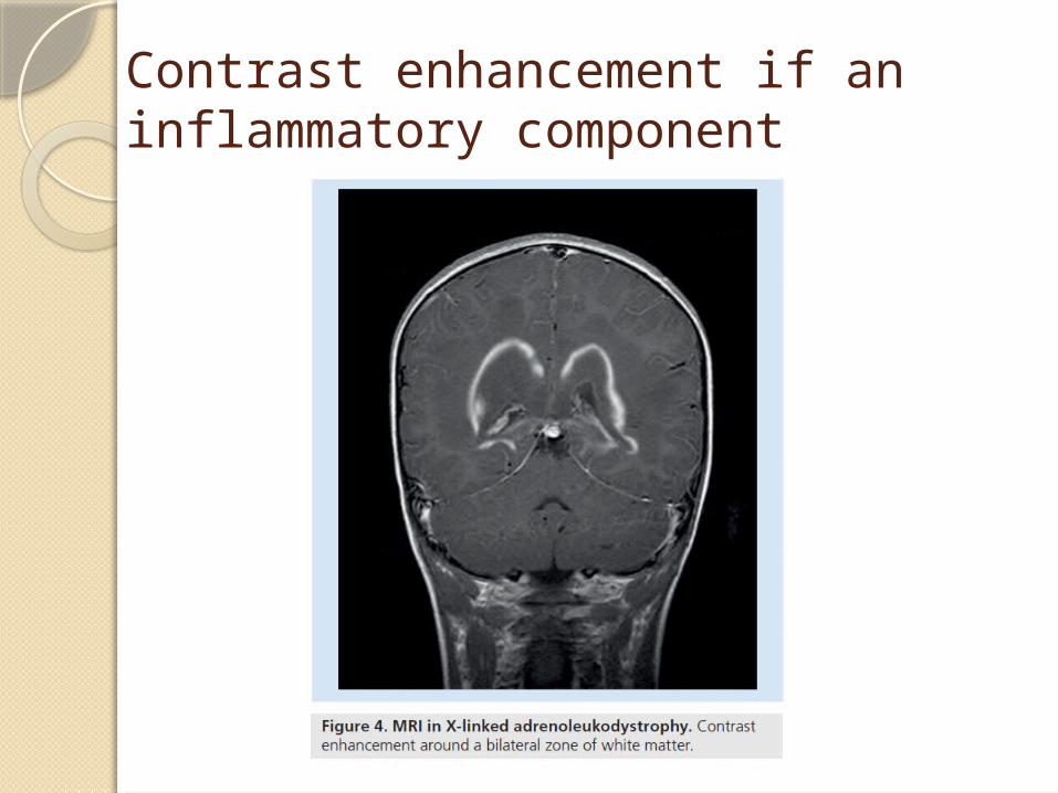
Contrast enhancement if an inflammatory component

FLAIR good for cysts

Additional imagingMRS
◦NAA elevated in Canavan◦Decreased NAA suggests neuronal
involvement in primary WM disease◦Lactate in “leukencephalopathy with
brainstem and spinal cord involvement and elevated lactate”
◦Other mitochondrial disorders
CT: better than MRI for calcifications

Globoid cell leukodystrophy
Swelling of optic nerves
Contrast enhancement of spinal roots
+/- peripheral nerve thickening

ElectrophysiologyNCS
◦Symmetric involvement of long spinal tracks and peripheral nerves
◦May help differentiate leukodystrophies Normal in X-ALD, usually abnormal with
metachromatic or globoid cell
◦Correlates with severity of clinical disease
Evoked potentials◦BAER abnormal first, then SSEP lower
limbs, then MEPs of lower limbs

Tests to consider early on in evaluation.
Low yield of done prior to exam and evaluation of imaging.

Other organ systemsOptho
◦ Cataracts Cerebrotendinous xanthomatosis Some forms of hypomyelination
◦ Cherry red spot: differentiate infantile/macrocephalic leukodystrophies from GM2 gangliosidosis Such as Tay-Sachs and Sandhoff
Endocrine◦ Addison’s disease +/- neuro invovlement in X-ALD◦ Ovarian failure
GI◦ Feeding and swallow issues are common.◦ Gallbladder papilloma in MLD
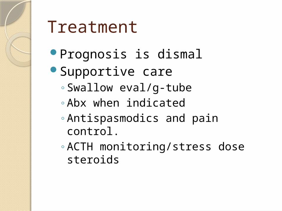
TreatmentPrognosis is dismalSupportive care
◦Swallow eval/g-tube◦Abx when indicated◦Antispasmodics and pain control. ◦ACTH monitoring/stress dose steroids

Treatment ContinuedLorenzo’s oil in X-ALD
◦ Erucic and oleic acid◦ Lowers VLC FAs◦ Benefits asymptomatic boys
Bone Marrow transplantation◦ Can halt progression in X-ALD, but…◦ 2/3 boys develop cerebral disease, and…◦ Successful only in early stages of disease.
Gene therapyExperimental
◦ Therapeutic window is narrow Asymptomatic____ Too far gone

Leukodystrophies, in Summary:Incurable with progressive motor
and mental disabilityLeukodystrophy if due to myelin
sheath, leukoencephalopathy if outside. (similar)
White matter and gray matter disease may overlap.
Definitive diagnosis is challenging, though timely diagnosis is required.

Summary continued,Diagnosis through:
◦Physical examination◦MRI imaging◦With help from targeted laboratory
testing
Important for family counseling and optimization of care◦Palliative◦experimental

ReferencesWelker and Patton. Assessment
of normal myelination with magnetic resonance imaging. Semin Neurol. 2012;32:15-28.
Kohlshutter and Eichler. Childhood leukodystrophies: a clinical perspective. Expert Rev. Neurother. 2011;11:1485-1496.