Embed Size (px)
Citation preview
Page 1/24
Co-transplantation of Hypoxia Pretreated HumanAdipose Derived Mesenchymal Stem Cells and CordBlood Mononuclear Cells to Treat Rats with AcuteMyocardial InfarctionHang Xiang
Chinese PLA General HospitalTianyuan Xiang
Chinese PLA General HospitalHongxia Zhang
USC: University of Southern CaliforniaAnn Xu
USC: University of Southern CaliforniaMatthew John Horwedel
BWH: Brigham and Women's HospitalQiang Zeng ( [email protected] )
BWH: Brigham and Women's HospitalYang Li
Chinese PLA General Hospital
Research
Keywords: adipose derived mesenchymal stem cells, cord blood mononuclear cells, co-transplantation,hypoxic precondition, myocardial infarction
Posted Date: November 17th, 2020
DOI: https://doi.org/10.21203/rs.3.rs-104757/v1
License: This work is licensed under a Creative Commons Attribution 4.0 International License. Read Full License
Page 2/24
AbstractBackground
Human adipose derived mesenchymal stem cells (ASCs) are ideal candidates for the treatment of acutemyocardial infarction (AMI), due to their favorable availability and regenerative potential. However, in vivostudies showed that ASCs are not resilient at the infarcted area, for a shortage of blood and oxygensupply. Hypoxic pretreatment was proven to be an effective way to enhance cell survival in ischemicatmosphere. Moreover, co-transplantation of stem cells was another promising strategy to improvecardiac function after transplantation. So, we hypothesized that hypoxic pretreated ASCs combined withproangiogenic cord blood mononuclear cells (CBMNCs) would promote treatment e�cacy after co-transplantation.
Methods
ASCs extracted from male volunteer were preconditioned in hypoxic condition (HP-ASC) for 24h, and totalRNA were extracted after that. Gene expressions were compared between HP-ASC and ASC. Then, wetransplanted stem cells to female Wistar rats which divided into different groups: (1) HP-ASCs group(n=10, 1x10⁶ASCs); (2) HP-ASCs + CBMNCs group (n=10, 0.5×10⁶ ASCs+0.5×10⁶ CBMNCs); (3) CBMNCsgroup (n=10, 1×10⁶ ASCs); (4) Control group (n=10, 40μL PBS); (5) Sham group (n=10). Echocardiogramwas performed before (0d) and after (30d) after cell transplantation. Hearts were harvested at 30d toanalyze the infarct size, myocardium apoptosis, stem cells viability and angiogenesis.
Results
In vitro study showed that HP-ASCs had a wide range of paracrine function, with the incretion growthfactors and their receptors, which would support the cell survivals. In addition, HP-ASCs also gainedpotentials in hypoxic adaptation (increased expression of HO-1 and SDF-1), as well as homing andimmigrating abilities (CXCR4, ICAM-1 and ICAM-2). In vivo studies showed that, 30 days aftertransplantation in AMI rats, the HP-ASCs group had a better improvement in cardiac function; reduction ofthe infarct size; and decrease of ASCs death than the other groups (HP-ASCs > HP-ASCs + CBMNCs ≧CBMNCs > PBS) (p<0.05). However, the combined group of HP-ASCs and CBMNCs had more signi�cantangiogenesis than the other groups (HP-ASCs + CBMNCs > CBMNCs > HP-ASCs > PBS) (p <0.05).
Conclusions
HP-ASCs alone had a greater potential in improving cardiac function in AMI rats. However, thecombination of HP-ASCs and CBMNCs had a better result in angiogenesis.
BackgroundMyocardial infarction (MI) is a life-threatening emergency which causes hospitalization and life spanreduction in US and worldwide [1]. The standard treatment of MI, including catheterization and

Page 3/24
medication, has shown signi�cant improvement in outcomes. However, the damaged heart tissues canonly be repaired with scars, which lost the myocardium characters of conductivity and contractility.Consequently, the �brotic scar leads to a chronic process of cardiac remodeling, which results incompromised ventricular performance and chronic heart failure. The positive correlation between the sizeof infarction and mortality urged us to �nd a new treatment approach for heart regeneration [2].Physiologically, the hearts of new-born babies have partial regenerative potential through the proliferationof pre-existing cardiomyocytes (CMs), but this does not exist in adults [3]. Recently, numerous studiesreported that stem/ progenitor cells can be used for the treatment of ischemic heart diseases [4, 5].Among those, adipose-derived stromal cells (ASCs) have shown encouraging outcomes in clinical trials[6, 7]. ASCs have more priorities than bone marrow stem cells for clinical use [8, 9], for their largequantities in isolation and minimal invasion, as well as high yield of progenitor cells per volume.
However, there are still some obstacles in improving ASCs survivals after transplantation, which resultfrom regional hostile microenvironment. The �eld is not suitable for transplanted cells to survive, as it issurrounded by necrotic cells and in�ammatory cells that increase the oxidative stress. As a consequence,stem cells generally display limited survival and low retention rate in injured tissues, reducing the bene�tof their therapeutic effects[10]. Therefore, we need to solve the problem of hypoxia adaption and bloodsupply, in order to elongate the cell lives in vivo.
To overcome the obstacle of hypoxia adaption, various precondition methods (e.g., hypoxia, heat shock,and exposure to oxidative stress) were used to accommodate the cells in vitro[11]. Hypoxia was afeasible and potential way to adapt cells before they were transplanted [12]. Exposure to a sub-lethalhypoxic would signi�cantly increase the tolerance and regenerative properties of stem/progenitor cells,resulting in marked protective effects against insults in the ischemic attack [13].
To establish a conducive vascular environment, scientists developed different co-transplantation systemsthat interacts synergistically to form stable vessels [14]. Human umbilical cord blood mononuclear cells(UCMNCs) have generated signi�cant attention in regenerative medicine for their e�cacy in treatingischemic diseases [15, 16]. And UCMNCs have been used with different types of stem cells in animalmodels [17]. Therefore, we hypothesized that co-transplantation of ASCs with UCMNCs would raise thesurvival e�cacy for the damaged tissue and seeded stem cells.
Materials And Methods
Isolation and Identi�cation of Human ASCsSubcutaneous abdominal adipose tissues were obtained from a healthy male who underwent aliposuction surgery. Adipose tissue was washed in phosphate-buffered saline (PBS) and minced, followedby digestion in 5 ml of type I Collagenase (1 mg/ml in 1% bovine serum albumin(BSA)/Hank’s balancedsaline solution; Life Technologies Japan) for 40 minutes at 37°C using a gentle MACS Dissociator(MiltenyiBiotec K.K., Tokyo, Japan) according to the manufacturer’s instructions. The digested tissue was
Page 4/24
�ltered through a 40-μm cell strainer (BD Falcon, Tokyo, Japan) and centrifuged at 450g for 10 minutes.The supernatant containing adipocytes and debris were discarded. Pelleted cells were rinsed twice withPBS, and then planted on the petri dishes with a density of ~2.0×10⁵/cm2. ASCs were cultured accordingto the standard protocol, with our modified media in Dulbecco’s modi�ed Eagle’s medium (DMEM)-F12(Gibco), supplemented with 10% FBS (HyClone), 100U/ml penicillin, and 100μg/ml streptomycin (Biolot).ASCs were subcultured after attaining 70-80% confluency. Passage 3-4 cells were used in the study.
ASCs were identi�ed with �owcytometry analysis by incubation with primary antibodies for 40 min at 4°C in phosphate-buffered saline (PBS) supplemented with 2% FBS and 2mM ethylenediaminetetraaceticacid (EDTA). The following direct conjugated antibodies (BD Pharmingen™) were used: anti-human (PE-CD34, PerCP-CD45, PE-CD90, APC-CD44, PE-CD73, and PE-CD105). All staining was controlled withappropriate isotope control antibodies. Analysis was performed on a SORP LSRII (Becton Dickinson)equipped with �ve lasers and data were collected with FACS DIVA software. Analysis was performedusing FlowJo™ 10.0.8 (Treestar, Ashland, OR). All measurements were performed with three biologicalreplicates.
Linage differentiation tests for osteogenesis, adipogenesis and chondrogenesis was performed asdescribed before [18, 19]. Osteogenic differentiation was evaluated by cellular alkaline phosphatase(ALP) activity (Alkaline Phosphatase kit, 86R; Sigma-Aldrich). Adipogenesis was con�rmed by Oil red Ostaining of intracellular lipids, and chondrogenesis was con�rmed by Alcian blue staining [19].
Hypoxia Precondition of ASCsHypoxic treatment protocol was referred as previously reported with some modi�cation [20]. A total of 3 x10⁶ ASCs (P3) were seeded on a T75 �ask and cultured at 2% O2 (hypoxia) in a ProOx-C-chamber system(Biospherix, Red�eld, NY), in comparation with the ambient oxygen tension 21% O2 (normoxia). Seventy-two hours later, cells were harvested for RNA expression analysis (Supplement Table 1).
Cord blood mononuclear cells (CBMNCs) IsolationIsolation of CBMNCs was performed as reported before [21]. In brief, 35ml of fresh cord blood wascollected from a healthy woman with an uncomplicated delivery. The blood was mixed with 7ml (1:5)Hetastarch solution (HetaSep™, STEM CELL TECH, US), and incubated at 37℃ for 30 min. Then, the cellsin the �oating layer were transferred to another tube, mixed with normal saline (NS) to 50ml, andcentrifuged for 10min (1500rpm). After having been washed for 2 times, the cord blood cells were mixedwith a 5ml lymphocyte separation medium (50494 LSM®, CappelTM, Shanghai), and centrifuged for20min (1500rpm). The thin layer of mononuclear cells was extracted carefully and washed 2 times. Cellviability was evaluated with a trypan blue exclusion test, and cell concentration was brought to 1.5-2.5×10⁶ cells/ml and used within 30 min.
Page 5/24
Surgical Procedure and TransplantationFemale Wistar rats (250–300g) were intubated under general anesthesia with 4% chloral hydrate (4mg/kg, intraperitoneally injection) and ventilated with room air by a small animal ventilator (SAR-830/AP,CWE, US). Myocardial infarction was induced by permanent ligation of the left anterior descendingcoronary artery with a 6–0 silk suture. Successful performance of coronary occlusion was veri�ed byblanching of the myocardium distal to the coronary ligation [22]. Stem cells or PBS were transplantedimmediately after ligation of the left anterior descending coronary artery. Transplanted groups weredivided into 5 groups: (1) HP-ASCs group (n=10, 1x10⁶ASCs); (2) HP-ASCs + CBMNCs group (n=10,0.5×106 ASCs+0.5×10⁶ EPCs); (3) CBMNCs group (n=10, 1×10⁶ ASCs); (4) Control group (n=10, 40μLPBS); (5) Sham group (n=10). A total amount of 40μL mixture was directly injected into 5 spots aroundthe ischemic region, except the sham group, which only underwent the thoracotomy without coronaryartery ligament or injection.
Measurement of Heart FunctionEchocardiography (Vevo770; Visualsonics, Toronto, ON, Canada) was performed at 0d and 30d aftermyocardial ischemia. Rats were anesthetized with 4% chloral hydrate (40 mg/kg, intraperitoneally), andimaged in the supine position at the fourth and �fth intercostal space with a 710B transducer. Both 2Dand M-mode images were used for measurements, and images were later analyzed by a trained blindreader using the cardiac analysis software (VisualSonics, version 2.2.3). The following variables aremeasured: ejection fraction (EF), fractional shortening (FS), stroke volume (SV), heart rate (HR), and leftventricle posterior wall thickening (LVPW) were measured.
Infarct Size MeasurementAll the hearts were harvested at 30d and embedded in optimal cutting temperature (OCT) compound(Sakura Finetek USA Inc, Torrance, Calif). The infarct and peri-infarct regions were cut into threetransverse sections then and stained by Masson trichrome and hematoxylin–eosin (HE). The stainedsections were measured and calculated for the average ratio of �brosis area (blue) to the entire LV area(percentage of �brosis area), and the average ratio of the reduced LV wall thickness in the scarred area tothe intact LV wall thickness from three different sites in each wall (LV wall thinning %). For each slice, 10randomly selected �elds were captured (× 100) and images were digitized and analysed with a digitalimage analyser (MIQAS, Qiuwei Co, China).
Living ASCs Cell Count and Vessel Count in Necrotic AreaTo count the living ASCs in heart sections, we used anti-human SRY antibody (Cat#MA5-17181, 1:200,Invitrogen, US) to trace the ASCs cells (ASCs used were only from human male)[23]. Six �elds per heartsection were randomly chosen and photographed under 40× magni�cation with a �uorescent
Page 6/24
microscope, and live cells (cells/mm2) were counted in every tenth heart section across the entire regionof interest.
For vascular counting, we used anti-human CD31 (Cat#ab32457, 1:200, Abcam, Shanghai) to stain thenewly formed capillaries. The number of capillaries was counted under a light microscope(magni�cation × 250, OLYMPUS BH2, Japan) for 10 random �elds in each transverse slice and presentedas the mean number of blood vessels per unit area (number/mm2). Both of these performances wererepeated in 8 separate sections per heart. Two independent observers were blinded to the identity of thetissues.
Statistical AnalysisStatistics were performed using SPSS 17.0 Software. Data are expressed as Mean ± Standard Error.Statistical comparisons were made using an unpaired t-test or one-way analysis of variance followed byBonferroni multiple comparison post hoc tests where appropriate. The results were consideredstatistically signi�cant when P<0.05.
ResultsCharacterization of ASCs and Differentiation
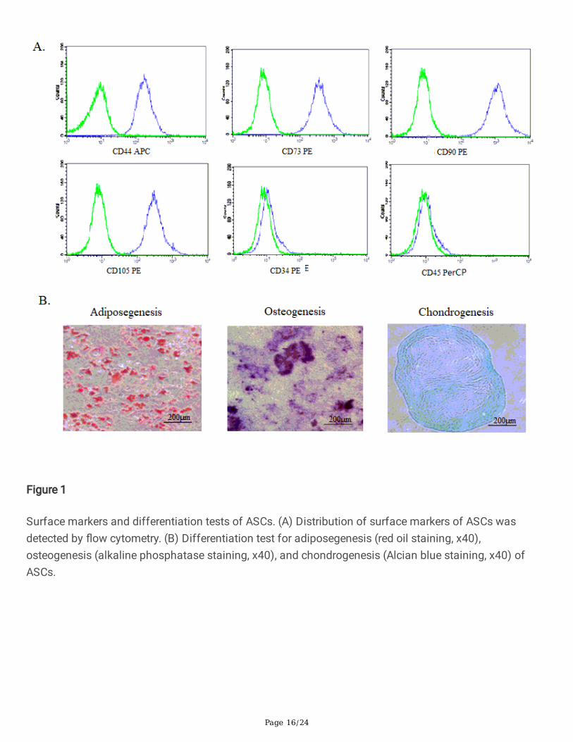
Morphology of ASCs was homogenously spindle-like after three passages (Fig.1A). Over 90% of thepopulation expressed the MSC-speci�c surface markers CD90 (+), CD105 (+), CD73 (+), CD44 (+), andwere negative for hematopoietic stem cell and endothelial surface marker CD45(-), CD34(-) (Fig.1B).
Differentiation tests showed that cultured ASCs (P3) have a strong potential of differentiation withosteogenesis, adiposegenesis and chondrogenesis, which were speci�c characteristics for mesenchymalstem cells (Fig.1A).
HP-ASCs Gene Expression after Hypoxia Pre-treatment in Vitro
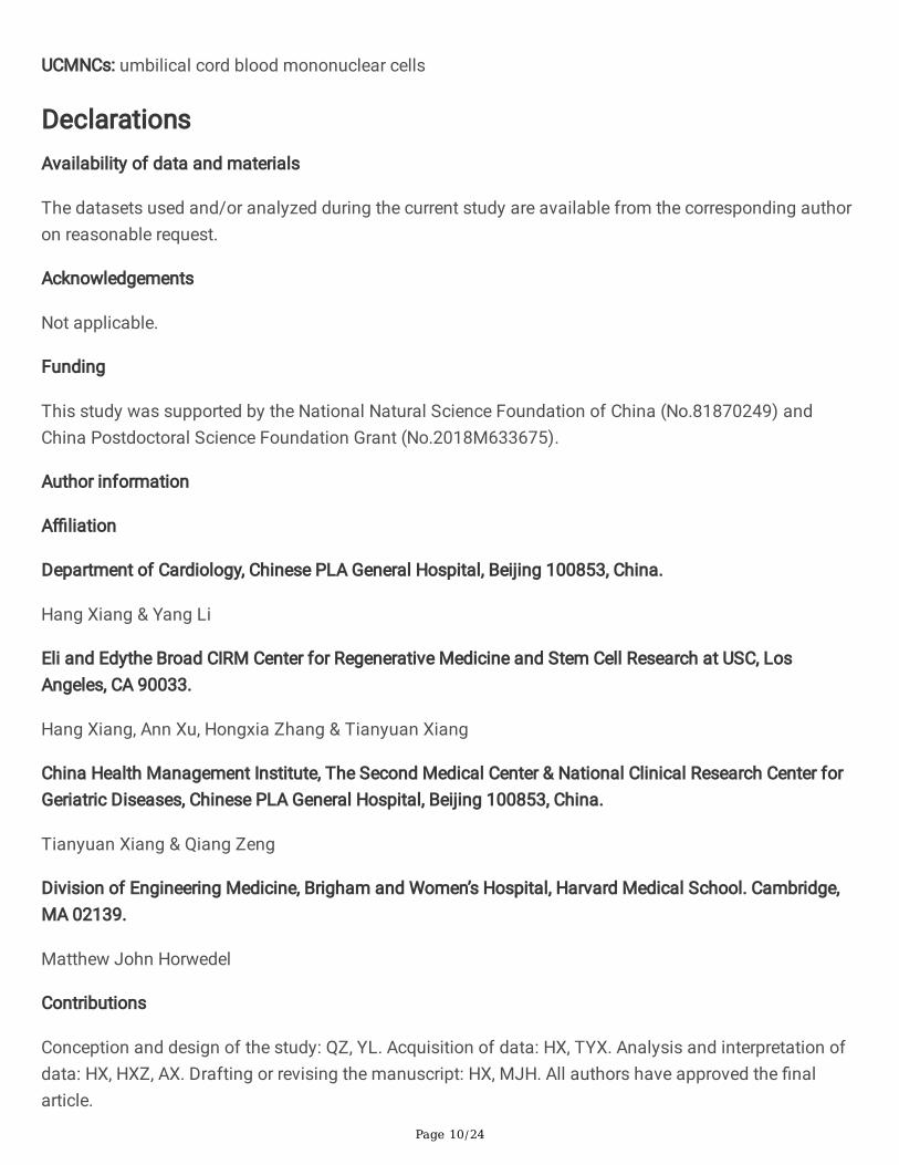
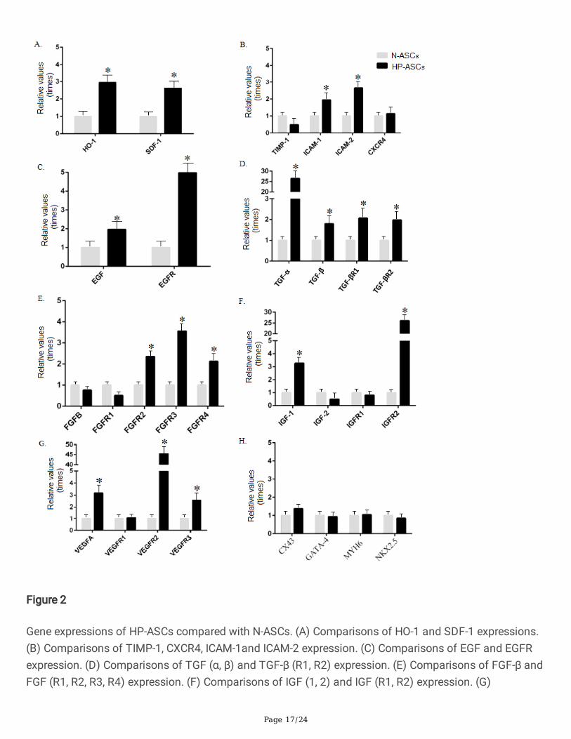
HP-ASCs displayed a strong adaptation for hypoxia after 72 hours of hypoxic treatment, by increasing theexpression of Heme Oxygenase-1 (HO-1) and Stromal cell Derived Factor-1 (SDF-1) (Fig.2A), as well asenhanced cell migration (CXCR4) and adhesion (ICAM-1and ICAM-2) potential(Fig.2B). HP-ASCs also hadboosted expression of growth factors and their receptors (EGF/EGFR, IGF-1/IGFR2; TGF(α, β)/TGF-β (R1,R2); VEGF-α/VEGF(R2, R3); FGF (R2, R3, R4) (Fig.2C-G). However, it did not show any evidence that HP-ASCs had been differentiation toward myocardocytes (Fig 2H).
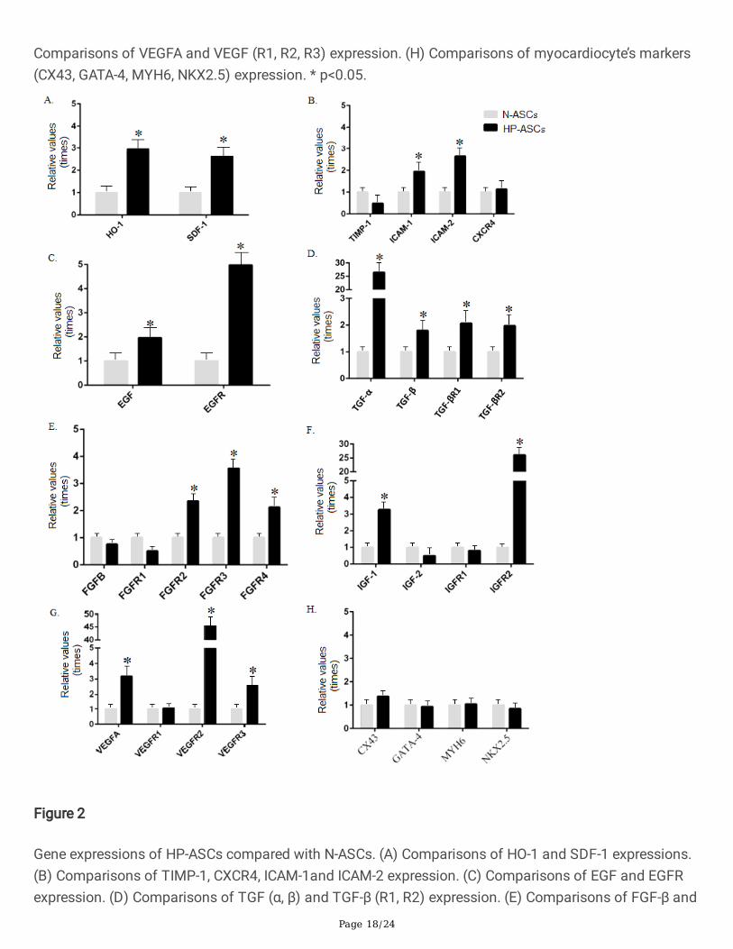
Cardiac function and haemodynamics after transplantation
Cardiac function was compared before and after cell transplantation (0d and 30d). The results showedthat there was no difference between these four groups before transplantation. At 30d, HP-ASCs group
Page 7/24
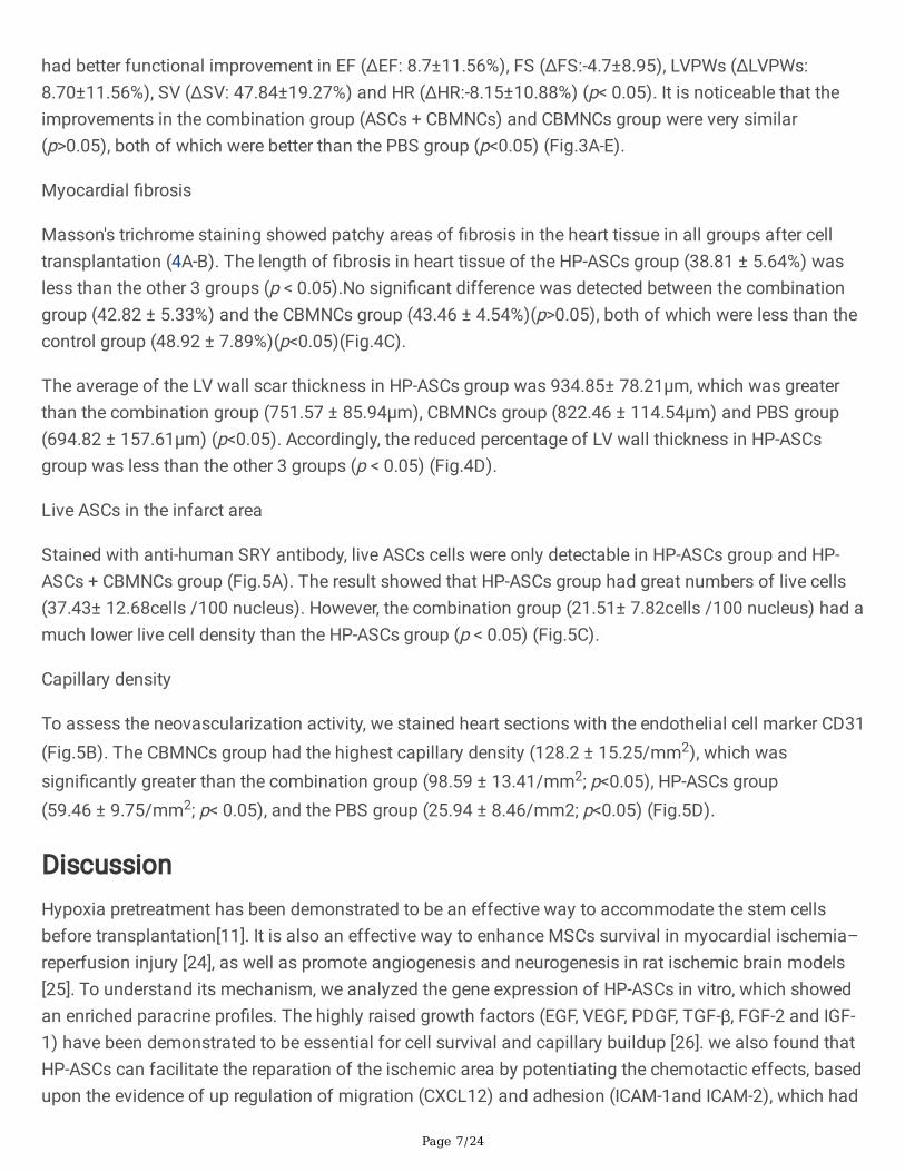
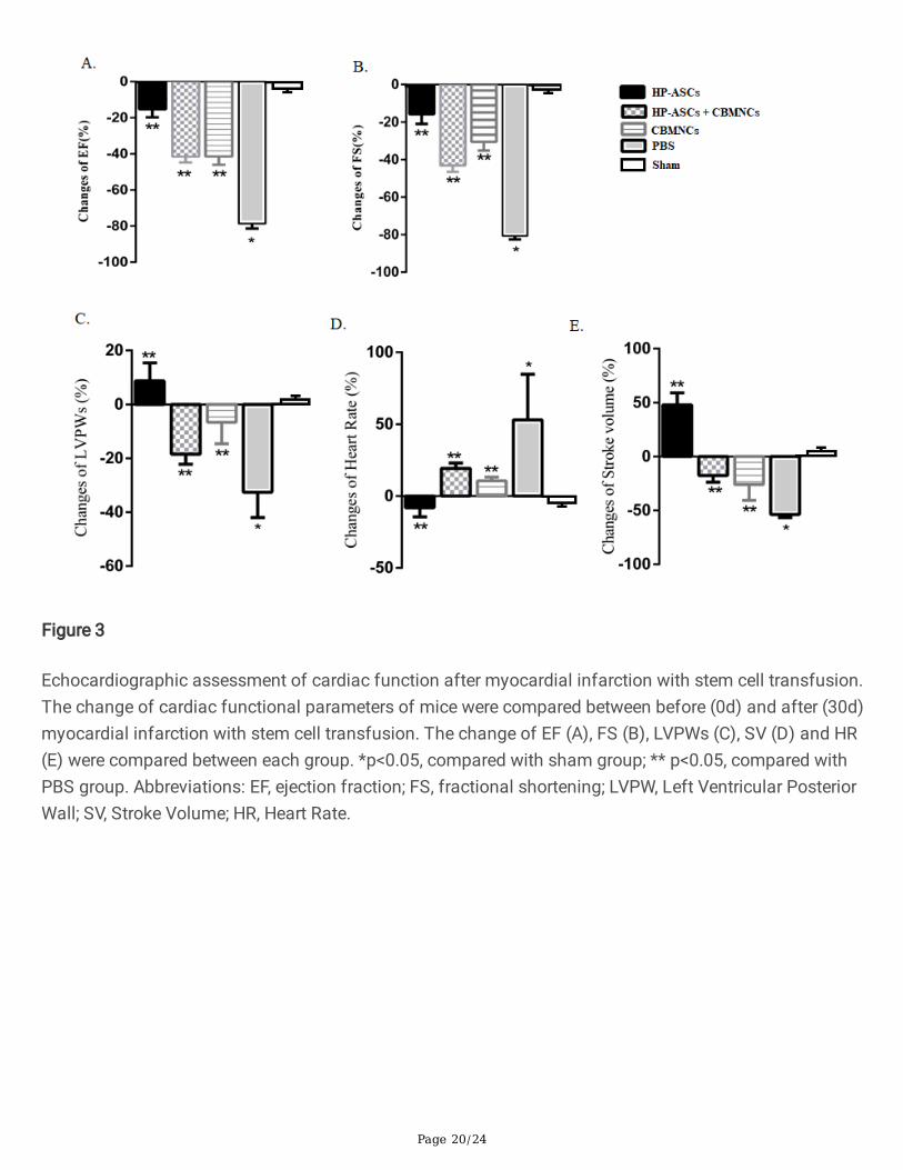
had better functional improvement in EF (ΔEF: 8.7±11.56%), FS (ΔFS:-4.7±8.95), LVPWs (ΔLVPWs:8.70±11.56%), SV (ΔSV: 47.84±19.27%) and HR (ΔHR:-8.15±10.88%) (p< 0.05). It is noticeable that theimprovements in the combination group (ASCs + CBMNCs) and CBMNCs group were very similar(p>0.05), both of which were better than the PBS group (p<0.05) (Fig.3A-E).
Myocardial �brosis
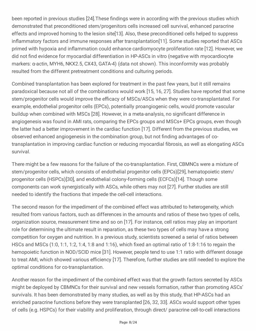
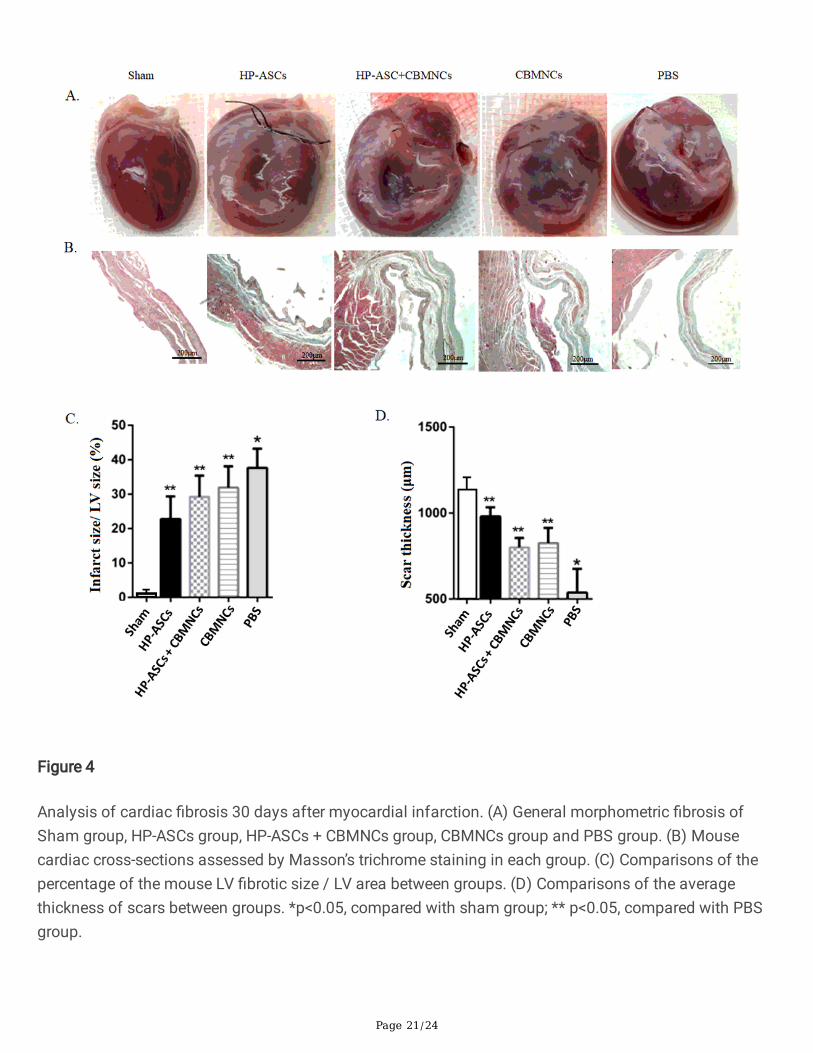
Masson's trichrome staining showed patchy areas of �brosis in the heart tissue in all groups after celltransplantation (4A-B). The length of �brosis in heart tissue of the HP-ASCs group (38.81 ± 5.64%) wasless than the other 3 groups (p < 0.05).No signi�cant difference was detected between the combinationgroup (42.82 ± 5.33%) and the CBMNCs group (43.46 ± 4.54%)(p>0.05), both of which were less than thecontrol group (48.92 ± 7.89%)(p<0.05)(Fig.4C).
The average of the LV wall scar thickness in HP-ASCs group was 934.85± 78.21μm, which was greaterthan the combination group (751.57 ± 85.94μm), CBMNCs group (822.46 ± 114.54μm) and PBS group(694.82 ± 157.61μm) (p<0.05). Accordingly, the reduced percentage of LV wall thickness in HP-ASCsgroup was less than the other 3 groups (p < 0.05) (Fig.4D).
Live ASCs in the infarct area
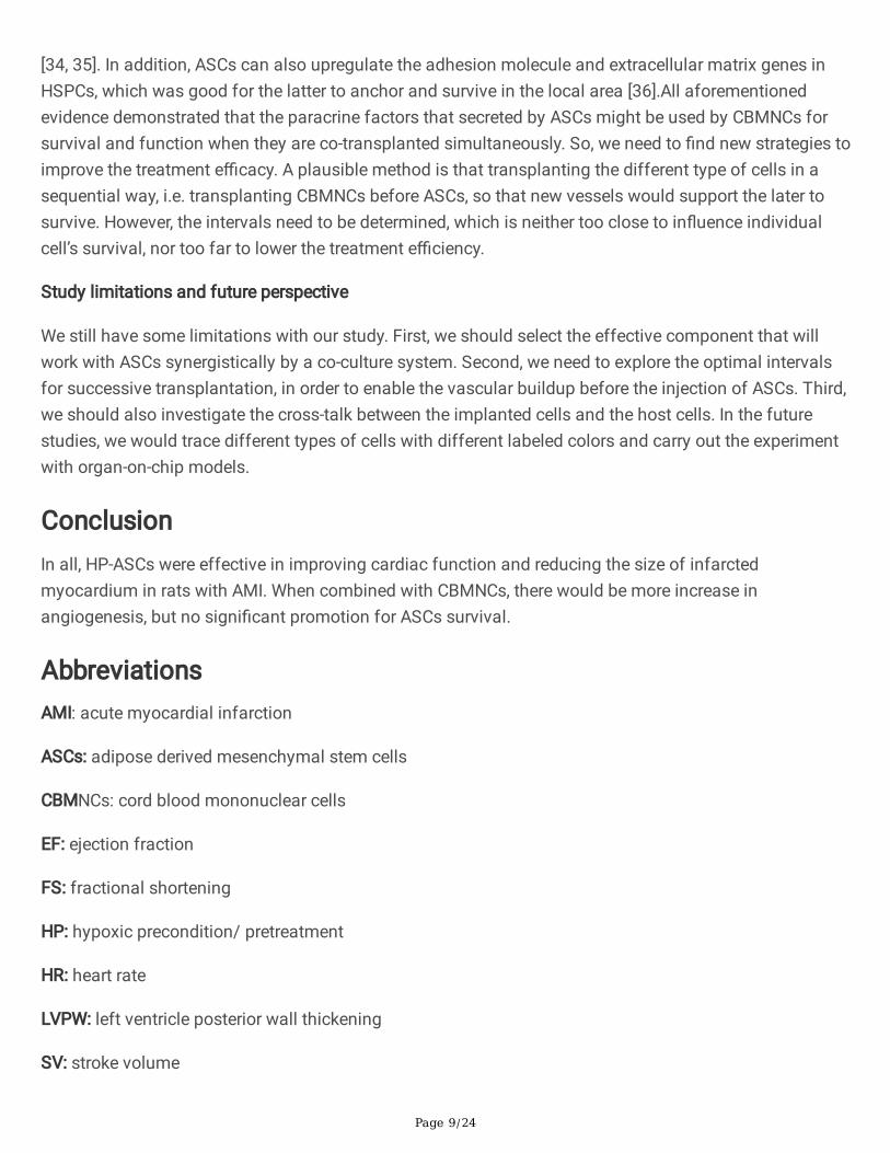
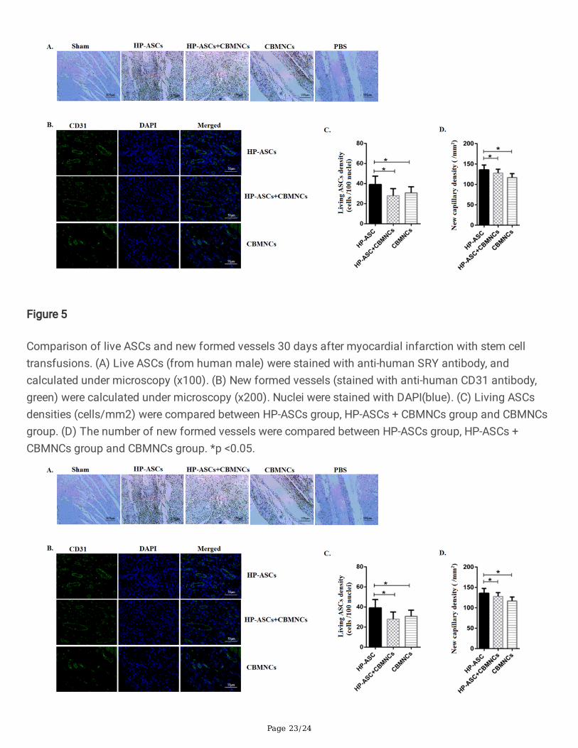
Stained with anti-human SRY antibody, live ASCs cells were only detectable in HP-ASCs group and HP-ASCs + CBMNCs group (Fig.5A). The result showed that HP-ASCs group had great numbers of live cells(37.43± 12.68cells /100 nucleus). However, the combination group (21.51± 7.82cells /100 nucleus) had amuch lower live cell density than the HP-ASCs group (p < 0.05) (Fig.5C).
Capillary density
To assess the neovascularization activity, we stained heart sections with the endothelial cell marker CD31(Fig.5B). The CBMNCs group had the highest capillary density (128.2 ± 15.25/mm2), which wassigni�cantly greater than the combination group (98.59 ± 13.41/mm2; p<0.05), HP-ASCs group(59.46 ± 9.75/mm2; p< 0.05), and the PBS group (25.94 ± 8.46/mm2; p<0.05) (Fig.5D).
DiscussionHypoxia pretreatment has been demonstrated to be an effective way to accommodate the stem cellsbefore transplantation[11]. It is also an effective way to enhance MSCs survival in myocardial ischemia–reperfusion injury [24], as well as promote angiogenesis and neurogenesis in rat ischemic brain models[25]. To understand its mechanism, we analyzed the gene expression of HP-ASCs in vitro, which showedan enriched paracrine pro�les. The highly raised growth factors (EGF, VEGF, PDGF, TGF-β, FGF-2 and IGF-1) have been demonstrated to be essential for cell survival and capillary buildup [26]. we also found thatHP-ASCs can facilitate the reparation of the ischemic area by potentiating the chemotactic effects, basedupon the evidence of up regulation of migration (CXCL12) and adhesion (ICAM-1and ICAM-2), which had
Page 8/24
been reported in previous studies [24].These �ndings were in according with the previous studies whichdemonstrated that preconditioned stem/progenitors cells increased cell survival, enhanced paracrineeffects and improved homing to the lesion site[13]. Also, these preconditioned cells helped to suppressin�ammatory factors and immune responses after transplantation[11]. Some studies reported that ASCsprimed with hypoxia and in�ammation could enhance cardiomyocyte proliferation rate [12]. However, wedid not �nd evidence for myocardial differentiation in HP-ASCs in vitro (negative with myocardiocytemarkers: α-actin, MYH6, NKX2.5, CX43, GATA-4) (data not shown). This inconformity was probablyresulted from the different pretreatment conditions and culturing periods.
Combined transplantation has been explored for treatment in the past few years, but it still remainsparadoxical because not all of the combinations would work [15, 16, 27]. Studies have reported that somestem/progenitor cells would improve the e�cacy of MSCs/ASCs when they were co-transplantated. Forexample, endothelial progenitor cells (EPCs), potentially proangiogenic cells, would promote vascularbuildup when combined with MSCs [28]. However, in a meta-analysis, no signi�cant difference inangiogenesis was found in AMI rats, comparing the EPCs groups and MSCs+ EPCs groups, even thoughthe latter had a better improvement in the cardiac function [17]. Different from the previous studies, weobserved enhanced angiogenesis in the combination group, but not �nding advantages of co-transplantation in improving cardiac function or reducing myocardial �brosis, as well as elongating ASCssurvival.
There might be a few reasons for the failure of the co-transplantation. First, CBMNCs were a mixture ofstem/progenitor cells, which consists of endothelial progenitor cells (EPCs)[29], hematopoietic stem/progenitor cells (HSPCs)[30], and endothelial colony-forming cells (ECFCs)[14]. Though somecomponents can work synergistically with ASCs, while others may not [27]. Further studies are stillneeded to identify the fractions that impede the cell-cell interactions.
The second reason for the impediment of the combined effect was attributed to heterogeneity, whichresulted from various factors, such as differences in the amounts and ratios of these two types of cells,organization source, measurement time and so on [17]. For instance, cell ratios may play an importantrole for determining the ultimate result in reparation, as these two types of cells may have a strongcompetition for oxygen and nutrition. In a previous study, scientists screened a serial of ratios betweenHSCs and MSCs (1:0, 1:1, 1:2, 1:4, 1:8 and 1:16), which �xed an optimal ratio of 1:8-1:16 to regain thehemopoietic function in NOD/SCID mice [31]. However, people tend to use 1:1 ratio with different dosageto treat AMI, which showed various e�ciency [17]. Therefore, further studies are still needed to explore theoptimal conditions for co-transplantation.
Another reason for the impediment of the combined effect was that the growth factors secreted by ASCsmight be deployed by CBMNCs for their survival and new vessels formation, rather than promoting ASCs’survivals. It has been demonstrated by many studies, as well as by this study, that HP-ASCs had anenriched paracrine functions before they were transplanted [26, 32, 33]. ASCs would support other typesof cells (e.g. HSPCs) for their viability and proliferation, through direct/ paracrine cell-to-cell interactions
Page 9/24
[34, 35]. In addition, ASCs can also upregulate the adhesion molecule and extracellular matrix genes inHSPCs, which was good for the latter to anchor and survive in the local area [36].All aforementionedevidence demonstrated that the paracrine factors that secreted by ASCs might be used by CBMNCs forsurvival and function when they are co-transplanted simultaneously. So, we need to �nd new strategies toimprove the treatment e�cacy. A plausible method is that transplanting the different type of cells in asequential way, i.e. transplanting CBMNCs before ASCs, so that new vessels would support the later tosurvive. However, the intervals need to be determined, which is neither too close to in�uence individualcell’s survival, nor too far to lower the treatment e�ciency.
Study limitations and future perspective
We still have some limitations with our study. First, we should select the effective component that willwork with ASCs synergistically by a co-culture system. Second, we need to explore the optimal intervalsfor successive transplantation, in order to enable the vascular buildup before the injection of ASCs. Third,we should also investigate the cross-talk between the implanted cells and the host cells. In the futurestudies, we would trace different types of cells with different labeled colors and carry out the experimentwith organ-on-chip models.
ConclusionIn all, HP-ASCs were effective in improving cardiac function and reducing the size of infarctedmyocardium in rats with AMI. When combined with CBMNCs, there would be more increase inangiogenesis, but no signi�cant promotion for ASCs survival.
AbbreviationsAMI: acute myocardial infarction
ASCs: adipose derived mesenchymal stem cells
CBMNCs: cord blood mononuclear cells
EF: ejection fraction
FS: fractional shortening
HP: hypoxic precondition/ pretreatment
HR: heart rate
LVPW: left ventricle posterior wall thickening
SV: stroke volume
Page 10/24
UCMNCs: umbilical cord blood mononuclear cells
DeclarationsAvailability of data and materials
The datasets used and/or analyzed during the current study are available from the corresponding authoron reasonable request.
Acknowledgements
Not applicable.
Funding
This study was supported by the National Natural Science Foundation of China (No.81870249) andChina Postdoctoral Science Foundation Grant (No.2018M633675).
Author information
A�liation
Department of Cardiology, Chinese PLA General Hospital, Beijing 100853, China.
Hang Xiang & Yang Li
Eli and Edythe Broad CIRM Center for Regenerative Medicine and Stem Cell Research at USC, LosAngeles, CA 90033.
Hang Xiang, Ann Xu, Hongxia Zhang & Tianyuan Xiang
China Health Management Institute, The Second Medical Center & National Clinical Research Center forGeriatric Diseases, Chinese PLA General Hospital, Beijing 100853, China.
Tianyuan Xiang & Qiang Zeng
Division of Engineering Medicine, Brigham and Women’s Hospital, Harvard Medical School. Cambridge,MA 02139.
Matthew John Horwedel
Contributions
Conception and design of the study: QZ, YL. Acquisition of data: HX, TYX. Analysis and interpretation ofdata: HX, HXZ, AX. Drafting or revising the manuscript: HX, MJH. All authors have approved the �nalarticle.
Page 11/24
Corresponding authors
Yang Li & Qiang Zeng.
Ethics declarations
Ethics approval and consent to participate
All experiments were carried out in compliance with the Helsinki Declaration. The animal experimentswere performed according to the Federation for Laboratory Animal Science Association’s guidelines andapproved by the Animal Care Committee of the PLA General Hospital (E2018-06-06). Human ASCs wereharvested from a healthy man who underwent a plastic abdominal surgery, and informed consent wasobtained from the donors according to the institutional guidelines. CBMNCs were isolated from cordblood of a normally delivered female baby. CBMNCs were collected and processed with the approval ofthe ‘Chinese PLA General Hospital Institutional Review Board’ (L2018-12-03) and manufactured tomononuclear cells according to our good manufacturing practice (GMP) process in the Human CellTherapy Laboratory, Chinese PLA General Hospital, China.
Consent for publication
Not applicable
Competing interest
The authors have no commercial, proprietary, or �nancial interest in the products or companies describedin this article.
References1. Mozaffarian D, Benjamin EJ, Go AS, Arnett DK, Blaha MJ, Cushman M, et al. Heart disease and stroke
statistics--2015 update: a report from the American Heart Association. Circulation. 2015;131(4):e29-322.
2. Yoshida K, Gould KL. Quantitative relation of myocardial infarct size and myocardial viability bypositron emission tomography to left ventricular ejection fraction and 3-year mortality with andwithout revascularization. Journal of the American College of Cardiology. 1993; 22(4):984-97.
3. Mollova M, Bersell K, Walsh S, Savla J, Das LT, Park SY, et al. Cardiomyocyte proliferation contributesto heart growth in young humans. Proceedings of the National Academy of Sciences of the UnitedStates of America. 2013; 110(4):1446-51.
4. Wollert KC, Meyer GP, Lotz J, Ringes-Lichtenberg S, Lippolt P, Breidenbach C, et al. Intracoronaryautologous bone-marrow cell transfer after myocardial infarction: the BOOST randomised controlledclinical trial. Lancet (London, England). 2004; 364(9429):141-8.
Page 12/24
5. Hare JM, Fishman JE, Gerstenblith G, DiFede Velazquez DL, Zambrano JP, Suncion VY, et al.Comparison of allogeneic vs autologous bone marrow–derived mesenchymal stem cells delivered bytransendocardial injection in patients with ischemic cardiomyopathy: the POSEIDON randomizedtrial. Jama. 2012; 308(22):2369-79.
�. Oguz E, Ayik F, Ozturk P, Engin C, Nalbantgil S, Yagdi T, et al. Long-term results of autologous stemcell transplantation in the treatment of patients with congestive heart failure. Transplantationproceedings. 2011; 43(3):931-4.
7. Losordo DW, Schatz RA, White CJ, Udelson JE, Veereshwarayya V, Durgin M, et al. Intramyocardialtransplantation of autologous CD34+ stem cells for intractable angina: a phase I/IIa double-blind,randomized controlled trial. Circulation. 2007; 115(25):3165-72.
�. Argentati C, Morena F, Bazzucchi M, Armentano I, Emiliani C, Martino S. Adipose Stem CellTranslational Applications: From Bench-to-Bedside. International journal of molecular sciences.2018; 19(11).
9. Minteer DM, Marra KG, Rubin JP. Adipose stem cells: biology, safety, regulation, and regenerativepotential. Clinics in plastic surgery. 2015; 42(2):169-79.
10. Lo EH, Wang X, Cuzner ML. Extracellular proteolysis in brain injury and in�ammation: role forplasminogen activators and matrix metalloproteinases. Journal of neuroscience research. 2002;69(1):1-9.
11. Sart S, Ma T, Li Y. Preconditioning stem cells for in vivo delivery. BioResearch open access. 2014;3(4):137-49.
12. Przybyt E, Krenning G, Brinker MG, Harmsen MC. Adipose stromal cells primed with hypoxia andin�ammation enhance cardiomyocyte proliferation rate in vitro through STAT3 and Erk1/2. Journalof translational medicine. 2013; 11:39.
13. Hu X, Yu SP, Fraser JL, Lu Z, Ogle ME, Wang JA, et al. Transplantation of hypoxia-preconditionedmesenchymal stem cells improves infarcted heart function via enhanced survival of implanted cellsand angiogenesis. The Journal of thoracic and cardiovascular surgery. 2008; 135(4):799-808.
14. Lin RZ, Moreno-Luna R, Li D, Jaminet SC, Greene AK, Melero-Martin JM. Human endothelial colony-forming cells serve as trophic mediators for mesenchymal stem cell engraftment via paracrinesignaling. Proceedings of the National Academy of Sciences of the United States of America. 2014;111(28):10137-42.
15. Oommen S, Yamada S, Cantero Peral S, Campbell KA, Bruinsma ES, Terzic A, et al. Human umbilicalcord blood-derived mononuclear cells improve murine ventricular function upon intramyocardialdelivery in right ventricular chronic pressure overload. Stem cell research & therapy. 2015; 6(1):50.
1�. Henning RJ, Abu-Ali H, Balis JU, Morgan MB, Willing AE, Sanberg PR. Human umbilical cord bloodmononuclear cells for the treatment of acute myocardial infarction. Cell transplantation. 2004; 13(7-8):729-39.
17. Sun K, Zhou Z, Ju X, Zhou Y, Lan J, Chen D, et al. Combined transplantation of mesenchymal stemcells and endothelial progenitor cells for tissue engineering: a systematic review and meta-analysis.
Page 13/24
Stem cell research & therapy. 2016; 7(1):151.
1�. Wystrychowski W, Patlolla B, Zhuge Y, Neofytou E, Robbins RC, Beygui RE. Multipotency andcardiomyogenic potential of human adipose-derived stem cells from epicardium, pericardium, andomentum. Stem cell research & therapy. 2016; 7(1):84.
19. Bwalya EC, Kim S, Fang J, Wijekoon HMS, Hosoya K, Okumura M. Effects of pentosan polysulfateand polysulfated glycosaminoglycan on chondrogenesis of canine bone marrow-derivedmesenchymal stem cells in alginate and micromass culture. The Journal of veterinary medicalscience. 2017; 79(7):1182-90.
20. Hsiao ST, Lokmic Z, Peshavariya H, Abberton KM, Dusting GJ, Lim SY, et al. Hypoxic conditioningenhances the angiogenic paracrine activity of human adipose-derived stem cells. Stem cells anddevelopment. 2013; 22(10):1614-23.
21. Maslova EV, Andreeva ER, Andrianova IV, Bobyleva PI, Romanov YA, Kabaeva NV, et al. Enrichment ofumbilical cord blood mononuclears with hemopoietic precursors in co-culture with mesenchymalstromal cells from human adipose tissue. Bulletin of experimental biology and medicine. 2014;156(4):584-9.
22. Ii M, Nishimura H, Iwakura A, Wecker A, Eaton E, Asahara T, et al. Endothelial progenitor cells arerapidly recruited to myocardium and mediate protective effect of ischemic preconditioning via"imported" nitric oxide synthase activity. Circulation. 2005; 111(9):1114-20.
23. Zhang X, Wei M, Zhu W, Han B. Combined transplantation of endothelial progenitor cells andmesenchymal stem cells into a rat model of isoproterenol-induced myocardial injury. Archives ofcardiovascular diseases. 2008; 101(5):333-42.
24. Tang YL, Zhu W, Cheng M, Chen L, Zhang J, Sun T, et al. Hypoxic preconditioning enhances thebene�t of cardiac progenitor cell therapy for treatment of myocardial infarction by inducing CXCR4expression. Circulation research. 2009; 104(10):1209-16.
25. Chen J, Yang Y, Shen L, Ding W, Chen X, Wu E, et al. Hypoxic Preconditioning Augments theTherapeutic E�cacy of Bone Marrow Stromal Cells in a Rat Ischemic Stroke Model. Cellular andmolecular neurobiology. 2017; 37(6):1115-29.
2�. Li X, Ma T, Sun J, Shen M, Xue X, Chen Y, et al. Harnessing the secretome of adipose-derived stemcells in the treatment of ischemic heart diseases. Stem cell research & therapy. 2019; 10(1):196.
27. Hong SJ, Kihlken J, Choi SC, March KL, Lim DS. Intramyocardial transplantation of human adipose-derived stromal cell and endothelial progenitor cell mixture was not superior to individual cell typetransplantation in improving left ventricular function in rats with myocardial infarction. Internationaljournal of cardiology. 2013; 164(2):205-11.
2�. Zigdon-Giladi H, Bick T, Lewinson D, Machtei EE. Co-transplantation of endothelial progenitor cellsand mesenchymal stem cells promote neovascularization and bone regeneration. Clinical implantdentistry and related research. 2015; 17(2):353-9.
29. Suuronen EJ, Price J, Veinot JP, Ascah K, Kapila V, Guo XW, et al. Comparative effects ofmesenchymal progenitor cells, endothelial progenitor cells, or their combination on myocardial
Page 14/24
infarct regeneration and cardiac function. The Journal of thoracic and cardiovascular surgery. 2007;134(5):1249-58.
30. Noort WA, Kruisselbrink AB, in't Anker PS, Kruger M, van Bezooijen RL, de Paus RA, et al.Mesenchymal stem cells promote engraftment of human umbilical cord blood-derived CD34(+) cellsin NOD/SCID mice. Experimental hematology. 2002; 30(8):870-8.
31. Kim DH, Yoo KH, Yim YS, Choi J, Lee SH, Jung HL, et al. Cotransplanted bone marrow derivedmesenchymal stem cells (MSC) enhanced engraftment of hematopoietic stem cells in a MSC-dosedependent manner in NOD/SCID mice. Journal of Korean medical science. 2006; 21(6):1000-4.
32. Rehman J, Traktuev D, Li J, Merfeld-Clauss S, Temm-Grove CJ, Bovenkerk JE, et al. Secretion ofangiogenic and antiapoptotic factors by human adipose stromal cells. Circulation. 2004;109(10):1292-8.
33. Dai W, Hale SL, Kloner RA. Role of a paracrine action of mesenchymal stem cells in the improvementof left ventricular function after coronary artery occlusion in rats. Regenerative medicine. 2007;2(1):63-8.
34. Andreeva ER, Andrianova IV, Gornostaeva AN, Gogiya BS, Buravkova LB. Evaluation of committedand primitive cord blood progenitors after expansion on adipose stromal cells. Cell and tissueresearch. 2018; 372(3):523-33.
35. Andreeva ER, Andrianova IV, Sotnezova EV, Buravkov SV, Bobyleva PI, Romanov YA, et al. Humanadipose-tissue derived stromal cells in combination with hypoxia effectively support ex vivoexpansion of cord blood haematopoietic progenitors. PloS one. 2014; 10(4):e0124939.
3�. Buravkova LB, Andreeva ER, Lobanova MV, Cotnezova EV, Grigoriev AI: The Differential Expression ofAdhesion Molecule and Extracellular Matrix Genes in Mesenchymal Stromal Cells after Interactionwith Cord Blood Hematopoietic Progenitors. Doklady Biochemistry and biophysics. 2018; 479(1):69-71.
Figures

Page 15/24
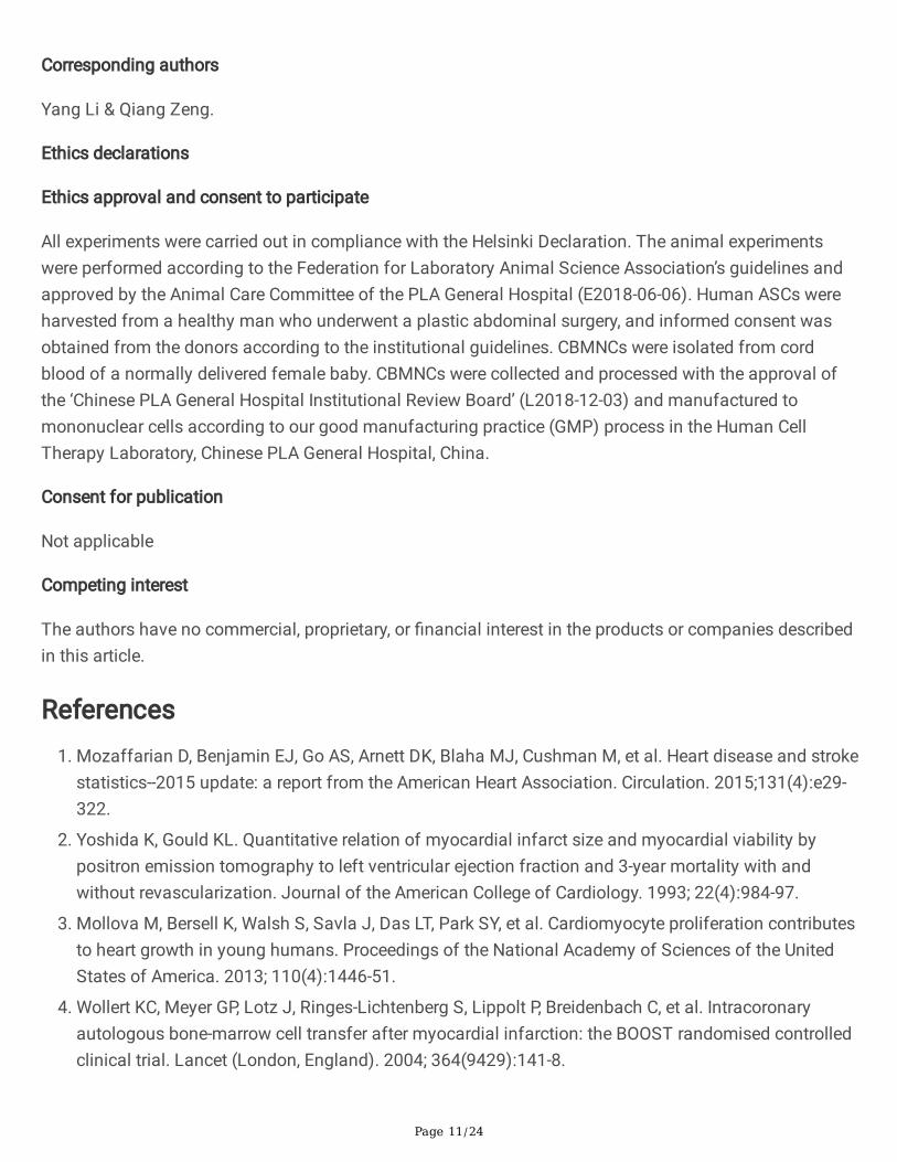
Figure 1
Surface markers and differentiation tests of ASCs. (A) Distribution of surface markers of ASCs wasdetected by �ow cytometry. (B) Differentiation test for adiposegenesis (red oil staining, x40),osteogenesis (alkaline phosphatase staining, x40), and chondrogenesis (Alcian blue staining, x40) ofASCs.
Page 16/24
Figure 1
Surface markers and differentiation tests of ASCs. (A) Distribution of surface markers of ASCs wasdetected by �ow cytometry. (B) Differentiation test for adiposegenesis (red oil staining, x40),osteogenesis (alkaline phosphatase staining, x40), and chondrogenesis (Alcian blue staining, x40) ofASCs.
Page 17/24
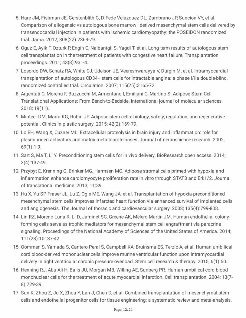
Figure 2
Gene expressions of HP-ASCs compared with N-ASCs. (A) Comparisons of HO-1 and SDF-1 expressions.(B) Comparisons of TIMP-1, CXCR4, ICAM-1and ICAM-2 expression. (C) Comparisons of EGF and EGFRexpression. (D) Comparisons of TGF (α, β) and TGF-β (R1, R2) expression. (E) Comparisons of FGF-β andFGF (R1, R2, R3, R4) expression. (F) Comparisons of IGF (1, 2) and IGF (R1, R2) expression. (G)
Page 18/24
Comparisons of VEGFA and VEGF (R1, R2, R3) expression. (H) Comparisons of myocardiocyte’s markers(CX43, GATA-4, MYH6, NKX2.5) expression. * p<0.05.
Figure 2
Gene expressions of HP-ASCs compared with N-ASCs. (A) Comparisons of HO-1 and SDF-1 expressions.(B) Comparisons of TIMP-1, CXCR4, ICAM-1and ICAM-2 expression. (C) Comparisons of EGF and EGFRexpression. (D) Comparisons of TGF (α, β) and TGF-β (R1, R2) expression. (E) Comparisons of FGF-β and
Page 19/24
FGF (R1, R2, R3, R4) expression. (F) Comparisons of IGF (1, 2) and IGF (R1, R2) expression. (G)Comparisons of VEGFA and VEGF (R1, R2, R3) expression. (H) Comparisons of myocardiocyte’s markers(CX43, GATA-4, MYH6, NKX2.5) expression. * p<0.05.
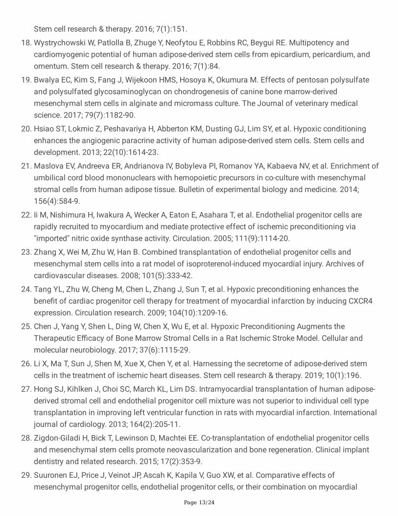
Figure 3
Echocardiographic assessment of cardiac function after myocardial infarction with stem cell transfusion.The change of cardiac functional parameters of mice were compared between before (0d) and after (30d)myocardial infarction with stem cell transfusion. The change of EF (A), FS (B), LVPWs (C), SV (D) and HR(E) were compared between each group. *p<0.05, compared with sham group; ** p<0.05, compared withPBS group. Abbreviations: EF, ejection fraction; FS, fractional shortening; LVPW, Left Ventricular PosteriorWall; SV, Stroke Volume; HR, Heart Rate.
Page 20/24
Figure 3
Echocardiographic assessment of cardiac function after myocardial infarction with stem cell transfusion.The change of cardiac functional parameters of mice were compared between before (0d) and after (30d)myocardial infarction with stem cell transfusion. The change of EF (A), FS (B), LVPWs (C), SV (D) and HR(E) were compared between each group. *p<0.05, compared with sham group; ** p<0.05, compared withPBS group. Abbreviations: EF, ejection fraction; FS, fractional shortening; LVPW, Left Ventricular PosteriorWall; SV, Stroke Volume; HR, Heart Rate.
Page 21/24
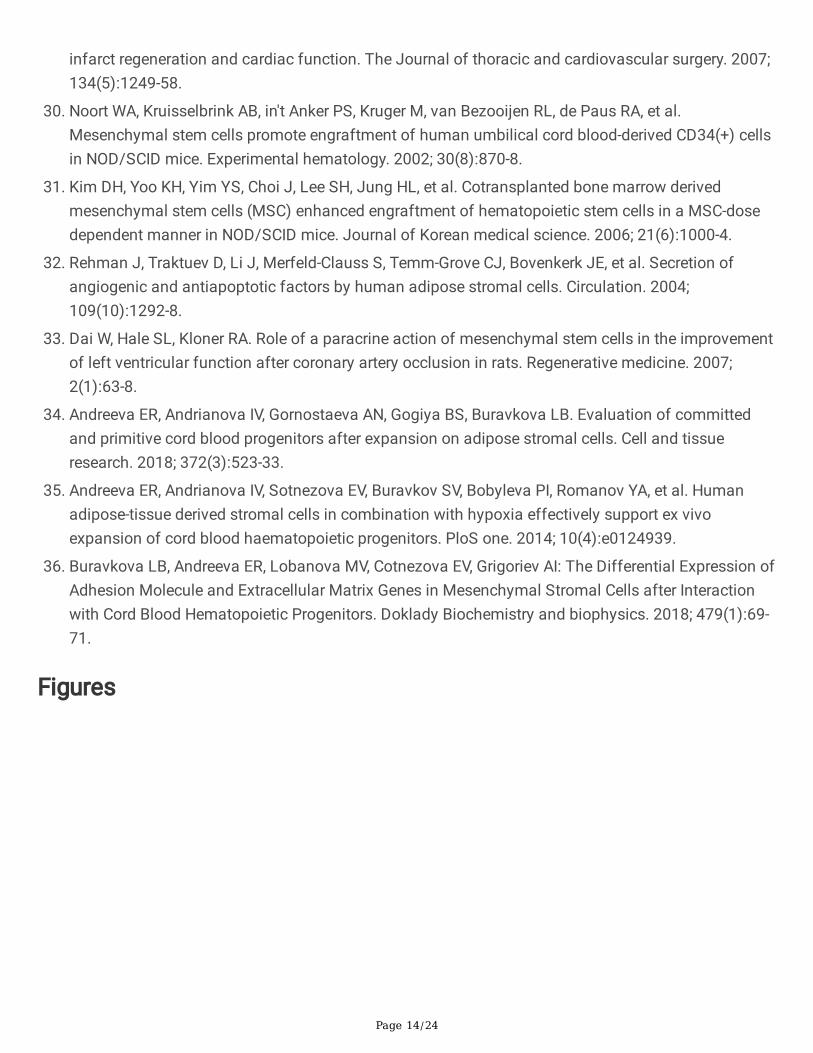
Figure 4
Analysis of cardiac �brosis 30 days after myocardial infarction. (A) General morphometric �brosis ofSham group, HP-ASCs group, HP-ASCs + CBMNCs group, CBMNCs group and PBS group. (B) Mousecardiac cross-sections assessed by Masson’s trichrome staining in each group. (C) Comparisons of thepercentage of the mouse LV �brotic size / LV area between groups. (D) Comparisons of the averagethickness of scars between groups. *p<0.05, compared with sham group; ** p<0.05, compared with PBSgroup.

Page 22/24
Figure 4
Analysis of cardiac �brosis 30 days after myocardial infarction. (A) General morphometric �brosis ofSham group, HP-ASCs group, HP-ASCs + CBMNCs group, CBMNCs group and PBS group. (B) Mousecardiac cross-sections assessed by Masson’s trichrome staining in each group. (C) Comparisons of thepercentage of the mouse LV �brotic size / LV area between groups. (D) Comparisons of the averagethickness of scars between groups. *p<0.05, compared with sham group; ** p<0.05, compared with PBSgroup.
Page 23/24
Figure 5
Comparison of live ASCs and new formed vessels 30 days after myocardial infarction with stem celltransfusions. (A) Live ASCs (from human male) were stained with anti-human SRY antibody, andcalculated under microscopy (x100). (B) New formed vessels (stained with anti-human CD31 antibody,green) were calculated under microscopy (x200). Nuclei were stained with DAPI(blue). (C) Living ASCsdensities (cells/mm2) were compared between HP-ASCs group, HP-ASCs + CBMNCs group and CBMNCsgroup. (D) The number of new formed vessels were compared between HP-ASCs group, HP-ASCs +CBMNCs group and CBMNCs group. *p <0.05.
Page 24/24
Figure 5
Comparison of live ASCs and new formed vessels 30 days after myocardial infarction with stem celltransfusions. (A) Live ASCs (from human male) were stained with anti-human SRY antibody, andcalculated under microscopy (x100). (B) New formed vessels (stained with anti-human CD31 antibody,green) were calculated under microscopy (x200). Nuclei were stained with DAPI(blue). (C) Living ASCsdensities (cells/mm2) were compared between HP-ASCs group, HP-ASCs + CBMNCs group and CBMNCsgroup. (D) The number of new formed vessels were compared between HP-ASCs group, HP-ASCs +CBMNCs group and CBMNCs group. *p <0.05.
Supplementary Files
This is a list of supplementary �les associated with this preprint. Click to download.
Supplementtable1.docx
Supplementtable1.docx
graphicabstract.tif
graphicabstract.tif