Embed Size (px)
Citation preview

Commentary 3733
IntroductionThe myosins are a superfamily of actin-based motor proteins thathave crucial roles in cellular and organismal physiology (Hartmanet al., 2011). Myosins typically consist of a head domain that canbind to actin filaments and generate force, a neck domain thatprovides binding sites for myosin light chains, and a tail that endows specific properties, such as dimerization and bindingto cargo. One particular group of myosins has tails that contain amyosin tail homology 4 (MyTH4) domain and a band 4.1, ezrin,radixin, moesin (FERM) domain. Members of this ‘MyTH-FERM’superclass of myosins are expressed in organisms ranging fromslime molds to humans (Breshears et al., 2010). Because the ciliateTetrahymena thermophila also expresses myosins that containMyTH4-FERM domains (Sugita et al., 2011; Williams and Gavin,2005), myosins with these domains either arose very early ineukaryotic evolution or independently in two different lineages.Importantly, studies in Dictyostelium discoideum indicate thatMyTH-FERM myosins have ancient and conserved roles inmediating membrane–cytoskeleton interactions in protrusivestructures such as filopodia (Tuxworth et al., 2001). Of the fourMyTH-FERM myosins that are expressed in humans – Myo7a,Myo7b, Myo10 and Myo15a – two are associated with humandisease, with mutations in Myo15a causing deafness (Wang et al.,1998) and mutations in Myo7a resulting in both deafness andblindness (Weil et al., 1995).
Myo10 is the founding member of the class X myosins and isthe only MyTH-FERM myosin with pleckstrin homology (PH)domains (Fig. 1). To follow the nomenclature convention forvertebrate myosins, which recommends that they be identified bythe gene symbol for their heavy chain (Gillespie et al., 2001), werefer to myosin-X as Myo10. As the gene names for conventionalmyosins in vertebrates use ‘Myh’ as a prefix, whereas the genenames for unconventional myosins use ‘Myo’ as a prefix, it is veryimportant to note that ‘Myh10’ corresponds to non-muscle myosin-IIb, not Myo10. Similarly, the Drosophila melanogaster myosin at
chromosomal locus 10A, which is sometimes referred to asDrosophila Myo10A, corresponds to a MyTH-FERM myosinknown as Myosin XV or Sisyphus.
Myo10 is present in organisms ranging from humans tochoanoflagellates (unicellular eukaryotes that are thought to beancestral to metazoa), but Myo10 appears to have been lost in thelineages leading to Drosophila and Caenorhabditis elegans(Odronitz and Kollmar, 2007). Although humans and mice expressonly a single Myo10 gene, zebrafish have three Myo10-like genes(Sittaramane and Chandrasekhar, 2008). Myo10 was originallydiscovered in a screen for myosins in the inner ear (Solc et al.,1994), but it is now known to be expressed in most cells andtissues (Berg et al., 2000; Yonezawa et al., 2000). Myo10 appearsto be expressed at quite low levels (Berg et al., 2000) and is thuslikely to be several orders of magnitude less abundant than themajor cellular myosins such as non-muscle myosin-II. Mostimportantly, growing evidence demonstrates that Myo10 is amolecular motor that has crucial functions in the slender actin-based extensions known as filopodia (Divito and Cheney, 2008).
Filopodia are cylindrical extensions of the plasma membranethat contain a bundle of parallel actin filaments at their core(Mattila and Lappalainen, 2008). Many cells appear to rely onthese finger-like organelles to probe and interact with theirsurroundings, especially in processes such as axon guidance andangiogenesis (Eilken and Adams, 2010; Koleske, 2003). The precisemechanisms that underlie filopodia formation and function,however, remain unclear. The actin filaments in filopodia areknown to have their barbed ends oriented towards the filopodial tip(Mattila and Lappalainen, 2008), and polymerization of actinmonomers at the tip leads to a constant rearwards movement ofactin filaments, which is known as retrograde flow or treadmilling(Medeiros et al., 2006). As discussed below, Myo10 is a MyTH-FERM myosin that localizes to filopodial tips, moves withinfilopodia and has potent filopodia-inducing activity (Berg et al.,2000; Bohil et al., 2006; Kerber et al., 2009). Here, we review key
SummaryMyosin-X (Myo10) is an unconventional myosin with MyTH4-FERM domains that is best known for its striking localization to thetips of filopodia and its ability to induce filopodia. Although the head domain of Myo10 enables it to function as an actin-based motor,its tail contains binding sites for several molecules with central roles in cell biology, including phosphatidylinositol (3,4,5)-trisphosphate,microtubules and integrins. Myo10 also undergoes fascinating long-range movements within filopodia, which appear to represent anewly recognized system of transport. Myo10 is also unusual in that it is a myosin with important roles in the spindle, a microtubule-based structure. Exciting new studies have begun to reveal the structure and single-molecule properties of this intriguing myosin, aswell as its mechanisms of regulation and induction of filopodia. At the cellular and organismal level, growing evidence demonstratesthat Myo10 has crucial functions in numerous processes ranging from invadopodia formation to cell migration.
Key words: Myosin-X, Myo10, Filopodia, MyTH4-FERM, Intrafilopodial motility
Journal of Cell Science 124, 3733–3741 © 2011. Published by The Company of Biologists Ltddoi:10.1242/jcs.023549
Myosin-X: a MyTH-FERM myosin at the tips offilopodiaMichael L. Kerber and Richard E. Cheney*Department of Cell and Molecular Physiology, School of Medicine, University of North Carolina at Chapel Hill, Chapel Hill, NC 27599-7545, USA*Author for correspondence ([email protected])
Jour
nal o
f Cel
l Sci
ence
progress in understanding Myo10, from its domain structure andbiophysical properties to its roles in cells and organisms.
Myo10 structure and biochemical propertiesThe Myo10 heavy chain has a molecular mass of ~237 kDa andcan be divided into a head, neck and tail (Fig. 1) (Berg et al., 2000;Yonezawa et al., 2000). The head consists of a conserved myosinmotor domain that can bind to F-actin, hydrolyze ATP and produceforce. The Myo10 head is most similar to that of Myo7a, withwhich it shares 45% identity. The Myo10 neck consists of three IQmotifs, each of which can bind to a calmodulin or calmodulin-likelight chain. The myosin neck domain is thought to act as a rigidlever that increases the myosin step size and is often involved inregulating motor activity. The Myo10 neck can bind to eithercalmodulin (Homma et al., 2001) or to an epithelia-specific proteinknown as calmodulin-like protein (CLP; encoded by CALML3)(Rogers and Strehler, 2001). CLP shares ~85% identity withcalmodulin, but exhibits approximately eightfold weaker bindingto Ca2+ (Rogers and Strehler, 2001). CLP binds preferentially tothe third IQ motif of Myo10 (Caride et al., 2010), and
overexpression of CLP increases Myo10 protein levels by severalfold, apparently by stabilizing the Myo10 heavy chain during itstranslation (Bennett et al., 2008; Bennett et al., 2007; Bennett andStrehler, 2008).
The Myo10 tail begins with a short segment of ~130 aminoacids that was initially predicted to form an -helical coiled-coilby studies using computer programs such as PAIRCOIL andCOILS. This suggested that Myo10 heavy chains can dimerize byway of a short stalk of coiled-coil (Berg et al., 2000). Detailedmanual analysis of this -helical region, however, revealed that itis unusually rich in charged residues and frequently lacks thehydrophobic residues that form the hydrophobic seam between thetwo -helices in a coiled-coil (Knight et al., 2005). The work byKnight et al. also indicated that at least the initial portion of the -helical region in Myo10 forms an intriguing structure known aseither a single -helix (SAH) (Knight et al., 2005) or an ER/Khelix (Sivaramakrishnan et al., 2008). Although an individual -helix was traditionally thought to be too compliant to stablyspan a large distance by itself, the alternating layers of positivelyand negatively charged residues found in a SAH endows greaterstability and stiffness, and there is a growing recognition that aSAH can act as a structural element to lengthen the myosin leverarm and increase the step size (Baboolal et al., 2009; Knight et al.,2005; Sivaramakrishnan et al., 2008). Importantly, a baculovirus-expressed Myo10 construct consisting of the head, neck and entire-helical region was largely monomeric when visualized byelectron microscopy, although ~10% of the molecules appeared toform dimers (Knight et al., 2005). These results indicate that theproximal portion of the -helical region forms a SAH, whereas thedistal portion might undergo regulated dimerization by forming acoiled-coil or related structure. This also indicates that Myo10constructs consisting of the head, neck and -helical region, whichare often referred to as ‘heavy meromyosin (HMM)-like’ becauseof their similarity to the dimeric HMM fragment of myosin-II, can exist as monomers, dimers or a mixture of both. Importantly,SAH domains are predicted in many proteins, including myosinssuch as Myo6 and Myo7a (Peckham and Knight, 2009;Sivaramakrishnan et al., 2008; Spink et al., 2008). Recent researchwith Myo6 indicates that its SAH uses electrostatic interactions toform parallel dimers that are offset by one turn of -helix (Kim et al., 2010), and rearrangements of more proximal -helicalsequences might create additional calmodulin-binding sites (Liu et al., 2011). Given that the length of the SAH in Myo10 and themechanism of dimerization are both unclear, an important goal forthe future will be to determine the precise structure(s) formed bythe -helical region.
The next segment of the Myo10 tail contains three PEST regions.PEST regions are sequences that are enriched in proline, glutamate,serine and threonine residues and are often associated with proteinsthat rapidly turnover (Rechsteiner and Rogers, 1996). Myo10 canbe cleaved by the calcium-dependent protease calpain at its PESTregions in vitro (Berg et al., 2000), but it is not yet known whetherthis occurs in vivo. Although the structure of the PEST regions inMyo10 is unknown, cleavage at the PEST regions would have thefunctionally interesting result of splitting a motorized HMM-likefragment away from the majority of the tail.
A cluster of three PH domains follows the PEST region (Berget al., 2000; Yonezawa et al., 2003). The PH2 domain in Myo10matches consensus sequences for binding to phosphatidylinositol(3,4,5)-trisphosphate [PtdIns(3,4,5)P3] (Isakoff et al., 1998; Park et al., 2008; Tacon et al., 2004), a key cell signaling molecule that
3734 Journal of Cell Science 124 (22)
Motor
α-helix
3� PEST 3� PH MyTH4 FERM
A
B1a 1b2 3
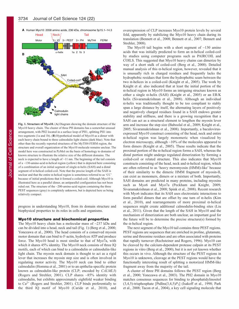
Human Myo10: 2058 amino acids, 236 kDa, chromosome 5p15.1–14.3
Head Neck Tail
Calmodulinlight chains
SAH
Coiled-coil?
3� IQ
Fig. 1. Structure of Myo10. (A)Diagram showing the domain structure of theMyo10 heavy chain. The cluster of three PH domains has a somewhat unusualarrangement, with PH2 located in a surface loop of PH1, splitting PH1 intotwo segments (1a and 1b). (B)Hypothetical model of Myo10 as a dimer witheach heavy chain bound to three calmodulin light chains (dark blue). Note thatother than the recently reported structures of the MyTH4-FERM region, thestructure and overall organization of the Myo10 molecule remains unclear. Themodel here was constructed in PyMol on the basis of homology to domains ofknown structure to illustrate the relative size of the different domains. Theneck is expected to have a length of ~11 nm. The beginning of the tail consistsof a ~130-amino-acid -helical region (yellow) that is depicted here consistingof a combination of an initial segment of single -helix (SAH) and a distalsegment of -helical coiled-coil. Note that the precise length of the SAH isunclear and that the entire -helical region is sometimes referred to as ‘CC’because of initial predictions that it formed a coiled-coil. Although Myo10 isillustrated here as a parallel dimer, an antiparallel configuration has not beenruled out. The structure of the ~200-amino-acid region containing the threePEST sequences (gray) is completely unknown, but is depicted here as beingrelatively compact.
Jour
nal o
f Cel
l Sci
ence

localizes primarily to the plasma membrane. Systematic screens ofPH domains also demonstrate that the PH2 domain of Myo10 canbe recruited to the plasma membrane by phosphoinositide 3-kinase(PI3K), the enzyme that generates PtdIns(3,4,5)P3 (Isakoff et al.,1998; Park et al., 2008). Although the PH2 domain of Myo10preferentially binds to PtdIns(3,4,5)P3 in lipid-binding assays, theisolated PH1 or PH2 domains show little interaction with otherphospholipids (Park et al., 2008; Plantard et al., 2010). Importantly,a construct consisting of all three PH domains is sufficient fortargeting to the plasma membrane, where it has a mean residencetime of ~20 seconds (Mashanov et al., 2004). Experiments withmacrophages show that endogenous Myo10 is recruited to thephagocytic cup downstream of PI3K and that Myo10 is requiredfor Fc-mediated phagocytosis (Cox et al., 2002). The PH domainsof Myo10 appear to contribute to its localization to filopodia(Plantard et al., 2010; Umeki et al., 2011), and exciting new datadiscussed below show that binding to PtdIns(3,4,5)P3 activatesMyo10 (Umeki et al., 2011).
The next domain in the Myo10 tail is a MyTH4 domain. MyTH4domains have a well-conserved primary structure and are almostalways located N-terminal to a FERM domain. MyTH4-FERMdomains are found in several proteins other than myosins, includinga family of plant kinesins in which they are implicated in targetingto microtubules (Narasimhulu and Reddy, 1998). The recentlypublished crystal structures of the MyTH4-FERM domains inMyo7a (Wu et al., 2011) and Myo10 (Hirano et al., 2011; Wei etal., 2011) are a major advance and the first to reveal the structureof a MyTH4 domain. These papers show that the MyTH4 domainforms an ~200-amino-acid helical bundle, and that the MyTH4 andFERM domains form a supramodule with highly conserved contactresidues. Functionally, the MyTH4 domain of Myo10 is sufficientfor binding to microtubules, although the MyTH4-FERMsupramodule showed stronger binding (Weber et al., 2004).Microtubule binding is mediated by a patch of positively chargedresidues in the MyTH4 domain that bind to the negatively charged E-hook region in the tails of - and -tubulin (Hirano et al., 2011). This ability to bind microtubules gives Myo10 theintriguing potential to act as a motorized link between actinfilaments and microtubules.
The tail of Myo10 ends in a FERM domain that can bind toseveral -integrins, a key family of cell surface receptors that areinvolved in cell adhesion and migration (Zhang et al., 2004). TheMyo10 FERM domain shares ~28% identity with the FERM
domain of talin, a major protein component of focal adhesions,which functions in ‘inside-out’ integrin signaling by binding to andactivating integrins. Like the FERM domain in talin, the FERMdomain in Myo10 binds to the NPXY motif that is present in thecytoplasmic tail of -integrins. Unlike talin, however, Myo10shows little or no localization to focal adhesions, the major sites ofintegrin localization in cells, and instead localizes to the tips offilopodia, where a small amount of integrin can be detected (Zhanget al., 2004). Together, these experiments indicate that Myo10 hasimportant roles in the early steps of integrin-mediated adhesion,where it appears to function in the transport and/or tethering ofintegrins in filopodia. Important unanswered questions are whetherMyo10 activates integrins and whether it competes or cooperateswith other integrin-binding proteins such as talin.
The Myo10 FERM domain also binds to the cytoplasmicdomains of the netrin receptors DCC (for deleted in colorectalcancer) and neogenin (Zhu et al., 2007). These guidance receptorshave key roles in many processes, and Myo10 has been shown tobe required for netrin-mediated axon guidance. The recently solvedcrystal structures of the Myo10 MyTH4-FERM supramodule boundto the P3 domain of DCC also provide the first views of aninteraction between Myo10 and a candidate cargo (Hirano et al.,2011; Wei et al., 2011). Unlike the talin–integrin interaction, inwhich a groove on the F3 lobe binds to a -strand formed by theNPXY motif, in the Myo10–DCC interaction the equivalent grooveon the F3 lobe binds to an -helix, thus revealing a new mode ofbinding for FERM domains. Biochemical experiments indicatethat DCC, integrin and microtubules all compete for binding to the MyTH4-FERM supramodule, strongly suggesting that thesupramodule can only bind to one of these candidate cargos at atime (Hirano et al., 2011).
Localization and dynamics of Myo10 infilopodiaAlthough the low abundance of endogenous Myo10 can makelocalization studies challenging, Myo10 clearly localizes to thetips of filopodia and retraction fibers (Fig. 2; supplementarymaterial Movie 1) (Berg et al., 2000; Tokuo and Ikebe, 2004;Zhang et al., 2004). In addition to filopodial tips, Myo10 alsoshows some localization to other regions of dynamic actin,including lamellipodia (Berg et al., 2000), invadopodia(Schoumacher et al., 2010) and phagocytic cups (Cox et al., 2002).Because Myo10 constructs that lack the motor domain fail to
3735The properties of myosin-X
5 μm 10 μm
B CControl siRNA Myo10 siRNAA Fig. 2. Myo10 localizes to filopodial tips and isrequired for filopodia formation.(A)Immunofluorescence image showing localization ofF-actin (red) and endogenous Myo10 (green) at the tipsof filopodia and along the leading edge of a bovine aorticendothelial cell. Image provided by Melinda DiVito,UNC Chapel Hill, NC. (B)Scanning electron microscopy(SEM) of a control HeLa cell showing a substrate-attached filopodium (arrowhead) and numerous dorsalfilopodia. (C)SEM of a Myo10-knockdown cell at thesame magnification showing a dramatic decrease indorsal filopodia. SEM images provided by Aparna Bohil,UNC Chapel Hill, NC. siRNA, small interfering RNA.
Jour
nal o
f Cel
l Sci
ence

localize to filopodial tips, it has been concluded that Myo10 usesits motor activity to transport itself along filopodial actin filamentsto the tips of filopodia (Berg and Cheney, 2002). Moreover, asHMM-like constructs consisting of the head, neck and -helicalregion are sufficient for tip localization, whereas a head–neckconstruct is not, tip localization was hypothesized to require bothmyosin motor activity and dimerization through some portion ofthe -helical region. Consistent with this, forced dimerization ofthe head–neck–SAH is indeed sufficient to trigger tip localization(Kerber et al., 2009; Tokuo et al., 2007).
Live-cell imaging shows that GFP–Myo10 is present at the tipsof filopodia as soon as they form and remains there as the filopodiaextend and retract (Berg and Cheney, 2002). Myo10 can alsoundergo several forms of movement within filopodia (Fig. 3). Thebright puncta of GFP–Myo10 that are normally present at the tipsof filopodia sometimes move slowly rearwards over a time courseof several minutes at rates of 10–20 nm/second, leading to thehypothesis that the slow rearwards movements are due to Myo10binding to filopodial actin filaments undergoing retrograde flow.This would provide a mechanism to allow Myo10 and anyassociated molecules to move rearwards, much like a package ona conveyor belt. Bright puncta of GFP–Myo10 also occasionallymove forwards towards the filopodial tip at velocities on the orderof ~80 nm/second, and this and other data indicate that Myo10uses its motor activity to move forwards along filopodial actin(Berg and Cheney, 2002; Sousa and Cheney, 2005; Tokuo andIkebe, 2004). Experiments with single-molecule sensitivity haverevealed a faster and more frequent form of forwards motility, inwhich extremely faint particles of GFP–Myo10 can be observedmoving towards the filopodial tip at ~600 nm/seconds, often alongthe entire length of a 5–10-m filopodium (Kerber et al., 2009).These experiments also indicate that each filopodial tip contains inthe order of 10–100 Myo10 molecules. Although it was unclearfrom these initial ‘single-molecule’ experiments whether the faintMyo10 particles corresponded to monomers or dimers, new dataindicate that the faint particles can exhibit two-step photobleaching,as expected for a dimer (Watanabe et al., 2010). In addition, invitro motility assays show that single molecules of HMM-like
forced dimers are capable of moving along actin at velocitiescomparable to those of the faint particles in cells (Nagy et al.,2008; Sun et al., 2010). The in vivo movements of the faintparticles of GFP–Myo10 provide one of the first examples of thedirect visualization of long-range movement of individual myosinmolecules in a living cell. These movements also raise the questionof whether other motors in cells are busily shuttling to-and-fro atthe single-molecule level.
Is Myo10 a component of an intrafilopodialtransport system?As filopodia and related structures, such as microvilli andstereocilia, generally lack microtubules and intracellular vesicles,directed transport in these structures is likely to depend on actinand actin-based motors (Kerber et al., 2009; Nambiar et al., 2010;Salles et al., 2009). One candidate cargo for Myo10 is vasodilator-stimulated phosphoprotein (VASP), a cytoplasmic protein that thatlocalizes to the tips of filopodia and lamellipodia. VASP has anti-capping activity and can stimulate filopodia formation (Applewhiteet al., 2007), and it binds to the tail of Myo10 (Tokuo and Ikebe,2004). Furthermore, in live-cell imaging experiments, bright punctaof VASP and Myo10 undergo co-transport in filopodia (Tokuo andIkebe, 2004), and single-molecule-level imaging shows that faintparticles of GFP–VASP move rapidly forwards in filopodia, muchlike faint particles of GFP–Myo10 (Kerber et al., 2009). Myo10also binds to and is implicated in the transport and/or localizationof several integral membrane proteins, including integrins (Zhanget al., 2004), the netrin receptors DCC and neogenin (Zhu et al.,2007), the bone morphogenetic protein (BMP) receptor activinreceptor-like kinase 6 (ALK6) (Pi et al., 2007) and vascularendothelial (VE)-cadherin (Almagro et al., 2010).
Motor and single-molecule propertiesThe localization and intrafilopodial motility of Myo10 raiseintriguing questions about its motor and biophysical properties.The initial studies with an HMM-like construct revealed thatMyo10, like most myosins, moves towards the barbed end of theactin filament (Homma et al., 2001). Detailed kinetic studies of a
3736 Journal of Cell Science 124 (22)
Extracellular matrix
Faint Myo10 particle
Cargo?
Bright Myo10puncta
Myo10tip spot
80 nm/s
600 nm/s
15 nm/sRetrograde actin flow
Filopodium
Fig. 3. Myo10 undergoes several forms of intrafilopodial motility. Model of Myo10 movements in filopodia. In HeLa cells, bright puncta or clusters of Myo10at tips of filopodia occasionally move rearwards within filopodia at the retrograde flow rate of ~15 nm/second. Bright puncta (areas of gray shading) have also beenobserved moving forwards within filopodia at ~80 nm/second. These puncta might include as-yet-undiscovered scaffolding components, actin polymerizationmachinery or cargos. Faint Myo10 particles (green), probably corresponding to dimers, move much faster (~600 nm/second) towards the filopodial tips. Forwardsmovements require Myo10 motor activity, whereas rearwards movements are hypothesized to be due to coupling to actin (red) undergoing retrograde flow. Myo10-interacting proteins (orange) include both cytoplasmic and membrane-associated proteins. See supplemental movie 5 of Kerber et al. for an animated model ofMyo10’s intrafilopodial motility (Kerber et al., 2009).
Jour
nal o
f Cel
l Sci
ence

head–3IQ construct reported a duty ratio of ~16% (Kovacs et al.,2005), and similar studies using a head–1IQ construct yielded aduty ratio of 60–70% (Homma and Ikebe, 2005). The duty ratiocorresponds to the fraction of the ATPase cycle that the motor istightly bound to the filament, so the value of 60–70% suggests thata Myo10 dimer can walk processively along a filament byalternating steps of its two heads. The kinetic studies also revealedthat the ‘weak’ binding state of Myo10 has an unusually strongaffinity for actin filaments, a property that could potentially keepit associated with actin and enhance processivity. Given the limitedspace for diffusion within a filopodium, even a moderatelyprocessive myosin might eventually succeed in moving to the tipas long as it moves fast enough to overcome retrograde flow.
In an exciting recent advance, full-length Myo10 has beenexpressed in baculovirus (Umeki et al., 2011). This revealed thatpurified Myo10 forms a compact monomer in which the Myo10head binds to the PH and FERM domains in the tail. An HMM-like construct that was lacking most of the tail had an actin-activated ATPase activity of 20 ATP molecules per second, fivefoldhigher than that of full-length Myo10. Although the ATPase activityof full-length Myo10 was found not to be regulated by Ca2+, it wasactivated by membranes containing either PtdIns(3,4,5)P3 orphosphatidylinositol (4,5)-bisphosphate [PtdIns(4,5)P2], andcrosslinking showed that binding to these membranes inducedMyo10 to form dimers (Umeki et al., 2011). This suggests ageneral model of Myo10 regulation in which membranes enrichedin PtdIns(3,4,5)P3 or PtdIns(4,5)P2 recruit Myo10 and convert itfrom a compact monomer into an active dimer (Fig. 4). This wouldbe analogous to the microtubule motor UNC-104/KIF1a, which isthought to exist as a monomer until it is targeted to PtdIns(4,5)P2-enriched vesicles by its PH domains, leading to an increased localconcentration and the formation of processive dimers (Tomishigeet al., 2002). Conversion from a compact monomer into an activedimer appears to be a general regulatory mechanism that is usedby many different motors, including Myo6 (Park et al., 2006; Yuet al., 2009) and Myo7a (Umeki et al., 2009; Yang et al., 2009).Intriguingly, recent studies with Myo7a indicate that binding to acargo induces this MyTH-FERM myosin to form dimers andlocalize to the tips of filopodia (Sakai et al., 2011).
A series of recent papers has begun to shed light, and controversy,on the single-molecule properties of Myo10 and the mechanismsthat target it to filopodia. Using in vitro motility assays and anHMM–Myo10 construct (amino acids 1–920) that was forced todimerize through a leucine zipper, single molecules were shown to move on individual actin filaments or actin bundles at velocitiesof 330–780 nm/second (Nagy et al., 2008). Interestingly, thisconstruct also moved along filopodial actin bundles in detergent-permeabilized cells, although it appeared to run off the ends withoutpausing at the tip (Brawley and Rock, 2009). This construct alsoshowed selectivity for actin bundles because its run length onbundles (630 nm) is fourfold longer than on single filaments,
whereas Myo5a, a well-studied processive myosin, has similar runlengths (~600 nm) on either form of actin. This raised the possibilitythat Myo10 moves on bundles using a ‘straddle’ mechanism, inwhich the two heads bind to separate filaments (Nagy and Rock,2010). The HMM–Myo10 forced dimer had an average step sizeof ~18 nm, too short to easily span the ~36 nm pseudohelicalrepeat of the actin filament (Nagy and Rock, 2010; Ricca andRock, 2010). Unlike the ~36 nm step of Myo5a, which is ideal forstepping along one face of a single filament, an ~18 nm step issterically awkward as it would force a myosin head to reach aroundto the back side of the filament. On a bundle, however, only theouter faces of actin filaments are available for binding and adjacentfilaments provide additional binding sites, so an ~18 nm stepwould favor ‘straddling’ and might help direct Myo10 to filopodia.
Work from another group using a different HMM-like forceddimer, however, showed little or no selectivity for bundles (Sunand Goldman, 2011; Sun et al., 2010). The construct used in theseexperiments included amino acids 1–939 and was forced todimerize through a coiled-coil sequence from Myo5a; similarresults were also reported for a ‘native’ HMM-like Myo10construct. Step-size measurements with the forced dimer showedthat it took large steps of ~34 nm on single filaments. On bundles,this forced dimer exhibited both ~34 nm and ~20 nm steps, withthe shorter steps attributed to stepping to adjacent filaments. Theseresults challenge the straddling model, and suggest that othermechanisms might target Myo10 to filopodia. Given thattropomyosin regulates the binding of myosin to actin in muscle, itwould be interesting to determine whether targeting of Myo10 tofilopodia involves tropomyosins or other actin-binding proteins.Although the basis for the differences in step size and selectivityreported by the two groups are unclear, it is possible that differences in the sites and methods of forced dimerization lead todifferences in the structure of the -helical region or the precisepositioning of the head domains. Sun and colleagues also usedsophisticated biophysical approaches to show that their forced-dimer construct uses a hand-over-hand mechanism to moveprocessively at ~200 nm/seconds with a run length of ~1000 nm(Sun et al., 2010). These authors also report the important findingthat binding to actin can induce their ‘native’ HMM-like Myo10 toform processive dimers (Sun et al., 2010), a result remarkablysimilar to the proximity-induced dimerization seen with Myo6(Park et al., 2006).
Myo10 induces filopodia by multiplemechanismsIn addition to its abilities to move on actin filaments, Myo10exhibits potent filopodia-inducing activity. Light microscopy wasinitially used to show that overexpressing full-length Myo10, butnot an HMM-like construct or the tail alone, increased the numberand length of substrate-attached filopodia (Berg and Cheney, 2002).The ability of Myo10 to promote substrate-attached filopodia
3737The properties of myosin-X
PtdIns(3,4,5)P3 Dimerization
Compact,inactive monomer
Extended monomer Active dimer
PtdIns(3,4,5)P3-rich plasma membrane
Fig. 4. Model for Myo10 regulation. The tail of Myo10 binds to itshead domain, forming a compact monomer that is enzymaticallyinactive (Umeki et al., 2011). Binding of the Myo10 tail tomembranes containing PtdIns(3,4,5)P3 or PtdIns(4,5)P2 disrupts theintramolecular interactions between the head and tail, inducing theformation of an extended monomer. This, plus the increased localconcentration of Myo10 resulting from its recruitment tomembranes, is thought to lead to the formation of an active dimerthat can move processively along actin.
Jour
nal o
f Cel
l Sci
ence
depends largely on its ability to bind to integrins through its FERMdomain (Zhang et al., 2004), indicating that Myo10 can act byusing a ‘sticky fingers’ mechanism that promotes adhesion offilopodia to the substrate and thus stabilizes them. Consistent withthis, an analysis of the dynamics of filopodia in cells transfectedwith either full-length Myo10 or a FERM deletion construct showsthat the full-length Myo10 increases the ability of filopodia that areextending along the substrate to undergo a second round ofextension by approximately fivefold (Watanabe et al., 2010).
To test whether Myo10 also promotes filopodia induction bymechanisms that are independent of adhesion, scanning electronmicroscopy (SEM) was used to visualize dorsal filopodia (i.e.filopodia that are not attached to the substrate) in cells transfectedwith Myo10 or with a FERM deletion construct. Both constructsled to massive increases in dorsal filopodia, demonstrating thatMyo10 can promote filopodia independently of adhesion or integrinbinding (Bohil et al., 2006). Myo10 also appeared to actdownstream of the Rho GTPase Cdc42 and independently of VASPproteins. Because an HMM-like construct failed to induce dorsalfilopodia, elements of the Myo10 tail upstream of the FERMdomain, such as the MyTH4 domain, appear to be important forthe formation or stabilization of dorsal filopodia (Bohil et al.,2006), and recent data indicate roles for the PH domains (Umekiet al., 2011). The Myo10 tail could potentially stimulate filopodiaformation through a variety of mechanisms, including regulatingmotor activity, inducing dimerization and/or transporting proteinsthat are important for filopodia formation.
Importantly, the induced dimerization of a Myo10 constructconsisting of the head, neck and SAH is sufficient to trigger bothtip localization and a pulse of short-lived filopodia (Tokuo et al.,2007). This result suggests that Myo10 dimers initiate filopodiaformation by using their two heads to link the barbed ends oflamellipodial actin filaments together, thus triggering the formationof filopodial actin bundles. Consistent with this role, GFP–Myo10has been observed in structures that resemble -precursors (the -shaped precursors to filopodial actin bundles) (Sousa et al.,2006), and GFP–Myo10 accumulates rapidly at sites of filopodialinitiation (Watanabe et al., 2010). Importantly, endogenous Myo10 is required for filopodia formation because knockdown ofMyo10 dramatically decreases the otherwise numerous dorsalfilopodia in HeLa cells (Bohil et al., 2006) (Fig. 2). New data showthat Myo10 is also required for the formation of invadopodia,actin-based protrusions that share several similarities with filopodiaand that function in the digestion of extracellular matrix andmetastasis of cancer cells (Schoumacher et al., 2010). Myo10function is also required for the patterning of podosomes(McMichael et al., 2010), invadopodia-like structures that areinvolved in integrin-dependent adhesion in cells such as osteoclasts.
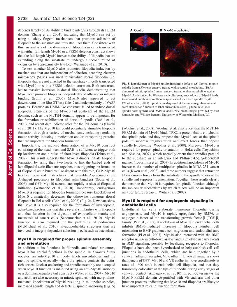
Myo10 is required for proper spindle assemblyand orientationIn addition to its functions in filopodia and related structures,Myo10 has crucial functions in the spindle. In Xenopus laevisoocytes, an anti-Myo10 antibody labels microtubules and themeiotic spindle, especially where the spindle contacts the actin-rich cortex. Nuclear anchoring and spindle assembly are disruptedwhen Myo10 function is inhibited using an anti-Myo10 antibodyor a dominant-negative tail construct (Weber et al., 2004). Myo10and actin are also necessary in mitotic spindles, with morpholino-mediated knockdown of Myo10 resulting in multipolar spindles,increased spindle length and defects in spindle anchoring (Fig. 5)
(Woolner et al., 2008). Woolner et al. also report that the MyTH4-FERM domain of Myo10 binds TPX2, a protein that is enriched inthe spindle pole, and they propose that Myo10 acts at the spindlepole to suppress fragmentation and exert forces that oppose spindle lengthening (Woolner et al., 2008). Moreover, Myo10 isrequired for proper spindle orientation in HeLa cells (Toyoshimaand Nishida, 2007), which normally orient their spindles parallelto the substrate in an integrin- and PtdIns(3,4,5)P3-dependentmanner (Toyoshima et al., 2007). In addition, knockdown of Myo10increases the number of multipolar spindles in MDA-231 cancercells (Kwon et al., 2008), and these authors suggest that retractionfibers convey forces from the substrate to the spindle to orient thespindle and suppress fragmentation. Together, these results clearlydemonstrate that Myo10 is required for spindle function, althoughthe molecular mechanisms by which it acts will be an importantarea for future research (Wuhr et al., 2008).
Myo10 is required for angiogenic signaling inendothelial cellsEndothelial tip cells elaborate numerous filopodia duringangiogenesis, and Myo10 is rapidly upregulated by BMP6, anangiogenic factor of the transforming growth factor- (TGF-)family (Pi et al., 2007). Knockdown of Myo10 in endothelial cellsinhibits BMP6-mediated increases in filopodia number, cellorientation to BMP gradients, cell migration and endothelial tubeformation (Pi et al., 2007). Myo10 also interacted with the BMPreceptor ALK6 in pull-down assays, and is involved in early eventsin BMP signaling, possibly by localizing receptors to filopodia.Filopodia have also been hypothesized to help establish cell–celljunctions in endothelial cells, which are held together by a cell–cell adhesion receptor, VE-cadherin. Live-cell imaging showsthat puncta of GFP–Myo10 and VE-cadherin move coordinately atrates of ~600 nm/s in endothelial-cell filopodia, and that theytransiently colocalize at the tips of filopodia during early stages ofcell–cell contact (Almagro et al., 2010). In pull-down assays theMyo10 FERM domain co-purified with VE-cadherin and otherjunction proteins, indicating that Myo10 and filopodia are likely tohave important roles in junction formation.
3738 Journal of Cell Science 124 (22)
A Control B Myo10 morpholino
5 μm
5 μm
Fig. 5. Knockdown of Myo10 results in spindle defects. (A)Normal mitoticspindle from a Xenopus embryo treated with a control morpholino. (B)Anabnormal mitotic spindle from an embryo treated with a morpholino againstMyo10. As described by Woolner and colleagues, knockdown of Myo10 leadsto increased numbers of multipolar spindles and increased spindle length(Woolner et al., 2008). Spindles are displayed at the same magnification andwere stained for -tubulin to label microtubules (red), -tubulin to labelspindle poles (green), and DAPI to label DNA (blue). Images provided by JoshSandquist and William Bement, University of Wisconsin, Madison, WI.
Jour
nal o
f Cel
l Sci
ence

Myo10 functions in melanosome transfer inmelanocytesIn addition to acting as cellular sensors and contact points, filopodiacan also serve as passageways for the transport of materials betweencells. For example, melanocytes generate filopodia-like extensionsthat allow Myo5a to transport melanosomes towards their tips, andin a striking example of inter-cellular organelle transfer, thesurrounding keratinocytes internalize the filopodia by a processthat appears to be similar to phagocytosis. Myo10 was recentlyreported to function in melanosome transfer (Singh et al., 2010),with exposure to UV light increasing Myo10 expression, thenumber of filopodia and melanosome transfer, whereas knockdownof Myo10 decreased the number of filopodia and melanosometransfer. Although Myo10 was required for induction of filopodiain melanocytes, it might also have a role in ‘phagocytosis’ by thekeratinocytes. Viruses such as HIV can also move from cell to cellusing filopodia-like structures (Lehmann et al., 2005; Sherer et al.,2007; Sherer and Mothes, 2008), although the role of Myo10 inthis process has not yet been tested.
Functions of Myo10 in neuronsFilopodia are thought to be especially important in neurons forgrowth cone guidance and synapse formation. Myo10 isdevelopmentally regulated in brain, with relatively high levels inmouse cerebrum during the first two weeks after birth (Sousa etal., 2006), a peak period of synapse formation, when neuronselaborate an estimated 50,000 filopodia per day (Portera-Cailliauet al., 2003). Myo10 expression then drops to much lower levelsin adult cerebrum. Brain also expresses a ‘headless’ form of Myo10that lacks most of the head domain and thus is unable to act as amotor, localize to filopodial tips or induce filopodia (Sousa et al.,2006). Headless Myo10 has been hypothesized to act either as ascaffolding molecule or as a naturally occurring dominant negative(Sousa et al., 2006). Although the numerous binding partners ofMyo10, such as integrin, can make interpretation of dominant-negative approaches challenging, motorless Myo10 constructsclearly inhibit netrin-dependent axon outgrowth (Zhu et al., 2007)as well as formation of focal adhesions and chemotaxis in aneuronal cell line (Wang et al., 2009). Axon guidance depends onboth actin and microtubules, with filopodia being especiallyimportant for axonal path-finding (Dent and Gertler, 2003).Although cultures of cortical neurons that lack all three membersof the VASP family fail to form filopodia or generate neurites,expressing full-length Myo10 rescues both filopodia formationand neuritogenesis (Dent et al., 2007). Myo10 is also upregulatedapproximately sevenfold following nerve injury, suggesting that itfunctions in nerve regeneration.
Myo10 is required for early development andneural crest migrationAlthough there are currently no Myo10-knockout animals, studieswith morpholino-mediated knockdown in Xenopus embryos hasrevealed that Myo10 has important functions in development(Hwang et al., 2009; Nie et al., 2009). During early development,Myo10 is detected primarily in neural crest and paraxial mesoderm.Neural crest cells are highly migratory cells that give rise to severalcell types, including craniofacial cartilage and bone, peripheralneurons and melanocytes. Although loss of maternal Myo10appeared to be lethal, knockdown of zygotic Myo10 inhibitedneural crest cell migration and resulted in a dramatic decrease incranium size (Hwang et al., 2009). Consistent with the ability of
Myo10 to bind integrins and promote filopodia, plating theknockdown cells on fibronectin revealed defects in adhesion, cellspreading, filopodia formation and cell migration (Hwang et al.,2009; Nie et al., 2009).
Conclusion and perspectivesResearch over the past decade clearly demonstrates that Myo10 isa MyTH-FERM myosin with central roles in filopodia. Thelocalization of Myo10 at the filopodial tip indicates that there is alargely uncharacterized filopodial tip complex that is likely tofunction in polymerization, adhesion and cell signaling. Much hasbeen revealed about the basic properties of Myo10, and themechanisms by which it targets to and induces filopodia arebeginning to be unraveled. Exciting new theoretical studies indicatethat the lengths of filopodia should be limited by the diffusion ofproteins, such as actin, that are consumed during filopodial growth;motor proteins, such as Myo10, could overcome these limits ifthey actively transport such proteins to the tip (Zhuravlev et al.,2010; Zhuravlev and Papoian, 2009). The intrafilopodialmovements of Myo10 raise intriguing questions about this form ofmotility and the identity of Myo10 cargos. In addition, these resultsraise the larger question of whether structures such as filopodia,microvilli and stereocilia use myosin motors to power transportsystems that are analogous to intraflagellar transport, a transportsystem that is central to the formation of cilia and that is defectivein a large number of human diseases. Biophysical studies of Myo10are revealing fundamental mechanisms of motor regulation, suchas induced dimerization and the roles of a new structural element,the SAH. Work with Myo10 also raises the question of the steppingpatterns myosins use when moving on an array of filaments, anissue that could be investigated by tagging the two heads of aMyo10 dimer with different colors. The ability of Myo10 to bindto microtubules, and its functions in spindle orientation, also raisefundamental questions about the roles of actin and myosin in thespindle. It also remains to be seen whether Myo10 interacts withmicrotubules in interphase cells. As a myosin that binds to integrins,acts downstream of PI3K and is required for invadopodia formation,it will be important to investigate the functions of Myo10 in cellsignaling and in disease processes such as cancer. Althoughmorpholino studies in Xenopus embryos demonstrate importantfunctions for Myo10 in developmental processes, such as neuralcrest migration, the generation of knockout or conditionally nullanimal models would greatly facilitate studies of the physiologicalfunctions of Myo10. Finally, research with Myo10 might be justthe tip of the iceberg, as a growing number of unconventionalmyosins, including Myo3a (Salles et al., 2009), Myo7a (Sakai et al., 2011) and Myo15a (Belyantseva et al., 2005), appear to actas barbed-end-tracking proteins and have important functions inbundle-based protrusions.
Note added in proofThe structure of the PH1–PH2 region has recently been reported,and this region forms a rigid supramodule whose lipid-bindingpockets are positioned side-by-side, allowing Myo10 to bind tophosphatidylinositol (3,4,5)-trisphosphate with high specificity andavidity (Lu et al., 2011).
Funding
Our work is supported by grants from the National Institutes of Heath,National Institute on Deafness and Other Communication Disorders[grant number R01-DC03299 to R.E.C.]; and National Institutes of
3739The properties of myosin-X
Jour
nal o
f Cel
l Sci
ence

Heath, National Heart, Lung, and Blood Institute [grant number P01-HL080166 to R.E.C.]. Deposited in PMC for release after 12 months.
Supplementary material available online athttp://jcs.biologists.org/lookup/suppl/doi:10.1242/jcs.023549/-/DC1
ReferencesAlmagro, S., Durmort, C., Chervin-Petinot, A., Heyraud, S., Dubois, M., Lambert,
O., Maillefaud, C., Hewat, E., Schaal, J. P., Huber, P. et al. (2010). The motor proteinmyosin-X transports VE-cadherin along filopodia to allow the formation of earlyendothelial cell-cell contacts. Mol. Cell. Biol. 30, 1703-1717.
Applewhite, D. A., Barzik, M., Kojima, S., Svitkina, T. M., Gertler, F. B. and Borisy,G. G. (2007). Ena/VASP proteins have an anti-capping independent function in filopodiaformation. Mol. Biol. Cell 18, 2579-2591.
Baboolal, T. G., Sakamoto, T., Forgacs, E., White, H. D., Jackson, S. M., Takagi, Y.,Farrow, R. E., Molloy, J. E., Knight, P. J., Sellers, J. R. et al. (2009). The SAHdomain extends the functional length of the myosin lever. Proc. Natl. Acad. Sci. USA106, 22193-22198.
Belyantseva, I. A., Boger, E. T., Naz, S., Frolenkov, G. I., Sellers, J. R., Ahmed, Z. M.,Griffith, A. J. and Friedman, T. B. (2005). Myosin-XVa is required for tip localizationof whirlin and differential elongation of hair-cell stereocilia. Nat. Cell Biol. 7, 148-156.
Bennett, R. D. and Strehler, E. E. (2008). Calmodulin-like protein enhances myosin-10translation. Biochem. Biophys. Res. Commun. 369, 654-659.
Bennett, R. D., Mauer, A. S. and Strehler, E. E. (2007). Calmodulin-like proteinincreases filopodia-dependent cell motility via up-regulation of myosin-10. J. Biol.Chem. 282, 3205-3212.
Bennett, R. D., Caride, A. J., Mauer, A. S. and Strehler, E. E. (2008). Interaction withthe IQ3 motif of myosin-10 is required for calmodulin-like protein-dependent filopodialextension. FEBS Lett. 582, 2377-2381.
Berg, J. S. and Cheney, R. E. (2002). Myosin-X is an unconventional myosin thatundergoes intrafilopodial motility. Nat. Cell Biol. 4, 246-250.
Berg, J. S., Derfler, B. H., Pennisi, C. M., Corey, D. P. and Cheney, R. E. (2000).Myosin-X, a novel myosin with pleckstrin homology domains, associates with regionsof dynamic actin. J. Cell Sci. 113, 3439-3451.
Bohil, A. B., Robertson, B. W. and Cheney, R. E. (2006). Myosin-X is a molecular motorthat functions in filopodia formation. Proc. Natl. Acad. Sci. USA 103, 12411-12416.
Brawley, C. M. and Rock, R. S. (2009). Unconventional myosin traffic in cells reveals aselective actin cytoskeleton. Proc. Natl. Acad. Sci. USA 106, 9685-9690.
Breshears, L. M., Wessels, D., Soll, D. R. and Titus, M. A. (2010). An unconventionalmyosin required for cell polarization and chemotaxis. Proc. Natl. Acad. Sci. USA 107,6918-6923.
Caride, A. J., Bennett, R. D. and Strehler, E. E. (2010). Kinetic analysis revealsdifferences in the binding mechanism of calmodulin and calmodulin-like protein to theIQ motifs of myosin-10. Biochemistry 49, 8105-8116.
Cox, D., Berg, J. S., Cammer, M., Chinegwundoh, J. O., Dale, B. M., Cheney, R. E.and Greenberg, S. (2002). Myosin X is a downstream effector of PI(3)K duringphagocytosis. Nat. Cell Biol. 4, 469-477.
Dent, E. W. and Gertler, F. B. (2003). Cytoskeletal dynamics and transport in growthcone motility and axon guidance. Neuron 40, 209-227.
Dent, E. W., Kwiatkowski, A. V., Mebane, L. M., Philippar, U., Barzik, M., Rubinson,D. A., Gupton, S., Van Veen, J. E., Furman, C., Zhang, J. et al. (2007). Filopodiaare required for cortical neurite initiation. Nat. Cell Biol. 9, 1347-1359.
Divito, M. M. and Cheney, R. E. (2008). Myosin10. In Myosins: A Superfamily ofMolecular Motors (ed. L. M. Coluccio), pp. 403-419. Dordrecht, The Netherlands:Springer.
Eilken, H. M. and Adams, R. H. (2010). Dynamics of endothelial cell behavior insprouting angiogenesis. Curr. Opin. Cell Biol. 22, 617-625.
Gillespie, P. G., Albanesi, J. P., Bahler, M., Bement, W. M., Berg, J. S., Burgess, D.R., Burnside, B., Cheney, R. E., Corey, D. P., Coudrier, E. et al. (2001). Myosin-Inomenclature. J. Cell Biol. 155, 703-704.
Hartman, M. A., Finan, D., Sivaramakrishnan, S. and Spudich, J. A. (2011). Principlesof unconventional myosin function and targeting. Annu. Rev. Cell Dev. Biol. 27, 133-155.
Hirano, Y., Hatano, T., Takahashi, A., Toriyama, M., Inagaki, N. and Hakoshima, T.(2011). Structural basis of cargo recognition by the myosin-X MyTH4-FERM domain.EMBO J. 30, 2734-2747.
Homma, K. and Ikebe, M. (2005). Myosin X is a high duty ratio motor. J. Biol. Chem.280, 29381-29391.
Homma, K., Saito, J., Ikebe, R. and Ikebe, M. (2001). Motor function and regulation ofmyosin X. J. Biol. Chem. 276, 34348-34354.
Hwang, Y. S., Luo, T., Xu, Y. and Sargent, T. D. (2009). Myosin-X is required for cranialneural crest cell migration in Xenopus laevis. Dev. Dyn. 238, 2522-2529.
Isakoff, S. J., Cardozo, T., Andreev, J., Li, Z., Ferguson, K. M., Abagyan, R., Lemmon,M. A., Aronheim, A. and Skolnik, E. Y. (1998). Identification and analysis of PHdomain-containing targets of phosphatidylinositol 3-kinase using a novel in vivo assayin yeast. EMBO J. 17, 5374-5387.
Kerber, M. L., Jacobs, D. T., Campagnola, L., Dunn, B. D., Yin, T., Sousa, A. D.,Quintero, O. A. and Cheney, R. E. (2009). A novel form of motility in filopodiarevealed by imaging myosin-X at the single-molecule level. Curr. Biol. 9, 967-973.
Kim, H., Hsin, J., Liu, Y., Selvin, P. R. and Schulten, K. (2010). Formation of saltbridges mediates internal dimerization of myosin VI medial tail domain. Structure 18,1443-1449.
Knight, P. J., Thirumurugan, K., Xu, Y., Wang, F., Kalverda, A. P., Stafford, W. F.,3rd, Sellers, J. R. and Peckham, M. (2005). The predicted coiled-coil domain ofmyosin 10 forms a novel elongated domain that lengthens the head. J. Biol. Chem. 280,34702-34708.
Koleske, A. J. (2003). Do filopodia enable the growth cone to find its way? Sci. STKE2003, pe20.
Kovacs, M., Wang, F. and Sellers, J. R. (2005). Mechanism of action of myosin X, amembrane-associated molecular motor. J. Biol. Chem. 280, 15071-15083.
Kwon, M., Godinho, S. A., Chandhok, N. S., Ganem, N. J., Azioune, A., Thery, M.and Pellman, D. (2008). Mechanisms to suppress multipolar divisions in cancer cellswith extra centrosomes. Genes Dev. 22, 2189-2203.
Lehmann, M. J., Sherer, N. M., Marks, C. B., Pypaert, M. and Mothes, W. (2005).Actin- and myosin-driven movement of viruses along filopodia precedes their entry intocells. J. Cell Biol. 170, 317-325.
Liu, Y., Hsin, J., Kim, H., Selvin, P. R. and Schulten, K. (2011). Extension of a three-helix bundle domain of myosin VI and key role of calmodulins. Biophys. J. 100, 2964-2973.
Lu, Q., Yu, J., Yan, J., Wei, Z. and Zhang, M. (2011). Structural basis of the myosin-XPH1N-PH2-PH1C tandem as a specific and acute cellular PI(3,4,5)P3 sensor. Mol. Biol.Cell (in press).
Mashanov, G. I., Tacon, D., Peckham, M. and Molloy, J. E. (2004). The spatial andtemporal dynamics of pleckstrin homology domain binding at the plasma membranemeasured by imaging single molecules in live mouse myoblasts. J. Biol. Chem. 279,15274-15280.
Mattila, P. K. and Lappalainen, P. (2008). Filopodia: molecular architecture and cellularfunctions. Nat. Rev. Mol. Cell Biol. 9, 446-454.
McMichael, B. K., Cheney, R. E. and Lee, B. S. (2010). Myosin X regulates sealing zonepatterning in osteoclasts through linkage of podosomes and microtubules. J. Biol.Chem. 285, 9506-9515.
Medeiros, N. A., Burnette, D. T. and Forscher, P. (2006). Myosin II functions in actin-bundle turnover in neuronal growth cones. Nat. Cell Biol. 8, 215-226.
Nagy, S. and Rock, R. S. (2010). Structured post-IQ domain governs selectivity of myosinX for fascin-actin bundles. J. Biol. Chem. 285, 26608-26617.
Nagy, S., Ricca, B. L., Norstrom, M. F., Courson, D. S., Brawley, C. M., Smithback,P. A. and Rock, R. S. (2008). A myosin motor that selects bundled actin for motility.Proc. Natl. Acad. Sci. USA 105, 9616-9620.
Nambiar, R., McConnell, R. E. and Tyska, M. J. (2010). Myosin motor function: theins and outs of actin-based membrane protrusions. Cell. Mol. Life Sci. 67, 1239-1254.
Narasimhulu, S. B. and Reddy, A. S. (1998). Characterization of microtubule bindingdomains in the Arabidopsis kinesin-like calmodulin binding protein. Plant Cell 10, 957-965.
Nie, S., Kee, Y. and Bronner-Fraser, M. (2009). Myosin-X is critical for migratoryability of Xenopus cranial neural crest cells. Dev. Biol. 335, 132-142.
Odronitz, F. and Kollmar, M. (2007). Drawing the tree of eukaryotic life based on theanalysis of 2,269 manually annotated myosins from 328 species. Genome Biol. 8, R196.
Park, H., Ramamurthy, B., Travaglia, M., Safer, D., Chen, L. Q., Franzini-Armstrong,C., Selvin, P. R. and Sweeney, H. L. (2006). Full-length myosin VI dimerizes andmoves processively along actin filaments upon monomer clustering. Mol. Cell 21, 331-336.
Park, W. S., Heo, W. D., Whalen, J. H., O’Rourke, N. A., Bryan, H. M., Meyer, T. andTeruel, M. N. (2008). Comprehensive identification of PIP3-regulated PH domainsfrom C. elegans to H. sapiens by model prediction and live imaging. Mol. Cell 30, 381-392.
Peckham, M. and Knight, P. J. (2009). When a predicted coiled coil is really a single -helix, in myosins and other proteins. Soft Matter 5, 2493-2503.
Pi, X., Ren, R., Kelley, R., Zhang, C., Moser, M., Bohil, A. B., Divito, M., Cheney, R.E. and Patterson, C. (2007). Sequential roles for myosin-X in BMP6-dependentfilopodial extension, migration, and activation of BMP receptors. J. Cell Biol. 179,1569-1582.
Plantard, L., Arjonen, A., Lock, J. G., Nurani, G., Ivaska, J. and Stromblad, S.(2010). PtdIns(3,4,5)P is a regulator of myosin-X localization and filopodia formation.J. Cell Sci. 123, 3525-3534.
Portera-Cailliau, C., Pan, D. T. and Yuste, R. (2003). Activity-regulated dynamicbehavior of early dendritic protrusions: evidence for different types of dendritic filopodia.J. Neurosci. 23, 7129-7142.
Rechsteiner, M. and Rogers, S. W. (1996). PEST sequences and regulation by proteolysis.Trends Biochem. Sci. 21, 267-271.
Ricca, B. L. and Rock, R. S. (2010). The stepping pattern of myosin X is adapted forprocessive motility on bundled actin. Biophys. J. 99, 1818-1826.
Rogers, M. S. and Strehler, E. E. (2001). The tumor-sensitive calmodulin-like protein isa specific light chain of human unconventional myosin X. J. Biol. Chem. 276, 12182-12189.
Sakai, T., Umeki, N., Ikebe, R. and Ikebe, M. (2011). Cargo binding activates myosinVIIA motor function in cells. Proc. Natl. Acad. Sci. USA 108, 7028-7033.
Salles, F. T., Merritt, R. C., Jr, Manor, U., Dougherty, G. W., Sousa, A. D., Moore, J.E., Yengo, C. M., Dose, A. C. and Kachar, B. (2009). Myosin IIIa boosts elongationof stereocilia by transporting espin 1 to the plus ends of actin filaments. Nat. Cell Biol.11, 443-450.
Schoumacher, M., Goldman, R. D., Louvard, D. and Vignjevic, D. M. (2010). Actin,microtubules, and vimentin intermediate filaments cooperate for elongation ofinvadopodia. J. Cell Biol. 189, 541-556.
Sherer, N. M. and Mothes, W. (2008). Cytonemes and tunneling nanotubules in cell-cellcommunication and viral pathogenesis. Trends Cell Biol. 18, 414-420.
3740 Journal of Cell Science 124 (22)
Jour
nal o
f Cel
l Sci
ence

Sherer, N. M., Lehmann, M. J., Jimenez-Soto, L. F., Horensavitz, C., Pypaert, M. andMothes, W. (2007). Retroviruses can establish filopodial bridges for efficient cell-to-cell transmission. Nat. Cell Biol. 9, 310-315.
Singh, S. K., Kurfurst, R., Nizard, C., Schnebert, S., Perrier, E. and Tobin, D. J.(2010). Melanin transfer in human skin cells is mediated by filopodia-a model forhomotypic and heterotypic lysosome-related organelle transfer. FASEB J. 24, 3756-3769.
Sittaramane, V. and Chandrasekhar, A. (2008). Expression of unconventional myosingenes during neuronal development in zebrafish. Gene Expr. Patterns 8, 161-170.
Sivaramakrishnan, S., Spink, B. J., Sim, A. Y., Doniach, S. and Spudich, J. A. (2008).Dynamic charge interactions create surprising rigidity in the ER/K alpha-helical proteinmotif. Proc. Natl. Acad. Sci. USA 105, 13356-13361.
Solc, C. K., Derfler, B. H., Duyk, G. M. and Corey, D. P. (1994). Molecular cloning ofmyosins from the bullfrog saccular macula: a candidate for the hair cell adaptationmotor. Auditory Neurosci. 1, 63-75.
Sousa, A. D. and Cheney, R. E. (2005). Myosin-X: a molecular motor at the cell’sfingertips. Trends Cell Biol. 15, 533-539.
Sousa, A. D., Berg, J. S., Robertson, B. W., Meeker, R. B. and Cheney, R. E. (2006).Myo10 in brain: developmental regulation, identification of a headless isoform anddynamics in neurons. J. Cell Sci. 119, 184-194.
Spink, B. J., Sivaramakrishnan, S., Lipfert, J., Doniach, S. and Spudich, J. A. (2008).Long single alpha-helical tail domains bridge the gap between structure and function ofmyosin VI. Nat. Struct. Mol. Biol. 15, 591-597.
Sugita, M., Iwataki, Y., Nakano, K. and Numata, O. (2011). Unique sequences andpredicted functions of myosins in Tetrahymena thermophila. Gene 480, 10-20.
Sun, Y. and Goldman, Y. E. (2011). Lever-arm mechanics of processive myosins. Biophys.J. 101, 1-11.
Sun, Y., Sato, O., Ruhnow, F., Arsenault, M. E., Ikebe, M. and Goldman, Y. E. (2010).Single-molecule stepping and structural dynamics of myosin X. Nat. Struct. Mol. Biol.17, 485-491.
Tacon, D., Knight, P. J. and Peckham, M. (2004). Imaging myosin 10 in cells. Biochem.Soc. Trans. 32, 689-693.
Tokuo, H. and Ikebe, M. (2004). Myosin X transports Mena/VASP to the tip of filopodia.Biochem. Biophys. Res. Commun. 319, 214-220.
Tokuo, H., Mabuchi, K. and Ikebe, M. (2007). The motor activity of myosin-X promotesactin fiber convergence at the cell periphery to initiate filopodia formation. J. Cell Biol.179, 229-238.
Tomishige, M., Klopfenstein, D. R. and Vale, R. D. (2002). Conversion of Unc104/KIF1Akinesin into a processive motor after dimerization. Science 297, 2263-2267.
Toyoshima, F. and Nishida, E. (2007). Integrin-mediated adhesion orients the spindleparallel to the substratum in an EB1- and myosin X-dependent manner. EMBO J. 26,1487-1498.
Toyoshima, F., Matsumura, S., Morimoto, H., Mitsushima, M. and Nishida, E. (2007).PtdIns(3,4,5)P3 regulates spindle orientation in adherent cells. Dev. Cell 13, 796-811.
Tuxworth, R. I., Weber, I., Wessels, D., Addicks, G. C., Soll, D. R., Gerisch, G. andTitus, M. A. (2001). A role for myosin VII in dynamic cell adhesion. Curr. Biol. 11,318-329.
Umeki, N., Jung, H. S., Watanabe, S., Sakai, T., Li, X. D., Ikebe, R., Craig, R. andIkebe, M. (2009). The tail binds to the head-neck domain, inhibiting ATPase activityof myosin VIIA. Proc. Natl. Acad. Sci. USA 106, 8483-8488.
Umeki, N., Jung, H. S., Sakai, T., Sato, O., Ikebe, R. and Ikebe, M. (2011). Phospholipid-dependent regulation of the motor activity of myosin X. Nat. Struct. Mol. Biol. 18, 783-788.
Wang, A., Liang, Y., Fridell, R. A., Probst, F. J., Wilcox, E. R., Touchman, J. W.,Morton, C. C., Morell, R. J., Noben-Trauth, K., Camper, S. A. et al. (1998).Association of unconventional myosin MYO15 mutations with human nonsyndromicdeafness DFNB3. Science 280, 1447-1451.
Wang, J. J., Fu, X. Q., Guo, Y. G., Yuan, L., Gao, Q. Q., Yu, H. L., Shi, H. L., Wang,X. Z., Xiong, W. C. and Zhu, X. J. (2009). Involvement of headless myosin X in themotility of immortalized gonadotropin-releasing hormone neuronal cells. Cell Biol. Int.33, 578-585.
Watanabe, T. M., Tokuo, H., Gonda, K., Higuchi, H. and Ikebe, M. (2010). Myosin-Xinduces filopodia by multiple elongation mechanism. J. Biol. Chem. 285, 19605-19614.
Weber, K. L., Sokac, A. M., Berg, J. S., Cheney, R. E. and Bement, W. M. (2004). Amicrotubule-binding myosin required for nuclear anchoring and spindle assembly.Nature 431, 325-329.
Wei, Z., Yan, J., Lu, Q., Pan, L. and Zhang, M. (2011). Cargo recognition mechanismof myosin X revealed by the structure of its tail MyTH4-FERM tandem in complex withthe DCC P3 domain. Proc. Natl. Acad. Sci. USA 108, 3572-3577.
Weil, D., Blanchard, S., Kaplan, J., Guilford, P., Gibson, F., Walsh, J., Mburu, P.,Varela, A., Levilliers, J., Weston, M. D. et al. (1995). Defective myosin VIIA generesponsible for Usher syndrome type 1B. Nature 374, 60-61.
Williams, S. A. and Gavin, R. H. (2005). Myosin genes in Tetrahymena. Cell Motil.Cytoskeleton 61, 237-243.
Woolner, S., O’Brien, L. L., Wiese, C. and Bement, W. M. (2008). Myosin-10 and actinfilaments are essential for mitotic spindle function. J. Cell Biol. 182, 77-88.
Wu, L., Pan, L., Wei, Z. and Zhang, M. (2011). Structure of MyTH4-FERM domains inmyosin VIIa tail bound to cargo. Science 331, 757-760.
Wuhr, M., Mitchison, T. J. and Field, C. M. (2008). Mitosis: new roles for myosin-Xand actin at the spindle. Curr. Biol. 18, R912-R914.
Yang, Y., Baboolal, T. G., Siththanandan, V., Chen, M., Walker, M. L., Knight, P. J.,Peckham, M. and Sellers, J. R. (2009). A FERM domain autoregulates Drosophilamyosin 7a activity. Proc. Natl. Acad. Sci. USA 106, 4189-4194.
Yonezawa, S., Kimura, A., Koshiba, S., Masaki, S., Ono, T., Hanai, A., Sonta, S.,Kageyama, T., Takahashi, T. and Moriyama, A. (2000). Mouse myosin X: moleculararchitecture and tissue expression as revealed by northern blot and in situ hybridizationanalyses. Biochem. Biophys. Res. Commun. 271, 526-533.
Yonezawa, S., Yoshizaki, N., Sano, M., Hanai, A., Masaki, S., Takizawa, T., Kageyama,T. and Moriyama, A. (2003). Possible involvement of myosin-X in intercellularadhesion: importance of serial pleckstrin homology regions for intracellular localization.Dev. Growth Differ. 45, 175-185.
Yu, C., Feng, W., Wei, Z., Miyanoiri, Y., Wen, W., Zhao, Y. and Zhang, M. (2009).Myosin VI undergoes cargo-mediated dimerization. Cell 138, 537-548.
Zhang, H., Berg, J. S., Li, Z., Wang, Y., Lang, P., Sousa, A. D., Bhaskar, A., Cheney,R. E. and Stromblad, S. (2004). Myosin-X provides a motor-based link betweenintegrins and the cytoskeleton. Nat. Cell Biol. 6, 523-531.
Zhu, X. J., Wang, C. Z., Dai, P. G., Xie, Y., Song, N. N., Liu, Y., Du, Q. S., Mei, L.,Ding, Y. Q. and Xiong, W. C. (2007). Myosin X regulates netrin receptors andfunctions in axonal path-finding. Nat. Cell Biol. 9, 184-192.
Zhuravlev, P. I. and Papoian, G. A. (2009). Molecular noise of capping protein bindinginduces macroscopic instability in filopodial dynamics. Proc. Natl. Acad. Sci. USA 106,11570-11575.
Zhuravlev, P. I., Der, B. S. and Papoian, G. A. (2010). Design of active transport mustbe highly intricate: a possible role of myosin and Ena/VASP for G-actin transport infilopodia. Biophys. J. 98, 1439-1448.
3741The properties of myosin-X
Jour
nal o
f Cel
l Sci
ence