Embed Size (px)
Citation preview

NERVOUS SYSTEMChapter 7

I. OVERVIEW
A. Three overlapping functions1. Monitor changes inside and outside of the body
(sensory input)2. Processes sensory input and makes decisions
about what to do (integration)3. Then effects a response called a motor output
B. Works with the endocrine system to maintain homeostasis
II. ORGANIZATION OF THE NERVOUS SYSTEM
A. Structural Classification1. Central nervous system – brain and spinal cord
a. Interpret incoming sensory information and issues instructions based on past experiences and current conditions
2. Peripheral nervous system – outside of CNSa. Spinal nerves – impulse to and from spinal cordb. Cranial nerves – impulse to and from brain

II. ORGANIZATION OF THE NERVOUS SYSTEM
B. Functional Classification1. Concerned only with PNS2. Sensory (afferent) division – convey impulses to
the CNS from sensory receptorsa. Somatic sensory fibers – from skin, skeletal, and
jointsb. Visceral sensory fibers – from visceral organs
3. Motor (efferent) division – carries impulses from the CNS to effector organs, muscles, and glands
a. Somatic nervous system (voluntary nervous system) – allows us to consciously, or voluntarily, control our skeletal muscles (except for those reflex muscles)
b. Autonomic nervous system (involuntary nervous system) – regulates events that are automatic or involuntary
i. sympathetic and parasympathetic

III. NERVOUS TISSUE: STRUCTURE AND FUNCTION
A. Supporting Cells1. Neuroglia (glia) –
literally “nerve glue”, generally support, insulate, and protect
a. Resemble neurons, but not able to transmit nerve impulses
b. Never lose ability to divide, thus most brain tumors are gliomas or formed from neuroglia

III. NERVOUS TISSUE: STRUCTURE AND FUNCTION
c. Astrocytes – abundant, star-shaped, nearly half of neural tissue
i. Numerous swollen projections that cling to neurons and anchor them to capillaries for nutrients
ii. Form a barrier between capillaries to protect the neurons from substances in the blood
iii. Control chemical environment by picking up excess ions and released neurotransmitters

III. NERVOUS TISSUE: STRUCTURE AND FUNCTION
d. Microglia – spider-like phagocytes that dispose of debris, including dead brain cells and bacteria
e. Ependymal cells – line cavities of brain and spinal cord, cilia help to circulate cerebrospinal fluid and forms a protective cushion around the CNS

III. NERVOUS TISSUE: STRUCTURE AND FUNCTION
f. Oligodendrocytes – wrap flat extensions tightly around nerve fibers, producing fatty insulating coverings called myelin sheath
g. PNS supporting cells e. Schwann cells – form
myelin sheathsf. Satellite cells – act as
protective, cushioning cells

III. NERVOUS TISSUE: STRUCTURE AND FUNCTION
B. Neurons1. Anatomy
a. Neurons (nerve cells) – highly specialized to transmit messages from one part of the body to another
i. All include a cell body and one or more processesb. Cell body is metabolic center of neuron containing
organelles except for centrioles
i. Particularly abundant are rough ER (Nissl substance), neurofibrils, and intermediate filaments for cell shape
III. NERVOUS TISSUE: STRUCTURE AND FUNCTION
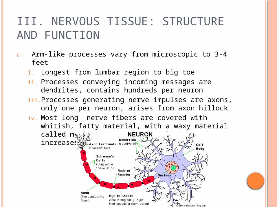
c. Arm-like processes vary from microscopic to 3-4 feeti. Longest from lumbar region to big toeii. Processes conveying incoming messages are dendrites,
contains hundreds per neuroniii. Processes generating nerve impulses are axons, only
one per neuron, arises from axon hillockiv. Most long nerve fibers are covered with whitish, fatty
material, with a waxy material called myelin – protects, insulates, and increases transmission rates
III. NERVOUS TISSUE: STRUCTURE AND FUNCTION
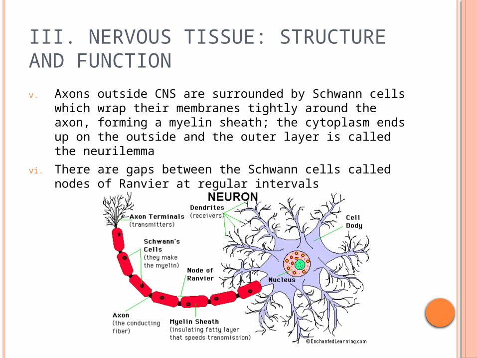
v. Axons outside CNS are surrounded by Schwann cells which wrap their membranes tightly around the axon, forming a myelin sheath; the cytoplasm ends up on the outside and the outer layer is called the neurilemma
vi. There are gaps between the Schwann cells called nodes of Ranvier at regular intervals
III. NERVOUS TISSUE: STRUCTURE AND FUNCTIONvii. Myelinated fibers also found
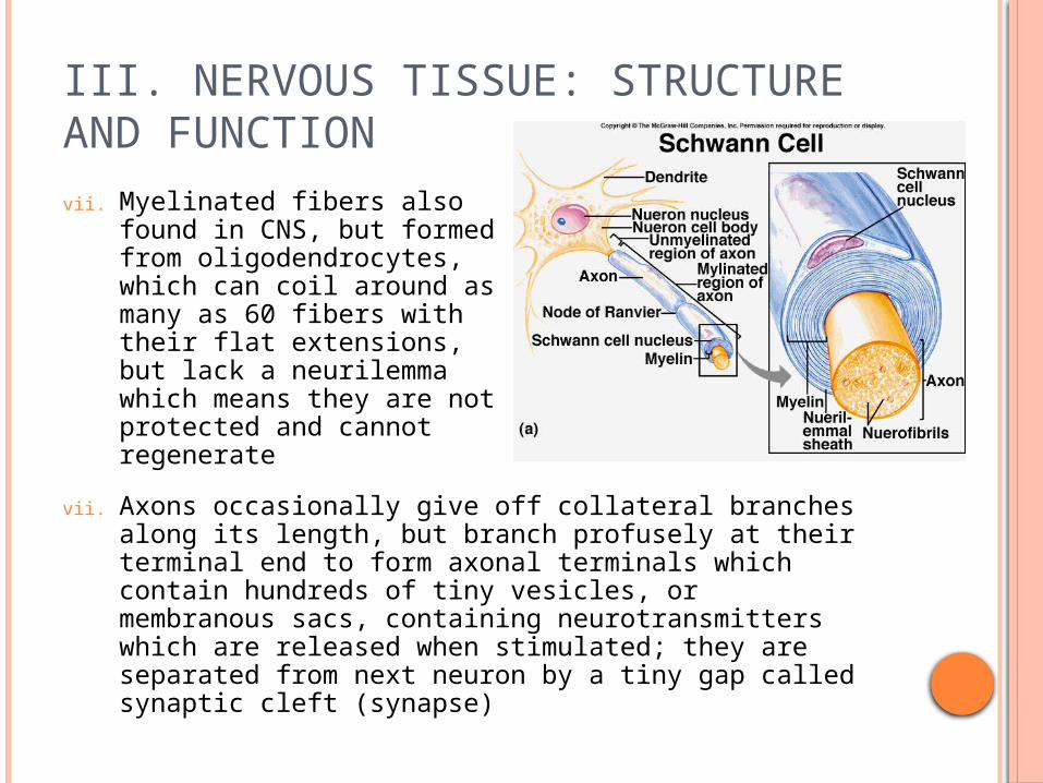
in CNS, but formed from oligodendrocytes, which can coil around as many as 60 fibers with their flat extensions, but lack a neurilemma which means they are not protected and cannot regenerate
vii. Axons occasionally give off collateral branches along its length, but branch profusely at their terminal end to form axonal terminals which contain hundreds of tiny vesicles, or membranous sacs, containing neurotransmitters which are released when stimulated; they are separated from next neuron by a tiny gap called synaptic cleft (synapse)
III. NERVOUS TISSUE: STRUCTURE AND FUNCTION
d. Clustered neuron cell bodies in the CNS are called nuclei, a few small clusters outside the CNS in the PNS are called ganglia
e. Bundles of nerve fibers running through CNS are called tracts and in PNS are called nerves
f. Cell body carries out most of the metabolic functions, so if it is damaged or dies it is not replaced
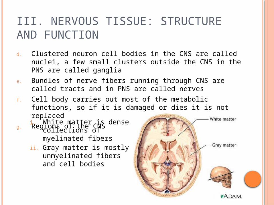
g. Regions of the CNS
i. White matter is dense collections of myelinated fibers
ii. Gray matter is mostly unmyelinated fibers and cell bodies
III. NERVOUS TISSUE: STRUCTURE AND FUNCTION
2. Homeostatic Imbalancea. Importance of myelin sheaths is seen in people
with multiple sclerosis (MS)i. Myelin sheaths are gradually destroyed, converted
to hardened sheaths (scleroses) and when this happen the electrical current is short-circuited, which causes loss of muscle control and they become increasingly disabled
3. Classificationa. Functional Classification
i. Groups neurons according to the direction the nerve impulse is traveling
III. NERVOUS TISSUE: STRUCTURE AND FUNCTION
ii. Sensory neurons – cell body found outside the CNS in a ganglion, dendrite endings have specialized receptors that are activated by changes nearby
a) Complex receptors of special sense organs (vision, hearing, equilibrium, taste, and smell)
b) Skin – cutaneous sense organs, extreme heat, cold or excessive pressure can be interpreted as pain
c) Muscles and tendons – proprioceptors, detect amount of stretch or tension so proper adjustments can be made to maintain balance and normal posture
d) Pain – bare dendrite endings, most numerous
iii. Motor neurons – carry nerve impulses from CNS to viscera, muscles, and/or glands, cell bodies are always located in CNS
iv. Associations neurons (interneurons) – connect motor and sensory neurons, cell bodies always located in CNS

III. NERVOUS TISSUE: STRUCTURE AND FUNCTION
b. Structural classification – based on number of processes extending from the cell body
i. Multipolar neuron – several processes, most common structural type, include all motor and association neurons
ii. Bipolar neurons – two processes (axon and dendrite), rare in adult, only in special sense organs (eye, ear)

III. NERVOUS TISSUE: STRUCTURE AND FUNCTION
iii. Unipolar neurons – single process, very short and divides almost immediately into proximal (central) and distal (peripheral) fibers; only the small branches at the end of the peripheral process are dendrites, remainder act as axons
a) Conduct nerve impulses toward and away from the cell body, included are sensory neurons in the PNS ganglia

4. Physiologya. Nerve impulses – two major functions
i. Irritability - the ability to respond to a stimulus and convert it into a nerve impulse
a) A neuron is polarized when it is resting, or inactive; this also means that there are fewer positive ions inside the plasma membrane than out
b) Ions inside are K+ and ions outside are Na+, and as long as the inside remains more negative the neuron stays inactive
III. NERVOUS TISSUE: STRUCTURE AND FUNCTION
III. NERVOUS TISSUE: STRUCTURE AND FUNCTION
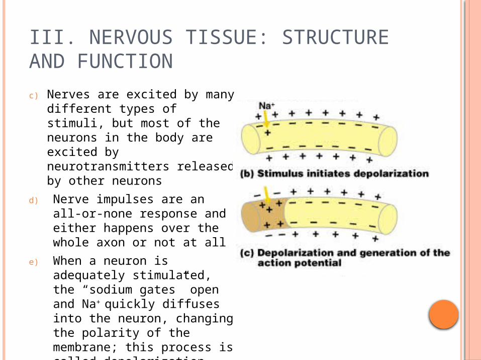
c) Nerves are excited by many different types of stimuli, but most of the neurons in the body are excited by neurotransmitters released by other neurons
d) Nerve impulses are an all-or-none response and either happens over the whole axon or not at all
e) When a neuron is adequately stimulated, the “sodium gates” open and Na+ quickly diffuses into the neuron, changing the polarity of the membrane; this process is called depolarization

III. NERVOUS TISSUE: STRUCTURE AND FUNCTION
f) If this stimulus is strong enough, and there’s a large enough Na+ in-rush, then depolarization activates the neuron to initiate and transmit an action potential (nerve impulse)
g) Once Na+ ions rush in the membrane permeability changes again and is no longer permeable to Na+, but permeable to K+, which diffuse out and the electrical charge is restored; this is called repolarization
III. NERVOUS TISSUE: STRUCTURE AND FUNCTION
h) Until repolarization occurs there cannot be another nerve impulse conducted
i) The initial concentrations must still be restored, so the Na-K pump is activated, which uses ATP
j) This nerve impulse is along unmyelinated fibers
k) Nerve impulses occur much faster though down myelinated fibers because it leaps from node to node since the current cannot flow across myelin insulation – this is called saltatory conduction
Nerve Impulses

III. NERVOUS TISSUE: STRUCTURE AND FUNCTION
ii. Conductivity – the ability to transmit the nerve impulse to other neurons, muscles, or glands
a) Impulses from one neuron travel across the synapse to another by means of a neurotransmitter
b) Dendrite of the next neuron receives the neurotransmitter and an action potential is then started
III. NERVOUS TISSUE: STRUCTURE AND FUNCTION
iii. Homeostatic Imbalance – impact on conduction of impulses
a) alcohol, sedatives, and anesthetics all reduce membrane permeability
b) Cold and continuous pressure interrupt blood circulation, so after warming up or pressure removed then prickly feeling comes when impulses are transmitted again

III. NERVOUS TISSUE: STRUCTURE AND FUNCTION
b. Reflex arc – direct route from a sensory neuron, to an interneuron, to an effector
i. Reflexes are rapid, predictable, and involuntary responses to stimuli that always travel in the same direction
ii. Autonomic reflexes regulate the activity of smooth muscles, the heart, and glands, saliva, eye pupils, digestion, elimination, blood pressure, sweating
iii. Somatic reflexes are all reflexes that stimulate skeletal muscles
IV. CENTRAL NERVOUS SYSTEM
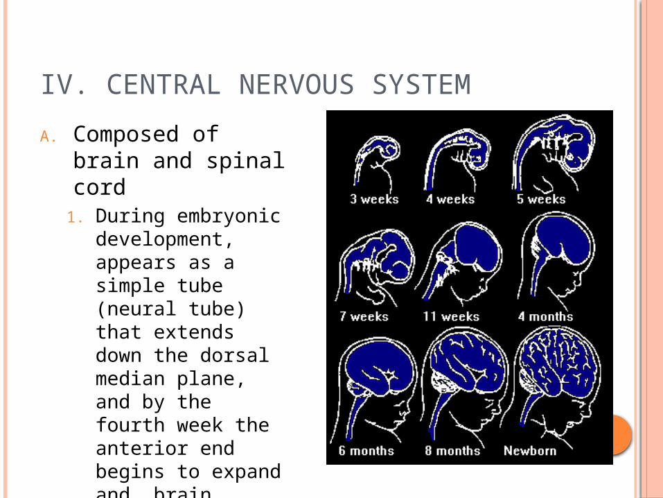
A. Composed of brain and spinal cord
1. During embryonic development, appears as a simple tube (neural tube) that extends down the dorsal median plane, and by the fourth week the anterior end begins to expand and brain formation begins
IV. CENTRAL NERVOUS SYSTEM
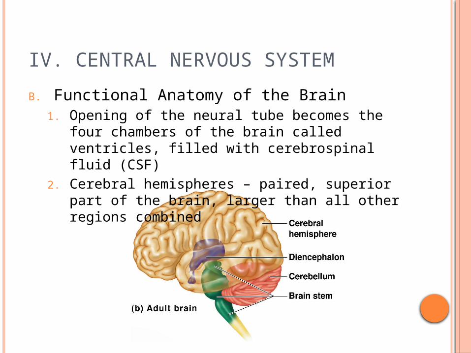
B. Functional Anatomy of the Brain1. Opening of the neural tube becomes the four
chambers of the brain called ventricles, filled with cerebrospinal fluid (CSF)
2. Cerebral hemispheres – paired, superior part of the brain, larger than all other regions combined

IV. CENTRAL NERVOUS SYSTEM
a. Surface has ridges called gyri seperated by shallow grooves called sulci, and few deeper grooves called fissures that separate large regions of the brain
i. these all serve as landmarks
ii. Single, deep fissure (longitudinal fissure) separates hemispheres
iii. Other fissures or sulci divide hemispheres into lobes named for cranial bones that lie over them

IV. CENTRAL NERVOUS SYSTEM
b. Parietal lobe lies posterior to the central sulcus and is home to the somatic sensory area
i. all of the body’s sensory receptors (except special senses) are localized and interpreted here, and all of the pathways are upside-down and backwards

IV. CENTRAL NERVOUS SYSTEM
c. Frontal lobe lies anterior to the central sulcus and is home to the primary motor area
i. Axons of these motor neurons form major voluntary motor tract – the pyramidal, or corticospinal tract, which descends to the cord
ii. Like the somatic sensory cortex, all of the pathways are upside-down and backwards

IV. CENTRAL NERVOUS SYSTEM
iii. Broca’s area is involved in the ability to speak, damage causes inability to say words properly
iv. Higher intellectual reasoning located in anterior of frontal lobe, and language comprehension
d. Speech area is at the junction of temporal, parietal, and occipital lobes, it allows you to sound out words
e. Cell bodies of neurons involved in cerebral hemisphere functions are found only in the outermost gray matter – the cerebral cortex
IV. CENTRAL NERVOUS SYSTEM
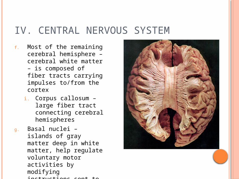
f. Most of the remaining cerebral hemisphere – cerebral white matter – is composed of fiber tracts carrying impulses to/from the cortex
i. Corpus callosum – large fiber tract connecting cerebral hemispheres
g. Basal nuclei – islands of gray matter deep in white matter, help regulate voluntary motor activities by modifying instructions sent to the skeletal muscles by the primary motor cortex
IV. CENTRAL NERVOUS SYSTEM
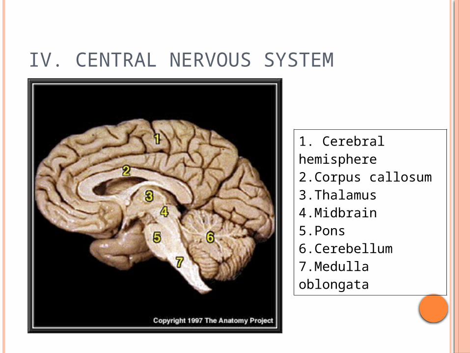
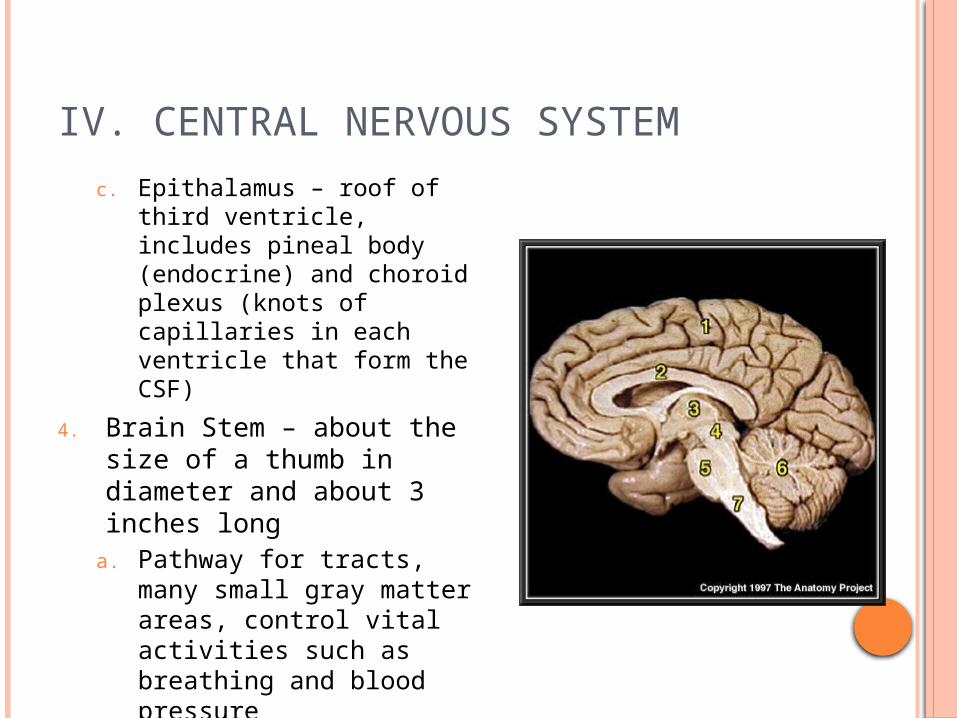
1. Cerebral hemisphere 2.Corpus callosum 3.Thalamus 4.Midbrain 5.Pons 6.Cerebellum 7.Medulla oblongata

IV. CENTRAL NERVOUS SYSTEM
3. Diencephalon (interbrain) – sits atop brain stem and enclosed by cerebral hemispheres
a. Thalamus – encloses shallow third ventricle, relay station for sensory impulses passing to sensory cortex and recognizes whether the impulse in pleasant or unpleasant

IV. CENTRAL NERVOUS SYSTEM
b. Hypothalamus makes up floor of diencephalon, plays role in regulation of body temperature, water balance, and metabolism
i. Also center for many drives and emotions, called limbic system – thirst, appetite, sex, pain, and pleasure centers
ii. Regulates pituitary gland or hypophysis (endocrine organ)
iii. Mammillary bodies involved in olfaction (smell) bulge from floor
IV. CENTRAL NERVOUS SYSTEM
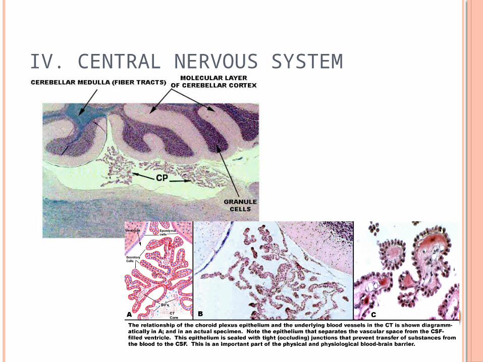
c. Epithalamus – roof of third ventricle, includes pineal body (endocrine) and choroid plexus (knots of capillaries in each ventricle that form the CSF)
4. Brain Stem – about the size of a thumb in diameter and about 3 inches long
a. Pathway for tracts, many small gray matter areas, control vital activities such as breathing and blood pressure
IV. CENTRAL NERVOUS SYSTEM
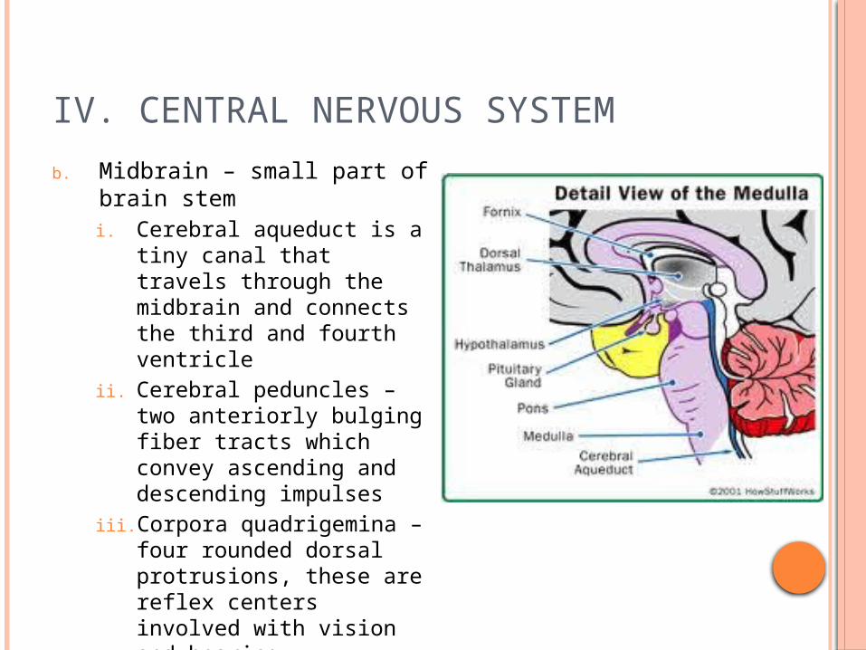
b. Midbrain – small part of brain stemi. Cerebral aqueduct is a tiny
canal that travels through the midbrain and connects the third and fourth ventricle
ii. Cerebral peduncles – two anteriorly bulging fiber tracts which convey ascending and descending impulses
iii. Corpora quadrigemina – four rounded dorsal protrusions, these are reflex centers involved with vision and hearing
IV. CENTRAL NERVOUS SYSTEM
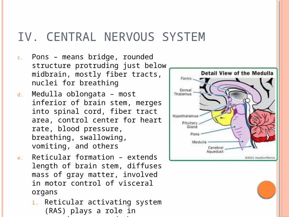
c. Pons – means bridge, rounded structure protruding just below midbrain, mostly fiber tracts, nuclei for breathing
d. Medulla oblongata – most inferior of brain stem, merges into spinal cord, fiber tract area, control center for heart rate, blood pressure, breathing, swallowing, vomiting, and others
e. Reticular formation – extends length of brain stem, diffuses mass of gray matter, involved in motor control of visceral organsi. Reticular activating system (RAS)
plays a role in consciousness and the awake/sleep cycles and damage to this area can result in coma

IV. CENTRAL NERVOUS SYSTEM
5. Cerebellum – large, cauliflower-like projection dorsally from under occipital lobe of cerebrum
a. Much like cerebrum – two hemispheres, convoluted surface, outer cortex of gray matter and inner white matter
b. Provides precise timing for skeletal muscle activity and controls balance and equilibrium
c. Fibers come here from the equilibrium apparatus of the inner ear, the eye, the proprioceptors, and other areas
IV. CENTRAL NERVOUS SYSTEM
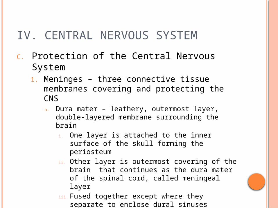
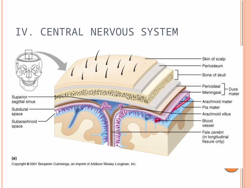
C. Protection of the Central Nervous System1. Meninges – three connective tissue
membranes covering and protecting the CNSa. Dura mater – leathery, outermost layer, double-
layered membrane surrounding the braini. One layer is attached to the inner surface of the
skull forming the periosteumii. Other layer is outermost covering of the brain
that continues as the dura mater of the spinal cord, called meningeal layer
iii. Fused together except where they separate to enclose dural sinuses (collects venous blood)
iv. Dural sinuses fold inward in several places to attach brain to cranial cavity – falx cerebri and tentorium cerebelli
IV. CENTRAL NERVOUS SYSTEM
IV. CENTRAL NERVOUS SYSTEM
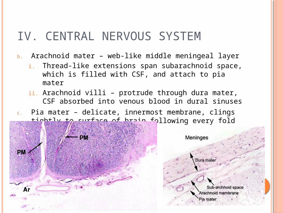
b. Arachnoid mater – web-like middle meningeal layeri. Thread-like extensions span subarachnoid space, which
is filled with CSF, and attach to pia materii. Arachnoid villi – protrude through dura mater, CSF
absorbed into venous blood in dural sinuses
c. Pia mater – delicate, innermost membrane, clings tightly to surface of brain following every fold
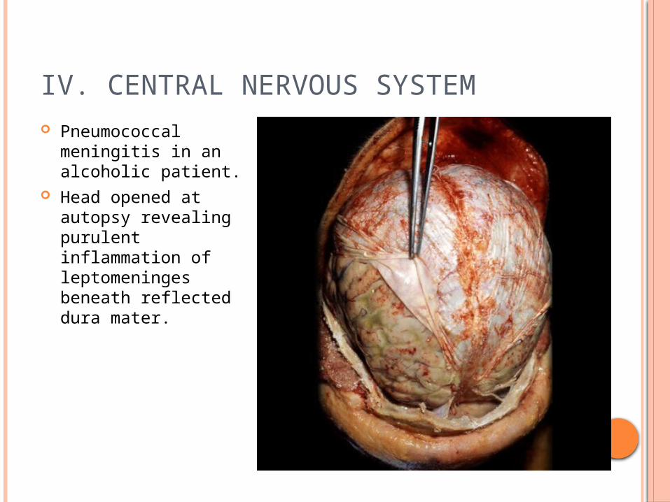
IV. CENTRAL NERVOUS SYSTEM Pneumococcal
meningitis in an alcoholic patient.
Head opened at autopsy revealing purulent inflammation of leptomeninges beneath reflected dura mater.

IV. CENTRAL NERVOUS SYSTEM
2. Cerebrospinal Fluid – similar to blood plasma, but less protein, more vitamin C, and different ion composition
a. Continually formed from blood by choroid plexuses, which are capillaries hanging from the top in each ventricle
b. Continually moving by circulating through lateral ventricles in cerebral hemispheres, third ventricle (diencephalon), and fourth ventricle (brain stem)
c. Some fluid reaching fourth ventricle travels down central canal of spinal cord, but most stays in subarachnoid space which leaves through holes in fourth ventricle
d. Fluid is recycled through arachnoid villi into blood in the dural sinuses
e. Normal volume is about 150 mL or ½ cup

IV. CENTRAL NERVOUS SYSTEM
f. Any changes in CSF can indicate a problem in brain or spinal cord, and a sample can be obtained through a lumbar (spinal) tap, and checked for blood cells
g. Since the fluid is decreased with this test, patient must remain horizontal for 6 – 12 hours to prevent a headache
IV. CENTRAL NERVOUS SYSTEM

IV. CENTRAL NERVOUS SYSTEM
3. Hydrocephalusa. If the CSF cannot drain it accumulates and causes pressure on
the brain, this is called hydrocephalus, which literally means water on the brain
b. In babies the head enlarges to allow for the extra spinal fluid because of the soft bones
c. In adults though the condition can lead to brain damage because of hardened skull
d. A shunt to drain excess fluid is then placed
IV. CENTRAL NERVOUS SYSTEM
4. The Blood-Brain Barriera. Separates neurons from blood-borne substances,
composed of least permeable capillaries in the whole body which provide most of the protection and the astrocytes help with this
b. Only water, glucose, and essential amino acids pass easily, but urea, toxins, proteins, and most drugs are prevented from entering
c. Useless against fats, respiratory gases, and other fat-soluble molecule (alcohol, nicotine, anesthetics)
IV. CENTRAL NERVOUS SYSTEM
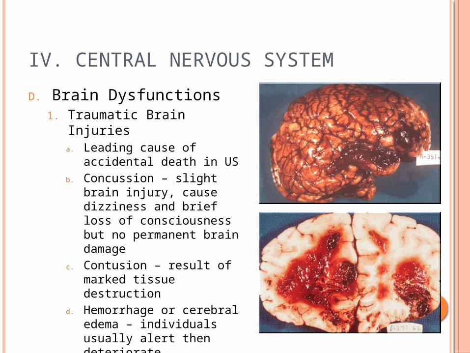
D. Brain Dysfunctions1. Traumatic Brain Injuries
a. Leading cause of accidental death in US
b. Concussion – slight brain injury, cause dizziness and brief loss of consciousness but no permanent brain damage
c. Contusion – result of marked tissue destruction
d. Hemorrhage or cerebral edema – individuals usually alert then deteriorate neurologically, caused from compression of vital brain tissue

IV. CENTRAL NERVOUS SYSTEM
2. Cerebrovascular Accident (CVAs) – strokesa. Third leading cause of death in the USb. Occur when blood circulation to a brain area is blocked by
clot, rupture, or tissue diesc. Area of brain damage can be determined by patient’s
symptomsd. Fewer than 1/3 of those that survive are alive three years
later, but not hopeless, undamaged neurons take over some of the lost functione. Not all strokes are “completed”, result in temporary restriction of blood flow called a transient ischemic attack (TIA) that last from 5 to 50 minutes, these are red flags though for impending more serious CVAs

IV. CENTRAL NERVOUS SYSTEM
E. Spinal Cord1. Approximately 17 inches long, glistening white
continuation of brain stem2. Provides two-way conduction pathway to/from brain3. Only extends to the 1st/2nd lumbar vertebrae because it
grows slower than vertebral column, spinal nerves at inferior end are called cauda equina
4. Cushioned and protected by meninges, which continue past end of spinal cord
5. 31 pairs of spinal nerves arise from cord
IV. CENTRAL NERVOUS SYSTEM
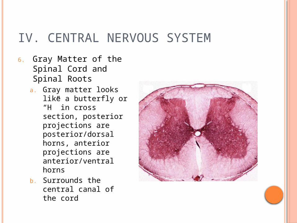
6. Gray Matter of the Spinal Cord and Spinal Roots
a. Gray matter looks like a butterfly or “H” in cross section, posterior projections are posterior/dorsal horns, anterior projections are anterior/ventral horns
b. Surrounds the central canal of the cord
IV. CENTRAL NERVOUS SYSTEM
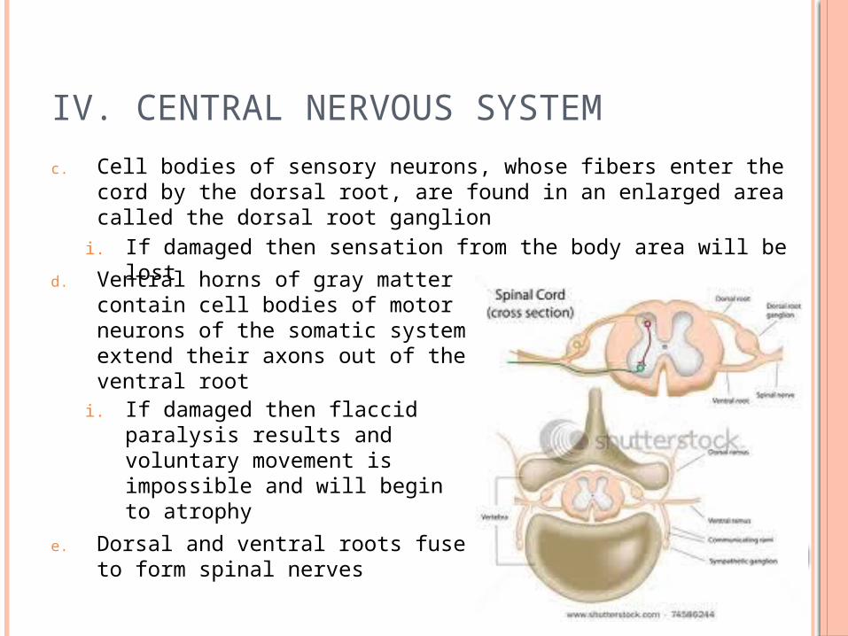
c. Cell bodies of sensory neurons, whose fibers enter the cord by the dorsal root, are found in an enlarged area called the dorsal root ganglion
i. If damaged then sensation from the body area will be lost
d. Ventral horns of gray matter contain cell bodies of motor neurons of the somatic system extend their axons out of the ventral root
i. If damaged then flaccid paralysis results and voluntary movement is impossible and will begin to atrophy
e. Dorsal and ventral roots fuse to form spinal nerves
IV. CENTRAL NERVOUS SYSTEM
7. White Matter of the Spinal Corda. Composed of myelinated fiber tractsb. Because of irregular shape of gray matter, white matter
divided into three regions i. Posterior column – ascending tracts that carry
sensory input to the brainii. Lateral and anterior columns – contain ascending and
descending motor tracts
8. Homeostatic Imbalancea. Transection or crushing of the cord results in spastic
paralysis where affected muscles stay healthy because of reflex arcs, meaning movements are involuntary
b. Quadriplegic – all 4 limbs affectedc. Paraplegic – only legs are paralyzed

V. PERIPHERAL NERVOUS SYSTEM
A. Structure of a Nerve1. Nerve – bundle of nerve
fibers outside the CNSa. Each process is wrapped in a
connective tissue sheath called the endoneurium
b. Groups of fibers are bound by coarser connective tissue called the perineurium to form fiber bundles or fascicles
c. Fascicles are bound together by a tough fibrous sheath called the epineurium for form a cord-like nerve

V. PERIPHERAL NERVOUS SYSTEM

V. PERIPHERAL NERVOUS SYSTEM
2. Like neurons, nerves are classified by direction in which impulses are transmitted
a. Mixed nerves carry both sensory and motor fibersb. Afferent or sensory nerves carry impulses toward the
CNSc. Efferent or motor nerves carry impulses away from
CNS
B. Autonomic Nervous System1. Motor subdivision of the PNS that controls body
activities automatically, thus also called the involuntary nervous system

V. PERIPHERAL NERVOUS SYSTEM
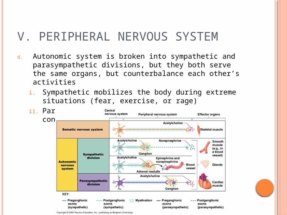
2. Somatic and Autonomic Nervous Systems Compared
a. Neuronsi. Somatic – cell bodies are inside the CNS, axons
extend all the way to the skeletal muscle they serveii. Autonomic – chain of two motor neurons, first is in
brain or spinal cord and its axon (preganglionic axon) leaves CNS to synapse with second motor neuron in a ganglion outside the CNS, the axon of this neuron is the postganglionic axon
b. Effector organsi. Somatic – skeletal muscleii. Autonomic – smooth muscle, cardiac muscle, glands
c. Neurotransmittersi. Somatic – acetylcholineii. Autonomic – acetylcholine, epinephrine,
norepinephrine
V. PERIPHERAL NERVOUS SYSTEM
d. Autonomic system is broken into sympathetic and parasympathetic divisions, but they both serve the same organs, but counterbalance each other’s activities
i. Sympathetic mobilizes the body during extreme situations (fear, exercise, or rage)
ii. Parasympathetic allows us to unwind and conserve energy
V. PERIPHERAL NERVOUS SYSTEM
3. Anatomy of the Sympathetic Divisiona. Originates from T1 through L2b. Ganglia are at the sympathetic trunk (near the spinal
cord)c. Short preganglionic neuron and long postganglionic
neuron transmit impulse from CNS to the effectord. Norepinephrine and epinephrine are neurotransmitters
to the effector organs
4. Anatomy of the Parasympathetic Divisiona. Originates from the brain stem and S1 through S4b. Terminal ganglia are at the effector organsc. Always uses acetylcholine as a neurotransmitter
V. PERIPHERAL NERVOUS SYSTEM
5. Autonomic Functioninga. Sympathetic – “fight-or-flight”
i. Response to unusual stimulusii. Takes over to increase activitiesiii. Remember as the “E” division – exercise, excitement,
emergency, and embarrassmentb. Parasympathetic – housekeeping activities
i. Conserves energyii. Maintains daily necessary body functionsiii. Remember as the “D” division – digestion,
defecation, diuresis

VI. DEVELOPMENTAL ASPECTS OF THE NERVOUS SYSTEM
A. Embryonic development1. Nervous system is formed during the first
month of embryonic development2. Any maternal infection can have extremely
harmful effects3. The hypothalamus is one of the last areas of the
brain to develop, which regulates body temperature, thus preemies have trouble with this
4. No more neurons are formed after birth, but growth and maturation continues for several years
5. The brain reaches maximum weight as a young adult

VI. DEVELOPMENTAL ASPECTS OF THE NERVOUS SYSTEM
The apparent enlargement of the ventricles seen here is due to atrophy of the head of the caudate from neuronal loss with Huntington's disease, an autosomal dominant condition characterized clinically by choreiform movements.