Embed Size (px)
Citation preview
This document is downloaded at: 2020-07-29T08:38:33Z
Title The effects of volar locking plates for distal radius fractures on the imagequality of high-resolution peripheral quantitative computed tomography
Author(s) 江良, 允
Citation Nagasaki University (長崎大学), 博士(医学) (2020-03-19)
Issue Date 2020-03-19
URL http://hdl.handle.net/10069/39710
Right © 2019 Elsevier Inc. All rights reserved.
NAOSITE: Nagasaki University's Academic Output SITE
http://naosite.lb.nagasaki-u.ac.jp
Contents lists available at ScienceDirect
Bone
journal homepage: www.elsevier.com/locate/bone
Full Length Article
The effects of volar locking plates for distal radius fractures on the imagequality of high-resolution peripheral quantitative computed tomography
Makoto Eraa, Ko Chibaa,⁎, Yuichiro Nishinoa, Narihiro Okazakia, Takashi Miyamotoa,Akihiko Yonekuraa, Masato Tomitaa, Toshiyuki Tsurumotob, Makoto Osakia
a Department of Orthopedic Surgery, Nagasaki University Graduate School of Biomedical Sciences, JapanbDepartment of Macroscopic Anatomy, Nagasaki University Graduate School of Biomedical Sciences, Japan
A R T I C L E I N F O
Keywords:High-resolution peripheral quantitativecomputed tomography (HR-pQCT)Fracture healingVolar locking plateImage quality
A B S T R A C T
Introduction: High-resolution peripheral quantitative computed tomography (HR-pQCT) has enabled us to ob-serve changes of bone microstructure during fracture healing. However, a method of analyzing the healingprocess after osteosynthesis has yet to be established due to implant artifacts. The purpose of this study was toinvestigate the effects of volar locking plates for distal radius fractures on the image quality of HR-pQCT.Methods: Four different types of plates for distal radius fractures were evaluated. The scan region of HR-pQCTwas the center of each plate, not including the screw insertion sites.
To assess plate-generated noise, each plate was fixed to the top of a water-filled rubber glove, scanned by HR-pQCT, and the signal-to-noise ratio (SNR) of the region under the plates was calculated. To investigate accuracy,12 cadaveric radii with and without each plate were scanned by HR-pQCT, and differences between the mea-sured values with and without the plate were evaluated. Differences between the measurements of the entirecircumference and of the dorsal third of the radius were also compared. Reproducibility of the in vivo mea-surement was investigated by repeated scans of 10 patients with distal radius fractures who had undergonesurgery with a volar locking plate.Results: The SNR was significantly higher away from the plate than immediately below the plate. Percentagedifferences of the measurement values between with and without the plate were 1.4%–3.2% for cortical bonemineral density (Ct.BMD) and 7.2%–9.8% for cortical bone thickness (Ct.Th) when the entire circumference wasmeasured. When the dorsal third was measured, they were 0.3%–1.7% for Ct.BMD and 1.8%–2.7% for Ct.Th.The root-mean-square coefficient of variation (RMS%CV) was 1.12% for Ct.BMD and 4.18% for Ct.Th.Conclusions: The accuracy and reproducibility of cortical bone measurements with a volar locking plate on HR-pQCT were acceptable when the dorsal third of the Ct.BMD was analyzed, and this method would be useful for invivo analysis of the fracture healing process after osteosynthesis.
1. Introduction
Fracture healing consists of inflammatory, repair, and remodelingphases [1]. In the inflammatory phase, growth factor migration andcapillary angiogenesis occur. In the repair phase, membranous ossifi-cation and endochondral ossification cause soft callus followed by hardcallus formation. In the remodeling phase, natural correction takesplace. Previous basic studies of these fracture healing processes havebeen performed using animal models, with no in vivo analyses in hu-mans.
Fracture healing of patients can be evaluated by plain X-rays and
clinical computed tomography (CT). However, plain X-rays providetwo-dimensional images that do not enable detailed analysis of fracturehealing. Clinical CT enables three-dimensional evaluation, but it canonly provide the macrostructure of the cortical bone at the fracture sitedue to its limited resolution.
High-resolution peripheral quantitative CT (HR-pQCT) is a quanti-tative CT method for human extremities that provides the highestavailable resolution (voxel size 61 μm) of any clinical imaging mod-alities [2–4]. Previously, analysis of human bone microstructure in vivohad required sample collection by invasive techniques such as bonebiopsy, which could not be used for longitudinal observations over
https://doi.org/10.1016/j.bone.2019.07.025Received 11 February 2019; Received in revised form 16 July 2019; Accepted 21 July 2019
⁎ Corresponding author at: Department of Orthopedic Surgery, Nagasaki University Graduate School of Biomedical Sciences, 1-7-1, Sakamoto, Nagasaki 852-8501,Japan.
E-mail address: [email protected] (K. Chiba).
Bone 127 (2019) 620–625
Available online 31 July 20198756-3282/ © 2019 Elsevier Inc. All rights reserved.
T
time. The advent of HR-pQCT has enabled observation of the changes ofhuman bone microstructure in vivo over time with low radiation ex-posure.
In previous studies by HR-pQCT, the healing process of distal radiusfractures under conservative treatment was observed [5,6]. It has beenthought that CT analysis after surgical treatment with implants ischallenging due to metal artifacts [7]. Thus, a method of analyzing thehealing process after osteosynthesis by HR-pQCT has yet to be estab-lished.
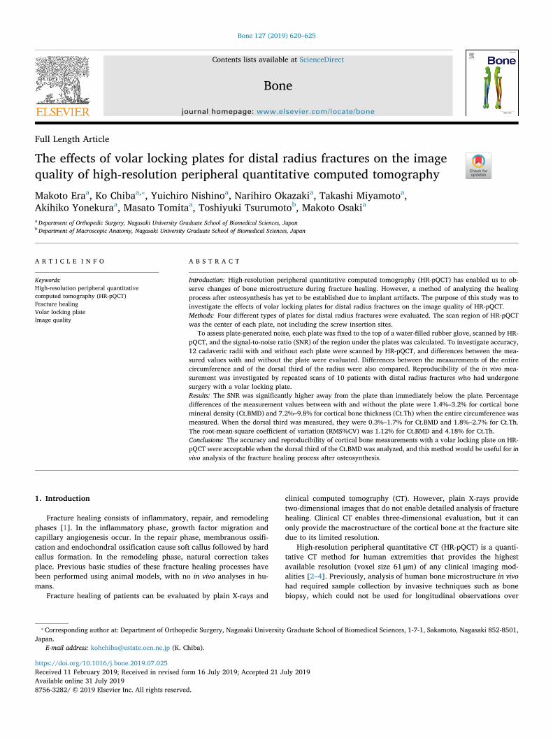
In preliminary experiments, we found that severe metal artifactswere present in bone at screw insertion areas (Fig. 1A), but that imagesof sufficient quality for observation were obtained from areas withoutscrews (Fig. 1B).
In this study, the effects of volar locking plates on the HR-pQCTimages were investigated, with the aim of establishing a method ofanalyzing the postoperative fracture healing process in vivo.
2. Methods
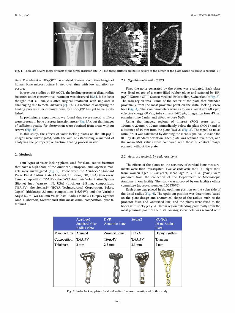
Four types of volar locking plates used for distal radius fracturesthat have a high share of the American, European, and Japanese mar-kets were investigated (Fig. 2). These were the Acu-Loc2® StandardVolar Distal Radius Plate (Acumed, Hillsboro, OR, USA) (thickness:2 mm; composition: Ti6Al4V); the DVR® Anatomic Volar Plating System(Biomet Inc., Warsaw, IN, USA) (thickness 2.5mm; composition:Ti6Al4V); the Stellar2® (HOYA Technosurgical Corporation, Tokyo,Japan) (thickness: 2.1 mm; composition: Ti6Al4V); and the VariableAngle LCP® Two-Column Volar Distal Radius Plate 2.4 (Depuy SynthesGmbH, Oberdorf, Switzerland) (thickness: 2 mm, composition: pure ti-tanium).
2.1. Signal-to-noise ratio (SNR)
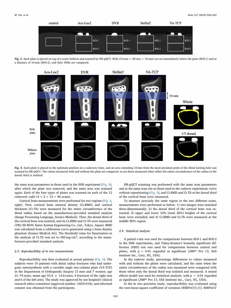
First, the noise generated by the plates was evaluated. Each platewas fixed on top of a water-filled rubber glove and scanned by HR-pQCT (Xtreme CT II, Scanco Medical, Brüttisellen, Switzerland) (Fig. 3).The scan region was 10mm of the center of the plate that extendedproximally from the most proximal point on the distal locking screwhole (Fig. 4). The scan parameters were as follows: voxel size 60.7 μm,effective energy 60 kVp, tube current 1470 μA, integration time 43ms,scanning time 2min, and effective dose 5 μSv.
Using the images, regions of interest (ROI) were set to10mm×20mm×10mm immediately below the plate (ROI-1) and ata distance of 10mm from the plate (ROI-2) (Fig. 3). The signal-to-noiseratio (SNR) was calculated by dividing the mean signal value inside theROI by its standard deviation. Each plate was scanned five times, andthe mean SNR values were compared with those of control imagesscanned without the plate.
2.2. Accuracy analysis by cadaveric bone
The effects of the plates on the accuracy of cortical bone measure-ments were then investigated. Twelve cadaveric radii (all right radiifrom women aged 61–78 years, mean age 71.7 ± 4.3 years) wereprepared from the collection of the Department of MacroscopicAnatomy in our facility. The study was approved by our facility's ethicscommittee (approval number: 15033076).
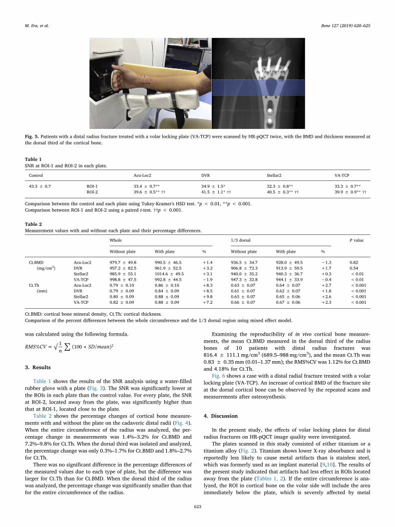
Each plate was placed in the optimum position on the volar side ofthe distal radius (Fig. 4). The optimum position was determined basedon the plate design and anatomical shape of the radius, such as thepronator fossa and watershed line, and the plates were fixed to thebones with sticky jelly. A 10-mm region extending proximally from themost proximal point of the distal locking screw hole was scanned with
Fig. 1. There are severe metal artifacts at the screw insertion site (A), but these artifacts are not so severe at the center of the plate where no screw is present (B).
Fig. 2. Volar locking plates for distal radius fractures investigated in this study.
M. Era, et al. Bone 127 (2019) 620–625
621
the same scan parameters as those used in the SNR experiment (Fig. 4),after which the plate was removed, and the same area was scannedagain. Each of the four types of plates was scanned on each of the 12cadaveric radii (4× 2×12=96 scans).
Cortical bone measurements were performed for two regions (Fig. 4,right). First, cortical bone mineral density (Ct.BMD) and corticalthickness (Ct.Th) were measured for the entire circumference of thedistal radius based on the manufacturer-provided standard analysis(Image Processing Language, Scanco Medical). Then, the dorsal third ofthe cortical bone was isolated, and its Ct.BMD and Ct.Th were measured(TRI/3D-BON: Ratoc System Engineering Co., Ltd., Tokyo, Japan). BMDwas calculated from a calibration curve generated using a bone densityphantom (Scanco Medical AG). The threshold value for binarization inthe analysis of Ct.Th was set to 450mg/cm3, according to the manu-facturer-provided standard analysis.
2.3. Reproducibility of in vivo measurements
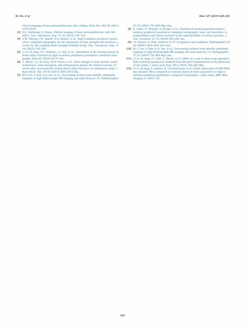
Reproducibility was then evaluated in actual patients (Fig. 5). Thesubjects were 10 patients with distal radius fractures who had under-gone osteosynthesis with a variable angle two-column plate (VA-TCP)in the Department of Orthopaedic Surgery (3 men and 7 women; age21–74 years, mean age 53.9 ± 14.8 years; 4 fractures of the right armand 6 of the left arm). The study was approved by our hospital's clinicalresearch ethics committee (approval number: 16031418), and informedconsent was obtained from the participants.
HR-pQCT scanning was performed with the same scan parametersand at the same scan site as those used in the cadaver experiment, twicewithout repositioning (Fig. 5), and Ct.BMD and Ct.Th at the dorsal thirdof the cortical bone were measured.
To measure precisely the same region in the two different scans,measurements were performed as below: 1) two images were matchedthree-dimensionally; 2) the dorsal third of the cortical bone was ex-tracted; 3) upper and lower 10% (total 20%) heights of the corticalbone were excluded; and 4) Ct.BMD and Ct.Th were measured at themiddle 80% region.
2.4. Statistical analysis
A paired t-test was used for comparisons between ROI-1 and ROI-2in the SNR experiments, and Tukey-Kramer's honestly significant dif-ference (HSD) test was used for comparisons between control andplates, with p < 0.01 regarded as significant (JMP® Pro 13, SASInstitute Inc., Cary, NC, USA).
In the cadaver study, percentage differences in values measuredwith and without the plates were calculated, and the rates when theentire circumference of the radius was measured were compared withthose when only the dorsal third was isolated and measured. A mixedeffects model was used for statistical analysis, with p < 0.01 regardedas significant (JMP® Pro 13, SAS Institute Inc., Cary, NC, USA).
In the in vivo precision study, reproducibility was evaluated usingthe root-mean-square coefficient of variation (RMS%CV) [8]. RMS%CV
Fig. 3. Each plate is placed on top of a water balloon and scanned by HR-pQCT. ROIs (10mm×20mm×10mm) are set immediately below the plate (ROI-1) and ata distance of 10mm (ROI-2), and their SNRs are compared.
Fig. 4. Each plate is placed in the optimum position on a cadaveric bone, and an area extending 10mm from the most proximal point of the distal locking hole wasscanned by HR-pQCT. The values measured with and without the plate are compared, as are those measured when either the entire circumference of the radius or thedorsal third is isolated.
M. Era, et al. Bone 127 (2019) 620–625
622
was calculated using the following formula.
∑= √ ∗RMS CVn
SD mean% 1 (100 / )2
3. Results
Table 1 shows the results of the SNR analysis using a water-filledrubber glove with a plate (Fig. 3). The SNR was significantly lower atthe ROIs in each plate than the control value. For every plate, the SNRat ROI-2, located away from the plate, was significantly higher thanthat at ROI-1, located close to the plate.
Table 2 shows the percentage changes of cortical bone measure-ments with and without the plate on the cadaveric distal radii (Fig. 4).When the entire circumference of the radius was analyzed, the per-centage change in measurements was 1.4%–3.2% for Ct.BMD and7.2%–9.8% for Ct.Th. When the dorsal third was isolated and analyzed,the percentage change was only 0.3%–1.7% for Ct.BMD and 1.8%–2.7%for Ct.Th.
There was no significant difference in the percentage differences ofthe measured values due to each type of plate, but the difference waslarger for Ct.Th than for Ct.BMD. When the dorsal third of the radiuswas analyzed, the percentage change was significantly smaller than thatfor the entire circumference of the radius.
Examining the reproducibility of in vivo cortical bone measure-ments, the mean Ct.BMD measured in the dorsal third of the radiusbones of 10 patients with distal radius fractures was816.4 ± 111.1 mg/cm3 (689.5–988mg/cm3), and the mean Ct.Th was0.83 ± 0.35mm (0.01–1.37mm); the RMS%CV was 1.12% for Ct.BMDand 4.18% for Ct.Th.
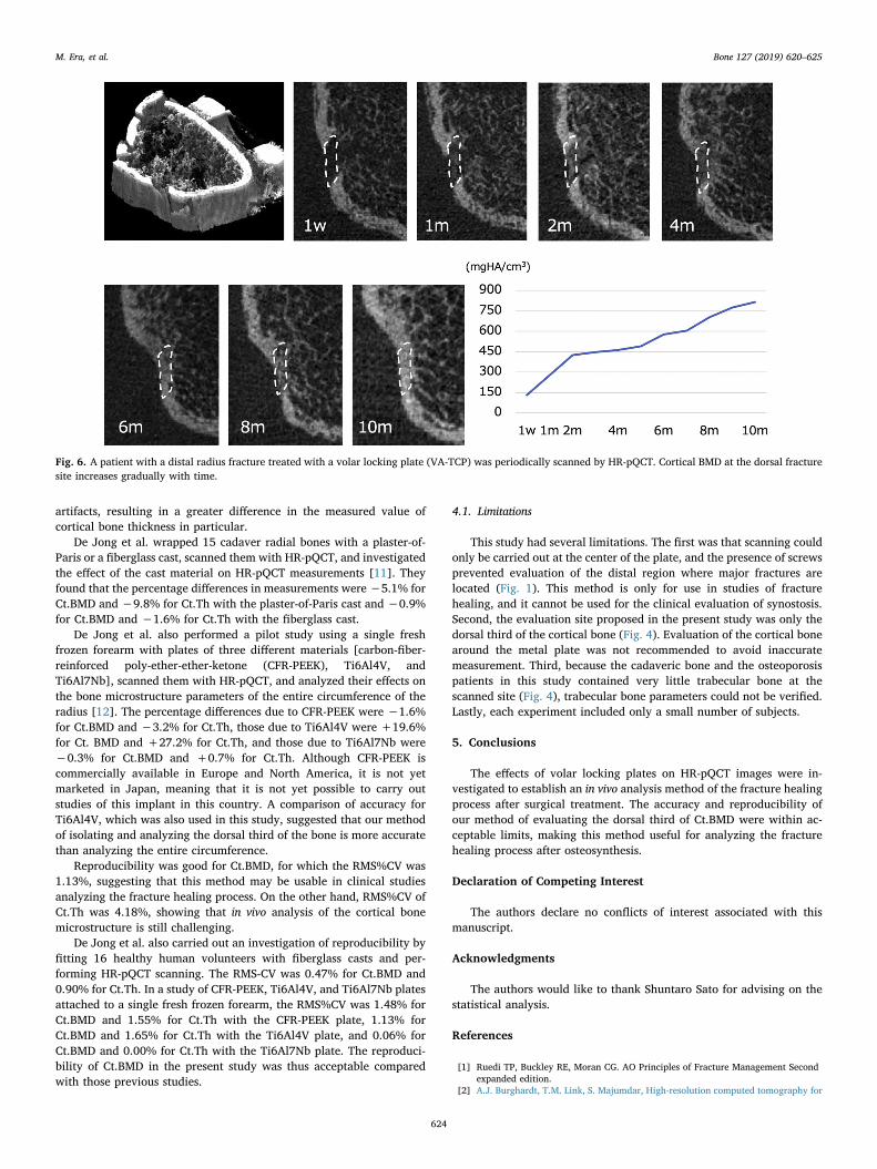
Fig. 6 shows a case with a distal radial fracture treated with a volarlocking plate (VA-TCP). An increase of cortical BMD of the fracture siteat the dorsal cortical bone can be observed by the repeated scans andmeasurements after osteosynthesis.
4. Discussion
In the present study, the effects of volar locking plates for distalradius fractures on HR-pQCT image quality were investigated.
The plates scanned in this study consisted of either titanium or atitanium alloy (Fig. 2). Titanium shows lower X-ray absorbance and isreportedly less likely to cause metal artifacts than is stainless steel,which was formerly used as an implant material [9,10]. The results ofthe present study indicated that artifacts had less effect in ROIs locatedaway from the plate (Tables 1, 2). If the entire circumference is ana-lyzed, the ROI in cortical bone on the volar side will include the areaimmediately below the plate, which is severely affected by metal
Fig. 5. Patients with a distal radius fracture treated with a volar locking plate (VA-TCP) were scanned by HR-pQCT twice, with the BMD and thickness measured atthe dorsal third of the cortical bone.
Table 1SNR at ROI-1 and ROI-2 in each plate.
Control Acu-Loc2 DVR Stellar2 VA-TCP
43.3 ± 0.7 ROI-1 33.4 ± 0.7** 34.9 ± 1.5* 32.3 ± 0.8** 33.3 ± 0.7**ROI-2 39.6 ± 0.5** †† 41.5 ± 1.1* †† 40.5 ± 0.3** †† 39.9 ± 0.9** ††
Comparison between the control and each plate using Tukey-Kramer's HSD test. *p < 0.01, **p < 0.001.Comparison between ROI-1 and ROI-2 using a paired t-test. ††p < 0.001.
Table 2Measurement values with and without each plate and their percentage differences.
Whole 1/3 dorsal P value
Without plate With plate % Without plate With plate %
Ct.BMD(mg/cm3)
Acu-Loc2 979.7 ± 49.8 990.5 ± 46.5 +1.4 936.5 ± 34.7 928.0 ± 49.5 −1.3 0.82DVR 957.2 ± 82.5 961.9 ± 52.5 +3.2 906.8 ± 72.3 913.9 ± 59.5 +1.7 0.54Stellar2 985.9 ± 55.1 1014.6 ± 49.5 +3.1 940.0 ± 35.2 940.3 ± 36.7 +0.3 <0.01VA-TCP 998.8 ± 47.5 992.8 ± 44.5 −1.9 947.3 ± 32.8 944.1 ± 33.9 −0.4 <0.01
Ct.Th(mm)
Acu-Loc2 0.79 ± 0.10 0.86 ± 0.10 +8.3 0.63 ± 0.07 0.64 ± 0.07 +2.7 <0.001DVR 0.79 ± 0.09 0.84 ± 0.09 +8.5 0.61 ± 0.07 0.62 ± 0.07 +1.8 <0.001Stellar2 0.80 ± 0.09 0.88 ± 0.09 +9.8 0.63 ± 0.07 0.65 ± 0.06 +2.6 <0.001VA-TCP 0.82 ± 0.09 0.88 ± 0.09 +7.2 0.66 ± 0.07 0.67 ± 0.06 +2.3 <0.001
Ct.BMD: cortical bone mineral density, Ct.Th: cortical thickness.Comparison of the percent differences between the whole circumference and the 1/3 dorsal region using mixed effect model.
M. Era, et al. Bone 127 (2019) 620–625
623
artifacts, resulting in a greater difference in the measured value ofcortical bone thickness in particular.
De Jong et al. wrapped 15 cadaver radial bones with a plaster-of-Paris or a fiberglass cast, scanned them with HR-pQCT, and investigatedthe effect of the cast material on HR-pQCT measurements [11]. Theyfound that the percentage differences in measurements were −5.1% forCt.BMD and −9.8% for Ct.Th with the plaster-of-Paris cast and −0.9%for Ct.BMD and −1.6% for Ct.Th with the fiberglass cast.
De Jong et al. also performed a pilot study using a single freshfrozen forearm with plates of three different materials [carbon-fiber-reinforced poly-ether-ether-ketone (CFR-PEEK), Ti6Al4V, andTi6Al7Nb], scanned them with HR-pQCT, and analyzed their effects onthe bone microstructure parameters of the entire circumference of theradius [12]. The percentage differences due to CFR-PEEK were −1.6%for Ct.BMD and −3.2% for Ct.Th, those due to Ti6Al4V were +19.6%for Ct. BMD and +27.2% for Ct.Th, and those due to Ti6Al7Nb were−0.3% for Ct.BMD and +0.7% for Ct.Th. Although CFR-PEEK iscommercially available in Europe and North America, it is not yetmarketed in Japan, meaning that it is not yet possible to carry outstudies of this implant in this country. A comparison of accuracy forTi6Al4V, which was also used in this study, suggested that our methodof isolating and analyzing the dorsal third of the bone is more accuratethan analyzing the entire circumference.
Reproducibility was good for Ct.BMD, for which the RMS%CV was1.13%, suggesting that this method may be usable in clinical studiesanalyzing the fracture healing process. On the other hand, RMS%CV ofCt.Th was 4.18%, showing that in vivo analysis of the cortical bonemicrostructure is still challenging.
De Jong et al. also carried out an investigation of reproducibility byfitting 16 healthy human volunteers with fiberglass casts and per-forming HR-pQCT scanning. The RMS-CV was 0.47% for Ct.BMD and0.90% for Ct.Th. In a study of CFR-PEEK, Ti6Al4V, and Ti6Al7Nb platesattached to a single fresh frozen forearm, the RMS%CV was 1.48% forCt.BMD and 1.55% for Ct.Th with the CFR-PEEK plate, 1.13% forCt.BMD and 1.65% for Ct.Th with the Ti6Al4V plate, and 0.06% forCt.BMD and 0.00% for Ct.Th with the Ti6Al7Nb plate. The reproduci-bility of Ct.BMD in the present study was thus acceptable comparedwith those previous studies.
4.1. Limitations
This study had several limitations. The first was that scanning couldonly be carried out at the center of the plate, and the presence of screwsprevented evaluation of the distal region where major fractures arelocated (Fig. 1). This method is only for use in studies of fracturehealing, and it cannot be used for the clinical evaluation of synostosis.Second, the evaluation site proposed in the present study was only thedorsal third of the cortical bone (Fig. 4). Evaluation of the cortical bonearound the metal plate was not recommended to avoid inaccuratemeasurement. Third, because the cadaveric bone and the osteoporosispatients in this study contained very little trabecular bone at thescanned site (Fig. 4), trabecular bone parameters could not be verified.Lastly, each experiment included only a small number of subjects.
5. Conclusions
The effects of volar locking plates on HR-pQCT images were in-vestigated to establish an in vivo analysis method of the fracture healingprocess after surgical treatment. The accuracy and reproducibility ofour method of evaluating the dorsal third of Ct.BMD were within ac-ceptable limits, making this method useful for analyzing the fracturehealing process after osteosynthesis.
Declaration of Competing Interest
The authors declare no conflicts of interest associated with thismanuscript.
Acknowledgments
The authors would like to thank Shuntaro Sato for advising on thestatistical analysis.
References
[1] Ruedi TP, Buckley RE, Moran CG. AO Principles of Fracture Management Secondexpanded edition.
[2] A.J. Burghardt, T.M. Link, S. Majumdar, High-resolution computed tomography for
Fig. 6. A patient with a distal radius fracture treated with a volar locking plate (VA-TCP) was periodically scanned by HR-pQCT. Cortical BMD at the dorsal fracturesite increases gradually with time.
M. Era, et al. Bone 127 (2019) 620–625
624
clinical imaging of bone microarchitecture, Clin. Orthop. Relat. Res. 469 (8) (2011)2179–2193.
[3] K.K. Nishiyama, E. Shane, Clinical imaging of bone microarchitecture with HR-pQCT, Curr. Osteoporos. Rep. 11 (2) (2013) 147–155.
[4] A.M. Cheung, J.D. Adachi, D.A. Hanley, et al., High-resolution peripheral quanti-tative computed tomography for the assessment of bone strength and structure: areview by the Canadian Bone Strength Working Group, Curr. Osteoporos. Rep. 11(2) (2013) 136–146.
[5] J.J.A. de Jong, P.C. Willems, J.J. Arts, et al., Assessment of the healing process indistal radius fractures by high resolution peripheral quantitative computed tomo-graphy, Bone 64 (2014) 65–74 Jul.
[6] U. Meyer, J.J. de Jong, S.G.P. Bours, et al., Early changes in bone density, micro-architecture, bone resorption, and inflammation predict the clinical outcome 12weeks after conservatively treated distal radius fractures: an exploratory study, J.Bone Miner. Res. 29 (9) (2014) 2065–2073 Sep.
[7] M.J. Lee, S. Kim, S.A. Lee, et al., Overcoming artifact from metallic orthopedicimplants at high-field-strength MR imaging and multi-detector CT, RadioGraphics
27 (3) (2007) 791–803 May-Jun.[8] K. Chiba, N. Okazaki, A. Kurogi, et al., Precision of second-generation high-re-
solution peripheral quantitative computed tomography: intra- and intertester re-producibilities and factors involved in the reproducibility of cortical porosity, J.Clin. Densitom. 21 (2) (2018) 295–302 Jun.
[9] J.F. Barrett, N. Keat, Artifacts in CT: recognition and avoidance, Radiographics 24(6) (2004) 1679–1691 Nov–Dec.
[10] M.-J. Lee, S. Kim, S.-A. Lee, et al., Overcoming artifacts from metallic orthopedicimplants at high-field-strength MR imaging and multi-detector CT, Radiographics27 (3) (2007) 791–803 May-Jun.
[11] J.J.A. de Jong, J.J. Arts, U. Meyer, et al., Effect of a cast on short-term reproduci-bility and bone parameters obtained from HR-pQCT measurements at the distal endof the radius, J. Bone Joint Surg. 98-A (2016) 356–362 Mar.
[12] J.J.A. de Jong, A. Lataster, B. van Rietbergen, et al., Distal radius plate of CFR-PEEKhas minimal effect compared to titanium plates on bone parameters in high-re-solution peripheral quantitative computed tomography: a pilot study, BMC Med.Imaging 17 (2017) 18.
M. Era, et al. Bone 127 (2019) 620–625
625


![TheRelationshipbetweenSideofOnsetandCerebralRegional ...2.4.ReHoCalculations. ReHo maps were generated using RESTPlusV1.2,withtheprocedurespublishedpreviously [29]. Kendall’s coefficient](https://img.pdfslide.net/doc/110x75/60c5627e811fd00c785dc493/therelationshipbetweensideofonsetandcerebralregional-24rehocalculations-reho.jpg)


![Prototype centrifugal natural gas cleaner CONFIDENTIAL · Nomenclature A area [m2] CD drag force coefficient [-] CM torque coefficient [-] D diameter [m] d diameter [m] dc channel](https://img.pdfslide.net/doc/110x75/5ecbd0d31495fb70d12dbcc0/prototype-centrifugal-natural-gas-cleaner-nomenclature-a-area-m2-cd-drag-force.jpg)