Embed Size (px)
Citation preview
Nematoda
1: Thalassomonhystera sp. from East Pacific Rise: 9°N, Tica; by M. Bright.
173
© Biologiezentrum Linz/Austria; download unter www.biologiezentrum.at
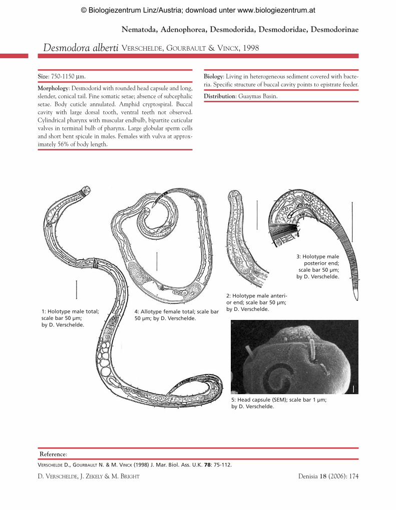
Desmodora alberti VERSCHELDE, GOURBAULT & VINCX, 1998
Size: 750-1150 µm.
Morphology: Desmodorid with rounded head capsule and long,slender, conical tail. Fine somatic setae; absence of subcephalicsetae. Body cuticle annulated. Amphid cryptospiral. Buccalcavity with large dorsal tooth, ventral teeth not observed.Cylindrical pharynx with muscular endbulb, bipartite cuticularvalves in terminal bulb of pharynx. Large globular sperm cellsand short bent spicule in males. Females with vulva at approx-imately 56% of body length.
Biology: Living in heterogeneous sediment covered with bacte-ria. Specific structure of buccal cavity points to epistrate feeder.
Distribution: Guaymas Basin.
Reference:
VERSCHELDE D., GOURBAULT N. & M. VINCX (1998) J. Mar. Biol. Ass. U.K. 78: 75-112.
D. VERSCHELDE, J. ZEKELY & M. BRIGHT Denisia 18 (2006): 174
2: Holotype male anteri-or end; scale bar 50 µm;by D. Verschelde.4: Allotype female total; scale bar
50 µm; by D. Verschelde.
5: Head capsule (SEM); scale bar 1 µm; by D. Verschelde.
Nematoda, Adenophorea, Desmodorida, Desmodoridae, Desmodorinae
1: Holotype male total;scale bar 50 µm; by D. Verschelde.
3: Holotype male posterior end;
scale bar 50 µm; by D. Verschelde.
© Biologiezentrum Linz/Austria; download unter www.biologiezentrum.at
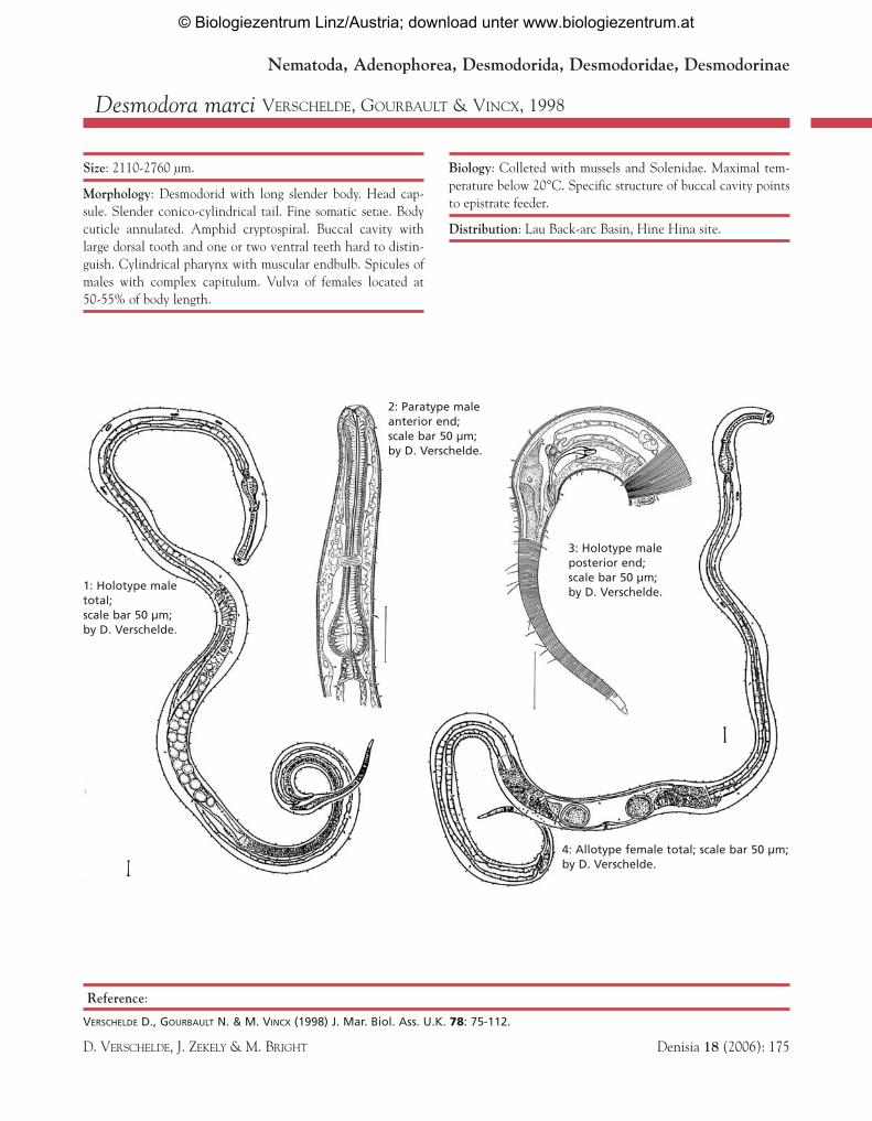
Desmodora marci VERSCHELDE, GOURBAULT & VINCX, 1998
Size: 2110-2760 µm.
Morphology: Desmodorid with long slender body. Head cap-sule. Slender conico-cylindrical tail. Fine somatic setae. Bodycuticle annulated. Amphid cryptospiral. Buccal cavity withlarge dorsal tooth and one or two ventral teeth hard to distin-guish. Cylindrical pharynx with muscular endbulb. Spicules ofmales with complex capitulum. Vulva of females located at 50-55% of body length.
Biology: Colleted with mussels and Solenidae. Maximal tem-perature below 20°C. Specific structure of buccal cavity pointsto epistrate feeder.
Distribution: Lau Back-arc Basin, Hine Hina site.
Reference:
VERSCHELDE D., GOURBAULT N. & M. VINCX (1998) J. Mar. Biol. Ass. U.K. 78: 75-112.
D. VERSCHELDE, J. ZEKELY & M. BRIGHT Denisia 18 (2006): 175
Nematoda, Adenophorea, Desmodorida, Desmodoridae, Desmodorinae
1: Holotype maletotal;scale bar 50 µm;by D. Verschelde.
4: Allotype female total; scale bar 50 µm;by D. Verschelde.
2: Paratype maleanterior end; scale bar 50 µm; by D. Verschelde.
3: Holotype maleposterior end; scale bar 50 µm;by D. Verschelde.
© Biologiezentrum Linz/Austria; download unter www.biologiezentrum.at
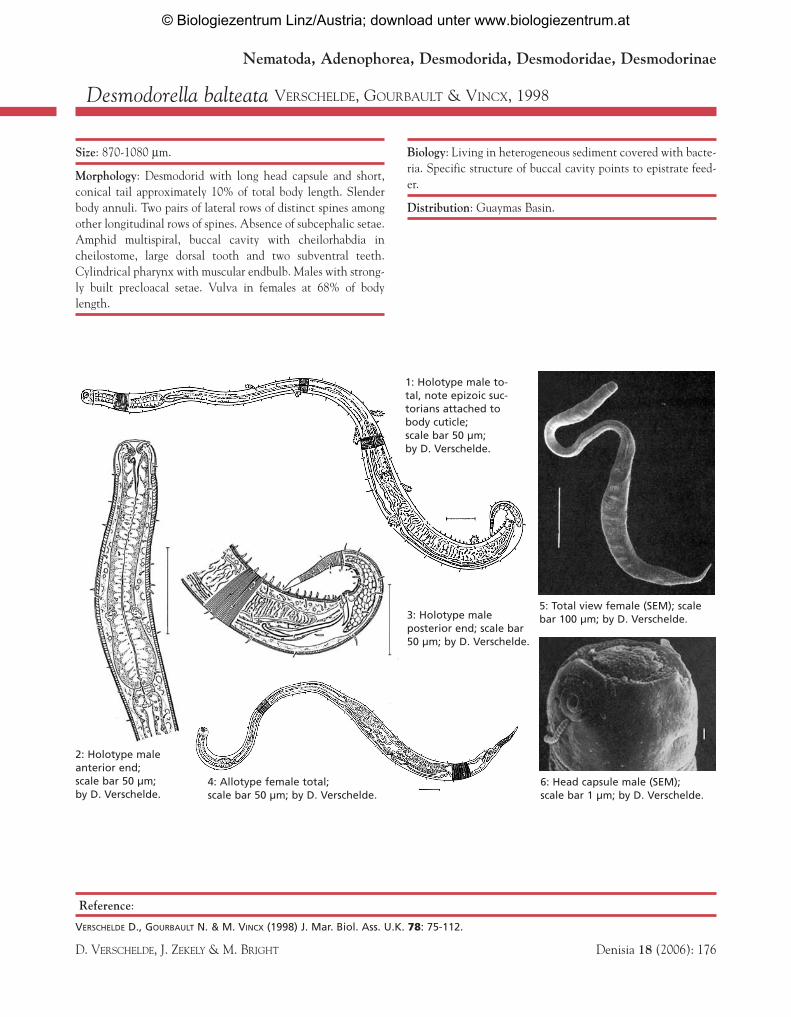
Desmodorella balteata VERSCHELDE, GOURBAULT & VINCX, 1998
Size: 870-1080 µm.
Morphology: Desmodorid with long head capsule and short,conical tail approximately 10% of total body length. Slenderbody annuli. Two pairs of lateral rows of distinct spines amongother longitudinal rows of spines. Absence of subcephalic setae.Amphid multispiral, buccal cavity with cheilorhabdia incheilostome, large dorsal tooth and two subventral teeth.Cylindrical pharynx with muscular endbulb. Males with strong-ly built precloacal setae. Vulva in females at 68% of bodylength.
Biology: Living in heterogeneous sediment covered with bacte-ria. Specific structure of buccal cavity points to epistrate feed-er.
Distribution: Guaymas Basin.
Reference:
VERSCHELDE D., GOURBAULT N. & M. VINCX (1998) J. Mar. Biol. Ass. U.K. 78: 75-112.
D. VERSCHELDE, J. ZEKELY & M. BRIGHT Denisia 18 (2006): 176
2: Holotype maleanterior end;scale bar 50 µm;by D. Verschelde.
5: Total view female (SEM); scalebar 100 µm; by D. Verschelde.
6: Head capsule male (SEM); scale bar 1 µm; by D. Verschelde.
Nematoda, Adenophorea, Desmodorida, Desmodoridae, Desmodorinae
1: Holotype male to-tal, note epizoic suc-torians attached tobody cuticle;scale bar 50 µm; by D. Verschelde.
3: Holotype maleposterior end; scale bar 50 µm; by D. Verschelde.
4: Allotype female total; scale bar 50 µm; by D. Verschelde.
© Biologiezentrum Linz/Austria; download unter www.biologiezentrum.at
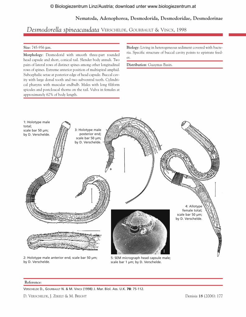
Desmodorella spineacaudata VERSCHELDE, GOURBAULT & VINCX, 1998
Size: 745-956 µm.
Morphology: Desmodorid with smooth three-part roundedhead capsule and short, conical tail. Slender body annuli. Twopairs of lateral rows of distinct spines among other longitudinalrows of spines. Extreme anterior position of multispiral amphid.Subcephalic setae at posterior edge of head capsule. Buccal cav-ity with large dorsal tooth and two subventral teeth. Cylindri-cal pharynx with muscular endbulb. Males with long filiformspicules and postcloacal thorns on the tail. Vulva in females atapproximately 62% of body length.
Biology: Living in heterogeneous sediment covered with bacte-ria. Specific structure of buccal cavity points to epistrate feed-er.
Distribution: Guaymas Basin.
Reference:
VERSCHELDE D., GOURBAULT N. & M. VINCX (1998) J. Mar. Biol. Ass. U.K. 78: 75-112.
D. VERSCHELDE, J. ZEKELY & M. BRIGHT Denisia 18 (2006): 177
Nematoda, Adenophorea, Desmodorida, Desmodoridae, Desmodorinae
1: Holotype maletotal; scale bar 50 µm;by D. Verschelde.
5: SEM micrograph head capsule male;scale bar 1 µm; by D. Verschelde.
4: Allotypefemale total;
scale bar 50 µm; by D. Verschelde.
2: Holotype male anterior end; scale bar 50 µm; by D. Verschelde.
3: Holotype maleposterior end;
scale bar 50 µm;by D. Verschelde.
© Biologiezentrum Linz/Austria; download unter www.biologiezentrum.at
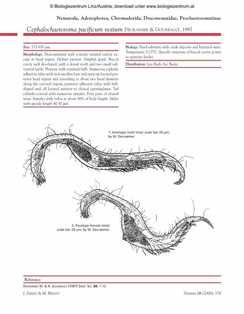
Cephalochaetosoma pacificum notium DECRAEMER & GOURBAULT, 1997
Size: 575-850 µm.
Morphology: Draconematid with coarsely striated cuticle ex-cept in head region. Helmet present. Amphid spiral. Buccalcavity well developed, with a dorsal tooth and two small sub-ventral teeth. Pharynx with terminal bulb. Numerous cephalicadhesion tubes with non-swollen base and open tip located pos-terior head region and extending to about two head diamtersalong the cervical region; posterior adhesion tubes with bell-shaped end, all located anterior to cloacal opening/anus. Tailcylindro-conoid with numerous annules. Four pairs of cloacalsetae. Females with vulva at about 48% of body length. Maleswith spicule length 40-50 µm.
Biology: Hard substrate with oxide deposits and bacterial mats.Temperature 5-17°C. Specific structure of buccal cavity pointsto epistrate feeder.
Distribution: Lau Back-Arc Basin.
Reference:
DECRAEMER W. & N. GOURBAULT (1997) Zool. Scr. 26: 1-12.
J. ZEKELY & M. BRIGHT Denisia 18 (2006): 178
Nematoda, Adenophorea, Chromadorida, Draconematidae, Prochaetosomatinae
1: Holotype male total; scale bar 20 µm;by W. Decraemer.
2: Paratype female total; scale bar 20 µm; by W. Decraemer.
© Biologiezentrum Linz/Austria; download unter www.biologiezentrum.at
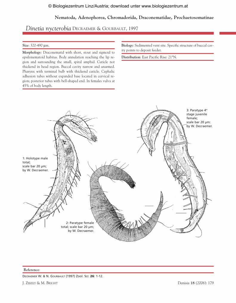
Dinetia nycterobia DECRAEMER & GOURBAULT, 1997
Size: 320-490 µm.
Morphology: Draconematid with short, stout and sigmoid toepsilonematoid habitus. Body annulation reaching the lip re-gion and surrounding the small, spiral amphid. Cuticle notthickend in head region. Buccal cavity narrow and unarmed.Pharynx with terminal bulb with thickend cuticle. Cephalicadhesion tubes without expanded base located in cervical re-gion; posterior tubes with bell-shaped end. In females vulva at45% of body length.
Biology: Sedimented vent site. Specific structure of buccal cav-ity points to deposit feeder.
Distribution: East Pacific Rise: 21°N.
Reference:
DECRAEMER W. & N. GOURBAULT (1997) Zool. Scr. 26: 1-12.
J. ZEKELY & M. BRIGHT Denisia 18 (2006): 179
Nematoda, Adenophorea, Chromadorida, Draconematidae, Prochaetosomatinae
3: Paratype 4th
stage juvenilefemale; scale bar 20 µm:by W. Decraemer.
2: Paratype femaletotal; scale bar 20 µm;
by W. Decraemer.
1: Holotype maletotal; scale bar 20 µm; by W. Decraemer.
© Biologiezentrum Linz/Austria; download unter www.biologiezentrum.at
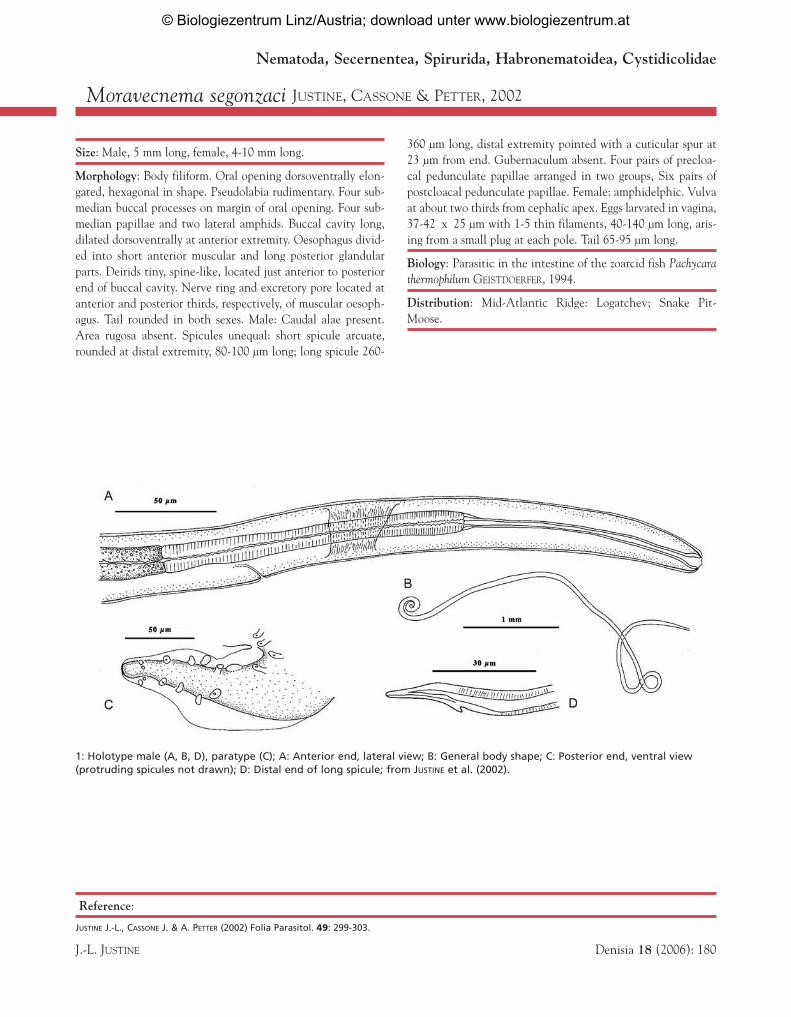
Moravecnema segonzaci JUSTINE, CASSONE & PETTER, 2002
Reference:
JUSTINE J.-L., CASSONE J. & A. PETTER (2002) Folia Parasitol. 49: 299-303.
J.-L. JUSTINE Denisia 18 (2006): 180
1: Holotype male (A, B, D), paratype (C); A: Anterior end, lateral view; B: General body shape; C: Posterior end, ventral view(protruding spicules not drawn); D: Distal end of long spicule; from JUSTINE et al. (2002).
Nematoda, Secernentea, Spirurida, Habronematoidea, Cystidicolidae
Size: Male, 5 mm long, female, 4-10 mm long.
Morphology: Body filiform. Oral opening dorsoventrally elon-gated, hexagonal in shape. Pseudolabia rudimentary. Four sub-median buccal processes on margin of oral opening. Four sub-median papillae and two lateral amphids. Buccal cavity long,dilated dorsoventrally at anterior extremity. Oesophagus divid-ed into short anterior muscular and long posterior glandularparts. Deirids tiny, spine-like, located just anterior to posteriorend of buccal cavity. Nerve ring and excretory pore located atanterior and posterior thirds, respectively, of muscular oesoph-agus. Tail rounded in both sexes. Male: Caudal alae present.Area rugosa absent. Spicules unequal: short spicule arcuate,rounded at distal extremity, 80-100 µm long; long spicule 260-
360 µm long, distal extremity pointed with a cuticular spur at23 µm from end. Gubernaculum absent. Four pairs of precloa-cal pedunculate papillae arranged in two groups, Six pairs ofpostcloacal pedunculate papillae. Female: amphidelphic. Vulvaat about two thirds from cephalic apex. Eggs larvated in vagina,37-42 x 25 µm with 1-5 thin filaments, 40-140 µm long, aris-ing from a small plug at each pole. Tail 65-95 µm long.
Biology: Parasitic in the intestine of the zoarcid fish Pachycarathermophilum GEISTDOERFER, 1994.
Distribution: Mid-Atlantic Ridge: Logatchev; Snake Pit-Moose.
© Biologiezentrum Linz/Austria; download unter www.biologiezentrum.at
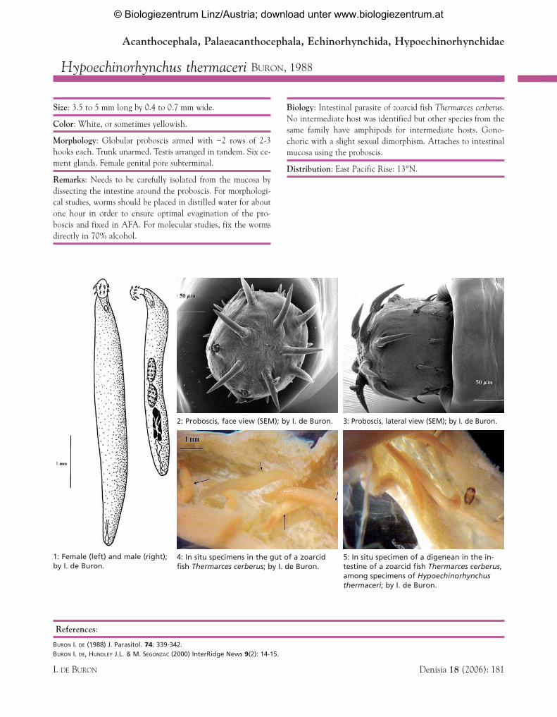
Hypoechinorhynchus thermaceri BURON, 1988
References:
BURON I. DE (1988) J. Parasitol. 74: 339-342.BURON I. DE, HUNDLEY J.L. & M. SEGONZAC (2000) InterRidge News 9(2): 14-15.
I. DE BURON Denisia 18 (2006): 181
1: Female (left) and male (right);by I. de Buron.
4: In situ specimens in the gut of a zoarcidfish Thermarces cerberus; by I. de Buron.
5: In situ specimen of a digenean in the in-testine of a zoarcid fish Thermarces cerberus,among specimens of Hypoechinorhynchusthermaceri; by I. de Buron.
2: Proboscis, face view (SEM); by I. de Buron. 3: Proboscis, lateral view (SEM); by I. de Buron.
Acanthocephala, Palaeacanthocephala, Echinorhynchida, Hypoechinorhynchidae
Size: 3.5 to 5 mm long by 0.4 to 0.7 mm wide.
Color: White, or sometimes yellowish.
Morphology: Globular proboscis armed with ~2 rows of 2-3hooks each. Trunk unarmed. Testis arranged in tandem. Six ce-ment glands. Female genital pore subterminal.
Remarks: Needs to be carefully isolated from the mucosa bydissecting the intestine around the proboscis. For morphologi-cal studies, worms should be placed in distilled water for aboutone hour in order to ensure optimal evagination of the pro-boscis and fixed in AFA. For molecular studies, fix the wormsdirectly in 70% alcohol.
Biology: Intestinal parasite of zoarcid fish Thermarces cerberus.No intermediate host was identified but other species from thesame family have amphipods for intermediate hosts. Gono-choric with a slight sexual dimorphism. Attaches to intestinalmucosa using the proboscis.
Distribution: East Pacific Rise: 13°N.
© Biologiezentrum Linz/Austria; download unter www.biologiezentrum.at

Thermonemertes valens ROGERS, GIBSON & TUNNICLIFFE, 1996
Reference:
ROGERS A.D., GIBSON R. & V. TUNNICLIFFE (1996) Deep-Sea Res. I 43(10): 1581-1599.
M. BISCOITO & A.J. ALMEIDA Denisia 18 (2006): 183
1: T. valens (preserved specimen); left anterior end, right total;from ROGERS et al. (1996).
2: Thermonemertes sp.; live specimen from East Pacific Rise:9°N; by M. Bright.
Nemertini, Hoplonemertini, Monostilifera
3: Thermonemertes sp.; live specimen anterior end with par-tially extruded proboscis; from East Pacific Rise: 13°N; by M.Bright
Size: 5 cm.
Color: Bright red; after fixation uniformly flesh colored.
Morphology: The head is spatuloate and dorsoventrally flat-tened. A single cephalic furrow on the ventral surface towardsthe rear of the head. Mouth and rhynchodeum open into a sin-gle opening behind the tip of the head. The body is slender anddorsoventrally compressed. In the posterior half the ventral sur-face appears distinctly concave.
Remarks: Another yet undescribed species of Thermonemerteswas found at East Pacific Rise: 9°N from inactive sulfide chim-neys and at 13°N from Riftia pachyptila aggregations.
Biology: Lives on rocks and bacterial mats; not associated withvestimentiferan tubeworms. The diet remains unknown. Mightbe able to swim. It has been suggested that this species is an ear-ly colonizer of vent communities, but is displaced in aging com-munities.
Distribution: Juan de Fuca Ridge.
© Biologiezentrum Linz/Austria; download unter www.biologiezentrum.at
Annelida, Polychaeta1
According to different countings, the polychaete worms, with111 presently described species, constitute 18-20% of the totalnumber of species fully identified from vent samples. 30% of thesebristle worms are belonging to scale worms (Polynoidae), 10% toalvinellids, 9% to siboglinids, 6% to spionids, 5% to hesionids anddorvilleids. Other families account for less than 5% of describedannelid species. The present recorded species are largely (94%)considered as ‘endemic’ of the deep-sea hydrothermal vent envi-ronment (or according to Andrey Gebruk, vent ‘obligate’), lessthan 4% are shared with other deep-sea reduced environments andca. 2% are ‘regular’ deep-sea species. However this high ratio of en-demism will probably be revised downward when the sampling ofthe peripheral areas will make progress accounting for opportunis-tic groups as dorvilleids or spionids which could be thriving in ad-jacent disturbed habitats.
Because of their odd biology and ecology, two species of an-nelids Riftia pachyptila (‘the giant tube worm’) and Alvinella pompe-jana (‘the Pompeii worm’) became emblematic of the vent researchand are still considered as ‘biological models’. Numerous researchprojects dealing with the functioning of endosymbiosis in Riftiapachyptila and the physiological adaptation of Alvinella pompejanato its extreme habitat are presently underway.
Nevertheless, the basic taxonomy of vent annelids is far to becompleted. Recent cruises which occurred in the Western Pacific(Lau Basin, Kermadec and Mariana Arcs) and on southern East Pa-cific Rise brought back a lot of new interesting specimens present-ly under study. For example, five new species of scale-worms fromLau Basin are being described belonging to Levensteiniella,Branchinotogluma, Lepidonotopodium and Harmothoe as well as onespecies of ampharetid, one sigalionid, one alvinellid (Paralvinella n.sp.), a new species of dorvilleid belonging to Parougia and one tere-bellid (cf. Polycirrus). From southern East Pacific Rise, severalspecies of scale worms, one species of alvinellid and one surprisingnew genus of spionid are currently under description. New familiesfor this environment were found and the description of new speciesof flabelligerid and sphaerodorid are under wording. Former collec-tions of worms from Mid-Atlantic Ridge and East Pacific Rise arestill not exhaustively studied and several families (e.g. dorvilleid,spionid, capitellid, cirratulid) need further taxonomic work.
The molecular identification of sibling species among wide-spread morphotypes (e.g. Amphisamytha galapagensis or Archinomerosacea) lead ‘classical’ taxonomists to dig back throughout formercollections looking for new diagnostic characters. For example, ajoint work between morphologists and molecular taxonomists al-
lows recognizing three different new species of Archinome and onenew genus of Amphinomidae among specimens previously identi-fied as A. rosacea (J. Kudenov, pers. comm.) Conversely, newrecords of previously described species from new locations (e.g. Hes-iolyra bergi on Mid-Atlantic Ridge vents or Hesiospina vestimentiferafrom Lau Basin) question the dispersal and allopatric speciation inresponse to major vicariance events. Because of the influence ofjoint occurrences in biogeographic analyses, the solution of thesetaxonomic riddle must be seriously considered and we urge scien-tists working at sea to focus on these widespread morphotypes andto build up parallel samples for classical and molecular taxonomy.
Polychaetes are delicate and fragile animals, and special careshould be taken when handling them. To be described, the speci-mens must be unfragmented and as intact as possible and damagedspecimens may be misidentified. Thus a gentle sorting is desirable assoon as possible after recovery of the samples, using soft pliers formacroscopic individuals and gentle sieving for others (avoid stackof sieves and split the samples before sieving). Specimens may be re-laxed prior to preservation (7.5% of magnesium chloride) and tubesof tubicolous annelids must be opened; fixation is best in bufferedformalin 5-10%. After a suitable time of fixation, depending of thesize (in general < 24 hours), the worms may be transferred to 80%ethyl or isopropyl alcohol (formaldehyde even buffered, is a verypoor preservative).
1 Even if the question is still strongly debated and the molecular information isnot consistent, we chose herein, following ROUSE & FAUCHALD (1997) to in-clude the Pogonophora (Perviata and Vestimentifera) within the family Si-boglinidae (Polychaeta: Sabellida). Contrarily SOUTHWARD et al. (2005) chosea more conservative standpoint and retain the class ‘Pogonophora’ within An-nelida, waiting for more conclusive information (HALANYCH 2005).
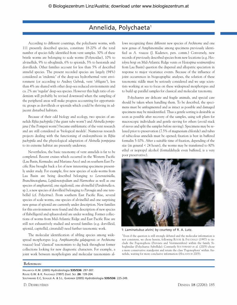
1: Laminatubus alvini; by courtesy of R. A. Lutz.
References:
HALANYCH K.M. (2005) Hydrobiologia 535/536: 297-307.ROUSE G.W. & K. FAUCHALD (1997) Zool. Scr. 26: 139-204.SOUTHWARD E.C, SCHULZE A. & S.L. GARDINER (2005) Hydrobiologia 535/536: 225-249.
D. DESBRUYÈRES DENISIA 18 (2006): 185
© Biologiezentrum Linz/Austria; download unter www.biologiezentrum.at