Embed Size (px)
Citation preview
General rights Copyright and moral rights for the publications made accessible in the public portal are retained by the authors and/or other copyright owners and it is a condition of accessing publications that users recognise and abide by the legal requirements associated with these rights.
Users may download and print one copy of any publication from the public portal for the purpose of private study or research.
You may not further distribute the material or use it for any profit-making activity or commercial gain
You may freely distribute the URL identifying the publication in the public portal If you believe that this document breaches copyright please contact us providing details, and we will remove access to the work immediately and investigate your claim.
Downloaded from orbit.dtu.dk on: Aug 11, 2020
Neodymium-140 DOTA-LM3Evaluation of an In Vivo Generator for PET with a Non-Internalizing Vector
Severin, Gregory; Kristensen, Lotte K.; Nielsen, Carsten H.; Fonslet, Jesper; Jensen, Andreas TueIngemann; Frellsen, Anders Floor; Jensen, K. M.; Elema, Dennis Ringkjøbing; Maecke, Helmut; Kjær,AndreasTotal number of authors:12
Published in:Frontiers in Medicine
Link to article, DOI:10.3389/fmed.2017.00098
Publication date:2017
Document VersionPublisher's PDF, also known as Version of record
Link back to DTU Orbit
Citation (APA):Severin, G., Kristensen, L. K., Nielsen, C. H., Fonslet, J., Jensen, A. T. I., Frellsen, A. F., Jensen, K. M., Elema,D. R., Maecke, H., Kjær, A., Johnston, K., & Köster, U. (2017). Neodymium-140 DOTA-LM3: Evaluation of an InVivo Generator for PET with a Non-Internalizing Vector. Frontiers in Medicine, 4, [98].https://doi.org/10.3389/fmed.2017.00098

July 2017 | Volume 4 | Article 981
Original researchpublished: 12 July 2017
doi: 10.3389/fmed.2017.00098
Frontiers in Medicine | www.frontiersin.org
Edited by: Jean-Pierre Pouget,
Institut national de la santé et de la recherche médicale
(INSERM), France
Reviewed by: Frank Roesch,
Johannes Gutenberg-Universität Mainz, Germany
Nicolas Lepareur, Centre Eugène Marquis, France
Clemens Decristoforo, Innsbruck Medical University, Austria
*Correspondence:Gregory W. Severin
Specialty section: This article was submitted
to Nuclear Medicine, a section of the journal
Frontiers in Medicine
Received: 06 January 2017Accepted: 20 June 2017Published: 12 July 2017
Citation: Severin GW, Kristensen LK,
Nielsen CH, Fonslet J, Jensen AI, Frellsen AF, Jensen KM, Elema DR, Maecke H, Kjær A, Johnston K and
Köster U (2017) Neodymium-140 DOTA-LM3: Evaluation of an
In Vivo Generator for PET with a Non-Internalizing Vector.
Front. Med. 4:98. doi: 10.3389/fmed.2017.00098
neodymium-140 DOTa-lM3: evaluation of an In Vivo generator for PeT with a non-internalizing VectorGregory W. Severin1,2,3*, Lotte K. Kristensen4, Carsten H. Nielsen4, Jesper Fonslet1, Andreas I. Jensen1, Anders F. Frellsen1, K. M. Jensen1, Dennis R. Elema1, Helmut Maecke5, Andreas Kjær 4, Karl Johnston6 and Ulli Köster6,7
1 Hevesy Laboratory, DTU Nutech, Technical University of Denmark, Roskilde, Denmark, 2 Department of Chemistry, Michigan State University, East Lansing, MI, United States, 3 Facility for Rare Isotope Beams, Michigan State University, East Lansing, MI, United States, 4 Department of Clinical Physiology, Nuclear Medicine & PET and Cluster for Molecular Imaging, Rigshospitalet, University of Copenhagen, Copenhagen, Denmark, 5 Department of Nuclear Medicine, University Hospital Freiburg, Freiburg, Germany, 6 ISOLDE, CERN, Geneva, Switzerland, 7 Institut Laue-Langevin, Grenoble, France
140Nd (t1/2 = 3.4 days), owing to its short-lived positron emitting daughter 140Pr (t1/2 = 3.4 min), has promise as an in vivo generator for positron emission tomography (PET). However, the electron capture decay of 140Nd is chemically disruptive to macro-cycle-based radiolabeling, meaning that an in vivo redistribution of the daughter 140Pr is expected before positron emission. The purpose of this study was to determine how the delayed positron from the de-labeled 140Pr affects preclinical imaging with 140Nd. To explore the effect, 140Nd was produced at CERN-ISOLDE, reacted with the somatostatin analogue, DOTA-LM3 (1,4,7,10- tetraazacyclododecane, 1,4,7- tri acetic acid, 10- acetamide N - p-Cl-Phecyclo(d-Cys-Tyr-d-4-amino-Phe(carbamoyl)-Lys-Thr-Cys)d-Tyr-NH2) and injected into H727 xenograft bearing mice. Comparative pre- and post-mortem PET imaging at 16 h postinjection was used to quantify the in vivo redistribution of 140Pr following 140Nd decay. The somatostatin receptor-positive pancreas exhibited the highest tissue accumulation of 140Nd-DOTA-LM3 (13% ID/g at 16 h) coupled with the largest observed redistribution rate, where 56 ± 7% (n = 4, mean ± SD) of the in situ produced 140Pr washed out of the pancreas before decay. Contrastingly, the liver, spleen, and lungs acted as strong sink organs for free 140Pr3+. Based upon these results, we conclude that 140Nd imaging with a non-internalizing vector convolutes the biodistribution of the tracer with the accumulation pattern of free 140Pr. This redistribution phenomenon may show promise as a probe of the cellular interaction with the vector, such as in determining tissue dependent internalization behavior.
Keywords: in vivo generator, 140nd, 140Pr, internalization, positron emission tomography, DOTa-lM3
inTrODUcTiOn
The demand for long-lived positron emitting radiolanthanides is growing due to the success of targeted internal radiotherapy with 177Lu, and the promise of other therapeutic lanthanides such as Auger electron emitters 165Er, and 135La or combined beta-/Auger electron emitters such as 161Tb (1–5). Neodymium-140 (140Nd, t1/2 = 3.4 days) decays to praseodymium-140 (140Pr, t1/2 = 3.4 min) by
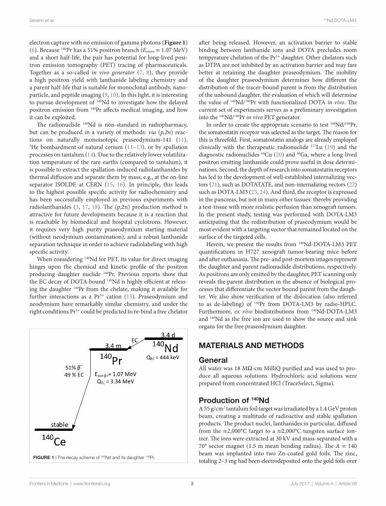
FigUre 1 | The decay scheme of 140Nd and its daughter 140Pr.
2
Severin et al. 140Nd DOTA-LM3
Frontiers in Medicine | www.frontiersin.org July 2017 | Volume 4 | Article 98
electron capture with no emission of gamma photons (Figure 1) (6). Because 140Pr has a 51% positron branch (Emean = 1.07 MeV) and a short half-life, the pair has potential for long-lived posi-tron emission tomography (PET) tracing of pharmaceuticals. Together as a so-called in vivo generator (7, 8), they provide a high positron yield with lanthanide labeling chemistry and a parent half-life that is suitable for monoclonal antibody, nano-particle, and peptide imaging (9, 10). In this light, it is interesting to pursue development of 140Nd to investigate how the delayed positron emission from 140Pr affects medical imaging, and how it can be exploited.
The radionuclide 140Nd is non-standard in radiopharmacy, but can be produced in a variety of methods: via (p,2n) reac-tions on naturally monoisotopic praseodymium-141 (11), 3He bombardment of natural cerium (11–13), or by spallation processes on tantalum (14). Due to the relatively lower volatiliza-tion temperature of the rare earths (compared to tantalum), it is possible to extract the spallation-induced radiolanthanides by thermal diffusion and separate them by mass, e.g., at the on-line separator ISOLDE at CERN (15, 16). In principle, this leads to the highest possible specific activity for radiochemistry and has been successfully employed in previous experiments with radiolanthanides (3, 17, 18). The (p,2n) production method is attractive for future developments because it is a reaction that is reachable by biomedical and hospital cyclotrons. However, it requires very high purity praseodymium starting material (without neodymium contamination), and a robust lanthanide separation technique in order to achieve radiolabeling with high specific activity.
When considering 140Nd for PET, its value for direct imaging hinges upon the chemical and kinetic profile of the positron producing daughter nuclide 140Pr. Previous reports show that the EC decay of DOTA-bound 140Nd is highly efficient at releas-ing the daughter 140Pr from the chelate, making it available for further interactions as a Pr3+ cation (13). Praseodymium and neodymium have remarkably similar chemistry, and under the right conditions Pr3+ could be predicted to re-bind a free chelator
after being released. However, an activation barrier to stable binding between lanthanide ions and DOTA precludes room temperature chelation of the Pr3+ daughter. Other chelators such as DTPA are not inhibited by an activation barrier and may fare better at retaining the daughter praseodymium. The mobility of the daughter praseodymium determines how different the distribution of the tracer-bound parent is from the distribution of the unbound daughter, the evaluation of which will determine the value of 140Nd/140Pr with functionalized DOTA in vivo. The current set of experiments serves as a preliminary investigation into the 140Nd/140Pr in vivo PET generator.
In order to create the appropriate scenario to test 140Nd/140Pr, the somatostatin receptor was selected as the target. The reason for this is threefold. First, somatostatin analogs are already employed clinically with the therapeutic radionuclide 177Lu (19) and the diagnostic radionuclides 64Cu (20) and 68Ga, where a long-lived positron emitting lanthanide could prove useful in dose determi-nations. Second, the depth of research into somatostatin receptors has led to the development of well-established internalizing vec-tors (21), such as DOTATATE, and non-internalizing vectors (22) such as DOTA-LM3 (23, 24). And third, the receptor is expressed in the pancreas, but not in many other tissues: thereby providing a test-tissue with more realistic perfusion than xenograft tumors. In the present study, testing was performed with DOTA-LM3 anticipating that the redistribution of praseodymium would be most evident with a targeting vector that remained located on the surface of the targeted cells.
Herein, we present the results from 140Nd-DOTA-LM3 PET quantifications in H727 xenograft tumor-bearing mice before and after euthanasia. The pre- and post-mortem images represent the daughter and parent radionuclide distributions, respectively. As positrons are only emitted by the daughter, PET scanning only reveals the parent distribution in the absence of biological pro-cesses that differentiate the vector bound parent from the daugh-ter. We also show verification of the dislocation (also referred to as de-labeling) of 140Pr from DOTA-LM3 by radio-HPLC. Furthermore, ex vivo biodistributions from 140Nd-DOTA-LM3 and 140Nd as the free ion are used to show the source and sink organs for the free praseodymium daughter.
MaTerials anD MeThODs
generalAll water was 18 MΩ-cm MilliQ purified and was used to pro-duce all aqueous solutions. Hydrochloric acid solutions were prepared from concentrated HCl (TraceSelect, Sigma).
Production of 140ndA 55 g/cm2 tantalum foil target was irradiated by a 1.4 GeV proton beam, creating a multitude of radioactive and stable spallation products. The product nuclei, lanthanides in particular, diffused from the ≈2,000°C target to a ≈2,000°C tungsten surface ion-izer. The ions were extracted at 30 kV and mass-separated with a 70° sector magnet (1.5 m mean bending radius). The A = 140 beam was implanted into two Zn-coated gold foils. The zinc, totaling 2–3 mg had been electrodeposited onto the gold foils over
3
Severin et al. 140Nd DOTA-LM3
Frontiers in Medicine | www.frontiersin.org July 2017 | Volume 4 | Article 98
approximately 0.5 cm2. The entire procedure is nearly identical to the methodology recently described for projects collecting Tb isotopes at ISOLDE (3, 17).
radiochemistryThe following methodology was carried out two times with small variations between each run.
The zinc layer containing 140Nd was etched briefly with aq. HCl (200 µL, 2 M), and the resulting solution was diluted to 2.2 mL with aq. HCl (2 M). This was heated to 98°C, cooled, and passed over AG1x8 anion exchange resin (300 mg, Biorad, 200–400 mesh, initially formate-form) packed in a 4 mm internal diameter (ID), fritted polypropylene column (Supelco) that had been prepped by three times sequential washing with water (3× bed volume), 2 M HCl (3× bed volume), and 6 M HCl (3× bed volume), finishing with equilibration in 2 M HCl. The resin has a high affinity for [ZnCl4]2− ions in 2 M HCl (25) and was intended to remove any zinc impurity from the 140Nd. An additional 0.5 mL of 2 M HCl was used to rinse residual 140Nd from the column, and the entire effluent was collected and adjusted to pH 5–6 with aq. ammonium acetate (1 M, pH 7) and aq. ammonium hydroxide (28%, TraceSelect, Sigma) to a final acetate concentration of roughly 200 mM. This solution was passed over a hydroxamate functionalized Waters CM resin bed (26) (100 mg, 4 mm ID) to trap the 140Nd and was washed with water (7 mL), then eluted with aq. HCl (600 µL, 0.1 M). The elu-ent was kept as the stock 140Nd solution for further radiolabeling and formulation.
For the DOTA-LM3 preparation, 220 µL of the 140Nd stock solution was added to aq. ammonium acetate (780 µL, 300 mM). Two productions were made, the first with 9 µg DOTA-LM3 (added from a stock solution of 1 mg/mL in water) and the second with 18 µg DOTA-LM3. The solution was heated in a sealed container to 95°C and incubated for 25 min. After cooling to room temperature, the labeling reaction was quenched with aq. DTPA (12 µL, 0.5 mM, pH 7) and let stand for 5 min. The solution was passed over a Waters C18 light sep-pak (prepped with 10 mL ethanol and 10 mL water) to trap the labeled product, rinsed with 1 mL water, and then eluted with 2 mL ethanol. The ethanol solution was taken almost to dryness (residual volume was approximately 50 µL) with the residual being diluted with 900 µL HEPES buffered isotonic saline (10 mM HEPES, 150 mM NaCl, pH 7.4) and was used directly for injections.
The neodymium “chloride” injections were prepared by dilut-ing 100 µL of 140Nd stock (in 0.1 M HCl) with 390 µL HEPES buffered isotonic saline (10 mM HEPES), and neutralizing with 10 µL 1 M Na-HEPES.
hPlc Verification of De-labeling and radioTlc for Determination of radiochemical PuritySamples of 140Nd-DOTA-LM3 were injected onto a reverse-phase C-18 (Luna 3uC18(2)(n) 100 A 100 × 2 mm 3 μm, Phenomenex) column at a flow rate of 0.5 mL/min starting from 0% acetonitrile in water, and reaching 100% over a 15 min gradient. Elution was
monitored with a radio detector. The entire effluent was collected in 1 min intervals (500 µL each) and quantified 4 days after collection by liquid scintillation counting on a HIDEX 300 SL spectrometer.
RadioTLC was performed by spotting 1 µL of the DOTA-LM3 solutions (before and after C-18 purification) onto aluminum-backed silica TLC sheets. The sheets were eluted in 10% (w/v) aq. CH3COONa:CH3OH (1:1). Unreacted 140Nd remained at the origin, and 140Nd-DOTA-LM3 moved to Rf ~0.5.
PeT imaging and Ex Vivo BiodistributionsNCI-H727 lung carcinoid cancer cells (ATCC CRL-5815, LGC Standards) were cultured in RPMI-1640 media supplemented with 10% fetal bovine serum and 1% penicillin-streptomycin (Invitrogen) at 37° C and 5% CO2. Cells in their exponential growth phase and at 80–90% confluence were harvested by trypsinization and resuspended in 1:1 media and matrigel (BD Biosciences) at 5 × 10⁷ cells/mL. Subcutaneous tumors were established in female NMRI nude mice (Taconic, Denmark) by inoculation of 5 × 10⁶ cells in 100 µL on each flank above the hind limbs in the subcutaneous space. All animal experiments were performed under a protocol approved by the National Animal Experiments Inspectorate of Denmark.
Longitudinal small animal PET/CT imaging (Inveon Multi-modality PET/CT scanner, Siemens) was performed with NCI-H727 tumor bearing mice injected intravenously with 3.3–4.3 MBq 140Nd-DOTA-LM3 (n = 8) or 2.7–3.1 MBq 140Nd- chloride (n = 3) in 150 µL. Mice were anesthetized with sevoflu-rane (Abbott Laboratories) during injection and PET/CT imag-ing. PET data was acquired for 600 s in list mode at 1, 3, and 16 h after injection. The mice were sacrificed after the 16 h time-point and a PET acquisition was performed 2 h post-mortem. Images were reconstructed using a 3D maximum a posteriori algorithm with CT based attenuation correction. CT images were acquired with the following settings: 300 projections, 65 kV, 500 µA, and 400 ms exposure, and reconstructed with an isotropic voxel size of 105 µm. Image analysis was performed using the Inveon Software (Siemens). Region of interests (ROIs) were drawn man-ually over the tumor regions and other organs based on the CT images and the uptake of 140Nd-DOTA-LM3 or 140Nd-chloride quantified as % injected dose per gram tissue (%ID/g).
Conventional ex vivo biodistribution was performed after the post-mortem scan. Tumors and organs were resected, weighted and the radioactivity was counted in a gamma counter (Wizard2, PerkinElmer).
resUlTs anD DiscUssiOn
isolation of 140nd at isOlDe, radiochemical Purification, and radiolabeling of 140nd-DOTa-lM3140Nd was produced by 1.4 GeV proton induced spallation of tantalum at ISOLDE. The process for vaporization and ionization of lanthanides at the ISOLDE facility is well described (15), and proceeded without complication. The electromagnetic separa-tion of proton rich A = 140 spallation products led to a total
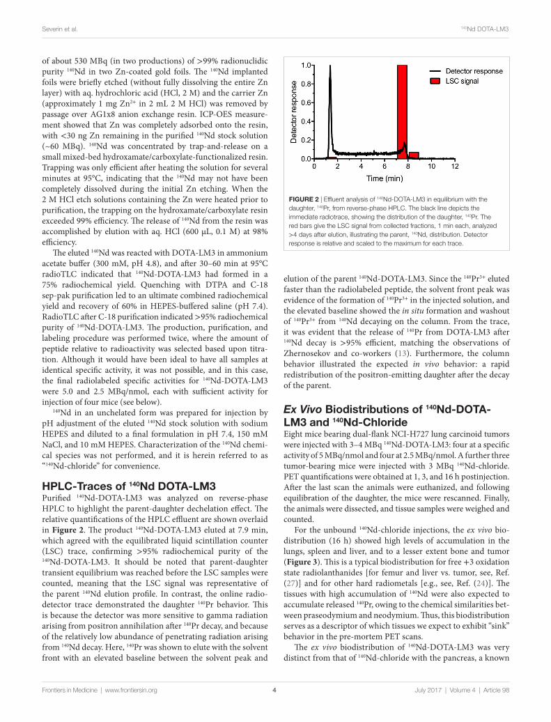
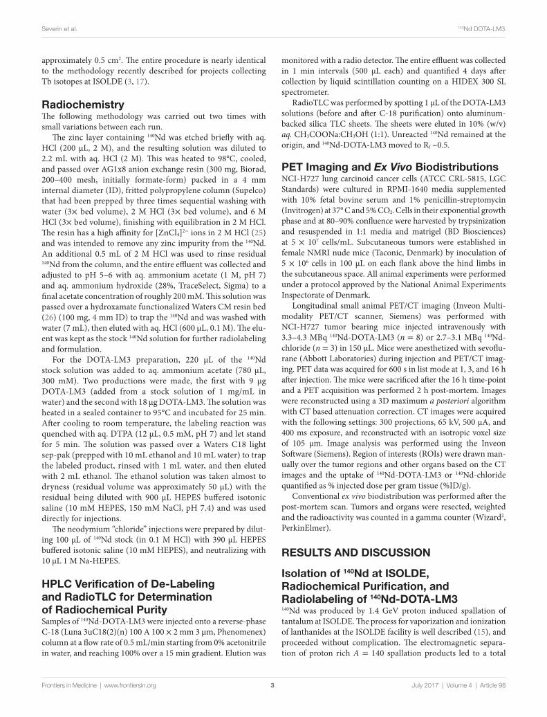
FigUre 2 | Effluent analysis of 140Nd-DOTA-LM3 in equilibrium with the daughter, 140Pr, from reverse-phase HPLC. The black line depicts the immediate radiotrace, showing the distribution of the daughter, 140Pr. The red bars give the LSC signal from collected fractions, 1 min each, analyzed >4 days after elution, illustrating the parent, 140Nd, distribution. Detector response is relative and scaled to the maximum for each trace.
4
Severin et al. 140Nd DOTA-LM3
Frontiers in Medicine | www.frontiersin.org July 2017 | Volume 4 | Article 98
of about 530 MBq (in two productions) of >99% radionuclidic purity 140Nd in two Zn-coated gold foils. The 140Nd implanted foils were briefly etched (without fully dissolving the entire Zn layer) with aq. hydrochloric acid (HCl, 2 M) and the carrier Zn (approximately 1 mg Zn2+ in 2 mL 2 M HCl) was removed by passage over AG1x8 anion exchange resin. ICP-OES measure-ment showed that Zn was completely adsorbed onto the resin, with <30 ng Zn remaining in the purified 140Nd stock solution (~60 MBq). 140Nd was concentrated by trap-and-release on a small mixed-bed hydroxamate/carboxylate-functionalized resin. Trapping was only efficient after heating the solution for several minutes at 95°C, indicating that the 140Nd may not have been completely dissolved during the initial Zn etching. When the 2 M HCl etch solutions containing the Zn were heated prior to purification, the trapping on the hydroxamate/carboxylate resin exceeded 99% efficiency. The release of 140Nd from the resin was accomplished by elution with aq. HCl (600 µL, 0.1 M) at 98% efficiency.
The eluted 140Nd was reacted with DOTA-LM3 in ammonium acetate buffer (300 mM, pH 4.8), and after 30–60 min at 95°C radioTLC indicated that 140Nd-DOTA-LM3 had formed in a 75% radiochemical yield. Quenching with DTPA and C-18 sep-pak purification led to an ultimate combined radiochemical yield and recovery of 60% in HEPES-buffered saline (pH 7.4). RadioTLC after C-18 purification indicated >95% radiochemical purity of 140Nd-DOTA-LM3. The production, purification, and labeling procedure was performed twice, where the amount of peptide relative to radioactivity was selected based upon titra-tion. Although it would have been ideal to have all samples at identical specific activity, it was not possible, and in this case, the final radiolabeled specific activities for 140Nd-DOTA-LM3 were 5.0 and 2.5 MBq/nmol, each with sufficient activity for injection of four mice (see below).
140Nd in an unchelated form was prepared for injection by pH adjustment of the eluted 140Nd stock solution with sodium HEPES and diluted to a final formulation in pH 7.4, 150 mM NaCl, and 10 mM HEPES. Characterization of the 140Nd chemi-cal species was not performed, and it is herein referred to as “140Nd-chloride” for convenience.
hPlc-Traces of 140nd DOTa-lM3Purified 140Nd-DOTA-LM3 was analyzed on reverse-phase HPLC to highlight the parent-daughter dechelation effect. The relative quantifications of the HPLC effluent are shown overlaid in Figure 2. The product 140Nd-DOTA-LM3 eluted at 7.9 min, which agreed with the equilibrated liquid scintillation counter (LSC) trace, confirming >95% radiochemical purity of the 140Nd-DOTA-LM3. It should be noted that parent-daughter transient equilibrium was reached before the LSC samples were counted, meaning that the LSC signal was representative of the parent 140Nd elution profile. In contrast, the online radio-detector trace demonstrated the daughter 140Pr behavior. This is because the detector was more sensitive to gamma radiation arising from positron annihilation after 140Pr decay, and because of the relatively low abundance of penetrating radiation arising from 140Nd decay. Here, 140Pr was shown to elute with the solvent front with an elevated baseline between the solvent peak and
elution of the parent 140Nd-DOTA-LM3. Since the 140Pr3+ eluted faster than the radiolabeled peptide, the solvent front peak was evidence of the formation of 140Pr3+ in the injected solution, and the elevated baseline showed the in situ formation and washout of 140Pr3+ from 140Nd decaying on the column. From the trace, it was evident that the release of 140Pr from DOTA-LM3 after 140Nd decay is >95% efficient, matching the observations of Zhernosekov and co-workers (13). Furthermore, the column behavior illustrated the expected in vivo behavior: a rapid redistribution of the positron-emitting daughter after the decay of the parent.
Ex Vivo Biodistributions of 140nd-DOTa-lM3 and 140nd-chlorideEight mice bearing dual-flank NCI-H727 lung carcinoid tumors were injected with 3–4 MBq 140Nd-DOTA-LM3: four at a specific activity of 5 MBq/nmol and four at 2.5 MBq/nmol. A further three tumor-bearing mice were injected with 3 MBq 140Nd-chloride. PET quantifications were obtained at 1, 3, and 16 h postinjection. After the last scan the animals were euthanized, and following equilibration of the daughter, the mice were rescanned. Finally, the animals were dissected, and tissue samples were weighed and counted.
For the unbound 140Nd-chloride injections, the ex vivo bio-distribution (16 h) showed high levels of accumulation in the lungs, spleen and liver, and to a lesser extent bone and tumor (Figure 3). This is a typical biodistribution for free +3 oxidation state radiolanthanides [for femur and liver vs. tumor, see, Ref. (27)] and for other hard radiometals [e.g., see, Ref. (24)]. The tissues with high accumulation of 140Nd were also expected to accumulate released 140Pr, owing to the chemical similarities bet-ween praseodymium and neodymium. Thus, this biodistribution serves as a descriptor of which tissues we expect to exhibit “sink” behavior in the pre-mortem PET scans.
The ex vivo biodistribution of 140Nd-DOTA-LM3 was very distinct from that of 140Nd-chloride with the pancreas, a known

FigUre 3 | Ex vivo biodistribution of 140Nd-DOTA-LM3 and 140Nd-chloride. Animals were euthanized 16 h after injection of 3.3–4.3 MBq 140Nd-DOTA-LM3 (5.0 MBq/nmol, 0.5 nmol injected, n = 4), 140Nd-DOTA-LM3 (2.5 MBq/nmol, 1 nmol injected, n = 4) and 2.7–3.1 MBq 140Nd-chloride (n = 3). Organ counting commenced after parent/daughter equilibrium was achieved (2 h after euthanasia). 140Nd-DOTA-LM3 quantifications give the “source” distribution for 140Pr in the 16 h positron emission tomography studies, and 140Nd-chloride qualitatively describes the “sink” behavior. Data are depicted as mean ± SEM.
5
Severin et al. 140Nd DOTA-LM3
Frontiers in Medicine | www.frontiersin.org July 2017 | Volume 4 | Article 98
somatostatin receptor positive organ, being particularly interest-ing (21). The pancreas was found to take up 140Nd-DOTA-LM3 without accumulating the free radiometal (13% ID/g with high specific activity 140Nd-DOTA-LM3, compared to 0.1% ID/g with the free 140Nd3+). Therefore, in the PET study, the pan-creas was expected to have a lower signal in the pre-mortem 140Nd-DOTA-LM3 scans compared to the post-mortem scans. The liver, spleen, and lungs had the opposite behavior, accumu-lating the free radiometal but not the peptide, and were expected to have a higher signal in the pre-mortem scans than in the post-mortem.
Fani et al. showed that in comparing the biodistribution of 68Ga-NODAGA-LM3, 68Ga-DOTA-LM3, 64Cu-NODAGA-LM3, and 64Cu-CBTE2A-LM3, tumor uptake relative to the pancreas, stomach, and kidney is highly variable depending on the metal and chelator used (23, 28). In the current work, we injected 300 pmol (5 MBq/nmol) or 600 pmol (2.5 MBq/nmol) in order to get enough signal for the PET scans. For the biodistributions from Fani et al., 10 pmol was injected, and blocking was per-formed with 200 nmol of excess DOTA-LM3. The overall effect of the blocking was to reduce the uptake in sst2 expressing tissues, which is exactly what was observed between the higher and lower specific activity injections of the current study: a modest reduc-tion in the uptake in sst2 expressing tissues relative to the kidney, and overall faster excretion.
Based upon the ex vivo biodistribution, the tumors had an intermediate behavior, weakly concentrating both the free 140Nd and the labeled peptide, meaning that the PET signal was expected to change little between the pre- and post-mortem scans (i.e., washout of 140Pr from the tumor volume could be compen-sated by uptake of 140Pr from the bloodstream). This behavior is in many ways an undesirable outcome, because signals arising from the tumors can be attributable to both the free metal ions
and the targeted peptide. Future work with a different vector/target system could give a result with a simpler interpretation. Nevertheless, the pancreas remains an interesting tissue within the present set of experiments.
PeT studies show Tissue Dependent redistribution of 140Pr after 140nd DecayPositron emission tomography data were analyzed to quantify the 140Pr signal from the tumors, pancreas, kidney, lung, and liver (Table 1). The PET signals for the tumors are given as the percent of the injected signal per gram (%IS/g) (Figure 4). This unit is non-standard and is described further in the Supplementary Material. %IS/g was chosen for two reasons: first because the PET scans quantify the redistributed daughter 140Pr, not the 140Nd-tracer, and second because the data were not corrected for point spreading due to extensive positron range. This means that there is a substantial partial volume distortion when converting from annihilation signal (%IS/g) to tracer concentration (%ID/g), and that the well-counter based ex vivo biodistribution is not directly comparable to the post mortem PET results. A discus-sion of the partial volume effect due to the high energy positrons from 140Pr is included in the Supplementary Material. The tumor time-activity curve shows that in this model (as expected from the ex vivo biodistribution) very little redistribution is observed. For the present case, the tumor was known to accumulate the trivalent lanthanide to some small degree (from the Nd-chloride injection data above), meaning that 140Pr3+ produced by decay in the tumor region had some tendency to remain. In other organs, however, due to the dechelation effect, there was a dramatic change in the PET signal in the pre- and post-mortem imaging quantifications. The redistribution effects are displayed in Figure 5 as the ratio of the mean organ signal between the post-mortem, and pre-mortem (16 h) images. This effect is most evident in the pancreas and liver for the high specific activity injections, where the decrease in liver signal is matched by an increase in the pancreas signal, confirming the behaviors of the peptide and free metal observed in the ex vivo biodistribution.
It should be noted that the injected mass of the tracer had a large effect upon the PET quantifications and ex vivo biodis-tribution (Figures 4 and 5). This indicates that the receptor-specific accumulation was becoming saturated as the injected mass increased from 0.5 nmol (5.0 MBq/nmol) to 1 nmol (2.5 MBq/nmol). This saturation behavior is non-desirable for probing the internalization behavior of the peptide as non-specific interactions begin to dominate the tracer distribution.
Figure 6 shows an example PET/CT reconstruction, quali-tatively illustrating the redistribution effect. The most striking differences in the images between the daughter distribution (16 h, left panels) and the parent distribution (post-mortem, right panels) are seen in the liver, lungs, and pancreas. As expected, the 16 h pre-mortem PET images more closely reflected the biodistribution observed for the free 140Nd-chloride injections, while the post-mortem images showed the biodistribution for the intact 140Nd-DOTA-LM3 tracer.
The most important result from the PET imaging was the pancreatic signal. In this case, in the pre-mortem image, it was
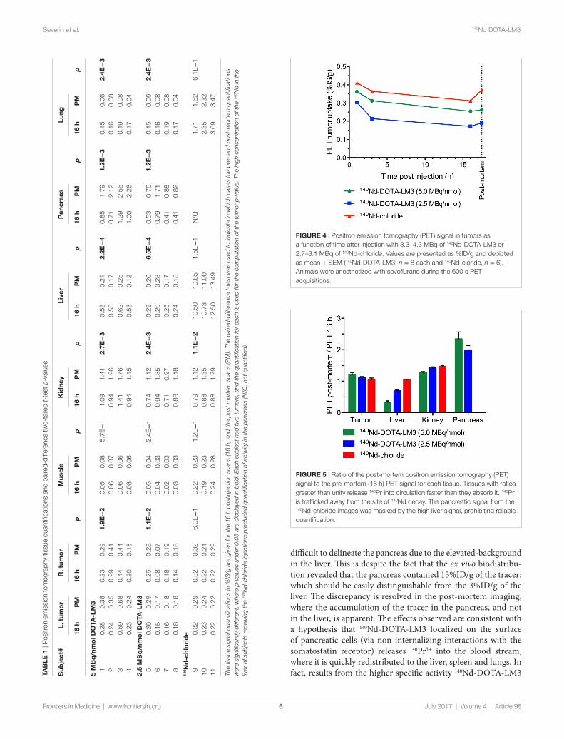
FigUre 5 | Ratio of the post-mortem positron emission tomography (PET) signal to the pre-mortem (16 h) PET signal for each tissue. Tissues with ratios greater than unity release 140Pr into circulation faster than they absorb it. 140Pr is trafficked away from the site of 140Nd decay. The pancreatic signal from the 140Nd-chloride images was masked by the high liver signal, prohibiting reliable quantification.
FigUre 4 | Positron emission tomography (PET) signal in tumors as a function of time after injection with 3.3–4.3 MBq of 140Nd-DOTA-LM3 or 2.7–3.1 MBq of 140Nd-chloride. Values are presented as %ID/g and depicted as mean ± SEM (140Nd-DOTA-LM3, n = 8 each and 140Nd-cloride, n = 6). Animals were anesthetized with sevoflurane during the 600 s PET acquisitions.
TaB
le 1
| P
ositr
on e
mis
sion
tom
ogra
phy
tissu
e qu
antifi
catio
ns a
nd p
aire
d-di
ffere
nce
two-
taile
d t-
test
p-v
alue
s.
sub
ject
#l.
tum
or
r. t
umo
rM
uscl
eK
idne
yli
ver
Pan
crea
slu
ng
16 h
PM
16 h
PM
p16
hP
Mp
16 h
PM
p16
hP
Mp
16 h
PM
p16
hP
Mp
5 M
Bq
/nm
ol D
OTa
-lM
31
0.28
0.38
0.23
0.29
1.9e
−2
0.05
0.08
5.7E
−1
1.09
1.41
2.7e
−3
0.53
0.21
2.2e
−4
0.85
1.79
1.2e
−3
0.15
0.06
2.4e
−3
20.
240.
350.
290.
410.
060.
070.
941.
260.
530.
170.
712.
120.
160.
083
0.59
0.68
0.44
0.44
0.06
0.06
1.41
1.76
0.62
0.25
1.29
2.56
0.19
0.08
40.
230.
240.
200.
180.
080.
060.
941.
150.
530.
121.
002.
260.
170.
04
2.5
MB
q/n
mo
l DO
Ta-l
M3
50.
260.
290.
250.
281.
1e−
20.
050.
042.
4E−
10.
741.
122.
4e−
30.
290.
206.
5e−
40.
530.
761.
2e−
30.
150.
062.
4e−
36
0.15
0.17
0.08
0.07
0.04
0.03
0.94
1.35
0.29
0.23
0.79
1.71
0.16
0.08
70.
160.
180.
180.
190.
020.
030.
710.
970.
250.
170.
410.
880.
190.
088
0.18
0.18
0.14
0.18
0.03
0.03
0.88
1.18
0.24
0.15
0.41
0.82
0.17
0.04
140 n
d-c
hlo
rid
e9
0.32
0.29
0.32
0.32
6.0E
−1
0.22
0.23
1.2E
−1
0.79
1.12
1.1e
−2
10.5
010
.85
1.5E
−1
N/Q
1.71
1.62
6.1E
−1
100.
230.
240.
220.
210.
190.
230.
881.
3510
.73
11.0
02.
352.
3211
0.22
0.22
0.22
0.29
0.24
0.28
0.88
1.29
12.5
013
.49
3.09
3.47
The
tissu
e si
gnal
qua
ntifi
catio
ns in
%IS
/g a
re g
iven
for
the
16 h
pos
tinje
ctio
n sc
ans
(16
h) a
nd th
e po
st m
orte
m s
cans
(PM
). Th
e pa
ired-
diffe
renc
e t-
test
was
use
d to
indi
cate
in w
hich
cas
es th
e pr
e- a
nd p
ost-
mor
tem
qua
ntifi
catio
ns
wer
e si
gnifi
cant
ly d
iffer
ent,
whe
re p
-val
ues
unde
r 0.
05 a
re d
ispl
ayed
in b
old.
Eac
h su
bjec
t had
two
tum
ors,
and
the
quan
tifica
tion
for
each
is u
sed
for
the
com
puta
tion
of th
e tu
mor
p-v
alue
. The
hig
h co
ncen
trat
ion
of th
e 14
0 Nd
in th
e liv
er o
f sub
ject
s re
ceiv
ing
the
140 N
d-ch
lorid
e in
ject
ions
pre
clud
ed q
uant
ifica
tion
of a
ctiv
ity in
the
panc
reas
(N/Q
, not
qua
ntifi
ed).
6
Severin et al. 140Nd DOTA-LM3
Frontiers in Medicine | www.frontiersin.org July 2017 | Volume 4 | Article 98
difficult to delineate the pancreas due to the elevated-background in the liver. This is despite the fact that the ex vivo biodistribu-tion revealed that the pancreas contained 13%ID/g of the tracer: which should be easily distinguishable from the 3%ID/g of the liver. The discrepancy is resolved in the post-mortem imaging, where the accumulation of the tracer in the pancreas, and not in the liver, is apparent. The effects observed are consistent with a hypothesis that 140Nd-DOTA-LM3 localized on the surface of pancreatic cells (via non-internalizing interactions with the somatostatin receptor) releases 140Pr3+ into the blood stream, where it is quickly redistributed to the liver, spleen and lungs. In fact, results from the higher specific activity 140Nd-DOTA-LM3
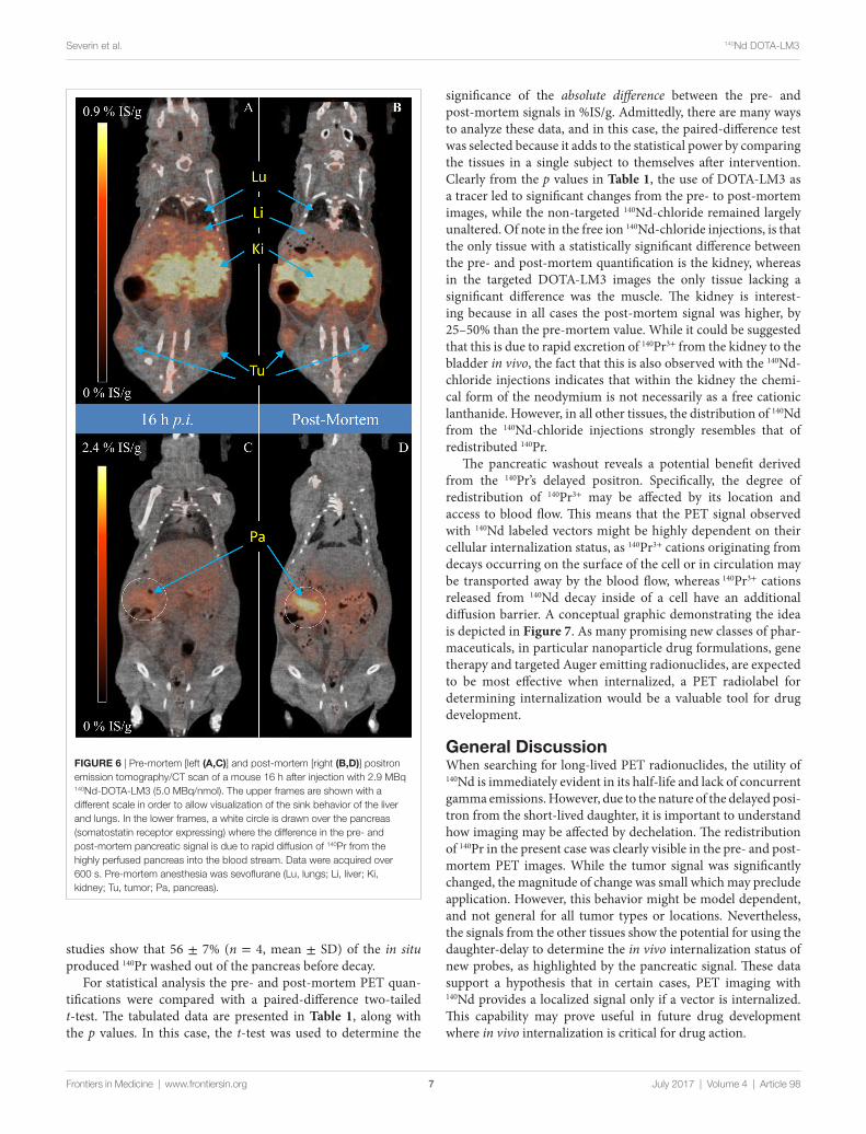
FigUre 6 | Pre-mortem [left (a,c)] and post-mortem [right (B,D)] positron emission tomography/CT scan of a mouse 16 h after injection with 2.9 MBq 140Nd-DOTA-LM3 (5.0 MBq/nmol). The upper frames are shown with a different scale in order to allow visualization of the sink behavior of the liver and lungs. In the lower frames, a white circle is drawn over the pancreas (somatostatin receptor expressing) where the difference in the pre- and post-mortem pancreatic signal is due to rapid diffusion of 140Pr from the highly perfused pancreas into the blood stream. Data were acquired over 600 s. Pre-mortem anesthesia was sevoflurane (Lu, lungs; Li, liver; Ki, kidney; Tu, tumor; Pa, pancreas).
7
Severin et al. 140Nd DOTA-LM3
Frontiers in Medicine | www.frontiersin.org July 2017 | Volume 4 | Article 98
studies show that 56 ± 7% (n = 4, mean ± SD) of the in situ produced 140Pr washed out of the pancreas before decay.
For statistical analysis the pre- and post-mortem PET quan-tifications were compared with a paired-difference two-tailed t-test. The tabulated data are presented in Table 1, along with the p values. In this case, the t-test was used to determine the
significance of the absolute difference between the pre- and post-mortem signals in %IS/g. Admittedly, there are many ways to analyze these data, and in this case, the paired-difference test was selected because it adds to the statistical power by comparing the tissues in a single subject to themselves after intervention. Clearly from the p values in Table 1, the use of DOTA-LM3 as a tracer led to significant changes from the pre- to post-mortem images, while the non-targeted 140Nd-chloride remained largely unaltered. Of note in the free ion 140Nd-chloride injections, is that the only tissue with a statistically significant difference between the pre- and post-mortem quantification is the kidney, whereas in the targeted DOTA-LM3 images the only tissue lacking a significant difference was the muscle. The kidney is interest-ing because in all cases the post-mortem signal was higher, by 25–50% than the pre-mortem value. While it could be suggested that this is due to rapid excretion of 140Pr3+ from the kidney to the bladder in vivo, the fact that this is also observed with the 140Nd- chloride injections indicates that within the kidney the chemi-cal form of the neodymium is not necessarily as a free cationic lanthanide. However, in all other tissues, the distribution of 140Nd from the 140Nd-chloride injections strongly resembles that of redistributed 140Pr.
The pancreatic washout reveals a potential benefit derived from the 140Pr’s delayed positron. Specifically, the degree of redistribution of 140Pr3+ may be affected by its location and access to blood flow. This means that the PET signal observed with 140Nd labeled vectors might be highly dependent on their cellular internalization status, as 140Pr3+ cations originating from decays occurring on the surface of the cell or in circulation may be transported away by the blood flow, whereas 140Pr3+ cations released from 140Nd decay inside of a cell have an additional diffusion barrier. A conceptual graphic demonstrating the idea is depicted in Figure 7. As many promising new classes of phar-maceuticals, in particular nanoparticle drug formulations, gene therapy and targeted Auger emitting radionuclides, are expected to be most effective when internalized, a PET radiolabel for determining internalization would be a valuable tool for drug development.
general DiscussionWhen searching for long-lived PET radionuclides, the utility of 140Nd is immediately evident in its half-life and lack of concurrent gamma emissions. However, due to the nature of the delayed posi-tron from the short-lived daughter, it is important to understand how imaging may be affected by dechelation. The redistribution of 140Pr in the present case was clearly visible in the pre- and post-mortem PET images. While the tumor signal was significantly changed, the magnitude of change was small which may preclude application. However, this behavior might be model dependent, and not general for all tumor types or locations. Nevertheless, the signals from the other tissues show the potential for using the daughter-delay to determine the in vivo internalization status of new probes, as highlighted by the pancreatic signal. These data support a hypothesis that in certain cases, PET imaging with 140Nd provides a localized signal only if a vector is internalized. This capability may prove useful in future drug development where in vivo internalization is critical for drug action.
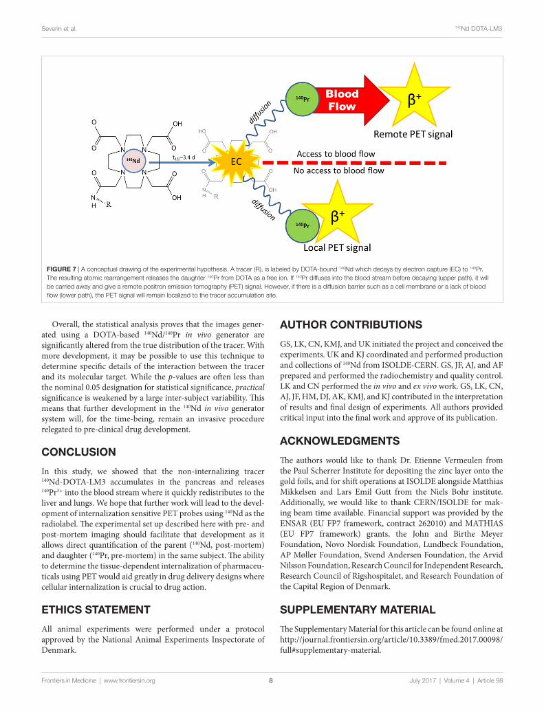
FigUre 7 | A conceptual drawing of the experimental hypothesis. A tracer (R), is labeled by DOTA-bound 140Nd which decays by electron capture (EC) to 140Pr. The resulting atomic rearrangement releases the daughter 140Pr from DOTA as a free ion. If 140Pr diffuses into the blood stream before decaying (upper path), it will be carried away and give a remote positron emission tomography (PET) signal. However, if there is a diffusion barrier such as a cell membrane or a lack of blood flow (lower path), the PET signal will remain localized to the tracer accumulation site.
8
Severin et al. 140Nd DOTA-LM3
Frontiers in Medicine | www.frontiersin.org July 2017 | Volume 4 | Article 98
Overall, the statistical analysis proves that the images gener-ated using a DOTA-based 140Nd/140Pr in vivo generator are significantly altered from the true distribution of the tracer. With more development, it may be possible to use this technique to determine specific details of the interaction between the tracer and its molecular target. While the p-values are often less than the nominal 0.05 designation for statistical significance, practical significance is weakened by a large inter-subject variability. This means that further development in the 140Nd in vivo generator system will, for the time-being, remain an invasive procedure relegated to pre-clinical drug development.
cOnclUsiOn
In this study, we showed that the non-internalizing tracer 140Nd-DOTA-LM3 accumulates in the pancreas and releases 140Pr3+ into the blood stream where it quickly redistributes to the liver and lungs. We hope that further work will lead to the devel-opment of internalization sensitive PET probes using 140Nd as the radiolabel. The experimental set up described here with pre- and post-mortem imaging should facilitate that development as it allows direct quantification of the parent (140Nd, post-mortem) and daughter (140Pr, pre-mortem) in the same subject. The ability to determine the tissue-dependent internalization of pharmaceu-ticals using PET would aid greatly in drug delivery designs where cellular internalization is crucial to drug action.
eThics sTaTeMenT
All animal experiments were performed under a protocol approved by the National Animal Experiments Inspectorate of Denmark.
aUThOr cOnTriBUTiOns
GS, LK, CN, KMJ, and UK initiated the project and conceived the experiments. UK and KJ coordinated and performed production and collections of 140Nd from ISOLDE-CERN. GS, JF, AJ, and AF prepared and performed the radiochemistry and quality control. LK and CN performed the in vivo and ex vivo work. GS, LK, CN, AJ, JF, HM, DJ, AK, KMJ, and KJ contributed in the interpretation of results and final design of experiments. All authors provided critical input into the final work and approve of its publication.
acKnOWleDgMenTs
The authors would like to thank Dr. Etienne Vermeulen from the Paul Scherrer Institute for depositing the zinc layer onto the gold foils, and for shift operations at ISOLDE alongside Matthias Mikkelsen and Lars Emil Gutt from the Niels Bohr institute. Additionally, we would like to thank CERN/ISOLDE for mak-ing beam time available. Financial support was provided by the ENSAR (EU FP7 framework, contract 262010) and MATHIAS (EU FP7 framework) grants, the John and Birthe Meyer Foundation, Novo Nordisk Foundation, Lundbeck Foundation, AP Møller Foundation, Svend Andersen Foundation, the Arvid Nilsson Foundation, Research Council for Independent Research, Research Council of Rigshospitalet, and Research Foundation of the Capital Region of Denmark.
sUPPleMenTarY MaTerial
The Supplementary Material for this article can be found online at http://journal.frontiersin.org/article/10.3389/fmed.2017.00098/full#supplementary-material.

9
Severin et al. 140Nd DOTA-LM3
Frontiers in Medicine | www.frontiersin.org July 2017 | Volume 4 | Article 98
reFerences
1. Grünberg J, Lindenblatt D, Dorrer H, Cohrs S, Zhernosekov K, Köster U, et al. Anti-L1CAM radioimmunotherapy is more effective with the radiolan-thanide terbium-161 compared to lutetium-177 in an ovarian cancer model. Eur J Nucl Med Mol Imaging (2014) 41:1907–15. doi:10.1007/s00259-014- 2798-3
2. Müller C, Reber J, Haller S, Dorrer H, Bernhardt P, Zhernosekov K, et al. Direct in vitro and in vivo comparison of 161Tb and 177Lu using a tumour- targeting folate conjugate. Eur J Nucl Med Mol Imaging (2014) 41:476–85. doi:10.1007/s00259-013-2563-z
3. Müller C, Zhernosekov K, Köster U, Johnston K, Dorrer H, Hohn A, et al. A unique matched quadruplet of terbium radioisotopes for PET and SPECT and for α- and β- radionuclide therapy: an in vivo proof-of-concept study with a new receptor-targeted folate derivative. J Nucl Med (2012) 53:1951–9. doi:10.2967/jnumed.112.107540
4. Bockisch A. Matched pairs for radionuclide-based imaging and therapy. Eur J Nucl Med Mol Imaging (2011) 38:1780–2. doi:10.1007/s00259-011- 1780-6
5. Uusijärvi H, Bernhardt P, Rösch F, Maecke HR, Forssell-Aronsson E. Electron-and positron-emitting radiolanthanides for therapy: aspects of dosimetry and production. J Nucl Med (2006) 47:807–14.
6. Nica N. Nuclear data sheets for A = 140. Nucl Data Sheets (2007) 108:1287–470. doi:10.1016/j.nds.2007.06.001
7. Mausner L. The in vivo generator for radioimmunotherapy. J Label (1989) 89:498–500. doi:10.1002/jlcr.25802601213
8. Edem PE, Fonslet J, Kjaer A, Herth M, Severin G. In vivo radionuclide generators for diagnostics and therapy. Bioinorg Chem Appl (2016) 2016: 6148357. doi:10.1155/2016/6148357
9. Rösch F, Forssell-Aronsson E. Radiolanthanides in nuclear medicine. Met Ions Biol Syst (2004) 42:77–108.
10. Hansen AE, Petersen AL, Henriksen JR, Boerresen B, Rasmussen P, Elema DR, et al. Positron emission tomography based elucidation of the enhanced permeability and retention effect in dogs with cancer using copper-64 liposomes. ACS Nano (2015) 9:6985–95. doi:10.1021/acsnano. 5b01324
11. Hilgers K. New cross section data for production of the therapeutic radionuclides 64Cu, 140Nd, and 192Ir. AIP Conf Proc (2005) 769:1631–3. doi:10.1063/1.1945319
12. Rösch F, Brockmann J, Lebedev NA, Qaim SM. Production and radiochemical separation of the Auger electron emitter 140Nd. Acta Oncol (2000) 39:727–30. doi:10.1080/028418600750063794
13. Zhernosekov KP, Filosofov DV, Qaim SM, Rösch F. A 140Nd/140Pr radionu-clide generator based on physico-chemical transitions in 140Pr complexes after electron capture decay of 140Nd-DOTA. Radiochim Acta (2007) 95: 319–27. doi:10.1524/ract.2007.95.6.319
14. Yakushev EA, Kovalík A, Filosofov DV, Korolev NA, Lebedev NA, Lubashevskia V, et al. An experimental comparison of the K- and L-Auger electron spectra generated in the decays of 140Nd and 111In. Appl Radiat Isot (2005) 62:451–6. doi:10.1016/j.apradiso.2004.06.012
15. Köster U. ISOLDE target and ion source chemistry. Radiochim Acta (2001) 89:749–56. doi:10.1524/ract.2001.89.11-12.749
16. Beyer GJ, Ruth TJ. The role of electromagnetic separators in the production of radiotracers for bio-medical research and nuclear medical application. Nucl
Instrum Methods Phys Res B (2003) 204:694–700. doi:10.1016/S0168-583X (03)00489-0
17. Müller C, Reber J, Haller S, Dorrer H, Köster U, Johnston K, et al. Folate receptor targeted alpha-therapy using terbium-149. Pharmaceuticals (Basel) (2014) 7:353–65. doi:10.3390/ph7030353
18. Müller C, Vermeulen C, Köster U, Johnston K, Türler A, Schibli R, et al. Alpha-PET with terbium-149: evidence and perspectives for radiotheragnos-tics. EJNMMI Radiopharm Chem (2017) 1:5. doi:10.1186/s41181-016-0008-2
19. Pfeifer AK, Gregersen T, Grønbaek H, Hansen CP, Müller-Brand J, Herskind Bruun K, et al. Peptide receptor radionuclide therapy with 90Y-DOTATOC and 177Lu-DOTATOC in advanced neuroendocrine tumors: results from a Danish cohort treated in Switzerland. Neuroendocrinology (2011) 93:189–96. doi:10.1159/000324096
20. Pfeifer A, Knigge U, Mortensen J, Oturai P, Berthelsen AK, Loft A, et al. Clinical PET of neuroendocrine tumors using 64Cu-DOTATATE: first- in-humans study. J Nucl Med (2012) 53:1207–15. doi:10.2967/jnumed.111.101469
21. Waser B, Tamma M-LL, Cescato R, Maecke HR, Reubi JC. Highly efficient in vivo agonist-induced internalization of sst2 receptors in somatostatin target tissues. J Nucl Med (2009) 50:936–41. doi:10.2967/jnumed.108.061457
22. Maecke HR, Reubi JC. Somatostatin receptors as targets for nuclear medicine imaging and radionuclide treatment. J Nucl Med (2011) 52:841–4. doi:10.2967/jnumed.110.084236
23. Fani M, Del Pozzo L, Abiraj K, Mansi R, Tamma ML, Cescato R, et al. PET of somatostatin receptor-positive tumors using 64Cu- and 68Ga- somatostatin antagonists: the chelate makes the difference. J Nucl Med (2011) 52:1110–8. doi:10.2967/jnumed.111.087999
24. Waser B, Cescato R, Tamma ML, Maecke HR, Reubi JC. Absence of soma-tostatin SST2 receptor internalization in vivo after intravenous SOM230 appli-cation in the AR42J animal tumor model. Eur J Pharmacol (2010) 644:257–62. doi:10.1016/j.ejphar.2010.07.005
25. Kraus KA, Nelson F. Anion exchange studies of the fission products. Proc Int Conf Peaceful Uses Energy (1956) 7:113–25.
26. Holland JP, Sheh Y, Lewis JS. Standardized methods for the production of high specific-activity zirconium-89. Nucl Med Biol (2009) 36:729–39. doi:10.1016/j.nucmedbio.2009.05.007
27. Beyer GJ, Bergmann R, Schomäcker K, Rösch F, Schäfer G, Kulikov EV, et al. Comparison of the biodistribution of 225Ac and radio- lanthanides as citrate complexes. Isotopenpraxis (1990) 26:111–4. doi:10.1080/ 10256019008624245
28. Fani M, Braun F, Waser B, Beetschen K, Cescato R, Erchegyi J, et al. Unexpected sensitivity of sst2 antagonists to N-terminal radiometal modifications. J Nucl Med (2012) 53:1481–9. doi:10.2967/jnumed.112.102764
Conflict of Interest Statement: The authors declare that the research was con-ducted in the absence of any commercial or financial relationships that could be construed as a potential conflict of interest.
Copyright © 2017 Severin, Kristensen, Nielsen, Fonslet, Jensen, Frellsen, Jensen, Elema, Maecke, Kjær, Johnston and Köster. This is an open-access article distributed under the terms of the Creative Commons Attribution License (CC BY). The use, distribution or reproduction in other forums is permitted, provided the original author(s) or licensor are credited and that the original publication in this journal is cited, in accordance with accepted academic practice. No use, distribution or reproduction is permitted which does not comply with these terms.