Embed Size (px)
Citation preview
Neonatal management and outcome in red cellalloimmunizationSmits-Wintjens, V.E.H.J.
CitationSmits-Wintjens, V. E. H. J. (2012, February 15). Neonatal management andoutcome in red cell alloimmunization. Retrieved fromhttps://hdl.handle.net/1887/18485 Version: Corrected Publisher’s Version
License:Licence agreement concerning inclusion of doctoralthesis in the Institutional Repository of the Universityof Leiden
Downloaded from: https://hdl.handle.net/1887/18485 Note: To cite this publication please use the final published version (ifapplicable).
Vivianne Smits - Wintjens
Neonatal management and outcome in red cell alloimmunization
Neonatal m
anagement and outcom
e in red cell alloimm
unization Vivianne Smits - W
intjens

Neonatal management and outcome
in red cell alloimmunization

ISBN/EAN: 978-90-9026589-6
Cover-photos: Mika van Zon, participant in the LOTUS study. Mika’s photos (a few days after birth and at the age of 5) are published with kind permission of his parents.
Layout and printing: Pasmans Offsetdrukkerij BV, Den Haag.
The printing of this thesis was financially supported by Willem-Alexander Kinderziekenhuis, Leiden.
© 2012 V.E.H.J. Smits-Wintjens, Leiden, The Netherlands.All rights reserved. No part of this publication may be reproduced, stored or transmitted in any way or by any means, without the prior permission of the author or the copyright-owning journals for previously published chapters.
Neonatal management and outcome
in red cell alloimmunization
Proefschrift
ter verkrijging vande graad van doctor aan de Universiteit Leiden,
op gezag van Rector Magnificus prof. mr. P.F. van der Heijden,volgens besluit van het College voor Promoties
te verdedigen op woensdag 15 februari 2012klokke 13:45 uur
door
Vivianne Elise Huberta Johanna Smits - Wintjens
geboren te Maastrichtin 1972

Promotiecommissie
Promotor : Prof. dr. F.J. Walther
Copromotor : dr. E. Lopriore
Overige leden : Prof. dr. A. Brand Prof. dr. R. Devlieger, Katholieke Universiteit Leuven, België dr. P.H. Dijk, Universitair Medisch Centrum Groningen dr. D. Oepkes

Aan mijn ouders,
Robert, Koen, Tijn en Lieke
Table of contents
Chapter 1 General introduction and outline of the thesis . . . . . . . . . . . . . . . . . . . . . . . . . . . . . . . . . . . . . . . . .
Chapter 2 Rhesus hemolytic disease of the newborn: postnatal management, associated morbidity and long-term outcome
Semin Fetal Neonatal Med 2008; 13:265-271 . . . . . . . . . . . . . . . . . . . . . . . . . . . . . . . . . . . . . . . . . . . . . . . . . .
Chapter 3 Intravenous immunoglobulin in neonates with Rhesus hemolytic
disease: a randomized controlled trial Pediatrics 2011; 127:680-686 . . . . . . . . . . . . . . . . . . . . . . . . . . . . . . . . . . . . . . . . . . . . . . . . . . . . . . . . . . . . . . . . . . . . . . . . . .
Chapter 4 Bacillus cereus cerebral abscesses in a term neonate with Rhesus hemolytic disease treated with exchange transfusion
J Pediatr Inf Dis 2010; 5(3):277-280 . . . . . . . . . . . . . . . . . . . . . . . . . . . . . . . . . . . . . . . . . . . . . . . . . . . . . . . . . . . . . . . . .
Chapter 5 Neonatal morbidity after exchange transfusion for red cell alloimmune hemolytic disease
Submitted . . . . . . . . . . . . . . . . . . . . . . . . . . . . . . . . . . . . . . . . . . . . . . . . . . . . . . . . . . . . . . . . . . . . . . . . . . . . . . . . . . . . . . . . . . . . . . . . . . . . . . .
Chapter 6 Cholestasis in neonates with red cell alloimmune hemolytic disease: incidence, risk factors and outcome
Neonatology; In press . . . . . . . . . . . . . . . . . . . . . . . . . . . . . . . . . . . . . . . . . . . . . . . . . . . . . . . . . . . . . . . . . . . . . . . . . . . . . . . . . . . . .
Chapter 7 Thrombocytopenia at birth in neonates with red cell alloimmune
hemolytic disease Vox Sang 2011; Epub ahead of print . . . . . . . . . . . . . . . . . . . . . . . . . . . . . . . . . . . . . . . . . . . . . . . . . . . . . . . . . . . . . . .
Chapter 8 Top-up transfusions in neonates with Rhesus hemolytic disease in relation to exchange transfusions
Vox Sang 2010; 99(1):65-70 . . . . . . . . . . . . . . . . . . . . . . . . . . . . . . . . . . . . . . . . . . . . . . . . . . . . . . . . . . . . . . . . . . . . . . . .
Chapter 9 Exchange transfusions and top-up transfusions in neonates with Kell hemolytic disease
Vox Sang 2010; 100(3):312-316 . . . . . . . . . . . . . . . . . . . . . . . . . . . . . . . . . . . . . . . . . . . . . . . . . . . . . . . . . . . . . . . . . . . . . . .
9
19
35
49
57
71
83
95
107
Chapter 10 Long-term neurodevelopmental outcome after intrauterine transfusion for fetal alloimmune anemia: the LOTUS study
Am J Obstet Gynecol 2011; Epub ahead of print . . . . . . . . . . . . . . . . . . . . . . . . . . . . . . . . . . . . . . . . . . . . .
Chapter 11 General discussion and future perspectives . . . . . . . . . . . . . . . . . . . . . . . . . . . . . . . . . . . . . . . . . . . . .
Chapter 12 Summary . . . . . . . . . . . . . . . . . . . . . . . . . . . . . . . . . . . . . . . . . . . . . . . . . . . . . . . . . . . . . . . . . . . . . . . . . . . . . . . . . . . . . . . . . . . . . . . . .
Chapter 13 Samenvatting . . . . . . . . . . . . . . . . . . . . . . . . . . . . . . . . . . . . . . . . . . . . . . . . . . . . . . . . . . . . . . . . . . . . . . . . . . . . . . . . . . . . . . . . . .
List of abbreviations . . . . . . . . . . . . . . . . . . . . . . . . . . . . . . . . . . . . . . . . . . . . . . . . . . . . . . . . . . . . . . . . . . . . . . . . . . . . . . . . . . . . . . . . . . . . . . . . . . . . .
Authors and affiliations . . . . . . . . . . . . . . . . . . . . . . . . . . . . . . . . . . . . . . . . . . . . . . . . . . . . . . . . . . . . . . . . . . . . . . . . . . . . . . . . . . . . . . . . . . . . . . . .
Curriculum Vitae . . . . . . . . . . . . . . . . . . . . . . . . . . . . . . . . . . . . . . . . . . . . . . . . . . . . . . . . . . . . . . . . . . . . . . . . . . . . . . . . . . . . . . . . . . . . . . . . . . . . . . . . . . .
Publications . . . . . . . . . . . . . . . . . . . . . . . . . . . . . . . . . . . . . . . . . . . . . . . . . . . . . . . . . . . . . . . . . . . . . . . . . . . . . . . . . . . . . . . . . . . . . . . . . . . . . . . . . . . . . . . . .
Dankwoord . . . . . . . . . . . . . . . . . . . . . . . . . . . . . . . . . . . . . . . . . . . . . . . . . . . . . . . . . . . . . . . . . . . . . . . . . . . . . . . . . . . . . . . . . . . . . . . . . . . . . . . . . . . . . . . . . .
117
133
147
153
163
165
167
169
171


1 General introduction
and outline of the thesis

10
General introduction and outline of the thesis
11
Introduction
Hemolytic disease of the fetus and newborn (HDFN) due to red cell alloimmunization has been a fascinating clinical picture for many centuries. The first report of a condition called hydrops fetalis dates back to 1609,1,2 when a French midwife described the delivery of twins. The first twin was hydropic and stillborn and the second suffered from jaundice and subse-quently died of kernicterus. These two conditions were not linked until 1932, when Diamond et al. described that hydrops and kernicterus were manifestations of the same disease, which they called erythroblastosis fetalis.3 However, the exact cause was still unknown. Since 1940, the analyses of Landsteiner and Wiener contributed largely to a better under-standing of the pathogenesis of HDFN.4-7 In their studies with rhesus monkeys Landsteiner and Wiener observed that agglutination of human red blood cells occurred in the presence of rhesus monkey red cell antiserum, whereas subjects who lacked the antigen on their red cells did not show agglutination.4,7 The authors called this antigen ‘Rhesus’ (Rh) and conse-quently the Rh-blood group system was born. Since then it has become clear that the most common cause of severe HDFN is ‘Rhesus disease’, resulting from maternal immunization to the Rhesus D (Rh D) antigen.8 However, more than 50 other red cell antigens associated with hemolytic disease have been described. Not only anti-Rh D, but also anti-Rh c and anti-Kell antibodies are associated with severe fetal and neonatal disease.9
Untreated HDFN is a major cause of perinatal mortality. In HDFN due to red cell alloim-munization, maternal immunoglobulin (IgG) antibodies directed against fetal red blood cells, pass the placenta into the fetal circulation and cause destruction of fetal red cells. The resulting progressive fetal anemia will then lead to fetal hydrops and perinatal death.10
During the last few decades prenatal care for patients with red cell alloimmunization has improved significantly. The introduction of Rh D prophylaxis in the late 1960s,11,12 the use of Doppler ultrasound to detect fetal anemia since the early 1970s13 and treatment with intra-uterine intravascular red cell transfusions (IUT) since the 1980s14-16 have led to a remarkable reduction in perinatal mortality. Before the introduction of Rh D prophylaxis and IUTs, peri-natal mortality was approximately 50% and the rate of perinatal morbidity was extremely high.1,17 The current most successful treatment, the use of IUTs, contributed largely to peri-natal survival rates exceeding 95% in experienced centers.10,18 As a result of improving pre-natal care strategies and consequently increased perinatal survival, attention is now shifting towards postnatal short-term and long-term management and morbidity.
Neonatal red cell alloimmunization may lead to excessive hyperbilirubinemia and per-manent brain damage due to kernicterus. Postnatal management is based mainly on the treatment of hyperbilirubinemia and consists of intensive phototherapy and exchange
Chapter 1
12
transfusion (ET).18 Despite improvement in neonatal intensive care, ET remains a high risk invasive procedure requiring the use of central lines and is associated with a significant rate of adverse reactions.19-26 Neonatal treatment with intravenous immunoglobulins (IVIg) has been suggested and used as an alternative therapy for ET.27 In a few small random-ized controlled trials IVIg reduced the need for ET and duration of phototherapy. However, these studies were restricted by several important methodological limitations.28-31 In 2002 a Cochrane review suggested that more well-designed trials were needed before routine use of IVIg could be recommended for treatment of HDN due to red cell alloimmunization.32
Postnatal management also consists of treatment of early and late anemia using top-up red blood cell transfusions.33 Several risk factors for neonatal anemia secondary to red cell alloimmunization have been reported, including IUT34, severity of HDN35,36, type of alloim-munization (including Kell versus Rh D)37 and the use of ET.18,38 However, only a limited number of studies (mostly case reports) have been published on differences in type of alloimmunization related to severity of neonatal anemia.37,39-41 In addition, the protective role of ET for neonatal anemia has only been demonstrated in one small study.38 Therefore, further research on anemia secondary to red cell alloimmunization is needed.
In the past, various other postnatal complications in neonatal red cell alloimmunization have been reported, including cholestatic liver disease and thrombocytopenia. The etiology of cholestasis in neonates with HDN due to red cell alloimmunization has been attributed to iron overload due to IUT.42-45 However, data on incidence, potential risk factors, neonatal management and outcome of cholestasis in red cell alloimmunization is scarce. Simultane-ously, only a few small studies have been published on incidence and severity of throm-bocytopenia in neonates with red cell alloimmunization.36,46-48 Therefore, more studies are needed to investigate these and other associated complications of neonatal red cell alloim-munization.
As perinatal survival improves, attention is shifting towards long-term outcome in survivors. One of the concerns of the successful use of IUTs is that a decrease in perinatal mortality may lead to an increase in children with long-term handicaps. Moreover, not much is known about the relation between the severity of fetal anemia and long-term neurodevelopmen-tal outcome.18 To date, only a few studies with small patient numbers have reported long-term neurodevelopmental outcome after IUT.49-56 Further research on this topic is needed to determine incidence of and risk factors for adverse neurodevelopmental outcome after IUT.
The aim of this thesis was to investigate various management options and to describe com-plications and outcome of neonatal red cell alloimmune hemolytic disease.
General introduction and outline of the thesis
13
Outline of the thesis
During this study period several study projects on management, complications and outcome in hemolytic disease of the newborn (HDN) due to red cell alloimmunization were performed, including the Leiden’s IVIg trial in Rhesus disease of the Neonate (LIVIN study) and the LOng-Term follow-up after intraUterine transfusionS (LOTUS) study. The LIVIN study is a randomized controlled trial (RCT) designed in collaboration with Sanquin Blood Bank (Southwest Region, Amsterdam) to determine whether the prophylactic use of IVIg reduces the need for ET in neonates with Rh D or c hemolytic disease. The LOTUS study is a large national cohort study designed in close collaboration with the Department of Obstetrics and the Department of ImmunoHematology and Blood Transfusion of the Leiden University Medical Center (LUMC). One of the aims of the LOTUS study was to evaluate the long-term neurodevelopmental outcome of children treated with IUT. The aims and outcomes of these and other studies are described in the following chapters:
Chapter 2Review of the literature on HDN due to red cell alloimmunization. This review focuses on postnatal management, associated morbidity and long-term outcome.
Chapter 3Randomized controlled trial on the use of IVIg in neonates with Rhesus HDN (LIVIN study), investigating the effect of IVIg on number of ETs. Chapter 4 Case report describing a term neonate with Rhesus hemolytic disease treated with ET and developing a rare but severe cerebral infection after umbilical cord catheterization.
Chapter 5 Study on morbidity associated with ETs in neonatal red cell alloimmune hemolytic disease.
Chapter 6 Study on cholestasis in neonates with red cell alloimmune hemolytic disease, describing incidence, risk factors and outcome. Chapter 7 Study on thrombocytopenia at birth in neonates with red cell alloimmune hemolytic disease, focusing on incidence and severity of and risk factors for thrombocytopenia at birth.
Chapter 1
14
Chapter 8Study on top-up red blood cell transfusions in neonates with Rhesus hemolytic disease in relation to ETs.
Chapter 9 Study on neonates with Kell hemolytic disease, focusing on ETs and top-up red blood cell transfusions.
Chapter 10 Study on long-term neurodevelopmental outcome after IUT for fetal alloimmune anemia (LOTUS study), to determine the incidence of and risk factors for neurodevelopmental impairment (NDI).
Chapter 11 General discussion and future perspectives
Chapter 12 Summary
Chapter 13Samenvatting
General introduction and outline of the thesis
15
References
1. Stockman JA, III. Overview of the state of the art of Rh disease: history, current clinical management, and recent progress. J Pediatr Hematol Oncol . 2001;23:385-393.
2. Dunn PM. Louise Bourgeois (1563-1636): royal midwife of France. Arch Dis Child Fetal Neonatal Ed . 2004;89:F185-F187.
3. Diamond LK, Blackfan KD, Baty JM. Erythroblastosis fetalis and its association with universal edema of the fetus, icterus gravis neonatotum and anemia of the newborn. J Pediatr . 1932;1:269-309.
4. Landsteiner K, Wiener AS. An agglutinable factor in human blood recognized by immune sera for rhesus blood. Proc Soc Exp Biol Med . 1940;43:223.
5. Urbaniak SJ, Greiss MA. Rh D haemolytic disease of the fetus and the newborn. Blood Rev . 2000;14:44-61.
6. Liumbruno GM, D’Alessandro A, Rea F, Piccinini V, Catalano L, Calizzani G, et al. The role of antenatal immunoprophylaxis in the prevention of maternal-foetal anti-Rh(D) alloimmunisation. Blood Transfus . 2010;8:8-16.
7. landsteiner K, Wiener AS. Studies on an agglutinogen (Rh) in human blood reacting with anti-rhesus sera and with human isoantibodies. J Exp Med . 1941;74:309-320.
8. Levine P, Katzin EM, Burnham M. Alloimmunization in pregnancy: its possible bearing on the etiology of erythroblastosis foetalis. JAMA . 1941;116:825-827.
9. Moise KJ. Fetal anemia due to non-Rhesus-D red-cell alloimmunization. Semin Fetal Neonatal Med . 2008;13:207-214.
10. Moise KJ, Jr. Management of rhesus alloimmunization in pregnancy. Obstet Gynecol . 2008;112:164-176.
11. Ascari WQ, Allen AE, Baker WJ, Pollack W. Rh-o (D) immune globulin (human). Evaluation in women at risk of Rh immunization. JAMA . 1968;205:71-74.
12. Prevention of Rh-haemolytic disease: final results of the “high-risk” clinical trial. A combined study from centres in England and Baltimore. Br Med J . 1971;2:607-609.
13. Jones MR. Ultrasonic B-scanning in rhesus incompatibility. J Clin Ultrasound . 1974;2:185-190.14. Liley AW. Intrauterine transfusion of foetus in haemolytic disease. Br Med J . 1963;2:1107-1109.15. Rodeck CH, Kemp JR, Holman CA, Whitmore DN, Karnicki J, Austin MA. Direct intravascular fetal blood
transfusion by fetoscopy in severe Rhesus isoimmunisation. Lancet . 1981;1:625-627.16. Rodeck CH, Nicolaides KH, Warsof SL, Fysh WJ, Gamsu HR, Kemp JR. The management of severe rhesus
isoimmunization by fetoscopic intravascular transfusions. Am J Obstet Gynecol . 1984;150:769-774.17. Bowman J. Thirty-five years of Rh prophylaxis. Transfusion . 2003;43:1661-1666.18. Smits-Wintjens VE, Walther FJ, Lopriore E. Rhesus haemolytic disease of the newborn: Postnatal man-
agement, associated morbidity and long-term outcome. Semin Fetal Neonatal Med . 2008;13:265-271.19. Jackson JC. Adverse events associated with exchange transfusion in healthy and ill newborns. Pediat-
rics . 1997;99:E7.20. Patra K, Storfer-Isser A, Siner B, Moore J, Hack M. Adverse events associated with neonatal exchange
transfusion in the 1990s. J Pediatr . 2004;144:626-631.21. Steiner LA, Bizzarro MJ, Ehrenkranz RA, Gallagher PG. A decline in the frequency of neonatal exchange
transfusions and its effect on exchange-related morbidity and mortality. Pediatrics . 2007;120:27-32.22. Badiee Z. Exchange transfusion in neonatal hyperbilirubinaemia: experience in Isfahan, Iran. Singa-
pore Med J . 2007;48:421-423.23. Keenan WJ, Novak KK, Sutherland JM, Bryla DA, Fetterly KL. Morbidity and mortality associated with
exchange transfusion. Pediatrics . 1985;75:417-421.24. Ip S, Chung M, Kulig J, O’Brien R, Sege R, Glicken S, et al. An evidence-based review of important issues
concerning neonatal hyperbilirubinemia. Pediatrics . 2004;114:e130-e153.
Chapter 1
16
25. Hosseinpour SS, Gharehbaghi MM. Exchange transfusion in severe hyperbilirubinemia: an experience in northwest Iran. Turk J Pediatr . 2010;52:367-371.
26. Davutoglu M, Garipardic M, Guler E, Karabiber H, Erhan D. The etiology of severe neonatal hyperbili-rubinemia and complications of exchange transfusion. Turk J Pediatr . 2010;52:163-166.
27. Management of hyperbilirubinemia in the newborn infant 35 or more weeks of gestation. Pediatrics . 2004;114:297-316.
28. Rubo J, Albrecht K, Lasch P, Laufkotter E, Leititis J, Marsan D, et al. High-dose intravenous immune globulin therapy for hyperbilirubinemia caused by Rh hemolytic disease. J Pediatr . 1992;121:93-97.
29. Gottstein R, Cooke RW. Systematic review of intravenous immunoglobulin in haemolytic disease of the newborn. Arch Dis Child Fetal Neonatal Ed . 2003;88:F6-10.
30. Alpay F, Sarici SU, Okutan V, Erdem G, Ozcan O, Gokcay E. High-dose intravenous immunoglobulin therapy in neonatal immune haemolytic jaundice. Acta Paediatr . 1999;88:216-219.
31. Dagoglu T, Ovali F, Samanci N, Bengisu E. High-dose intravenous immunoglobulin therapy for rhesus haemolytic disease. J Int Med Res . 1995;23:264-271.
32. Alcock GS, Liley H. Immunoglobulin infusion for isoimmune haemolytic jaundice in neonates. Cochrane Database Syst Rev . 2002;CD003313.
33. Rath ME, Smits-Wintjens VE, Walther FJ, Lopriore E. Hematological morbidity and management in neonates with hemolytic disease due to red cell alloimmunization. Early Hum Dev . 2011;87:583-588.
34. De Boer I, Zeestraten EC, Lopriore E, Van K, I, Kanhai HH, Walther FJ. Pediatric outcome in Rhesus hemolytic disease treated with and without intrauterine transfusion. Am J Obstet Gynecol . 2008;198:54. e1-54.e4.
35. Pessler F, Hart D. Hyporegenerative anemia associated with Rh hemolytic disease: treatment failure of recombinant erythropoietin. J Pediatr Hematol Oncol . 2002;24:689-693.
36. Koenig JM, Ashton RD, De Vore GR, Christensen RD. Late hyporegenerative anemia in Rh hemolytic disease. J Pediatr . 1989;115:315-318.
37. Babinszki A, Lapinski RH, Berkowitz RL. Prognostic factors and management in pregnancies compli-cated with severe kell alloimmunization: experiences of the last 13 years. Am J Perinatol . 1998;15:695-701.
38. Al-Alaiyan S, al OA. Late hyporegenerative anemia in neonates with rhesus hemolytic disease. J Perinat Med . 1999;27:112-115.
39. Wenk RE, Goldstein P, Felix JK. Kell alloimmunization, hemolytic disease of the newborn, and perinatal management. Obstet Gynecol . 1985;66:473-476.
40. Dhodapkar KM, Blei F. Treatment of hemolytic disease of the newborn caused by anti-Kell antibody with recombinant erythropoietin. J Pediatr Hematol Oncol . 2001;23:69-70.
41. Manoura A, Korakaki E, Hatzidaki E, Saitakis E, Maraka S, Papamastoraki I, et al. Use of recombinant erythropoietin for the management of severe hemolytic disease of the newborn of a K0 phenotype mother. Pediatr Hematol Oncol . 2007;24:69-73.
42. Lasker MR, Eddleman K, Toor AH. Neonatal hepatitis and excessive hepatic iron deposition following intrauterine blood transfusion. Am J Perinatol . 1995;12:14-17.
43. Aygun C, Tekinalp G, Gurgey A. Increased fetal iron load in rhesus hemolytic disease. Pediatr Hematol Oncol . 2004;21:329-333.
44. Berger HM, Lindeman JH, van Zoeren-Grobben D, Houdkamp E, Schrijver J, Kanhai HH. Iron overload, free radical damage, and rhesus haemolytic disease. Lancet . 1990;335:933-936.
45. Nasrat HA, Nicolini U, Nicolaidis P, Letsky EA, Gau G, Rodeck CH. The effect of intrauterine intravas-cular blood transfusion on iron metabolism in fetuses with Rh alloimmunization. Obstet Gynecol . 1991;77:558-562.
46. Saade GR, Moise KJ, Jr., Copel JA, Belfort MA, Carpenter RJ, Jr. Fetal platelet counts correlate with the severity of the anemia in red-cell alloimmunization. Obstet Gynecol . 1993;82:987-991.
General introduction and outline of the thesis
17
47. Van den Akker ES, de Haan TR, Lopriore E, Brand A, Kanhai HH, Oepkes D. Severe fetal thrombocytope-nia in Rhesus D alloimmunized pregnancies. Am J Obstet Gynecol . 2008;199:387-4.
48. Van den Akker ES, Klumper FJ, Brand A, Kanhai HH, Oepkes D. Kell alloimmunization in pregnancy: associated with fetal thrombocytopenia? Vox Sang . 2008;95:66-69.
49. Janssens HM, de Haan MJ, Van Kamp IL, Brand R, Kanhai HH, Veen S. Outcome for children treated with fetal intravascular transfusions because of severe blood group antagonism. J Pediatr . 1997;131:373-380.
50. Doyle LW, Kelly EA, Rickards AL, Ford GW, Callanan C. Sensorineural outcome at 2 years for survivors of erythroblastosis treated with fetal intravascular transfusions. Obstet Gynecol . 1993;81:931-935.
51. Hudon L, Moise KJ, Jr., Hegemier SE, Hill RM, Moise AA, Smith EO, et al. Long-term neurodevelopmen-tal outcome after intrauterine transfusion for the treatment of fetal hemolytic disease. Am J Obstet Gynecol . 1998;179:858-863.
52. Harper DC, Swingle HM, Weiner CP, Bonthius DJ, Aylward GP, Widness JA. Long-term neurodevelop-mental outcome and brain volume after treatment for hydrops fetalis by in utero intravascular trans-fusion. Am J Obstet Gynecol . 2006;195:192-200.
53. Grab D, Paulus WE, Bommer A, Buck G, Terinde R. Treatment of fetal erythroblastosis by intravascular transfusions: outcome at 6 years. Obstet Gynecol . 1999;93:165-168.
54. Farrant B, Battin M, Roberts A. Outcome of infants receiving in-utero transfusions for haemolytic disease. N Z Med J . 2001;114:400-403.
55. Stewart G, Day RE, Del PC, Whittle MJ, Turner TL, Holland BM. Developmental outcome after intra-vascular intrauterine transfusion for rhesus haemolytic disease. Arch Dis Child Fetal Neonatal Ed . 1994;70:F52-F53.
56. Weisz B, Rosenbaum O, Chayen B, Peltz R, Feldman B, Lipitz S. Outcome of severely anaemic fetuses treated by intrauterine transfusions. Arch Dis Child Fetal Neonatal Ed . 2009;94:F201-F204.

18

2 Rhesus hemolytic disease of the newborn:
postnatal management, associated morbidity and long-term outcome
Vivianne EHJ Smits-WintjensFrans J WaltherEnrico Lopriore
Semin Fetal Neonatal Med 2008; 13:265-271
Chapter 2
20
Abstract
Rhesus hemolytic disease of the newborn can lead to complications such as hyperbiliru-binemia, kernicterus and anemia. Postnatal management consists mainly of intensive pho-totherapy, exchange transfusion and blood transfusion. During the last decades, significant progress in prenatal care strategies for patients with Rhesus hemolytic disease has occurred. New prenatal management options have led to a remarkable reduction in perinatal mor-tality. As a result of the increase in perinatal survival, attention is now shifting towards short-term and long-term morbidity. This review focuses on the management of neonatal and pediatric complications associated with Rhesus hemolytic disease, discusses postna-tal treatment options and summarizes the results of studies on short-term and long-term outcome.
Review on neonatal Rhesus hemolytic disease
21
Introduction
Rhesus hemolytic disease of the newborn (RHDN) results from maternal red-cell alloimmu-nization. Production of maternal antibodies directed against the fetal red blood cells occurs when fetal red blood cells positive for a certain antigen, usually Rhesus D (Rh D), pass into the blood circulation of a mother negative for that particular antigen. Maternal immuno-globulin (IgG) antibodies might then cross the placenta into the fetal circulation and cause a wide variety of symptoms in the fetus, ranging from mild to severe hemolytic anemia and fetal hydrops. During the last few decades, a significant evolution in prenatal care strategies for patients with RHDN has occurred, including the introduction of Rh D prophylaxis, use of Doppler ultrasound to detect fetal anemia and in particular treatment with intrauterine blood transfusions (IUTs). These new management options have led to a dramatic decrease in perinatal mortality. Before the introduction of Rh D prophylaxis and IUTs, perinatal mor-tality was approximately 50% and the rate of perinatal morbidity was extremely high.1,2 With the use of IUTs, overall perinatal mortality in severe RHDN has decreased to less than 10%.3 As a result of the increase in perinatal survival, attention is now shifting towards short-term and long-term morbidity. Postnatal management of RHDN is based mainly on the treat-ment of hyperbilirubinemia and consists of intensive phototherapy (PT) and exchange transfusions (ETs) to prevent kernicterus. Various other postnatal complications in RHDN have been reported, including early and late anemia, thrombocytopenia, cholestasis and adverse long-term neurodevelopmental outcome. However, few studies have focused on postnatal management and outcome of RHDN.2,4-7
This chapter focuses on the management of neonatal and pediatric complications associ-ated with RHDN, discusses the efficacy of various postnatal treatment options and summa-rizes the results of studies on short-term and long-term outcome in RHDN.
Management of hyperbilirubinemia and the prevention of kernicterus
RHDN can lead to severe hyperbilirubinemia, acute bilirubin encephalopathy and even-tually chronic bilirubin encephalopathy, also known as kernicterus. Prevention of kernict-erus is considered to be the primary goal of postnatal treatment of RHDN.8
The acute stage of bilirubin encephalopathy is divided into three phases. In the early phase, affected infants become lethargic, hypotonic and suck poorly; the intermediate phase is characterized by stupor, irritability, fever, high-pitched cry and alternating hypertonia
Chapter 2
22
and hypotonia; the advanced phase is characterized by irreversible damage to the central nervous system (CNS) resulting in pronounced retrocollis and opisthotonus, shrill cry, no feeding, apnea, fever, deep stupor to coma, sometimes seizures and death. The chronic stage of bilirubin encephalopathy, i.e. kernicterus, refers to the clinical CNS findings caused by bilirubin toxicity to the basal ganglia and various brain stem nuclei. In kernicterus, infants develop a severe form of athetoid cerebral palsy, hearing problems and psychomotor handicaps.8-10
Phototherapy Treatment of neonatal hyperbilirubinemia with phototherapy (PT) was introduced in the early 1970s.8,11 PT lowers serum bilirubin levels through photo-oxidation and converts bili-rubin to a water-soluble substance. To deliver conventional PT, the infant is nursed under halogen or fluorescent lamps and the eyes are covered with a mask to prevent retinal damage. However, in the last 10 years new devices for delivering PT have been developed, these are the so-called fibreoptic PT devices. The infant is nursed on a blanket containing optical fibres that deliver light to the back. A recent Cochrane review suggests that even though fibreoptic PT has a place in the management of physiological neonatal hyperbili-rubinemia, the use of fibreoptic PT in the treatment of pathological neonatal hyperbilirubi-nemia due to hemolysis should be investigated in a randomized controlled clinical trial.11 The efficacy of PT is dependent on a number of factors, including spectral qualities of the delivered light (optimal wavelength range 400-520 nm), irradiance (intensity of light), surface area receiving PT, distance from the light to the skin (the optimal distance is approx-imately 30 cm), skin pigmentation, total serum bilirubin concentration at the start of PT and duration of exposure.4,12,13 PT is required when total serum bilirubin levels exceed the predefined bilirubin thresholds. Recently, new PT guidelines for bilirubin thresholds for chil-dren with RHDN have been published by the American Academy of Pediatrics (AAP).8
In RHDN, intensive PT should be started immediately after birth, as bilirubin levels may rise sharply after birth. Prompt and intensive PT treatment might prevent the need for ETs. Intensive PT implies the use of high levels of irradiance (430-490 nm, i.e. usually 30 µW/cm2 per nm or higher) distributed to as much of the infant’s surface as possible. In intensive PT the nappy (diaper) should be removed to achieve optimal surface area exposure. Additional surface area exposure can be achieved by lining the sides of the bassinet with aluminium foil or a white cloth. Intensive PT consists of (at least) two PT lamps above and a fibreoptic pad under the infant.8
Antenatal treatment with IUTs can reduce the need for PT. In a recent study performed at our center, we compared postnatal outcomes in 89 term and near-term infants with RHDN
Review on neonatal Rhesus hemolytic disease
23
treated with and without IUTs.7 We found a significant reduction in duration of PT in the IUT group: the duration of PT in the IUT and no-IUT groups was 3.8 and 5.1 days, respec-tively (p = 0.01). Replacement of fetal red blood cells by donor adult red blood cells via IUT reduces hemolysis and hence also the need for PT. We found no correlation between the number of IUTs and the duration of PT.
In some forms of RHDN, in particular Kell alloimmunization, anemia is more prominent than hyperbilirubinemia. Kell alloimmunization is the second most common alloimmunization after anti-D and accounts for 10% of the cases of severe hemolytic disease of the neonate. In Kell alloimmunization, a trilineage pancytopenia due to suppressed hematopoiesis is seen. Therefore, antibodies against antigens of the Kell blood group system should be consid-ered as a potential cause of unexplained inhibition of myelopoiesis14. Kell alloimmuniza-tion might therefore lead to severe fetal and neonatal anemia secondary to bone marrow depression rather than hemolysis. Consequently, only minimal PT might be necessary, despite severe anemia.4,15,16
Exchange transfusion Treatment of neonatal hyperbilirubinemia with exchange transfusion (ET) was introduced in the early 1950s.17 RHDN is one of the most common indications for ET. ET prevents kernict-erus by removing bilirubin from the circulation. In infants with RHDN, ET has the additional benefits of removing maternal antibodies (thus limiting further hemolysis) and correcting the associated anemia. ETs are performed with double volume transfusion (160 mL/kg) using irradiated, leukocyte-depleted erythrocytes (cross-matched against the mother and compatible with the infant) via an intravenous catheter, usually an umbilical vein.
ET is required in RHDN when intensive PT management fails and serum bilirubin levels approach the threshold for ET. The percentage of children with RHDN requiring treatment with ET ranges from 20% to more than 70%.7 This wide range is partly due to the use of dif-ferent serum bilirubin thresholds for ET. Comparison of the incidence of ET in children with RHDN between the various studies is therefore difficult to accomplish. Several studies man-dated the use of early ET, resulting in a higher rate of ET. Until 2005, management guidelines for RHDN at our center also mandated the use of early ET. The incidence of ET in term infants with RHDN delivered at our center before 2005 was 69%.7 Most ETs (88%) were performed within the first 12 h of birth. In 2004, the AAP published new ET guidelines and serum bili-rubin thresholds for children with RHDN.8 These new guidelines do not recommend the use of early ET. Our center adopted these new guidelines in 2005 and the incidence of infants with RHDN requiring treatment with ET dropped significantly from 69% to less than 20% thereafter.
Chapter 2
24
Whether treatment with IUT also reduces the need for ET in infants with RHDN is not clear. Neonates treated with IUT have a high percentage of adult (donor) red blood cells and a lower percentage of fetal red blood cells,18 which should result in reduced hemolysis and less need for ET. In a recent retrospective study performed at our center, we compared the need for ET in infants with RHD treated with or without IUT.7 We found that the median number of ETs was similar in the IUT and no-IUT groups. However, this paradoxical result might be related to the specific early ET criteria used in this study.7,19,20
Despite improvement in neonatal intensive care, ET still remains a high-risk invasive pro-cedure associated with a significant rate of adverse reactions. Although the mortality rate associated with ET is nowadays less then 0.3% in term infants, morbidity rates may reach 24%.21-25 Morbidity associated with ET includes, in particular, catheter-related complications. Umbilical venous catheterization is often required to perform an ET. Although umbilical venous catheterization is a common procedure in the management of sick neonates, it can lead to serious complications, including dislodgement, thrombosis, hemorrhage, arrhyth-mias, (pericardial) effusions, portal hypertension and infection.25-30 The incidence of sepsis related to umbilical venous catheterization is reported to range between 6% and 24%.31
Intravenous Immunoglobulin The new AAP guidelines recommend the use of 0.5–1.0 g/kg intravenous immunoglobu-lin (IVIg) in RHDN in case of failure of PT to reduce the need for ET.8 The exact mechanism for IVIg in RHDN is yet incompletely unraveled and is the subject of ongoing research. IVIg might increase IgG catabolism, resulting in a shorter half-life of antibodies (including anti-Rh antibodies); IVIg might also block the IgG-receptor on macrophages, resulting in a decreased removal of anti-Rh-coated erythrocytes from the circulation. A third hypothesis is the presence of anti-idiotypic antibodies as a result of IVIg treatment neutralising anti-Rh antibodies.32
A few small, randomized controlled trials have suggested that IVIg combined with PT reduces serum bilirubin levels and the need for ET in neonates with RHDN compared with PT alone. In these studies, treatment with IVIg also reduced the duration of PT and length of hospital-ization, but increased the need for late red blood cell transfusions. However, the number of patients included in these randomized controlled trials was small and the study-design and inclusion criteria varied considerably. One study included infants with ABO incompatibility;33 an unexpected and large number of these children with ABO incompatibility and hemo-lytic disease required an ET. Finally, the criteria for ET were discordant between the various studies.33-36 A recent Cochrane review suggested that the results of further trials of higher quality should be awaited and stated that: “further well designed studies are needed before
Review on neonatal Rhesus hemolytic disease
25
routine use of IVIg can be recommended for the treatment of isoimmune hemolytic jaun-dice”.37 In view of this dilemma, we recently started a prospective, randomized double-blind, placebo-controlled trial to assess the short- and long-term effects of the prophylactic use of IVIg in neonates with RHDN who were delivered at our center (the LIVIN study: http://www.controlledtrials.com/ISRCTN14013064/). A total of 40 patients is required in both arms of the study to detect a reduction in ETs from 30% in the placebo group to 6% in the IVIg group. To date, 38 patients have been enrolled, and final results will be awaited by the end of 2009.
AlbuminBilirubin is transported in the plasma bound to albumin. The fraction of bilirubin that is not bound to albumin can more readily cross the blood-brain barrier and may cause bili-rubin encephalopathy. The recent AAP guidelines suggest that serum albumin levels should be measured routinely, as an albumin level of less than 3 g/dL can be considered as a risk factor for lowering the threshold for PT.8 The AAP also recommends that if an ET is being considered, the bilirubin/albumin ratio should be used to determine the need for an ET. Administration of albumin before ET may increase the efficacy of the ET, because more bili-rubin will be mobilised from the tissues into the blood and excreted. However, evidence that albumin infusion increases the long-term outcome in infants with severe hyperbiliru-binemia is not available and thus routine use of albumin is not recommended.8
PhenobarbitalPhenobarbital increases bilirubin uptake, conjugation and excretion. A few studies in the 1970s and 1980s suggested that administration of phenobarbital at birth to a child with hyperbilirubinemia might decrease the need for ET.38-40 However, recent studies suggest that postnatal administration of phenobarbital does not offer additional advantage over routine use of intensive PT.5,41
A recent retrospective study by Trevett et al. showed that antenatal maternal administration of phenobarbital significantly reduces the need for ET in neonates affected with RHDN.42 The incidence of ET in neonates with and without antenatal phenobarbital administration was 9% versus 52%, respectively (p < 0.01). As suggested by the authors, further study in a randomized controlled trial is necessary to confirm these results.42
MetalloporphyrinsIn recent years, various metalloporphyrins (also known as heme oxygenase inhibitors) have been used to prevent and treat unconjugated hyperbilirubinemia. Metalloporphyrins act by inhibiting the enzyme heme oxygenase, the rate-limiting step in the catabolism of heme to bilirubin. Metalloporphyrins are natural or synthetic heme analogues, which reduce the
Chapter 2
26
production of bilirubin. By preventing the formation of bilirubin, metalloporphyrins have the potential to reduce the level of unconjugated bilirubin and thus decrease the need for PT and hospitalization. However, routine treatment is not recommended at present. A recent Cochrane review suggests that randomized controlled trials are required to compare metalloporphyrin treatment with placebo and to report on important outcomes such as severe hyperbilirubinemia, neonatal kernicterus, ET and long-term neurodevelopmental impairment.5,43,44
Hydration PT increases insensible water loss through the skin and raises the fluid requirements of infants undergoing PT.45-47 In addition, by-products of PT are eliminated in the urine. If oral hydration is inadequate, intravenous hydration may be necessary.48 However, there is no unequivocal evidence that increased fluid administration affects serum bilirubin concen-tration. An exception should be made for infants who are dehydrated, as they might need supplemental fluid intake to correct their dehydration. For breastfed infants with evidence of dehydration, supplementation with a milk-based formula inhibits the enterohepatic cir-culation of bilirubin and can improve the efficacy of PT.13
Management of anemia
Anemia may be present at birth (early anemia) or not until 1 - 3 weeks of age (late anemia). The degree of anemia varies in infants with RHDN. Late (hyporegenerative) anemia present-ing 1 week - 3 months after birth is a common problem in neonates with RHDN. Late anemia in RHDN is characterised by a reduction in reticulocyte count and low serum erythropoietin (EPO) levels.49 Other causal factors include reduction of the half-life of the transfused eryth-rocytes in infants who received IUTs and red blood cell transfusion or ET postnatally.7,49-52
The incidence of late anemia in neonates with RHDN ranges from 71 to 83%.7,49 Late anemia in RHDN usually resolves by the third month of life.5,7 In a recent study performed at our center we found that neonates with RHDN treated with IUTs required more top-up red cell transfusions during the first 6 months of life than neonates with RHDN not treated with IUTs (77% and 26.5% respectively; p < 0.01). Infants treated with IUTs had a significantly lower median reticulocyte count at birth than infants without IUT (7 ‰ versus 73 ‰, respectively; p < 0.01). The association between low reticulocyte count and increased need for top-up transfusions indicates that IUTs could result in suppression of erythropoiesis and in bone marrow hypo activity.Infants with RHDN treated with IUT must receive irradiated blood transfusions to prevent
Review on neonatal Rhesus hemolytic disease
27
the risk of transfusion associated graft-versus-host disease. Infants must be checked for the rate of hemoglobin fall once or twice a week (depending on the level of the hemoglobin concentration) until 3 months of age. However, international guidelines for red blood cell transfusion in infants during the first months after birth are not available and consensus on appropriate transfusion triggers and the volume of blood to be transfused is not available. Most countries use a transfusion protocol based on clinical condition, mechanical ventila-tion, gestational age, oxygen use and hematocrit or hemoglobin levels. In our center term neonates with RHDN are treated with transfusions of red blood cells when hemoglobin levels fall below 8.0 g/dL (5.0 mmol/L) or below 9.6 g/dL (6.0 mmol/L) when clinical symp-toms of anemia are present (need of extra oxygen, poor feeding, tachycardia and/or tac-hypnoea).
ErythropoietinErythropoietin (EPO) can be used to prevent late anemia and reduce the need for top-up transfusions of red blood cells. Recently, several studies and meta-analyses of EPO adminis-tration showed insufficient evidence to comment on the possible advantages of EPO. Routine use of EPO in infants with late anemia due to RHDN is therefore not recommended.53-56
Folic acidThe administration of folic acid until 3 months of age might - hypothetically - decrease the need for top-up transfusions of red blood cells by stimulating erythropoiesis. Suggested dosages vary from 25 to 1000 µg daily. However, there are no available data in the litera-ture to support or refute this hypothesis. Current studies do not provide any evidence that administration of folic acid reduces the need for top-up transfusions of red blood cells. The available data are therefore insufficient to directly guide routine clinical practice.51
IronAs discussed above, neonates with RHDN often require IUTs and (multiple) transfusions of red blood cells. The risks and potential consequences of iron overload due to these multiple transfusions are poorly recognized. High levels of cord blood ferritine have been reported in infants with RHDN.57 As infants with RHDN already have high iron storage, supplementation of iron is not recommended and should not be used.57
Chapter 2
28
Management of other associated morbidity
Hydrops fetalisSevere fetal anemia in RHDN can lead to hydrops fetalis. The immediate postnatal manage-ment of a newborn with hydrops fetalis constitutes one of the major challenges in neonatal medicine. Newborns with RHDN and hydrops fetalis have generalized subcutaneous edema and – often – fluid collections in pericardial, pleural or peritoneal spaces. Hydropic infants tolerate labour poorly and are usually depressed at birth. Intubation is often required and can be difficult because of edema. High pressures might be required during mechanical ventilation because of pulmonary edema and pulmonary hypoplasia (secondary to pleural effusions and ascites). Immediate drainage of ascites and pleural effusions in the delivery room might be life-saving procedures. If pulmonary hypertension induces severe hypox-emia, other treatment options should be envisaged, such as mechanical ventilation with high-frequency ventilation, inhaled nitric oxide or extra corporal membrane oxygenation. Circulatory insufficiency is often present and requires adequate inotropic support. As new-borns with fetal hydrops frequently show signs of cardiac decompensation, correction of chronic anemia should preferably be performed with a partial ET rather than with a simple transfusion accompanied by administration of diuretics.58
ThrombocytopeniaRecent literature suggests an association between low fetal platelet counts and fetal hydrops in severe Rh D alloimmunized pregnancies.59-61 Fetal thrombocytopenia may have grave consequences, such as intracranial hemorrhage and prolonged, possibly life-threat-ening bleeding from the puncture site of the IUT. In a recent study performed at our center, fetal platelet counts were measured prior to 914 IUTs in Rh D alloimmunized pregnancies. Severe fetal thrombocytopenia (platelet count < 50x109/L) was found in 3% of all fetal blood samplings and in 23% of severely hydropic fetuses. Perinatal mortality in fetuses with severe thrombocytopenia was 36% (van den Akker, personal communication). The incidence and severity of thrombocytopenia during the neonatal period is not known.
Cholestasis Several case reports describe elevated levels of conjugated bilirubin in neonates with RHDN.8,62-65 The incidence and pathogenesis of cholestasis and bronze baby syndrome in newborns with RHDN is not known. Cholestatic liver disease in neonates with RHDN treated with multiple IUTs, red blood cell transfusions and/or ETs can result from hyperferritine-mia and liver iron overload.8,62-65 Accumulation of copper porphyrines in serum and tissues, found in bronze baby syndrome, can be responsible for a lower plasma albumin concen-tration and less binding of bilirubin, posing a greater risk for kernicterus in patients with bronze baby syndrome.64
Review on neonatal Rhesus hemolytic disease
29
The current AAP guidelines state that thresholds for the treatment of hyperbilirubinemia should be based on the total serum bilirubin levels, without subtracting the conjugated bili-rubin fraction. Nevertheless, when conjugated bilirubin is significantly elevated (i.e. > 50%) there is no bilirubin threshold at which intervention is recommended. Bilirubin encephal-opathy is due to unbound, unconjugated bilirubin. Whether neonates with elevated conju-gated bilirubin are at increased risk for kernicterus is not known and management remains controversial. Treatment should ideally be based on the measurement of unbound, uncon-jugated bilirubin, but these measurements have not yet been adapted for clinical use.8,62-65
Long-term neurodevelopmental outcome and morbidity
Most study groups, including ours, nowadays report perinatal survival rates in red blood cell alloimmunization treated with IUTs above 90%.3 As perinatal survival improves, attention is shifting towards long-term outcome in survivors. To date, only a few small studies have reported on the long-term neurodevelopmental outcome in RHDN. The main limitation of these studies is the small number of patients included (range 16-69). Moreover, although hydrops fetalis is associated with increased mortality, not much is known about the associa-tion between the severity of fetal anemia and long-term neurodevelopmental outcome.66-70 Doyle et al. reported on the sensorineural outcome at 2 years of age in 38 survivors of fetal IUTs. The majority of these infants (92%) showed no sensorineural disability at 2 years of age.66 In a follow-up study performed at our center, Janssens et al. found that the neurode-velopmental outcome for children with RHDN treated with IUTs compared favorably with a group of high-risk, very-low-birth- weight infants (10% versus 18%, respectively) and less favorably with a healthy control group (10% versus 6%, respectively).70 Hudon et al. studied the neurodevelopmental outcome in 40 infants with RHDN treated with IUT. All infants showed normal developmental outcome at the age of 62 months.69 Grab et al. described 35 infants treated with IUTs for severe erythroblastosis. At 6 years of age no moderate or severe neurologic impairment was observed.68 Harper et al. evaluated long-term outcome in 18 hydropic fetuses treated with IUT. Death or major neurological morbidity occurred in 22% of the fetuses and 12% of the survivors had major neurologic sequele.67
In 2008, we will perform a follow-up study of all the children (n=350) with RHDN treated with IUTs at our center between 1992 and 2007. Our primary objective is to assess long-term neuromotor development, cognitive development and psychosocial well-being in the largest cohort reported so far. Our secondary objective is to investigate the association between adverse long-term outcome with risk factors, including gestational age at birth, cause and severity of fetal anemia, presence and severity of hydrops fetalis, and number of IUT procedures. Results of this study will be available by 2010.
Chapter 2
30
Practice points
• New guidelines for PT and ET thresholds have been published recently by the AAP. • The new AAP guidelines do not recommend the use of early ET.• Supplementation of iron in infants with RHDN is not recommended in consideration of
the high iron storage.
Research agenda
• Well-designed randomized controlled trials are necessary to address the efficacy and safety of IVIg use in newborns with RHDN.
• Randomized controlled studies in RHDN are required to evaluate the efficacy of various interventions, such as EPO and folic acid administration, to reduce the need of top-up red blood cell transfusions.
• Short-term outcome studies in infants with RHDN treated with IUTs are needed to inves-tigate the incidence of associated morbidities, including cholestasis.
• Long-term follow-up studies in large cohorts of infants with RHDN treated with IUTs are required to determine their neurodevelopmental outcome and determine potential risk factors.
References
1. Bowman J. Thirty-five years of Rh prophylaxis. Transfusion 2003; 43: 1661-1666. 2. Stockman J A, III. Overview of the state of the art of Rh disease: history, current clinical management,
and recent progress. J Pediatr Hematol Oncol 2001; 23: 385-393. 3. Van Kamp I L, Klumper F J, Oepkes D et al. Complications of intrauterine intravascular transfusion for
fetal anemia due to maternal red-cell alloimmunization. Am J Obstet Gynecol 2005; 192: 171-177. 4. Murray N A, Roberts I A. Hemolytic disease of the newborn. Arch Dis Child Fetal Neonatal Ed 2007; 92:
F83-F88. 5. Greenough A. Rhesus disease: postnatal management and outcome. Eur J Pediatr 1999; 158: 689-
693. 6. Urbaniak S J, Greiss M A. Rh D hemolytic disease of the fetus and the newborn. Blood Rev 2000; 14:
44-61. 7. De Boer, I, Zeestraten E C, Lopriore E, Van K, I, Kanhai H H, Walther F J. Pediatric outcome in Rhesus
hemolytic disease treated with and without intrauterine transfusion. Am J Obstet Gynecol 2008; 198: 54. e1-54. e4.
8. Management of hyperbilirubinemia in the newborn infant 35 or more weeks of gestation. Pediatrics 2004; 114: 297-316.
9. Hansen T W. Kernicterus: an international perspective. Semin Neonatol 2002; 7: 103-109.10. Hansen T W. Mechanisms of bilirubin toxicity: clinical implications. Clin Perinatol 2002; 29: 765-78, viii.
Review on neonatal Rhesus hemolytic disease
31
11. Mills J F, Tudehope D. Fibreoptic phototherapy for neonatal jaundice. Cochrane Database Syst Rev 2001; CD002060.
12. Sarici S U, Alpay F, Unay B, Ozcan O, Gokcay E. Double versus single phototherapy in term newborns with significant hyperbilirubinemia. J Trop Pediatr 2000; 46: 36-39.
13. Stokowski L A. Fundamentals of phototherapy for neonatal jaundice. Adv Neonatal Care 2006; 6: 303-312.
14. Wagner T, Resch B, Reiterer F, Gassner C, Lanzer G. Pancytopenia due to suppressed hematopoiesis in a case of fatal hemolytic disease of the newborn associated with anti-K supported by molecular K1 typing. J Pediatr Hematol Oncol 2004; 26: 13-15.
15. Weiner C P, Widness J A. Decreased fetal erythropoiesis and hemolysis in Kell hemolytic anemia. Am J Obstet Gynecol 1996; 174: 547-551.
16. Vaughan J I, Manning M, Warwick R M, Letsky E A, Murray N A, Roberts I A. Inhibition of erythroid progenitor cells by anti-Kell antibodies in fetal alloimmune anemia. N Engl J Med 1998; 338: 798-803.
17. BUHOT S. [Remarks on the clinical prognosis of hemolytic disease of the newborn treated by exchange transfusion.]. Rev Hematol 1950; 5: 469-470.
18. Egberts J, Van Kamp I L, Kanhai H H, Meerman R H, Giordano P C, Gravenhorst J B. The disappearance of fetal and donor red blood cells in alloimmunised pregnancies: a reappraisal. Br J Obstet Gynecol 1997; 104: 818-824.
19. Weiner C P, Williamson R A, Wenstrom K D et al. Management of fetal hemolytic disease by cordocen-tesis. II. Outcome of treatment. Am J Obstet Gynecol 1991; 165: 1302-1307.
20. Farrant B, Battin M, Roberts A. Outcome of infants receiving in-utero transfusions for hemolytic disease. N Z Med J 2001; 114: 400-403.
21. Keenan W J, Novak K K, Sutherland J M, Bryla D A, Fetterly K L. Morbidity and mortality associated with exchange transfusion. Pediatrics 1985; 75: 417-421.
22. Patra K, Storfer-Isser A, Siner B, Moore J, Hack M. Adverse events associated with neonatal exchange transfusion in the 1990s. J Pediatr 2004; 144: 626-631.
23. Jackson J C. Adverse events associated with exchange transfusion in healthy and ill newborns. Pediatrics 1997; 99: E7.
24. Steiner L A, Bizzarro M J, Ehrenkranz R A, Gallagher P G. A decline in the frequency of neonatal exchange transfusions and its effect on exchange-related morbidity and mortality. Pediatrics 2007; 120: 27-32.
25. Thayyil S, Milligan D W. Single versus double volume exchange transfusion in jaundiced newborn infants. Cochrane Database Syst Rev 2006; CD004592.
26. Inglis G D, Davies M W. Prophylactic antibiotics to reduce morbidity and mortality in neonates with umbilical venous catheters. Cochrane Database Syst Rev 2005; CD005251.
27. Tren M, Schepens E, Laroche S, van Overmeire B. Cardiac tamponade and pericardial effusion due to venous umbilical catheterization. Acta Pediatr 2005; 94: 626-628.
28. Sinha A, Fernandes C J, Kim J J, Fenrich A L, Jr., Enciso J. Atrial flutter following placement of an umbili-cal venous catheter. Am J Perinatol 2005; 22: 275-277.
29. Mohan M S, Patole S K. Neonatal ascites and hyponatremia following umbilical venous catheteriza-tion. J Pediatr Child Health 2002; 38: 612-614.
30. Kim J H, Lee Y S, Kim S H, Lee S K, Lim M K, Kim H S. Does umbilical vein catheterization lead to portal venous thrombosis? Prospective US evaluation in 100 neonates. Radiology 2001; 219: 645-650.
31. Butler-O’Hara M, Buzzard C J, Reubens L, McDermott M P, DiGrazio W, D’Angio C T. A randomized trial comparing long-term and short-term use of umbilical venous catheters in premature infants with birth weights of less than 1251 grams. Pediatrics 2006; 118: e25-e35.
32. Van Kamp I L, Klumper F J, Meerman R H, Brand A, Bennebroek G J, Kanhai H H. [Blood group immu-nization: results of treatment of fetal anemia with intra-uterine intravascular blood transfusion in the Netherlands, 1987-1995]. Ned Tijdschr Geneeskd 1999; 143: 2527-2531.
Chapter 2
32
33. Alpay F, Sarici S U, Okutan V, Erdem G, Ozcan O, Gokcay E. High-dose intravenous immunoglobulin therapy in neonatal immune hemolytic jaundice. Acta Pediatr 1999; 88: 216-219.
34. Gottstein R, Cooke R W. Systematic review of intravenous immunoglobulin in hemolytic disease of the newborn. Arch Dis Child Fetal Neonatal Ed 2003; 88: F6-10.
35. Dagoglu T, Ovali F, Samanci N, Bengisu E. High-dose intravenous immunoglobulin therapy for rhesus hemolytic disease. J Int Med Res 1995; 23: 264-271.
36. Rubo J, Wahn V. [High-dose immunoglobulin therapy of hyperbilirubinemia in rhesus incompatibil-ity]. Infusionsther Transfusionsmed 1993; 20 Suppl 1: 104-108.
37. Alcock G S, Liley H. Immunoglobulin infusion for isoimmune hemolytic jaundice in neonates. Cochrane Database Syst Rev 2002; CD003313.
38. Boreus L O, Jalling B, Wallin A. Plasma concentrations of phenobarbital in mother and child after com-bined prenatal and postnatal administration for prophylaxis of hyperbilirubinemia. J Pediatr 1978; 93: 695-698.
39. Trolle D. Decrease of total serum-bilirubin concentration in newborn infants after phenobarbitone treatment. Lancet 1968; 2: 705-708.
40. Trolle D. Decrease in the mortality rates for low-birth-weight infants after phenobarbitone treatment. Acta Obstet Gynecol Scand 1976; 55: 13-20.
41. Valdes O S, Maurer H M, Shumway C N, Draper D A, Hossaini A A. Controlled clinical trial of phenobar-bital and-or light in reducing neonatal hyperbilirubinemia in a predominantly Negro population. J Pediatr 1971; 79: 1015-1017.
42. Trevett T N, Jr., Dorman K, Lamvu G, Moise K J, Jr. Antenatal maternal administration of phenobarbi-tal for the prevention of exchange transfusion in neonates with hemolytic disease of the fetus and newborn. Am J Obstet Gynecol 2005; 192: 478-482.
43. Suresh G K, Martin C L, Soll R F. Metalloporphyrins for treatment of unconjugated hyperbilirubinemia in neonates. Cochrane Database Syst Rev 2003; CD004207.
44. Hansen T W. Recent advances in the pharmacotherapy for hyperbilirubinemia in the neonate. Expert Opin Pharmacother 2003; 4: 1939-1948.
45. Benders M J, van Bel F, van de Bor M. Hemodynamic consequences of phototherapy in term infants. Eur J Pediatr 1999; 158: 323-328.
46. Grunhagen D J, de Boer M G, de Beaufort A J, Walther F J. Transepidermal water loss during halogen spotlight phototherapy in preterm infants. Pediatr Res 2002; 51: 402-405.
47. Maayan-Metzger A, Yosipovitch G, Hadad E, Sirota L. Transepidermal water loss and skin hydration in preterm infants during phototherapy. Am J Perinatol 2001; 18: 393-396.
48. Mehta S, Kumar P, Narang A. A randomized controlled trial of fluid supplementation in term neonates with severe hyperbilirubinemia. J Pediatr 2005; 147: 781-785.
49. Al-Alaiyan S, al O A. Late hyporegenerative anemia in neonates with rhesus hemolytic disease. J Perinat Med 1999; 27: 112-115.
50. Burk C D, Malatack J J, Ramsey G. Misleading Rh phenotype and severe prolonged anemia in hemo-lytic disease of the newborn. Am J Dis Child 1987; 141: 712-713.
51. Koenig J M, Ashton R D, De Vore G R, Christensen R D. Late hyporegenerative anemia in Rh hemolytic disease. J Pediatr 1989; 115: 315-318.
52. Millard D D, Gidding S S, Socol M L et al. Effects of intravascular, intrauterine transfusion on prenatal and postnatal hemolysis and erythropoiesis in severe fetal isoimmunization. J Pediatr 1990; 117: 447-454.
53. Aher S, Ohlsson A. Late erythropoietin for preventing red blood cell transfusion in preterm and/or low birth weight infants. Cochrane Database Syst Rev 2006; 3: CD004868.
54. Ohlsson A, Aher S M. Early erythropoietin for preventing red blood cell transfusion in preterm and/or low birth weight infants. Cochrane Database Syst Rev 2006; 3: CD004863.
Review on neonatal Rhesus hemolytic disease
33
55. Ovali F, Samanci N, Dagoglu T. Management of late anemia in Rhesus hemolytic disease: use of recom-binant human erythropoietin (a pilot study). Pediatr Res 1996; 39: 831-834.
56. Aher S M, Ohlsson A. Early versus late erythropoietin for preventing red blood cell transfusion in preterm and/or low birth weight infants. Cochrane Database Syst Rev 2006; 3: CD004865.
57. Yilmaz S, Duman N, Ozer E et al. A case of rhesus hemolytic disease with hemophagocytosis and severe iron overload due to multiple transfusions. J Pediatr Hematol Oncol 2006; 28: 290-292.
58. Naulers G, Barten S, Vanhole C, Verheghe J, Devlieger H. Management of severe neonatal anemia due to fetomaternal transfusion. Am J Perinatol 1999; 16: 193-196.
59. Segal M, Manning F A, Harman C R, Menticoglou S. Bleeding after intravascular transfusion: experi-mental and clinical observations. Am J Obstet Gynecol 1991; 165: 1414-1418.
60. Saade G R, Moise K J, Jr., Copel J A, Belfort M A, Carpenter R J, Jr. Fetal platelet counts correlate with the severity of the anemia in red-cell alloimmunization. Obstet Gynecol 1993; 82: 987-991.
61. Van den Hof M C, Nicolaides K H. Platelet count in normal, small, and anemic fetuses. Am J Obstet Gynecol 1990; 162: 735-739.
62. Grobler J M, Mercer M J. Kernicterus associated with elevated predominantly direct-reacting bilirubin. S Afr Med J 1997; 87: 1146.
63. Ebbesen F. Low reserve albumin for binding of bilirubin in neonates with deficiency of bilirubin excre-tion and bronze baby syndrome. Acta Pediatr Scand 1982; 71: 415-420.
64. Bertini G, Dani C, Fonda C, Zorzi C, Rubaltelli F F. Bronze baby syndrome and the risk of kernicterus. Acta Pediatr 2005; 94: 968-971.
65. Ahlfors C E. Measurement of plasma unbound unconjugated bilirubin. Anal Biochem 2000; 279: 130-135.
66. Doyle L W, Kelly E A, Rickards A L, Ford G W, Callanan C. Sensorineural outcome at 2 years for survivors of erythroblastosis treated with fetal intravascular transfusions. Obstet Gynecol 1993; 81: 931-935.
67. Harper D C, Swingle H M, Weiner C P, Bonthius D J, Aylward G P, Widness J A. Long-term neurodevel-opmental outcome and brain volume after treatment for hydrops fetalis by in utero intravascular transfusion. Am J Obstet Gynecol 2006; 195: 192-200.
68. Grab D, Paulus W E, Bommer A, Buck G, Terinde R. Treatment of fetal erythroblastosis by intravascular transfusions: outcome at 6 years. Obstet Gynecol 1999; 93: 165-168.
69. Hudon L, Moise K J, Jr., Hegemier S E et al. Long-term neurodevelopmental outcome after intrauterine transfusion for the treatment of fetal hemolytic disease. Am J Obstet Gynecol 1998; 179: 858-863.
70. Janssens H M, de Haan M J, Van Kamp I L, Brand R, Kanhai H H, Veen S. Outcome for children treated with fetal intravascular transfusions because of severe blood group antagonism. J Pediatr 1997; 131: 373-380.

34

3 Intravenous immunoglobulin in neonates
with Rhesus hemolytic disease: a randomized controlled trial
Vivianne EHJ Smits-WintjensFrans J WaltherMirjam EA Rath
Irene TM LindenburgArjan B te Pas
Christine M KramerDick Oepkes
Anneke BrandEnrico Lopriore
Pediatrics 2011; 127:680-686
Chapter 3
36
Abstract
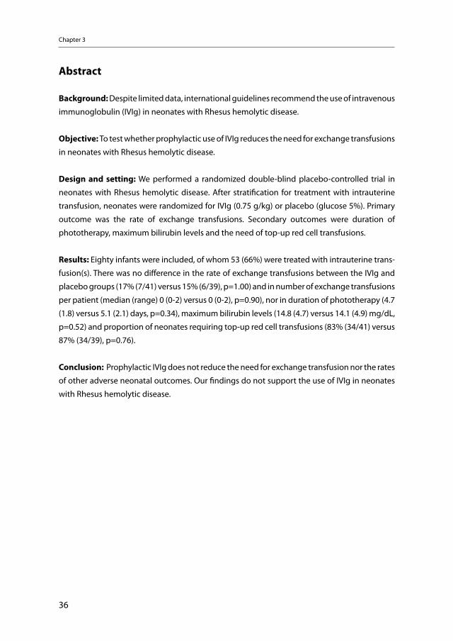
Background: Despite limited data, international guidelines recommend the use of intravenous
immunoglobulin (IVIg) in neonates with Rhesus hemolytic disease.
Objective: To test whether prophylactic use of IVIg reduces the need for exchange transfusions
in neonates with Rhesus hemolytic disease.
Design and setting: We performed a randomized double-blind placebo-controlled trial in
neonates with Rhesus hemolytic disease. After stratification for treatment with intrauterine
transfusion, neonates were randomized for IVIg (0.75 g/kg) or placebo (glucose 5%). Primary
outcome was the rate of exchange transfusions. Secondary outcomes were duration of
phototherapy, maximum bilirubin levels and the need of top-up red cell transfusions.
Results: Eighty infants were included, of whom 53 (66%) were treated with intrauterine trans-
fusion(s). There was no difference in the rate of exchange transfusions between the IVIg and
placebo groups (17% (7/41) versus 15% (6/39), p=1.00) and in number of exchange transfusions
per patient (median (range) 0 (0-2) versus 0 (0-2), p=0.90), nor in duration of phototherapy (4.7
(1.8) versus 5.1 (2.1) days, p=0.34), maximum bilirubin levels (14.8 (4.7) versus 14.1 (4.9) mg/dL,
p=0.52) and proportion of neonates requiring top-up red cell transfusions (83% (34/41) versus
87% (34/39), p=0.76).
Conclusion: Prophylactic IVIg does not reduce the need for exchange transfusion nor the rates
of other adverse neonatal outcomes. Our findings do not support the use of IVIg in neonates
with Rhesus hemolytic disease.
RCT on IVIg in neonatal Rhesus hemolytic disease
37
Introduction
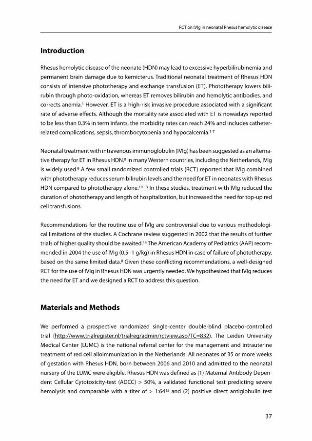
Rhesus hemolytic disease of the neonate (HDN) may lead to excessive hyperbilirubinemia and
permanent brain damage due to kernicterus. Traditional neonatal treatment of Rhesus HDN
consists of intensive phototherapy and exchange transfusion (ET). Phototherapy lowers bili-
rubin through photo-oxidation, whereas ET removes bilirubin and hemolytic antibodies, and
corrects anemia.1 However, ET is a high-risk invasive procedure associated with a significant
rate of adverse effects. Although the mortality rate associated with ET is nowadays reported
to be less than 0.3% in term infants, the morbidity rates can reach 24% and includes catheter-
related complications, sepsis, thrombocytopenia and hypocalcemia.1-7
Neonatal treatment with intravenous immunoglobulin (IVIg) has been suggested as an alterna-
tive therapy for ET in Rhesus HDN.8 In many Western countries, including the Netherlands, IVIg
is widely used.9 A few small randomized controlled trials (RCT) reported that IVIg combined
with phototherapy reduces serum bilirubin levels and the need for ET in neonates with Rhesus
HDN compared to phototherapy alone.10-13 In these studies, treatment with IVIg reduced the
duration of phototherapy and length of hospitalization, but increased the need for top-up red
cell transfusions.
Recommendations for the routine use of IVIg are controversial due to various methodologi-
cal limitations of the studies. A Cochrane review suggested in 2002 that the results of further
trials of higher quality should be awaited.14 The American Academy of Pediatrics (AAP) recom-
mended in 2004 the use of IVIg (0.5–1 g/kg) in Rhesus HDN in case of failure of phototherapy,
based on the same limited data.8 Given these conflicting recommendations, a well-designed
RCT for the use of IVIg in Rhesus HDN was urgently needed. We hypothesized that IVIg reduces
the need for ET and we designed a RCT to address this question.
Materials and Methods
We performed a prospective randomized single-center double-blind placebo-controlled
trial (http://www.trialregister.nl/trialreg/admin/rctview.asp?TC=832). The Leiden University
Medical Center (LUMC) is the national referral center for the management and intrauterine
treatment of red cell alloimmunization in the Netherlands. All neonates of 35 or more weeks
of gestation with Rhesus HDN, born between 2006 and 2010 and admitted to the neonatal
nursery of the LUMC were eligible. Rhesus HDN was defined as (1) Maternal Antibody Depen-
dent Cellular Cytotoxicity-test (ADCC) > 50%, a validated functional test predicting severe
hemolysis and comparable with a titer of > 1:6415 and (2) positive direct antiglobulin test
Chapter 3
38
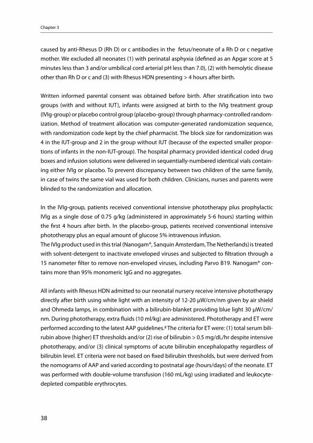
caused by anti-Rhesus D (Rh D) or c antibodies in the fetus/neonate of a Rh D or c negative
mother. We excluded all neonates (1) with perinatal asphyxia (defined as an Apgar score at 5
minutes less than 3 and/or umbilical cord arterial pH less than 7.0), (2) with hemolytic disease
other than Rh D or c and (3) with Rhesus HDN presenting > 4 hours after birth.
Written informed parental consent was obtained before birth. After stratification into two
groups (with and without IUT), infants were assigned at birth to the IVIg treatment group
(IVIg-group) or placebo control group (placebo-group) through pharmacy-controlled random-
ization. Method of treatment allocation was computer-generated randomization sequence,
with randomization code kept by the chief pharmacist. The block size for randomization was
4 in the IUT-group and 2 in the group without IUT (because of the expected smaller propor-
tions of infants in the non-IUT-group). The hospital pharmacy provided identical coded drug
boxes and infusion solutions were delivered in sequentially-numbered identical vials contain-
ing either IVIg or placebo. To prevent discrepancy between two children of the same family,
in case of twins the same vial was used for both children. Clinicians, nurses and parents were
blinded to the randomization and allocation.
In the IVIg-group, patients received conventional intensive phototherapy plus prophylactic
IVIg as a single dose of 0.75 g/kg (administered in approximately 5-6 hours) starting within
the first 4 hours after birth. In the placebo-group, patients received conventional intensive
phototherapy plus an equal amount of glucose 5% intravenous infusion.
The IVIg product used in this trial (Nanogam®, Sanquin Amsterdam, The Netherlands) is treated
with solvent-detergent to inactivate enveloped viruses and subjected to filtration through a
15 nanometer filter to remove non-enveloped viruses, including Parvo B19. Nanogam® con-
tains more than 95% monomeric IgG and no aggregates.
All infants with Rhesus HDN admitted to our neonatal nursery receive intensive phototherapy
directly after birth using white light with an intensity of 12-20 µW/cm/nm given by air shield
and Ohmeda lamps, in combination with a bilirubin-blanket providing blue light 30 µW/cm/
nm. During phototherapy, extra fluids (10 ml/kg) are administered. Phototherapy and ET were
performed according to the latest AAP guidelines.8 The criteria for ET were: (1) total serum bili-
rubin above (higher) ET thresholds and/or (2) rise of bilirubin > 0.5 mg/dL/hr despite intensive
phototherapy, and/or (3) clinical symptoms of acute bilirubin encephalopathy regardless of
bilirubin level. ET criteria were not based on fixed bilirubin thresholds, but were derived from
the nomograms of AAP and varied according to postnatal age (hours/days) of the neonate. ET
was performed with double-volume transfusion (160 mL/kg) using irradiated and leukocyte-
depleted compatible erythrocytes.
RCT on IVIg in neonatal Rhesus hemolytic disease
39
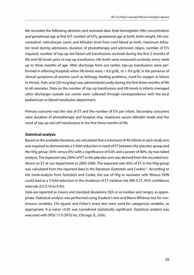
We recorded the following obstetric and neonatal data: fetal hemoglobin (Hb) concentration
and gestational age at first IUT, number of IUTs, gestational age at birth, birth weight, Hb con-
centration, reticulocyte count and bilirubin level from cord blood at birth, maximum biliru-
bin level during admission, duration of phototherapy and admission (days), number of ETs
required, number of top-up red blood cell transfusions received during the first 3 months of
life and Hb levels prior to top-up transfusion. Hb levels were measured routinely every week
up to three months of age. After discharge from our center, top-up transfusions were per-
formed in referring hospitals when Hb levels were < 8.0 g/dL, or < 9.6 g/dL in the presence of
clinical symptoms of anemia (such as lethargy, feeding problems, need for oxygen or failure
to thrive). Folic acid (50 mcg/day) was administered orally during the first three months of life
to all neonates. Data on the number of top-up transfusions and Hb levels in infants managed
(after discharge) outside our center were collected through correspondence with the local
pediatrician or blood transfusion department.
Primary outcome was the rate of ET and the number of ETs per infant. Secondary outcomes
were duration of phototherapy and hospital stay, maximum serum bilirubin levels and the
need of top-up red cell transfusions in the first three months of life.
Statistical analysisBased on the available literature, we calculated that a minimum of 40 infants in each study arm
was required to demonstrate a 5-fold reduction in need of ET between the placebo-group and
the IVIg-group (30% versus 6%) with a significance of 0.05 and a power of 80%, by two-tailed
analysis. The expected rate (30%) of ET in the placebo-arm was derived from the recorded inci-
dence on ET at our department in 2005-2006. The expected rate (6%) of ET in the IVIg-group
was calculated from the reported data in the literature (Gottstein and Cooke)11. According to
the meta-analysis from Gottstein and Cooke, the use of IVIg in neonates with Rhesus HDN
could lead to a 5-fold reduction in the incidence of ET (relative risk (RR) 0.21, 95% confidence
intervals (CI) 0.10 to 0.45).
Data are reported as means and standard deviations (SD) or as median and ranges, as appro-
priate. Statistical analysis was performed using Student-t-test and Mann-Whitney test for con-
tinuous variables. Chi square and Fisher’s exact test were used for categorical variables, as
appropriate. A p-value <0.05 was considered statistically significant. Statistical analysis was
executed with SPSS 17.0 (SPSS Inc, Chicago, IL, USA).
Chapter 3
40
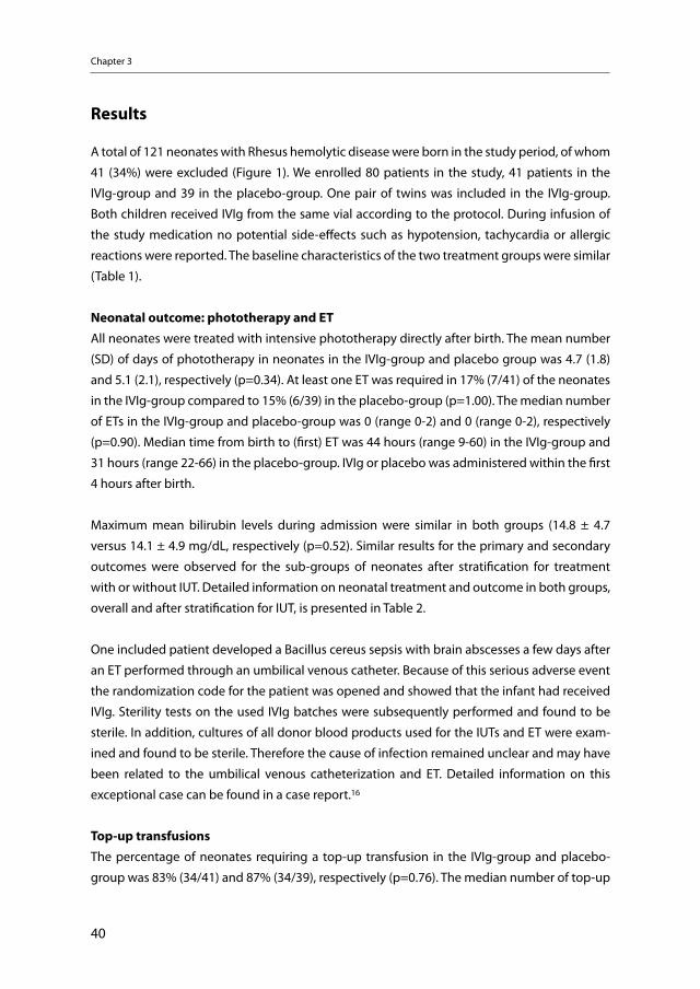
Results
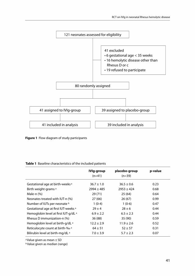
A total of 121 neonates with Rhesus hemolytic disease were born in the study period, of whom
41 (34%) were excluded (Figure 1). We enrolled 80 patients in the study, 41 patients in the
IVIg-group and 39 in the placebo-group. One pair of twins was included in the IVIg-group.
Both children received IVIg from the same vial according to the protocol. During infusion of
the study medication no potential side-effects such as hypotension, tachycardia or allergic
reactions were reported. The baseline characteristics of the two treatment groups were similar
(Table 1).
Neonatal outcome: phototherapy and ETAll neonates were treated with intensive phototherapy directly after birth. The mean number
(SD) of days of phototherapy in neonates in the IVIg-group and placebo group was 4.7 (1.8)
and 5.1 (2.1), respectively (p=0.34). At least one ET was required in 17% (7/41) of the neonates
in the IVIg-group compared to 15% (6/39) in the placebo-group (p=1.00). The median number
of ETs in the IVIg-group and placebo-group was 0 (range 0-2) and 0 (range 0-2), respectively
(p=0.90). Median time from birth to (first) ET was 44 hours (range 9-60) in the IVIg-group and
31 hours (range 22-66) in the placebo-group. IVIg or placebo was administered within the first
4 hours after birth.
Maximum mean bilirubin levels during admission were similar in both groups (14.8 ± 4.7
versus 14.1 ± 4.9 mg/dL, respectively (p=0.52). Similar results for the primary and secondary
outcomes were observed for the sub-groups of neonates after stratification for treatment
with or without IUT. Detailed information on neonatal treatment and outcome in both groups,
overall and after stratification for IUT, is presented in Table 2.
One included patient developed a Bacillus cereus sepsis with brain abscesses a few days after
an ET performed through an umbilical venous catheter. Because of this serious adverse event
the randomization code for the patient was opened and showed that the infant had received
IVIg. Sterility tests on the used IVIg batches were subsequently performed and found to be
sterile. In addition, cultures of all donor blood products used for the IUTs and ET were exam-
ined and found to be sterile. Therefore the cause of infection remained unclear and may have
been related to the umbilical venous catheterization and ET. Detailed information on this
exceptional case can be found in a case report.16
Top-up transfusionsThe percentage of neonates requiring a top-up transfusion in the IVIg-group and placebo-
group was 83% (34/41) and 87% (34/39), respectively (p=0.76). The median number of top-up
RCT on IVIg in neonatal Rhesus hemolytic disease
41
Table 1 Baseline characteristics of the included patients
IVIg-group placebo-group p-value
(n=41) (n=39)
Gestational age at birth-weeks a 36.7 ± 1.0 36.5 ± 0.6 0.23Birth weight-grams a 2994 ± 485 2953 ± 424 0.68Male-n (%) 29 (71) 25 (64) 0.64Neonates treated with IUT-n (%) 27 (66) 26 (67) 0.99Number of IUTs per neonate b 1 (0-4) 1 (0-6) 0.47Gestational age at first IUT-weeks a 29 ± 4 28 ± 6 0.44Hemoglobin level at first IUT-g/dL a 6.9 ± 2.2 6.5 ± 2.3 0.44Rhesus D immunization-n (%) 36 (88) 35 (90) 0.59Hemoglobin level at birth-g/dL a 12.2 ± 2.9 11.9 ± 2.6 0.52Reticulocyte count at birth-‰ a 64 ± 51 52 ± 57 0.31Bilirubin level at birth-mg/dL a 7.0 ± 3.9 5.7 ± 2.3 0.07
a Value given as mean ± SDb Value given as median (range)
121 neonates assessed for eligibility
80 randomly assigned
41 assigned to IVIg-group 39 assigned to placebo-group
41 included in analysis 39 included in analysis
41 excluded• 6 gestational age < 35 weeks• 16 hemolytic disease other than Rhesus D or c• 19 refused to participate
Figure 1 Flow diagram of study participants
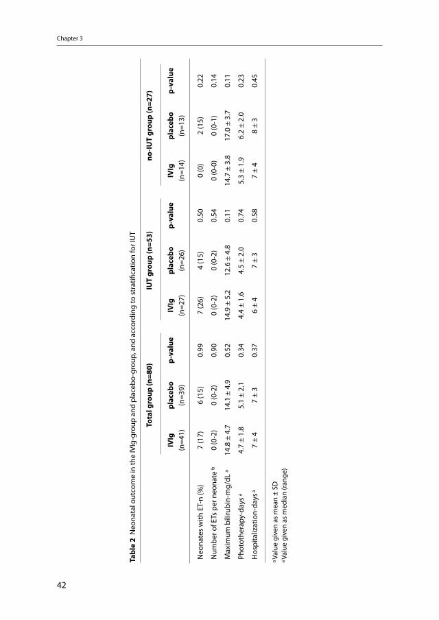
Chapter 3
42
Tabl
e 2
Neo
nata
l out
com
e in
the
IVIg
-gro
up a
nd p
lace
bo-g
roup
, and
acc
ordi
ng to
str
atifi
catio
n fo
r IU
T
To
tal g
roup
(n=8
0)IU
T gr
oup
(n=5
3)no
-IUT
grou
p (n
=27)
IVIg
plac
ebo
p-va
lue
IVIg
plac
ebo
p-va
lue
IVIg
plac
ebo
p-va
lue
(n
=41)
(n=3
9)
(n=2
7)(n
=26)
(n
=14)
(n=1
3)
Neo
nate
s w
ith E
T-n
(%)
7 (1
7)6
(15)
0.99
7 (2
6)4
(15)
0.50
0 (0
)2
(15)
0.22
Num
ber o
f ETs
per
neo
nate
b0
(0-2
)0
(0-2
)0.
900
(0-2
)0
(0-2
)0.
540
(0-0
)0
(0-1
)0.
14
Max
imum
bili
rubi
n-m
g/dL
a14
.8 ±
4.7
14
.1 ±
4.9
0.52
14.9
± 5
.212
.6 ±
4.8
0.11
14.7
± 3
.817
.0 ±
3.7
0.11
Phot
othe
rapy
-day
s a
4.7
± 1.
85.
1 ±
2.1
0.34
4.4
± 1.
64.
5 ±
2.0
0.74
5.3
± 1.
96.
2 ±
2.0
0.23
Hos
pita
lizat
ion-
days
a7
± 4
7 ±
30.
376
± 4
7 ±
30.
587
± 4
8 ±
30.
45
a Va
lue
give
n as
mea
n ±
SDa
Valu
e gi
ven
as m
edia
n (r
ange
)
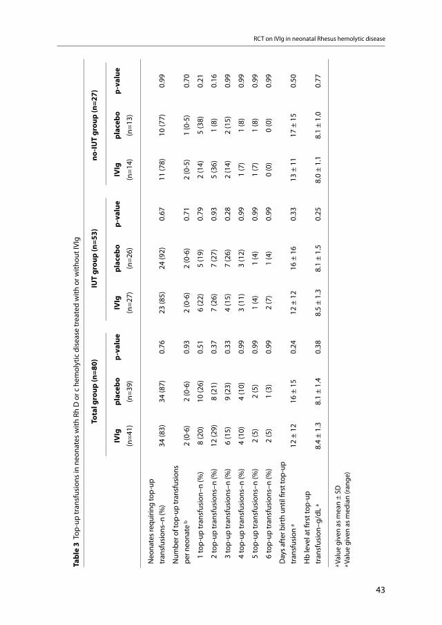
RCT on IVIg in neonatal Rhesus hemolytic disease
43
Tabl
e 3
Top-
up tr
ansf
usio
ns in
neo
nate
s w
ith R
h D
or c
hem
olyt
ic d
isea
se tr
eate
d w
ith o
r with
out I
VIg
To
tal g
roup
(n=8
0)IU
T gr
oup
(n=5
3)no
-IUT
grou
p (n
=27)
IVIg
pl
aceb
o p-
valu
eIV
Ig
plac
ebo
p-va
lue
IVIg
pl
aceb
op-
valu
e
(n=4
1)(n
=39)
(n
=27)
(n=2
6)
(n=1
4)(n
=13)
Neo
nate
s req
uirin
g to
p-up
tr
ansf
usio
ns–n
(%)
34 (8
3)34
(87)
0.76
23 (8
5)24
(92)
0.67
11 (7
8)10
(77)
0.99
Num
ber o
f top
-up
tran
sfus
ions
pe
r neo
nate
b2
(0-6
)2
(0-6
)0.
932
(0-6
)2
(0-6
)0.
712
(0-5
)1
(0-5
)0.
70
1 to
p-up
tran
sfus
ion–
n (%
)8
(20)
10 (2
6)0.
516
(22)
5 (1
9)0.
792
(14)
5 (3
8)0.
21
2 to
p-up
tran
sfus
ions
–n (%
)12
(29)
8 (2
1)0.
377
(26)
7 (2
7)0.
935
(36)
1 (8
)0.
16
3 to
p-up
tran
sfus
ions
–n (%
)6
(15)
9 (2
3)0.
334
(15)
7 (2
6)0.
282
(14)
2 (1
5)0.
99
4 to
p-up
tran
sfus
ions
–n (%
)4
(10)
4 (1
0)0.
993
(11)
3 (1
2)0.
991
(7)
1 (8
)0.
99
5 to
p-up
tran
sfus
ions
–n (%
)2
(5)
2 (5
)0.
991
(4)
1 (4
)0.
991
(7)
1 (8
)0.
99
6 to
p-up
tran
sfus
ions
–n (%
) 2
(5)
1 (3
)0.
992
(7)
1 (4
)0.
990
(0)
0 (0
)0.
99
Day
s aft
er b
irth
until
firs
t top
-up
tran
sfus
ion
a12
± 1
216
± 1
50.
2412
± 1
216
± 1
60.
3313
± 1
117
± 1
50.
50
Hb
leve
l at fi
rst t
op-u
p tr
ansf
usio
n–g/
dL a
8.4
± 1.
38.
1 ±
1.4
0.38
8.5
± 1.
38.
1 ±
1.5
0.25
8.0
± 1.
18.
1 ±
1.0
0.77
a Va
lue
give
n as
mea
n ±
SD
a Va
lue
give
n as
med
ian
(ran
ge)
Chapter 3
44
transfusions per neonate in the IVIg-group and placebo-group was 2 (range 0-6) and 2 (range
0-6), respectively (p=0.93). Mean hemoglobin level at first top-up transfusion and median
number of days until first top-up transfusion were similar in both groups. Detailed informa-
tion on the use of top-up transfusions in the IVIg-group and the placebo-group is presented
in Table 3.
Comment
In this RCT we have shown that prophylactic treatment with IVIg in neonates with Rhesus
hemolytic disease did not reduce the need for ET nor the rates of other adverse neonatal
outcomes. Our results do not support the recommendation to give IVIg in Rhesus hemolytic
disease, as stated in recent AAP guidelines8. Our study adds to the Cochrane analysis that
there is no evidence to recommend routine use of IVIg.14
In the past, several studies have suggested a positive effect of IVIg in reducing the rate of
hemolysis in Rhesus hemolytic disease.10,12,13,17-19 Although the exact mechanism of action of
IVIg remains unclear, IVIg has been reported to block Fc-receptors on macrophages, result-
ing in a decreased removal of anti-Rh antibody coated erythrocytes from the circulation. IVIg
might increase IgG catabolism, resulting in a shorter half-life of antibodies (including anti-Rh
antibodies). A third hypothesis is the presence of anti-idiotypic antibodies in IVIg neutralizing
anti-Rh antibodies.12,20-22
Our results are in contrast with the most recent recommendations of the AAP to use 0.5-1.0
g/kg IVIg in Rhesus hemolytic disease in case of failure of phototherapy.8 These guidelines
were published in 2004 and based on a limited number of small RCTs. Several important
methodological limitations hampered the interpretation of these studies, including sub-opti-
mal study-designs and the wide range of inclusion criteria.18,19 The Cochrane Collaboration
performed a review on three studies, in which a total of 189 infants were included.10,12-14 Rubo
et al.10 included 32 infants with Rhesus hemolytic disease in a multicenter RCT. No details on
IUT and gestational age were given. Several years later Dagoglu et al.13 included 29 preterm
and 12 term infants in a RCT. Cut-off for prematurity and criteria for top-up red cell transfu-
sions were not defined. In 1999 Alpay et al.12 enrolled 116 infants, predominantly with ABO
incompatibility (n=93), but also neonates with Rhesus hemolytic disease (n=16) and both Rh
and ABO incompatibility (n=7) were included. However, results were not given for each group
separately. None of the studies described detailed phototherapy guidelines and none of them
used a placebo in the control-group or described any method of blinding the intervention
after allocation concealment. According to the Cochrane review, none of the trials fulfilled
RCT on IVIg in neonatal Rhesus hemolytic disease
45
criteria for high quality study. Our study is the first well-designed randomized double-blind
placebo-controlled trial on this topic.
In 4 other studies, infants with ABO incompatibility were included.12,17,18,23 In general, com-
pared to Rhesus immunization, ABO incompatibility causes less severe hemolysis and there-
fore less neonatal morbidity.24 For that reason, we included only neonates with Rhesus disease.
These important methodological differences between our study and the previous ones may
explain the discordant results.
Several other explanations can be envisaged to explain the lack of effect of IVIg in our study.
A possible explanation could be the treatment with intensive and prophylactic phototherapy
starting immediately after birth, thereby reducing the risk of severe hyperbilirubinemia. In
addition, the majority of infants included in our study were treated with IUT. By IUT, Rhesus
incompatible erythrocytes of the fetus are replaced by Rhesus compatible cells of the donor.
Dependent on the interval between the last IUT and delivery, these donor cells are still present
after birth, resulting in less or more delayed hemolysis.25, 26 However, several groups includ-
ing ours have shown that even after IUT, neonates with Rhesus hemolytic disease still often
require ET.27,28 In our study, IVIg was neither effective in the IUT group nor in the group without
IUT. However, the number of patients included in the subgroup without IUT (n=27) may be too
small to draw firm conclusions. Recently, a research group from Brazil finalized a similar RCT on
IVIg for neonates with Rhesus hemolytic disease and, in accordance with our results, found no
difference between both groups on the rate of ET. Importantly, in their study the vast majority
of patients (n=80) had no prior treatment with IUT (ClinicalTrials.gov NCT00288600).29 There-
fore, both our RCT and the RCT from the Brazilian research group failed to show any effect
of IVIg in Rhesus hemolytic disease infants, irrespective of whether or not the infants were
treated with IUT. Care should be taken when interpreting our results, particularly the sub-
group analyses, due to the relatively limited number of patients. In addition, caution should be
used before applying the results of this study to all Rhesus isoimmunized infants. There may
be a subset of Rhesus isoimmunized infants with (inappropriate) delayed start of intensive
phototherapy, for whom IVIg might be effective. More studies are needed to study the effect
of IVIg in this specific subset of infants.
Although IVIg is considered to be an extremely safe product, adverse events can not be totally
eliminated. Rare but serious side effects such as transfusion transmitted diseases, anaphylaxis,
hypersensitivity, thrombosis, pulmonary emboli and renal failure have been reported.21,22
Recently, Figueras-Aloy et al30 reported a higher incidence of necrotizing enterocolitis (NEC)
in near-term infants with Rhesus hemolytic disease treated with IVIg compared to a control
group managed without IVIg. The authors correctly suggest that their results must be inter-
Chapter 3
46
preted with care given the retrospective nature of the study. Whether occurrence of NEC was
related to the administration of IVIg or to the fact that infants receiving IVIg were more ill than
the control group is not clear. Nevertheless, since potential (but rare) adverse effects associ-
ated with the use of IVIg can not be ruled out, the authors call for more caution when using IVIg
in neonates with Rhesus hemolytic disease. IVIg is a blood product prepared by separating the
gamma-globulin fraction from the plasma pooled from multiple donors. The manufacturing
of IVIg, including fractionation and filtration of viruses is an extremely intensive and expensive
process. Therefore, the use of IVIg for indications that are not confirmed by well-designed RCTs
should be restricted.22
Conclusion
Prophylactic treatment with IVIg (in a dosage of 0.75 g/kg) did not reduce the need for ET nor
the rates of other adverse neonatal outcomes. Our findings do not support the current recom-
mendations of the AAP to use IVIg in neonates with Rhesus hemolytic disease. In view of the
absence of beneficial effects, the use of IVIg for this indication should be discouraged.
Acknowledgement
Nanogam was provided by Sanquin Blood Supply Foundation (Amsterdam, The Netherlands).
References
1. Smits-Wintjens VE, Walther FJ, Lopriore E. Rhesus haemolytic disease of the newborn: Postnatal man-agement, associated morbidity and long-term outcome. Semin Fetal Neonatal Med . 2008;13:265-271.
2. Keenan WJ, Novak KK, Sutherland JM, Bryla DA, Fetterly KL. Morbidity and mortality associated with exchange transfusion. Pediatrics . 1985;75:417-421.
3. Patra K, Storfer-Isser A, Siner B, Moore J, Hack M. Adverse events associated with neonatal exchange transfusion in the 1990s. J Pediatr . 2004;144:626-631.
4. Jackson JC. Adverse events associated with exchange transfusion in healthy and ill newborns. Pediat-rics . 1997;99:E7.
5. Steiner LA, Bizzarro MJ, Ehrenkranz RA, Gallagher PG. A decline in the frequency of neonatal exchange transfusions and its effect on exchange-related morbidity and mortality. Pediatrics . 2007;120:27-32.
6. Thayyil S, Milligan DW. Single versus double volume exchange transfusion in jaundiced newborn infants. Cochrane Database Syst Rev . 2006;CD004592.
7. Hovi L, Siimes MA. Exchange transfusion with fresh heparinized blood is a safe procedure. Experiences from 1 069 newborns. Acta Paediatr Scand . 1985;74:360-365.
8. Management of hyperbilirubinemia in the newborn infant 35 or more weeks of gestation. Pediatrics . 2004;114:297-316.
RCT on IVIg in neonatal Rhesus hemolytic disease
47
9. New HV, Stanworth SJ, Engelfriet CP et al. Neonatal transfusions. Vox Sang . 2009;96:62-85.10. Rubo J, Albrecht K, Lasch P et al. High-dose intravenous immune globulin therapy for hyperbilirubine-
mia caused by Rh hemolytic disease. J Pediatr . 1992;121:93-97.11. Gottstein R, Cooke RW. Systematic review of intravenous immunoglobulin in haemolytic disease of the
newborn. Arch Dis Child Fetal Neonatal Ed . 2003;88:F6-10.12. Alpay F, Sarici SU, Okutan V, Erdem G, Ozcan O, Gokcay E. High-dose intravenous immunoglobulin
therapy in neonatal immune haemolytic jaundice. Acta Paediatr . 1999;88:216-219.13. Dagoglu T, Ovali F, Samanci N, Bengisu E. High-dose intravenous immunoglobulin therapy for rhesus
haemolytic disease. J Int Med Res . 1995;23:264-271.14. Alcock GS, Liley H. Immunoglobulin infusion for isoimmune haemolytic jaundice in neonates. Cochrane
Database Syst Rev . 2002;CD003313.15. Oepkes D, Van Kamp IL, Simon MJ, Mesman J, Overbeeke MA, Kanhai HH. Clinical value of an antibody-
dependent cell-mediated cytotoxicity assay in the management of Rh D alloimmunization. Am J Obstet Gynecol . 2001;184:1015-1020.
16. Smits-Wintjens VE, Steggerda SJ, Oepkes D, Van Kamp IL, Kramer CM, Walther FJ et al. Bacillus cereus cerebral abscesses in a term neonate with rhesus hemolytic disease treated with exchange transfusion. J Pediatr Inf Dis. 2010;5:277-280.
17. Nasseri F, Mamouri GA, Babaei H. Intravenous immunoglobulin in ABO and Rh hemolytic diseases of newborn. Saudi Med J . 2006;27:1827-1830.
18. Tanyer G, Siklar Z, Dallar Y, Yildirmak Y, Tiras U. Multiple dose IVIg treatment in neonatal immune hemo-lytic jaundice. J Trop Pediatr . 2001;47:50-53.
19. Voto LS, Sexer H, Ferreiro G et al. Neonatal administration of high-dose intravenous immunoglobulin in rhesus hemolytic disease. J Perinat Med . 1995;23:443-451.
20. Kriplani A, Malhotra SB, Mandal K. Fetal intravenous immunoglobulin therapy in rhesus hemolytic disease. Gynecol Obstet Invest . 2007;63:176-180.
21. Walsh S, Molloy EJ. Towards evidence based medicine for paediatricians. Is intravenous immunoglobulin superior to exchange transfusion in the management of hyperbilirubinaemia in term neonates? Arch Dis Child . 2009;94:739-741.
22. Kumar A, Teuber SS, Gershwin ME. Intravenous immunoglobulin: striving for appropriate use. Int Arch Allergy Immunol . 2006;140:185-198.
23. Miqdad AM, Abdelbasit OB, Shaheed MM, Seidahmed MZ, Abomelha AM, Arcala OP. Intravenous immu-noglobulin G (IVIg) therapy for significant hyperbilirubinemia in ABO hemolytic disease of the newborn. J Matern Fetal Neonatal Med . 2004;16:163-166.
24. Murray NA, Roberts IA. Haemolytic disease of the newborn. Arch Dis Child Fetal Neonatal Ed . 2007;92:F83-F88.
25. Oepkes D, Adama van SP. Intrauterine fetal transfusions in the management of fetal anemia and fetal thrombocytopenia. Semin Fetal Neonatal Med . 2007;12:432-438.
26. Egberts J, Van Kamp IL, Kanhai HH, Meerman RH, Giordano PC, Gravenhorst JB. The disappearance of fetal and donor red blood cells in alloimmunised pregnancies: a reappraisal. Br J Obstet Gynaecol . 1997;104:818-824.
27. De Boer I, Zeestraten EC, Lopriore E, Van K, I, Kanhai HH, Walther FJ. Pediatric outcome in Rhesus hemo-lytic disease treated with and without intrauterine transfusion. Am J Obstet Gynecol . 2008;198:54. e1-54.e4.
28. Rath ME, Smits-Wintjens VE, Lindenburg I et al. Top-up transfusions in neonates with Rh hemolytic disease in relation to exchange transfusions. Vox Sang . 2010;99(1):65-70.
29. Santos MC, Sa, Gomes, Camacho, Moreira. High-dose intravenous immunoglobulin therapy for hyper-bilirubinemia due Rh hemolytic disease: a randomized clinical trial. Pediatric Academic Societies-annual meeting-Vancouver 2010 . 2010;143.
30. Figueras-Aloy J, Rodriguez-Miguelez JM, Iriondo-Sanz M, Salvia-Roiges MD, Botet-Mussons F, Carbonell-Estrany X. Intravenous immunoglobulin and necrotizing enterocolitis in newborns with hemolytic disease. Pediatrics . 2010;125:139-144.

48

4 Bacillus cereus cerebral abscesses in a term neonate
with Rhesus hemolytic disease treated with exchange transfusion
Vivianne EHJ Smits-WintjensSylke J Steggerda
Dick OepkesInge L van Kamp
Christine M KramerFrans J WaltherEnrico Lopriore
J Pediatr Inf Dis 2010; 5(3):277-280
Chapter 4
50
Abstract
Exchange transfusion (ET) is the most effective method for treatment of severe hyperbiliru-binemia and is often required in Rhesus hemolytic disease of the newborn (RHDN). The use of ET is also associated with adverse reactions, including severe catheter-related infectious complications. We report a term neonate with RHDN treated with an ET through an umbili-cal venous catheter who developed brain abscesses due to a Bacillus cereus sepsis. This severe complication has not previously been reported. We discuss possible causes for this severe infection and provide suggestions on prevention.
Cerebral abscesses in a neonate with Rhesus hemolytic disease
51
Introduction
Exchange transfusion (ET) plays an important role in the treatment of hyperbilirubinemia of the newborn. Rhesus associated hemolytic disease of the newborn (RHDN) is one of the most common indications for ET. The main therapeutic effect of ET is removal of excess bili-rubin and maternal red blood cell antibodies in order to prevent kernicterus and to reduce hemolysis. Despite improvement in neonatal intensive care, ET still remains a high-risk inva-sive procedure associated with a significant rate of adverse reactions. Although the mortal-ity rate associated with ET is nowadays less then 0.3% in term infants, morbidity rates may reach 5%.1 Morbidity includes in particular catheter-related complications such as thrombo-sis, sepsis and necrotizing enterocolitis.1
We report a term neonate with RHDN treated with an ET who developed a rare but severe cerebral infection after umbilical cord catheterization.
Case report
A 36-year-old gravida 2 para 1 with severe Rh D alloimmunization required 3 intrauterine transfusions with filtered red blood cell concentrate for fetal anemia. These procedures were uncomplicated. The last transfusion before birth was done at 34 weeks’ gestation, with a post-transfusion hemoglobin level of 15.1 g/dl. The first labour had been protracted and was complicated by a complete perineal laceration after vacuum assisted delivery. For this reason the parents required a caesarean delivery, which was planned 3 weeks after the final intrauterine transfusion at a gestation of 37 weeks. A male infant was born with a birth weight of 2400 grams (25th percentile for gestational age) and Apgar scores of 10, 10 and 10 at 1, 5 and 10 min, respectively. No abnormalities were detected on physical examination, except a pale skin color. Laboratory investigations at birth showed a hemo-globin value of 9.6 g/dL, a total bilirubin concentration of 11.8 mg/dL and a conjugated bilirubin concentration of 11.3 mg/dL. Treatment with intensive phototherapy was started immediately after birth. After obtaining parental informed consent, the infant was included in a randomized placebo controlled trial to assess the short and long term effects of pro-phylactic use of intravenous immunoglobulin (IVIg) in neonates with RHDN (LIVIN trial: http://www.controlled-trials.com/ISRCTN14013064/). For this study a ready for use 5% IVIg solution (Nanogam® Sanquin, The Netherlands) or glucose 5% (placebo) was used. Our patient was randomized for IVIg which he received within four hours after birth. Due to per-sisting elevated bilirubin concentrations and possible need for an ET an umbilical venous catheterization was performed. Despite intensive phototherapy, the total serum bilirubin
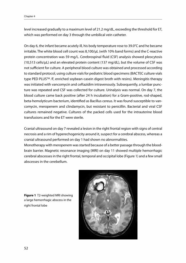
Chapter 4
52
level increased gradually to a maximum level of 21.2 mg/dL, exceeding the threshold for ET, which was performed on day 3 through the umbilical vein catheter.
On day 6, the infant became acutely ill, his body temperature rose to 39.0°C and he became irritable. The white blood cell count was 8,100/μL (with 10% band forms) and the C-reactive protein concentration was 59 mg/L. Cerebrospinal fluid (CSF) analysis showed pleocytosis (10,513 cells/μL) and an elevated protein content (137 mg/dL), but the volume of CSF was not sufficient for culture. A peripheral blood culture was obtained and processed according to standard protocol, using culture vials for pediatric blood specimens (BACTEC culture vials type PED PLUSTM /F, enriched soybean-casein digest broth with resins). Meningitis therapy was initiated with vancomycin and ceftazidim intravenously. Subsequently, a lumbar punc-ture was repeated and CSF was collected for culture. Urinalysis was normal. On day 7, the blood culture came back positive (after 24 h incubation) for a Gram-positive, rod-shaped, beta-hemolyticum bacterium, identified as Bacillus cereus. It was found susceptible to van-comycin, meropenem and clindamycin, but resistant to penicillin. Bacterial and viral CSF cultures remained negative. Cultures of the packed cells used for the intrauterine blood transfusions and for the ET were sterile.
Cranial ultrasound on day 7 revealed a lesion in the right frontal region with signs of central necrosis and a rim of hyperechogenicity around it, suspect for a cerebral abscess, whereas a cranial ultrasound performed on day 1 had shown no abnormalities. Monotherapy with meropenem was started because of a better passage through the blood- brain barrier. Magnetic resonance imaging (MRI) on day 11 showed multiple hemorrhagic cerebral abscesses in the right frontal, temporal and occipital lobe (Figure 1) and a few small abscesses in the cerebellum.
Figure 1 T2-weighted MRI showing a large hemorrhagic abscess in the
right frontal lobe
Cerebral abscesses in a neonate with Rhesus hemolytic disease
53
On day 21 the child again became irritable and developed a seizure (stretching of both arms and legs). Because of the short duration of the seizure (less than 1 min) and a normal elec-troencephalogram (EEG), anticonvulsant medication was not required. On day 29 the infant had a recurrent generalized convulsion and anticonvulsant treatment (phenobarbital) was initiated. Again, the EEG showed no epileptic activity. A MRI performed a few days after the seizures showed no changes in the cerebral lesions.
After 6 weeks of intravenous meropenem administration, antibiotic treatment was discon-tinued. The infant was discharged home on day 61 with anticonvulsant therapy. At 2 years of age, the infant had no neurologic sequelae on physical examination. Mental and psy-chomotor development indexes, assessed at 2 years of age with the Bayley Scales of Infant Development, were within normal ranges.
Discussion
ET has been used in neonatal intensive care units for more than 50 years and is still the most effective method for treatment of severe hyperbilirubinemia. However, ET is also asso-ciated with severe adverse reactions, including catheter-related infectious complications.2 We report a term infant with cerebral abscesses due to a Bacillus cereus sepsis, a severe complication which has not previously been reported.
Bacillus cereus is a probably ubiquitous soil bacterium and an opportunistic pathogen that is a common cause of food poisoning. In (premature) neonates, toxins produced by Bacillus cereus can cause necrosis of infected tissue. Bacillus cereus infections in neonates include meningoencephalitis and brain abscesses, characterized by liquefactive necrosis. A few sporadic cases have been reported in the literature and usually involved premature infants, suggesting an association with decreased immune response. Most cases of neo-natal meningoencephalitis and cerebral abscesses with Bacillus cereus described in the literature resulted in neonatal death.3-7
Several potential risk factors may have led to the Bacillus cereus infection in this infant. Because of severe RHDN, three intrauterine blood transfusions and one ET were required. Multiple blood transfusions are associated with an increased risk of infection. However, cul-tures of all donor blood used for the transfusions were sterile. Intrauterine transfusions have also been associated with several procedure-related complications, including, although rarely, intrauterine infections. In the largest published series on complications of intrauter-ine transfusions, only two infections with E. Coli, both resulting in fetal distress within 48 h
Chapter 4
54
after the procedure, were found in 740 procedures.8 In the current case, no signs of infection were present prenatally, and there was no premature rupture of membranes prior to the elective caesarean section. It seems therefore highly unlikely that the Bacillus cereus infec-tion had a prenatal origin. Moreover, cranial ultrasound on day 1 showed no abnormalities, excluding a link between the cerebral abscesses and antenatal invasive procedures.
Although very unlikely, another potential cause for infection in this case could be the IVIg solution. All IVIg preparations are routinely tested for sterility. The production of IVIg is an aseptical process. Up till now more than 80 batches resulting in approximately 100.000 vials of Nanogam® have been produced. Sterility tests on all these batches were negative.
Lastly, the most probable cause for infection in this case was the umbilical venous catheter-ization. Although umbilical venous catheterization is a common procedure in the manage-ment of sick neonates, it can lead to serious complications. Sequelae include thrombosis, embolization, hemorrhage, arrhythmias, (pericardial) effusions, portal hypertension and infection.9 In the literature the incidence of sepsis related to umbilical venous catheteriza-tion is reported between 6% and 24%.2
Umbilical venous catheterization was required in this case because an ET was deemed necessary to reduce the elevated bilirubin levels and the risk of bilirubin encephalopathy. However, whether catheterization and ET was strictly unavoidable is questionable.
First, hyperbilirubinemia was mainly due to elevated levels of the conjugated bilirubin frac-tion. Elevated levels of conjugated bilirubin are not a rare phenomenon in neonates with RHDN, although the etiology is not clear. The current AAP guidelines state that thresholds for treatment in hyperbilirubinemia should be based on the total bilirubin levels, without subtracting the conjugated bilirubin fraction.1 Nevertheless, when conjugated bilirubin is significantly elevated (i.e. > 50%) there is no bilirubin threshold at which intervention is rec-ommended. Bilirubin encephalopathy is due to unbound, unconjugated bilirubin. Whether neonates with increased conjugated bilirubin are at increased risk for kernicterus is not known and management remains controversial. Management should ideally be based on the measurement of unbound, unconjugated bilirubin, but these measurements have not yet been adapted to clinical use.
Second, the AAP guidelines recommend the use of IVIg in case of failure of intensive photo-therapy in order to reduce the need for ET.1 The evidence to recommend routine prophy-lactic treatment with IVIg is considered insufficient.10,11 A recent Cochrane review suggests that the results of further trials of higher quality should be awaited.10 Although the risk for
Cerebral abscesses in a neonate with Rhesus hemolytic disease
55
adverse effects of umbilical venous catheterization and ET may decrease in the future, it will never completely disappear. The best way to prevent procedure-related complications in RHDN is to reduce the need for ET.
References
1. American Academy of Pediatrics Subcommittee on Hyperbilirubinemia. Management of hyperbiliru-binemia in the newborn infant 35 or more weeks of gestation. Pediatrics 2004;114:297-316.
2. Butler-O’Hara M, Buzzard CJ, Reubens L, McDermott MP, DiGrazio W, D’Angio CT. A randomized trial comparing long-term and short-term use of umbilical venous catheters in premature infants with birth weights of less than 1251 grams. Pediatrics 2006;118:e25-e35.
3. Lequin MH, Vermeulen JR, van Elburg RM et al. Bacillus cereus meningoencephalitis in preterm infants: neuroimaging characteristics. AJNR Am J Neuroradiol 2005;26:2137-2143.
4. Chu WP, Que TL, Lee WK, Wong SN. Meningoencephalitis caused by Bacillus cereus in a neonate. Hong Kong Med J 2001;7:89-92.
5. Hilliard NJ, Schelonka RL, Waites KB. Bacillus cereus bacteremia in a preterm neonate. J Clin Microbiol 2003;41:3441-3444.
6. Gray J, George RH, Durbin GM, Ewer AK, Hocking MD, Morgan ME. An outbreak of Bacillus cereus respiratory tract infections on a neonatal unit due to contaminated ventilator circuits. J Hosp Infect 1999;41:19-22.
7. Van Der Zwet WC, Parlevliet GA, Savelkoul PH et al. Outbreak of Bacillus cereus infections in a neonatal intensive care unit traced to balloons used in manual ventilation. J Clin Microbiol 2000;38:4131-4136.
8. Van Kamp IL, Klumper FJ, Oepkes D et al. Complications of intrauterine intravascular transfusion for fetal anemia due to maternal red-cell alloimmunization. Am J Obstet Gynecol 2005;192:171-177.
9. Korver AM, Walther FJ, van der Molen AJ, de Beaufort AJ. Serious complications of umbilical venous catheterisation. Ned Tijdschr Geneeskd 2007;151:2219-2223.
10. Alcock GS, Liley H. Immunoglobulin infusion for isoimmune haemolytic jaundice in neonates. Cochrane Database Syst Rev 2002;CD003313.
11. Smits-Wintjens VE, Walther FJ, Lopriore E. Rhesus haemolytic disease of the newborn: Postnatal man-agement, associated morbidity and long-term outcome. Semin Fetal Neonatal Med 2008;13:265-271.

56

5 Neonatal morbidity after exchange transfusion
for red cell alloimmune hemolytic disease
Vivianne EHJ Smits-Wintjens and Mirjam EA RathErik W van Zwet
Dick OepkesAnneke BrandFrans J WaltherEnrico Lopriore
submitted
Chapter 5
58
Abstract
Objective: Our aim was to study the type and rate of complications associated with exchange transfusion (ET) in a large series of neonates with hemolytic disease of the newborn (HDN) due to red cell alloimmunization.
Patients and Methods: All neonates with HDN due to red cell alloimmunization admitted to our center between January 2001 and June 2011 were eligible for this study. We recorded the number and rate of complications during admission in the group of neonates treated with (ET-group) and without ET (no-ET-group). Multivariate logistic regression analysis was performed to measure independent risk of complications of ET treatment.
Results: A total of 347 infants with red cell alloimmune hemolytic disease were included, 39% (134/347) was treated with at least one ET during admission (ET-group) and 61% (213/347) did not require ET (no-ET-group). Comparison between the ET-group and no-ET-group showed that ET treatment was independently associated with : proven sepsis (8% versus 1% respectively, odds ratio (OR) 8.3, 95% confidence interval (CI) 1.7-40.3, p = 0.009), leukocytopenia (88% versus 23% respectively, OR 36.0, 95% CI 17.5-73.8, p < 0.001), severe thrombocytopenia (platelet count < 50×109/L) (63% versus 8% respectively, OR 31.4, 95% CI 14.0-70.4, p < 0.001), hypocalcemia (22% versus 1% respectively, OR 27.4, 95% CI 5.9-126.8, p < 0.001) and hypernatremia (8% versus 0% respectively, p < 0.001). Neonatal death did not occur in the group treated with ET.
Conclusion: ET in neonates with HDN is associated with increased risk of sepsis, leukocyt-openia, thrombocytopenia, hypocalcemia and hypernatremia.
Neonatal morbidity after exchange transfusions
59
Introduction
Hemolytic disease of the newborn (HDN) due to red cell alloimmunization may lead to excessive unconjugated hyperbilirubinemia. Neonatal treatment consists of intensive pho-totherapy and exchange transfusion (ET) to prevent kernicterus. After introduction in the late 1940s1-3 neonatal treatment with ET became one of the most commonly performed neonatal procedures. However, ET is a high-risk invasive procedure requiring the use of central lines and is associated with a significant rate of adverse events. Several studies have reported on mortality and morbidity rates associated with ET. Although the contemporary mortality rate is reported to be less than 2%, rates of morbidity and ET-related adverse events can reach 74%.4-11 Reported adverse events include mainly catheter-related com-plications (malposition, sepsis), complications related to the use of blood products (throm-boembolization, graft versus host reactions, infection), metabolic derangements (acidosis, disturbance of serum levels of sodium, calcium, potassium and glucose) and cardio-respira-tory reactions (including cardiac arrhythmias, cardiac arrest and apnea).4-11
In nearly all previous studies a heterogeneous group of infants with HDN treated with ET was included, varying from red cell alloimmunization to ABO-incompatibility.4-6,8,10,11 Hemolysis caused by ABO-incompatibility is usually less severe compared to red cell alloimmunization and therefore associated with a reduced rate of neonatal morbidity. Furthermore, different indications for ET were used, including anemia, idiopatic hyperbilirubinemia and metabolic/intrinsic erythrocyte disorders (pyruvate kinase deficiency, glucose-6-phosphate-dehydro-genase deficiency and Gilbert’s disease).4,10,11 In addition, most studies were limited by a relative small number of included patients.4-11
The aim of this study was to evaluate the type and rate of complications associated with ET in a large series of neonates with HDN due to red cell alloimmunization exclusively.
Patients and methods
All term and preterm neonates with HDN due to maternal red cell alloimmunization treated with or without ET, admitted to our center between January 2001 and June 2011 were eligi-ble for this retrospective observational study. The Leiden University Medical Center (LUMC) is the national referral center for the management and intrauterine treatment of red cell alloimmunization in the Netherlands. Neonatal outcome in part of this group was reported in previous studies.12-16
Chapter 5
60
Guidelines for the management of neonates with HDN admitted to our nursery (includ-ing intensive phototherapy and exchange transfusion) have previously been described.12,13 The guidelines for ET used in our neonatal center were revised in December 2005. Before December 2005, criteria for ET included: (1) bilirubin level at birth > 3.5 mg/dL (so-called early criterion) and/or (2) total serum bilirubin level above ET thresholds (rise of bilirubin value > 0.5 mg/dL/h despite intensive phototherapy). In neonates not treated with IUT, a hemoglobin level at birth of < 12.9 g/dL was also considered as an early criterion for ET.14 In December 2005 a new guideline of the American Academy of Pediatrics (AAP) with higher bilirubin thresholds for phototherapy and ET was implemented in our nursery.17 The criteria for ET after December 2005 were: (1) total serum bilirubin above (higher) ET thresholds17 and/or (2) rise of bilirubin > 0.5 mg/dL/h despite intensive phototherapy, and/or (3) clinical symptoms of acute bilirubin encephalopathy regardless of bilirubin level.
ET was performed with double-volume transfusion (160 mL/kg). In our center ET was performed with heparinized blood until December 2000. As of January 2001 heparinized blood was replaced by citrated plasma from a non-transfused male donor, lacking irregular erythrocyte antibodies, which was added to irradiated, leukocyte-depleted (< 1×10⁶) and thrombocyte-reduced red cell concentrate, compatible with maternal antibodies. Citrated plasma contains nearly no free calcium and physiologic levels of potassium and glucose, with an increased concentration of sodium (168 mEq/L). Because of the well-known differ-ences in complications between heparinized and citrated blood,18,19 we started including patients from the moment citrated plasma was used (January 2001).
Primary outcome of this study was the rate of complications during admission in the group treated with ET (ET-group) and without ET (no-ET-group).
An adverse event was defined as any complication that occurred during admission. The following events were recorded (definitions are described between brackets): hypocalce-mia (total serum calcium < 8 mg/dL), hypoglycemia (serum glucose < 46.8 mg/dL), hyper-kalemia (serum potassium > 6.5 mEq/L), hypokalemia (serum potassium < 3.0 mEq/L), hypernatremia (serum sodium > 150 mEq/L), hyponatremia (serum sodium < 130 mEq/L), metabolic acidosis (requiring treatment with bicarbonate), respiratory failure (requiring respiratory support with continuous positive airway pressure (CPAP) and/or mechanical ven-tilation), apnea (cessation of respiration for > 20 seconds), pulmonary hemorrhage, cardiac arrest (sudden cessation of heartbeat and cardiac function treated with cardiac resuscita-tion with either epinephrine and/or chest compressions), hypertension (high blood pres-sure requiring treatment with antihypertensive medication), hypotension (low blood pres-sure requiring treatment with intravenous fluids or vasopressors), necrotizing enterocolitis
Neonatal morbidity after exchange transfusions
61
(classified according to Bell’s criteria20), proven sepsis (clinical and/or biochemical signs of infection with a positive blood culture), suspected sepsis (clinical and/or biochemical signs of infection without a positive blood culture), disseminated intravascular coagulation (DIC) (requiring treatment with fresh frozen plasma), seizures (clinical evidence of seizure-like activity treated with anti-epileptic medication), leukocytopenia (leukocyte count in the first 24 hours after birth < 9x10⁹/L and after 24 hours < 5×10⁹/L)21, thrombocytopenia (platelet count < 150×109/L, defined as severe if platelet count < 50×109/L and very severe if platelet count < 20×109/L), intraventricular hemorrhage (classified according to Volpe22) or other cerebral hemorrhage and neonatal death. In our center, platelet transfusions are given when platelet counts fall below the following thresholds: 1) < 100×109/L before planned ET and 2) < 50×109/L after ET.
We recorded the following obstetric and neonatal data: type of red cell alloimmunization, number of intrauterine red cell transfusions (IUT), gestational age at birth, birth weight, presence of hydrops at birth, hemoglobin level, reticulocyte count, leukocyte count and bilirubin level at birth, days of admission, occurrence of the above mentioned complicati-ons during admission and time of occurrence in relation to ET, number of ETs and presence of umbilical venous catheter. Data are reported as means and standard deviations (SD) or as medians and interquartile ranges (IQR). Statistical analysis was performed using Student-t test and Mann-Whitney test for continuous variables. Chi-square and Fisher’s-exact test were used for categorical vari-ables. A p-value < .05 was considered to indicate statistical significance. Of all statistically significant complications identified with univariate analysis between the ET-group and no-ET-group a multivariate logistic regression analysis was performed to measure the indepen-dent effect of ET(s). The results of the logistic regression models were expressed as odds ratios (OR) and 95% confidence intervals (CI). Statistical analysis was performed using SPSS 17.0 (SPSS Inc., Chicago, IL, USA).
Results
During this 10-year study, 348 neonates with HDN due to red cell alloimmunization were admitted to our neonatal nursery. We excluded one preterm neonate (born at 29 weeks’ gestation) because he died immediately after caesarean section due to severe perinatal asphyxia following unsuccessful IUT. A total of 347 patients were included in this study, 134 (39%) in the group with at least one ET during admission (ET-group) and 213 (61%) in the group without ET (no-ET-group). Baseline characteristics of both groups are summarized in
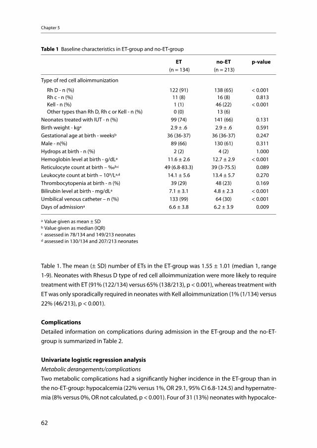
Chapter 5
62
Table 1. The mean (± SD) number of ETs in the ET-group was 1.55 ± 1.01 (median 1, range 1-9). Neonates with Rhesus D type of red cell alloimmunization were more likely to require treatment with ET (91% (122/134) versus 65% (138/213), p < 0.001), whereas treatment with ET was only sporadically required in neonates with Kell alloimmunization (1% (1/134) versus 22% (46/213), p < 0.001).
ComplicationsDetailed information on complications during admission in the ET-group and the no-ET-group is summarized in Table 2.
Univariate logistic regression analysisMetabolic derangements/complicationsTwo metabolic complications had a significantly higher incidence in the ET-group than in the no-ET-group: hypocalcemia (22% versus 1%, OR 29.1, 95% CI 6.8-124.5) and hypernatre-mia (8% versus 0%, OR not calculated, p < 0.001). Four of 31 (13%) neonates with hypocalce-
Table 1 Baseline characteristics in ET-group and no-ET-group
ET (n = 134)
no-ET (n = 213)
p-value
Type of red cell alloimmunization
Rh D - n (%) 122 (91) 138 (65) < 0.001 Rh c - n (%) 11 (8) 16 (8) 0.813 Kell - n (%) 1 (1) 46 (22) < 0.001 Other types than Rh D, Rh c or Kell - n (%) 0 (0) 13 (6)Neonates treated with IUT - n (%) 99 (74) 141 (66) 0.131Birth weight - kga 2.9 ± .6 2.9 ± .6 0.591Gestational age at birth - weeksb 36 (36-37) 36 (36-37) 0.247Male - n(%) 89 (66) 130 (61) 0.311Hydrops at birth - n (%) 2 (2) 4 (2) 1.000Hemoglobin level at birth - g/dLa 11.6 ± 2.6 12.7 ± 2.9 < 0.001Reticulocyte count at birth – ‰b,c 49 (6.8-83.3) 39 (3-75.5) 0.089Leukocyte count at birth – 109/La,d 14.1 ± 5.6 13.4 ± 5.7 0.270Thrombocytopenia at birth - n (%) 39 (29) 48 (23) 0.169Bilirubin level at birth - mg/dLa 7.1 ± 3.1 4.8 ± 2.3 < 0.001Umbilical venous catheter – n (%) 133 (99) 64 (30) < 0.001Days of admissiona 6.6 ± 3.8 6.2 ± 3.9 0.009
a Value given as mean ± SDb Value given as median (IQR)c assessed in 78/134 and 149/213 neonatesd assessed in 130/134 and 207/213 neonates
Neonatal morbidity after exchange transfusions
63
mia needed calcium replacement therapy and 3 of 11 (27%) needed treatment (additional sodium-free intravenous fluid) for hypernatremia. Severe symptoms of hypernatremia (sei-zures) did not occur.
Cardio-respiratory complicationsNo significant differences were seen between the two groups in respiratory support, apneas, cardiac arrest, and hypotension (Table 2). No cases of cardiac rhythm disorders, pulmonary hemorrhage and hypertension (requiring treatment) occurred.
Infectious complicationsProven sepsis occurred significantly more often in the ET-group than in the no-ET-group (8% versus 1%, OR 6.3, 95% CI 1.7-22.9). In the 14 neonates with proven sepsis, bacterial cultures were positive for Staphylococcus aureus in 7/14 (50%), coagulase-negative Staphylococcus in 3/14 (22%), beta-hemolytic Streptococcus in 1/14 (7%), Klebsiella pneumonia in 1/14 (7%), Escherichia coli in 1/14 (7%) and Bacillus cereus in 1/14 (7%). This last patient de veloped a Bacillus cereus sepsis with brain abscesses after an ET performed through an umbilical venous catheter. This exceptional case has previously been reported.23
The rate of leukocytopenia was significantly higher in the ET-group than in the no-ET-group (88% versus 23%, OR 24.7, 95% CI 13.4-45.5). All ET-treated neonates with proven sepsis had leukocytopenia during admission and in 55% (6/11) leukocytopenia occurred after ET.Umbilical venous catheterization was performed significantly more often in the ET-group than in the no-ET-group (99% versus 30%) (Table 1).
Hematological complicationsThe rate of thrombocytopenia (platelet count < 150×109/L) was significantly higher in the ET-group than in the no-ET-group (99% versus 32%, OR 143.8, 95% CI 34.6-598.6), and this was also true for severe thrombocytopenia and very severe thrombocytopenia (Table 2). Seventy-five of 134 ET-treated neonates (57%, 2 missing values) were treated with at least one platelet transfusion. One near-term neonate with Rhesus D alloimmunization born at 36 weeks’ gestation received a platelet transfusion after ET on day one because of a post-ET platelet count of 39×109/L. On day 2 a cranial ultrasound showed a hemorrhage in the right parieto-occipital periventricular white matter. This hemorrhage was not seen on antenatal ultrasounds. Magnetic resonance imaging showed no signs of sinus thrombosis and coa-gulation was normal. At one year of age, the infant had no neurologic sequelae on phy-sical examination. In the no-ET-group 3 neonates had signs of intracerebral hemorrhage on cranial ultrasound and/or MRI of whom one neonate had severe thrombocytopenia for which he received 4 platelet transfusions. This neonate died during admission (see below).
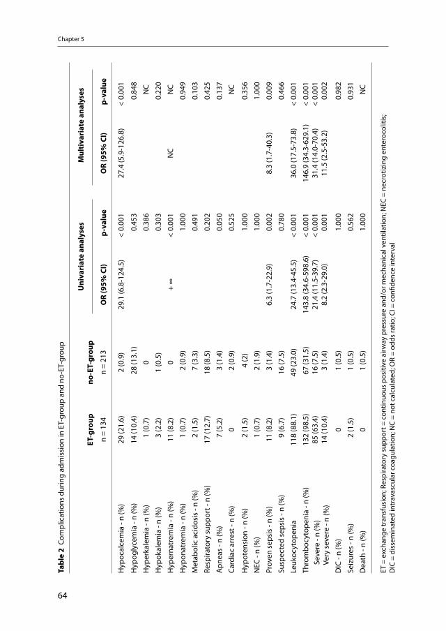
Chapter 5
64
Tabl
e 2
Com
plic
atio
ns d
urin
g ad
mis
sion
in E
T-gr
oup
and
no-E
T-gr
oup
ET-g
roup
n =
134
no-E
T-gr
oup
n =
213
Uni
vari
ate
anal
yses
Mul
tiva
riat
e an
alys
es
OR
(95%
CI)
p-va
lue
OR
(95%
CI)
p-va
lue
Hyp
ocal
cem
ia -
n (%
)29
(21.
6)2
(0.9
)29
.1 (6
.8-1
24.5
)<
0.00
127
.4 (5
.9-1
26.8
)<
0.00
1
Hyp
ogly
cem
ia -
n (%
)14
(10.
4) 2
8 (1
3.1)
0.
453
0.84
8
Hyp
erka
lem
ia -
n (%
) 1
(0.7
)0
0.38
6N
C
Hyp
okal
emia
- n
(%)
3 (2
.2)
1 (0
.5)
0.30
30.
220
Hyp
erna
trem
ia -
n (%
)11
(8.2
)0
+ ∞
< 0.
001
NC
NC
Hyp
onat
rem
ia -
n (%
) 1
(0.7
) 2
(0.9
)1.
000
0.94
9
Met
abol
ic a
cido
sis
- n (%
) 2
(1.5
) 7
(3.3
)0.
491
0.10
3
Resp
irato
ry s
uppo
rt -
n (%
) 17
(12.
7)18
(8.5
)0.
202
0.42
5
Apn
eas
- n (%
) 7
(5.2
) 3
(1.4
)0.
050
0.13
7
Card
iac
arre
st -
n (%
)0
2 (0
.9)
0.52
5N
C
Hyp
oten
sion
- n
(%)
2 (1
.5)
4 (2
)1.
000
0.35
6
NEC
- n
(%)
1 (0
.7)
2 (1
.9)
1.00
01.
000
Prov
en s
epsi
s - n
(%)
11 (8
.2)
3 (1
.4)
6.3
(1.7
-22.
9)0.
002
8.3
(1.7
-40.
3)0.
009
Susp
ecte
d se
psis
- n
(%)
9 (6
.7)
16 (7
.5)
0.78
00.
466
Leuk
ocyt
open
ia11
8 (8
8.1)
49 (2
3.0)
24.7
(13.
4-45
.5)
< 0.
001
36.0
(17.
5-73
.8)
< 0.
001
Thro
mbo
cyto
peni
a - n
(%)
S
ever
e - n
(%)
V
ery
seve
re -
n (%
)
132
(98.
5)85
(63.
4)14
(10.
4)
67
(31.
5)16
(7.5
) 3
(1.4
)
143.
8 (3
4.6-
598.
6)21
.4 (1
1.5-
39.7
)8.
2 (2
.3-2
9.0)
< 0.
001
< 0.
001
0.00
1
146.
9 (3
4.3-
629.
1)31
.4 (1
4.0-
70.4
)11
.5 (2
.5-5
3.2)
< 0.
001
< 0.
001
0.00
2
DIC
- n
(%)
0 1
(0.5
)1.
000
0.98
2
Seiz
ures
- n
(%)
2 (1
.5)
1 (0
.5)
0.56
20.
931
Dea
th -
n (%
)0
1 (0
.5)
1.00
0N
C
ET =
exc
hang
e tr
ansf
usio
n; R
espi
rato
ry s
uppo
rt =
con
tinuo
us p
ositi
ve a
irway
pre
ssur
e an
d/or
mec
hani
cal v
entil
atio
n; N
EC =
nec
rotiz
ing
ente
roco
litis
; D
IC =
dis
sem
inat
ed in
trav
ascu
lar c
oagu
latio
n; N
C =
not c
alcu
late
d; O
R =
odds
ratio
; CI =
con
fiden
ce in
terv
al
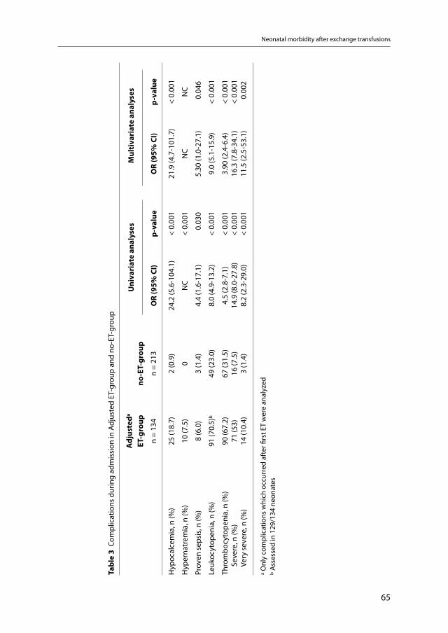
Neonatal morbidity after exchange transfusions
65
Tabl
e 3
Com
plic
atio
ns d
urin
g ad
mis
sion
in A
djus
ted
ET-g
roup
and
no-
ET-g
roup
Adj
uste
da
ET-g
roup
n =
134
no-E
T-gr
oup
n =
213
Uni
vari
ate
anal
yses
Mul
tiva
riat
e an
alys
es
OR
(95%
CI)
p-va
lue
OR
(95%
CI)
p-va
lue
Hyp
ocal
cem
ia, n
(%)
25 (1
8.7)
2 (0
.9)
24.2
(5.6
-104
.1)
< 0.
001
21.9
(4.7
-101
.7)
< 0.
001
Hyp
erna
trem
ia, n
(%)
10 (7
.5)
0N
C<
0.00
1N
CN
C
Prov
en s
epsi
s, n
(%)
8 (6
.0)
3 (1
.4)
4.4
(1.6
-17.
1)
0.03
05.
30 (1
.0-2
7.1)
0.0
46
Leuk
ocyt
open
ia, n
(%)
91 (7
0.5)
b49
(23.
0)8.
0 (4
.9-1
3.2)
< 0.
001
9.0
(5.1
-15.
9)<
0.00
1
Thro
mbo
cyto
peni
a, n
(%)
S
ever
e, n
(%)
V
ery
seve
re, n
(%)
90 (6
7.2)
71 (5
3)14
(10.
4)
67 (3
1.5)
16 (7
.5)
3 (1
.4)
4.5
(2.8
-7.1
)14
.9 (8
.0-2
7.8)
8.2
(2.3
-29.
0)
< 0.
001
< 0.
001
< 0.
001
3.90
(2.4
-6.4
)16
.3 (7
.8-3
4.1)
11.5
(2.5
-53.
1)
< 0.
001
< 0.
001
0.0
02
a Onl
y co
mpl
icat
ions
whi
ch o
ccur
red
afte
r firs
t ET
wer
e an
alyz
edb A
sses
sed
in 1
29/1
34 n
eona
tes
Chapter 5
66
Neonatal mortalityOnly one of the 347 neonates died during admission. This neonate from the no-ET-group, born at 30 weeks’ gestation, suffered from severe fetal hydrops and died on day 11 due to multiple organ failure including respiratory failure due to respiratory distress syndrome and severe pulmonary hypertension, bilateral intraventricular hemorrhage grade 2 and renal failure.
ET guideline changeTo measure the possible effect of ET guideline change12 on the rate of the previously men-tioned complications (Table 2) we performed a sub-analysis of ET treated neonates before and after guideline change. The rates of complications were not significantly different between both groups (data not shown).
Multivariate logistic regression analysisComplications during entire admission On multivariate logistic regression analysis we corrected for the following covariates: hemo-globin level at birth, type of red cell alloimmunization, days of admission, and gestational age at birth. We corrected for the first two because of the significant differences at baseline (Table 1) and for the latter because preterm neonates are more susceptible for ET-related complications.10,24 Since the presence of an umbilical venous catheter and ET are correlated (Spearman correlation coefficient r = 0.680, p = <0.001), we did not correct for the presence of an umbilical venous catheter. Multivariate regression analysis demonstrated that four complications, i.e. proven sepsis (OR 8.3, 95% CI 1.7-40.3), severe thrombocytopenia (OR 31.4, 95% CI 14.0-70.4), leukocytopenia (OR 36.0, 95% CI 17.5-73.8) and hypocalcemia (OR 27.4, 95% CI 5.9-126.8), had a higher incidence in the ET-group than in the no-ET-group (Table 2). Because none of the neonates in the no-ET-group had hypernatremia, additional multivariate logistic regression analyses could not be performed.
Complications after first ET In Table 2 all complications during entire admission in both groups are reported. Since our main interest is in ET-related complications, we performed a sub-analysis excluding all com-plications in the ET-group that were observed before (first) ET (adjusted-ET-group). Results are shown in Table 3.
Neonatal morbidity after exchange transfusions
67
Discussion
This study demonstrates that treatment with ET in neonates with HDN is associated with an increased risk of sepsis, thrombocytopenia, leukocytopenia, hypocalcemia and hypernatre-mia. Treatment with ET was not associated with neonatal death in our cohort.
In the last three decades several studies have been published on neonatal morbidity due to treatment with ET.4-11 One of the known risk factors associated with ET is development of invasive bacterial infections. The reported incidence of ET-related sepsis ranges from 0% to 11%.6,8,10 The incidence of proven sepsis detected in this study (8%) is in accordance with these previous reports. We found that the independent risk of sepsis was more than eight times higher in the ET-group than in the no-ET-group. The exact cause of the increased risk of infection is not fully understood, but is most probably related to the use of umbilical lines for ET. Umbilical catheters are a well known risk factor for nosocomial infection.25 Sepsis in the ET-group may also be caused by administration of infected blood products. Although the current risk of transmission of infectious diseases is relatively low, it is not completely negligible.7,26 Furthermore, ET-related wash out of leukocytes may also play a role in the higher incidence of sepsis in the ET-group, since in double volume ET more than 90% of cir-culating blood is replaced by leukocyte-depleted donor-blood.19,26-28 Finally, limited data on leukocytopenia in neonates with HDN due to red cell alloimmunization show that the risk of leukocytopenia is increased in severe Rhesus HDN.29-32 In this study 86% (12/14) of neonates with proven sepsis had leukocytopenia of whom 11 were treated with ET. In 55% (6/11) leu-kocytopenia occurred after treatment with ET. In the remaining 45% leukocytopenia might be caused by bone marrow suppression due to increased erythropoiesis.
Another reported risk associated with ET is thrombocytopenia due to the use of a blood product which consists of thrombocyte-free erythrocytes.18,19,33 Previous studies reported incidences of ET-related thrombocytopenia ranging from 6% to 44%.4-6,8,10,11 In our study, the incidence of thrombocytopenia (< 150×109/L) in the ET-group was much higher, almost all neonates (99%) had low platelet counts and 63% had severe thrombocytopenia (plate-lets < 50×109/L). The differences in incidence can be explained by several factors including methodological differences and differences in study population. We only included neo-nates with red cell alloimmune HDN which is known to be associated with an increased risk for thrombocytopenia, even without ET.16,34-36 Because other studies included neo-nates with ABO-incompatibility, their incidence of thrombocytopenia should be lower. In this study, the risk of severe thrombocytopenia is 21-fold higher in the group treated with ET. Neonatologists must be aware of this potentially devastating complication as massive hemorrhage may arise after ET. Complications can be prevented by prophylactic platelet
Chapter 5
68
transfusion before, during or after ET in case of low platelet counts. In our study, platelet transfusions were administered in 57% of neonates before, during or after ET.
A third known complication of ET is hypocalcemia, resulting from the use of citrated blood which contains almost no free calcium.18,19,33,37 Previous studies reported incidences of hypocalcemia ranging from 3% to 42%.4,8,10,11 In our study, 22% of neonates in the ET-group had a serum calcium < 8 mg/dL, and 13% of all hypocalcemic infants needed replacement therapy. We found that treatment with ET was independently associated with an almost 30-fold increased risk of hypocalcemia. If left untreated, hypocalcemia can lead to poten-tially devastating complications such as seizures and cardiac arrhythmias. It is therefore crucial to measure calcium levels during the ET-procedure and act accordingly.
Another metabolic complication which appears to be related to ET is hypernatremia. Hypernatremia probably results from an increased level of sodium in citrated blood (in our center 168 mEq/L). Only few studies have reported hypernatremia resulting from ET, and the exact incidence is unknown.38,39 Because hypernatremia can lead to serious complica-tions, we recommend frequent measurements of serum sodium levels during and after ET.
Finally, previous studies have reported that ET may also lead to neonatal death. Neonatal mortality attributable to ET ranges between 0.5% and 2%.4,8-10 In accordance with our find-ings, three recent studies reported no neonatal deaths after ET.5,6,11 However, all these studies (as well as ours) were not powered to detect a difference in neonatal mortality. Differences between the reported rates can be explained by methodological differences such as dif-ferent sizes of the study cohorts and differences in disease-severity between the cohorts. Neonates included in previous studies were often more premature than the near term-age population in our cohort.9-11 Another explanation could be that our center is the national referral center for intrauterine treatment of red cell alloimmunization. Consequently nearly all severely affected neonates with HDN due to red cell alloimmunization are born and treated in our center. As a result, ET is a frequently performed and standardized procedure in our unit and part of routine practice. We speculate that this may have contributed to the low level of severe morbidity or mortality in the ET-group.
To our knowledge this is the first large study comparing neonatal complications in a group of infants with red cell alloimmunization treated with ET and a control-group without ET. Nevertheless, the results of this study should be interpreted with care because of the retro-spective study design.
Neonatal morbidity after exchange transfusions
69
In conclusion, sepsis, leukocytopenia, thrombocytopenia, hypernatremia and hypocalce-mia are common complications in neonates with HDN due to red cell alloimmunization treated with ET. In experienced hands severe permanent morbidity and mortality rates due to ET-procedures can be reduced to a minimum.
References
1. Diamond LK, Allen FH, Jr., Thomas WO, Jr. Erythroblastosis fetalis. VII. Treatment with exchange trans-fusion. N Engl J Med . 1951;244:39-49.
2. Wallerstein H. Treatment of severe erythroblastosis by simultaneous removal and replacement of the blood of the newborn infant. Science . 1946;103:583-584.
3. Wiener AS, Wexler IB, Grundfast TH. Therapy of erythroblastosis fetalis with exchange transfusion. Bull N Y Acad Med . 1947;23:207-220.
4. Badiee Z. Exchange transfusion in neonatal hyperbilirubinaemia: experience in Isfahan, Iran. Singa-pore Med J . 2007;48:421-423.
5. Davutoglu M, Garipardic M, Guler E, Karabiber H, Erhan D. The etiology of severe neonatal hyperbili-rubinemia and complications of exchange transfusion. Turk J Pediatr . 2010;52:163-166.
6. Hosseinpour SS, Gharehbaghi MM. Exchange transfusion in severe hyperbilirubinemia: an experience in northwest Iran. Turk J Pediatr . 2010;52:367-371.
7. Ip S, Chung M, Kulig J, O’Brien R, Sege R, Glicken S, et al. An evidence-based review of important issues concerning neonatal hyperbilirubinemia. Pediatrics . 2004;114:e130-e153.
8. Jackson JC. Adverse events associated with exchange transfusion in healthy and ill newborns. Pedia-trics . 1997;99:E7.
9. Keenan WJ, Novak KK, Sutherland JM, Bryla DA, Fetterly KL. Morbidity and mortality associated with exchange transfusion. Pediatrics . 1985;75:417-421.
10. Patra K, Storfer-Isser A, Siner B, Moore J, Hack M. Adverse events associated with neonatal exchange transfusion in the 1990s. J Pediatr . 2004;144:626-631.
11. Steiner LA, Bizzarro MJ, Ehrenkranz RA, Gallagher PG. A decline in the frequency of neonatal exchange transfusions and its effect on exchange-related morbidity and mortality. Pediatrics . 2007;120:27-32.
12. Rath ME, Smits-Wintjens VE, Lindenburg I, Brand A, Oepkes D, Walther FJ, et al. Top-up transfusions in neonates with Rh hemolytic disease in relation to exchange transfusions. Vox Sang . 2010;99:65-70.
13. Rath ME, Smits-Wintjens VE, Lindenburg IT, Brand A, Van Kamp IL, Oepkes D, et al. Exchange transfusi-ons and top-up transfusions in neonates with Kell haemolytic disease compared to Rh D haemolytic disease. Vox Sang . 2011;100:312-316.
14. De Boer I, Zeestraten EC, Lopriore E, Van K, I, Kanhai HH, Walther FJ. Pediatric outcome in Rhesus hemo-lytic disease treated with and without intrauterine transfusion. Am J Obstet Gynecol . 2008;198:54e1-e4.
15. Smits-Wintjens VE, Walther FJ, Rath ME, Lindenburg IT, te Pas AB, Kramer CM, et al. Intravenous im-munoglobulin in neonates with rhesus hemolytic disease: a randomized controlled trial. Pediatrics . 2011;127:680-686.
16. Rath ME, Smits-Wintjens VE, Oepkes D, van Zwet EW, Van Kamp IL, Brand A, et al. Thrombocytopenia at birth in neonates with red cell alloimmune haemolytic disease. Vox Sang . 2011;doi: 10.1111/j.1423-0410.2011.01539.x.
17. American Academy of Pediatrics Subcommittee on Hyperbilirubinemia. Management of hyperbiliru-binemia in the newborn infant 35 or more weeks of gestation. Pediatrics . 2004;114:297-316.
18. Petaja J, Johansson C, Andersson S, Heikinheimo M. Neonatal exchange transfusion with heparinised whole blood or citrated composite blood: a prospective study. Eur J Pediatr . 2000;159:552-553.
Chapter 5
70
19. Gharehbaghi MM, Hosseinpour SS. Exchange transfusion in neonatal hyperbilirubinaemia: a compa-rison between citrated whole blood and reconstituted blood. Singapore Med J . 2010;51:641-644.
20. Bell MJ, Ternberg JL, Feigin RD, Keating JP, Marshall R, Barton L, et al. Neonatal necrotizing enterocoli-tis. Therapeutic decisions based upon clinical staging. Ann Surg . 1978;187:1-7.
21. Roberts IAG. Haematological values in the newborn (appendix 1). In: Rennie JM, editor. Roberton’s Textbook of Neonatology. 4 ed. Philadelphia: Elsevier; 2011. 1287.
22. Volpe JJ. Intracranial hemorrhage: germinal matrix-intraventricular hemorrhage of the premature in-fant. In: Volpe JJ, editor. Neurology of the newborn. 4th ed. Philadelphia: Saunders; 2001. 428-493.
23. Smits-Wintjens VE, Steggerda SJ, Oepkes D, Van Kamp IL, Kramer CM, Walther FJ, et al. Bacillus cereus cerebral abscesses in a term neonate with rhesus hemolytic disease treated with exchange transfu-sion. J Pediatr Inf Dis . 2010;5:277-280.
24. Maisels MJ, Watchko JF. Treatment of jaundice in low birthweight infants. Arch Dis Child Fetal Neonatal Ed . 2003;88:F459-F463.
25. Inglis GD, Davies MW. Prophylactic antibiotics to reduce morbidity and mortality in neonates with umbilical venous catheters. Cochrane Database Syst Rev . 2005;CD005251.
26. Fergusson D, Hebert PC, Barrington KJ, Shapiro SH. Effectiveness of WBC reduction in neonates: what is the evidence of benefit? Transfusion . 2002;42:159-165.
27. Liem RI, O’Gorman MR, Brown DL. Effect of red cell exchange transfusion on plasma levels of inflam-matory mediators in sickle cell patients with acute chest syndrome. Am J Hematol . 2004;76:19-25.
28. Xanthou M, Nicolopoulos D, Gizas A, Matsaniotis N. The response of leukocytes in the peripheral blood during and following exchange transfusion in the newborn. Pediatrics . 1973;51:570-574.
29. Rath ME, Smits-Wintjens VE, Walther FJ, Lopriore E. Hematological morbidity and management in neonates with hemolytic disease due to red cell alloimmunization. Early Hum Dev . 2011;87:583-588.
30. Koenig JM, Christensen RD. Neutropenia and thrombocytopenia in infants with Rh hemolytic disease. J Pediatr . 1989;114:625-631.
31. Segal N, Leibovitz E, Juster-Reicher A, Even-Tov S, Mogilner B, Barak Y. Neutropenia complicating Rh-hydrops fetalis: the effect of treatment with recombinant human granulocyte colony-stimulating fac-tor (rhG-CSF). Pediatr Hematol Oncol . 1998;15:193-197.
32. Blanco E, Johnston DL. Neutropenia in infants with hemolytic disease of the newborn. Pediatr Blood Cancer . 2011;doi: 10.1002/pbc.23233.
33. Samsom JF, Groenendijk MG, van der Lei J, Okken A. Exchange transfusion in the neonate, a compari-son between citrate-, heparinized- and reconstituted whole blood. Eur J Haematol . 1991;47:153-154.
34. Saade GR, Moise KJ, Jr., Copel JA, Belfort MA, Carpenter RJ, Jr. Fetal platelet counts correlate with the severity of the anemia in red-cell alloimmunization. Obstet Gynecol . 1993;82:987-991.
35. Van den Akker ES, de Haan TR, Lopriore E, Brand A, Kanhai HH, Oepkes D. Severe fetal thrombocytope-nia in Rhesus D alloimmunized pregnancies. Am J Obstet Gynecol . 2008;199:387e1-e4.
36. Van den Akker ES, Klumper FJ, Brand A, Kanhai HH, Oepkes D. Kell alloimmunization in pregnancy: associated with fetal thrombocytopenia? Vox Sang . 2008;95:66-69.
37. Maisels MJ, Li TK, Piechocki JT, Werthman MW. The effect of exchange transfusion on serum ionized calcium. Pediatrics . 1974;53:683-686.
38. Doyle PE, Eidelman AI, Lee K, Daum C, Gartner LM. Exchange transfusion and hypernatremia: possible role in intracranial hemorrhage in very-low-birth-weight infants. J Pediatr . 1978;92:848-849.
39. Steele AM, Brown DL, Lipsitz PJ. Relationship of exchange transfusion to hypernatremia. J Pediatr . 1979;94:168-169.

6 Cholestasis in neonates with red cell alloimmune
hemolytic disease: incidence, risk factors and outcome
Vivianne EHJ Smits-WintjensMirjam EA Rath
Irene TM LindenburgDick Oepkes
Erik W van ZwetFrans J WaltherEnrico Lopriore
Neonatology; In press
Chapter 6
72
Abstract
Background: Etiology of cholestatic liver disease in neonates with hemolytic disease of the newborn (HDN) has been associated with iron overload due to intrauterine red cell transfu-sions (IUTs). Data on the incidence and severity of cholestasis in neonates with HDN is scarce, and little is known about pathogenesis, risk factors, neonatal management and outcome.
Objective: To evaluate incidence, risk factors, management and outcome of cholestasis in neonates with red cell alloimmune hemolytic disease.
Methods: All (near-) term neonates with HDN due to red cell alloimmunization admitted to our center between January 2000 and July 2010 were included in this observational study. Liver function tests (including conjugated bilirubin) were routinely performed in the neo-natal period. We recorded the presence of cholestasis, investigated several potential risk factors and evaluated the management and outcome in affected neonates.
Results: A total of 313 infants with red cell alloimmune hemolytic disease treated with or without IUTs were included. The incidence of cholestasis was 13% (41/313). Two risk fac-tors were independently associated with cholestasis: treatment with at least one IUT (OR 5.81, 95% CI 1.70-19.80, p=0.005) and Rhesus D type of alloimmunization (OR 4.66, 95% CI 1.05-20.57, p=0.042). Additional diagnostic tests to investigate possible causes of cholesta-sis were all negative. In five infants (12%) supportive medical and nutritional therapy was started and one neonate required iron chelation therapy.
Conclusion: Cholestasis occurs in 13% of neonates with HDN due to red cell alloimmuniza-tion and is independently associated with IUT treatment and Rhesus D type of alloimmu-nization.
Cholestasis in red cell alloimmune HDN
73
Introduction
Hemolytic disease of the newborn (HDN) due to red cell alloimmunization may lead to excessive unconjugated hyperbilirubinemia, anemia and iron overload.1 A few studies have reported an association between HDN and the development of conjugated hyperbilirubine-mia, i.e. cholestasis.2-5 Some of these studies (mostly case reports) describe that cholestasis in neonates with HDN is uncommon and usually mild and transient.3-5 Other reports how-ever detail severe and protracted courses of cholestasis.2,4,6,7 The etiology of cholestatic liver disease in neonates with HDN has been associated with iron overload due to intrauterine transfusions (IUTs).6,8-10 Data on the incidence and severity of cholestasis in neonates with red cell alloimmune hemolytic disease is scarce, and little is known about pathogenesis, risk factors, neonatal management and outcome.
The aim of this study was to evaluate incidence, potential risk factors, management and out-come of cholestasis in a large series of neonates with HDN due to red cell alloimmunization.
Methods
All consecutive cases of (near-) term neonates (≥35 weeks of gestation) with HDN due to maternal red cell alloimmunization admitted to our center between January 2000 and July 2010 were included in this retrospective study. Neonatal outcome in part of this group was described in previous studies.11-14 The Leiden University Medical Center (LUMC) is the national referral center for the management and intrauterine treatment of red cell allo-immunization in the Netherlands. We excluded all preterm neonates (<35 weeks of gesta-tion) and neonates in whom conjugated bilirubin tests were not performed. The guidelines for the management of neonates with HDN admitted to our nursery (including intensive phototherapy and exchange transfusion (ET)) have previously been described.12,13
In addition to frequent total bilirubin levels, extensive diagnostic evaluations are routinely performed at birth and during the first week of life in infants with red cell alloimmune HDN admitted to our neonatal nursery. These evaluations include hematologic tests (complete blood counts), liver function tests (liver enzymes and total and conjugated bilirubin) and blood group, Coombs and irregular antibody tests.
Primary outcome of this study was the incidence of cholestatic icterus in neonates with HDN due to red cell alloimmunization. Secondary outcomes were management and out-come of cholestasis.
Chapter 6
74
Cholestasis or conjugated hyperbilirubinemia was defined as a conjugated serum bilirubin level above 1.0 mg/dL if total serum bilirubin level is less than 5 mg/dL, or a value of conju-gated bilirubin that represents more than 20% of total bilirubin if the total bilirubin level is greater than 5 mg/dL.15 Severe cholestasis was defined as a conjugated bilirubin level >50% of the total serum bilirubin concentration.
In neonates with cholestasis we recorded the following data: symptoms of cholestasis (such as discolored stools and dark urine), duration of conjugated hyperbilirubinemia, (type of ) therapy (including Ursodeoxycholic acid (15 mg/kg/d), Vitamin A (2500-5000 IU/d, in pre-matures 1000-1500 IU/kg/d), vitamin D (800 IU/d, in prematures 400 IU/d), vitamin E (5-10 mg/kg/d, in prematures 10-20 mg/kg/d), vitamin K (1 mg/d, birth weight < 1500 grams: 0.5 mg/d) and formula with medium chain triglycerides) and investigations performed to establish a specific cause for the cholestatic icterus. Possible causes for neonatal cholestasis are (1) infections (sepsis, urinary tract infection, toxoplasmosis, rubella, cytomegalo virus, human herpes virus 6, syphilis, parvovirus B19, echovirus, adenovirus, coxsackie virus, hep-atitis B and C); (2) bile duct anomalies, including biliary atresia and choledochal cyst; (3) inborn errors of metabolism, including alpha-1-antitrypsin deficiency, galactosemia, cystic fibrosis, tyrosinemia and progressive familial intrahepatic cholestasis, and (4) endocrinopa-thies (hypothyroidism and hypopituitarism).
We recorded the following obstetric and neonatal data: type of red cell alloimmunization, number of IUTs, gestational age at birth, birth weight, total bilirubin level and conjugated bilirubin level at birth, maximum total bilirubin level and maximum conjugated bilirubin level during admission, time until cholestasis disappeared (within 1 week, between 1 week and 1 month or after 1 month), maximum ferritin level during admission, duration of photo-therapy, number of ETs required and number of top up red blood cell transfusions received during the first 3 months of life.
Data are reported as means and standard deviations (SD) or as medians and ranges. Sta-tistical analysis was performed using Student-t test and Mann-Whitney test for continuous variables. Chi-square and Fisher’s-exact test were used for categorical variables. To assess the relationship between ferritin level and treatment with IUT a Spearman correlation was calculated. A p-value <0.05 was considered to indicate statistical significance. All predicting risk factors for cholestasis identified with univariate analysis were included in a multivariate logistic regression model to measure independent effects. The results of the logistic models were expressed as odds ratios (OR). Statistical analysis was performed using SPSS 17.0 (SPSS Inc., Chicago, IL, USA).
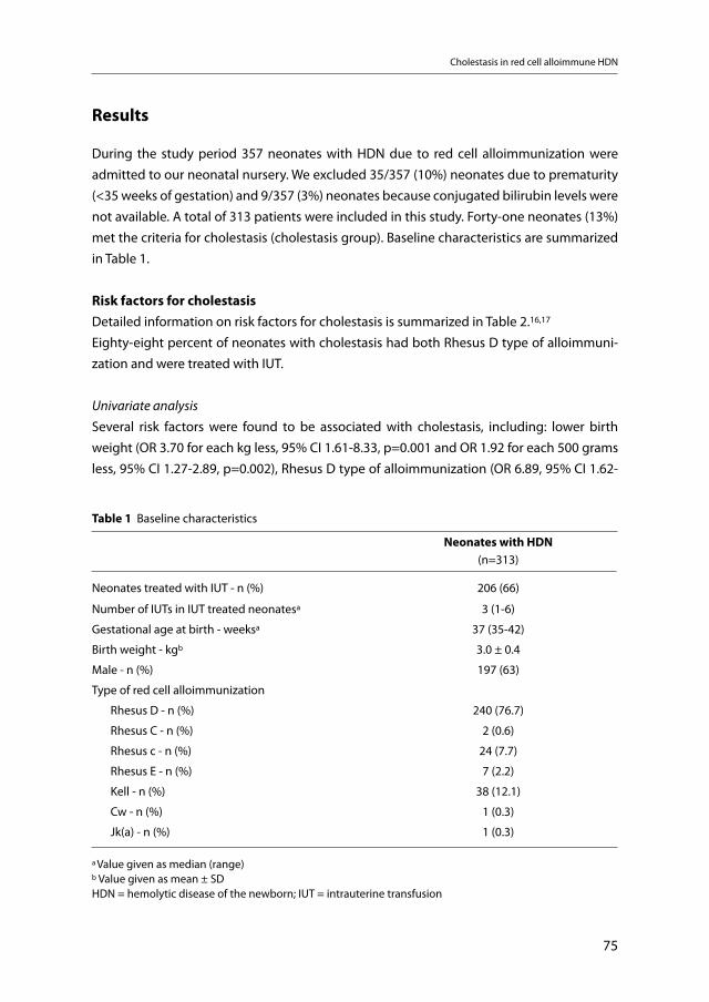
Cholestasis in red cell alloimmune HDN
75
Results
During the study period 357 neonates with HDN due to red cell alloimmunization were admitted to our neonatal nursery. We excluded 35/357 (10%) neonates due to prematurity (<35 weeks of gestation) and 9/357 (3%) neonates because conjugated bilirubin levels were not available. A total of 313 patients were included in this study. Forty-one neonates (13%) met the criteria for cholestasis (cholestasis group). Baseline characteristics are summarized in Table 1.
Risk factors for cholestasisDetailed information on risk factors for cholestasis is summarized in Table 2.16,17
Eighty-eight percent of neonates with cholestasis had both Rhesus D type of alloimmuni-zation and were treated with IUT.
Univariate analysisSeveral risk factors were found to be associated with cholestasis, including: lower birth weight (OR 3.70 for each kg less, 95% CI 1.61-8.33, p=0.001 and OR 1.92 for each 500 grams less, 95% CI 1.27-2.89, p=0.002), Rhesus D type of alloimmunization (OR 6.89, 95% CI 1.62-
Table 1 Baseline characteristics
Neonates with HDN (n=313)
Neonates treated with IUT - n (%) 206 (66)
Number of IUTs in IUT treated neonatesa 3 (1-6)
Gestational age at birth - weeksa 37 (35-42)
Birth weight - kgb 3.0 ± 0.4
Male - n (%) 197 (63)
Type of red cell alloimmunization
Rhesus D - n (%) 240 (76.7)
Rhesus C - n (%) 2 (0.6)
Rhesus c - n (%) 24 (7.7)
Rhesus E - n (%) 7 (2.2)
Kell - n (%) 38 (12.1)
Cw - n (%) 1 (0.3)
Jk(a) - n (%) 1 (0.3)
a Value given as median (range)b Value given as mean ± SDHDN = hemolytic disease of the newborn; IUT = intrauterine transfusion
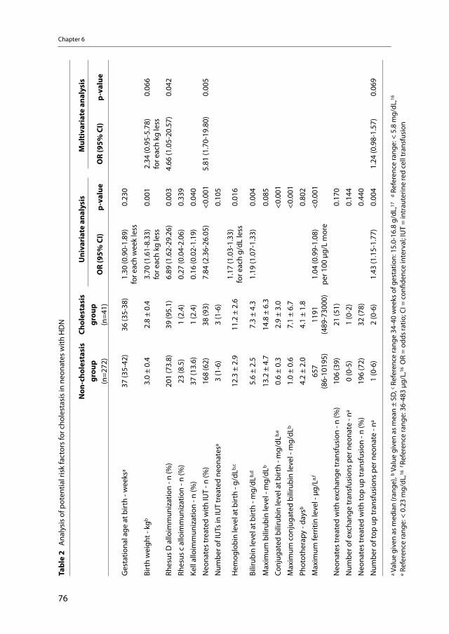
Chapter 6
76
Tabl
e 2
Ana
lysi
s of
pot
entia
l ris
k fa
ctor
s fo
r cho
lest
asis
in n
eona
tes
with
HD
N
Non
-cho
lest
asis
gr
oup
(n=2
72)
Chol
esta
sis
grou
p(n
=41)
Uni
vari
ate
anal
ysis
Mul
tiva
riat
e an
alys
is
OR
(95%
CI)
p-va
lue
OR
(95%
CI)
p-va
lue
Ges
tatio
nal a
ge a
t birt
h - w
eeks
a37
(35-
42)
36 (3
5-38
)1.
30 (0
.90-
1.89
)fo
r eac
h w
eek
less
0.23
0
Birt
h w
eigh
t - k
gb3.
0 ±
0.4
2.8
± 0.
43.
70 (1
.61-
8.33
)fo
r eac
h kg
less
0.00
12.
34 (0
.95-
5.78
)fo
r eac
h kg
less
0.06
6
Rhes
us D
allo
imm
uniz
atio
n - n
(%)
201
(73.
8)39
(95.
1) 6
.89
(1.6
2-29
.26)
0.00
34.
66 (1
.05-
20.5
7)0.
042
Rhes
us c
allo
imm
uniz
atio
n - n
(%)
23 (8
.5)
1 (2
.4)
0.27
(0.0
4-2.
06)
0.33
9
Kell
allo
imm
uniz
atio
n - n
(%)
37 (1
3.6)
1 (2
.4)
0.16
(0.0
2-1.
19)
0.04
0
Neo
nate
s tr
eate
d w
ith IU
T - n
(%)
168
(62)
38 (9
3) 7
.84
(2.3
6-26
.05)
<0.0
015.
81 (1
.70-
19.8
0)0.
005
Num
ber o
f IU
Ts in
IUT
trea
ted
neon
ates
a3
(1-6
)3
(1-6
)0.
105
Hem
oglo
bin
leve
l at b
irth
- g/d
Lb,c
12.3
± 2
.911
.2 ±
2.6
1.17
(1.0
3-1.
33)
for e
ach
g/dL
less
0.01
6
Bilir
ubin
leve
l at b
irth
- mg/
dLb,
d5.
6 ±
2.5
7.3
± 4.
31.
19 (1
.07-
1.33
)0.
004
Max
imum
bili
rubi
n le
vel -
mg/
dLb
13.2
± 4
.714
.8 ±
6.3
0.08
5
Conj
ugat
ed b
iliru
bin
leve
l at b
irth
- mg/
dLb,
e0.
6 ±
0.3
2.9
± 3.
0<0
.001
Max
imum
con
juga
ted
bilir
ubin
leve
l - m
g/dL
b1.
0 ±
0.6
7.1
± 6.
7<0
.001
Phot
othe
rapy
- da
ysb
4.2
± 2.
04.
1 ±
1.8
0.80
2
Max
imum
ferr
itin
leve
l - μ
g/La,
f65
7(8
6-10
195)
1191
(489
-730
00)
1.04
(0.9
9-1.
08)
per 1
00 μ
g/L
mor
e<0
.001
Neo
nate
s tr
eate
d w
ith e
xcha
nge
tran
sfus
ion
- n (%
)10
6 (3
9)21
(51)
0.17
0
Num
ber o
f exc
hang
e tr
ansf
usio
ns p
er n
eona
te -
na0
(0-5
)1
(0-2
)0.
144
Neo
nate
s tr
eate
d w
ith to
p up
tran
sfus
ion
- n (%
)19
6 (7
2)32
(78)
0.44
0
Num
ber o
f top
up
tran
sfus
ions
per
neo
nate
- na
1 (0
-6)
2 (0
-6)
1.43
(1.1
5-1.
77)
0.00
41.
24 (0
.98-
1.57
)0.
069
a Val
ue g
iven
as
med
ian
(ran
ge),
b Val
ue g
iven
as
mea
n ±
SD, c R
efer
ence
rang
e 34
-40
wee
ks o
f ges
tatio
n: 1
5.0-
16.8
g/d
L,17
d R
efer
ence
rang
e: <
5.8
mg/
dL,16
e Ref
eren
ce ra
nge:
< 0
.23
mg/
dL,16
f Ref
eren
ce ra
nge:
36-
483
μg/L
,16 O
R =
odds
ratio
; CI =
con
fiden
ce in
terv
al; I
UT
= in
trau
terin
e re
d ce
ll tr
ansf
usio
n
Cholestasis in red cell alloimmune HDN
77
29.26, p=0.009), treatment with IUT (OR 7.84, 95% CI 2.36-26.05, p<0.001), total serum bili-rubin level at birth (OR 1.19, 95% CI 1.07-1.33, p=0.004), maximum ferritin level (OR 1.04 per 100 μg/L more, 95% CI 0.99-1.08, p<0.001) and number of top up transfusions (OR 1.43, 95% CI 1.15-1.77, p=0.004).
Multivariate analysisOn multivariate analysis, the following risk factors were independently associated with cholestasis: Rhesus D type of alloimmunization (OR 4.66, 95% CI 1.05-20.57, p=0.042) and treatment with IUT (OR 5.81, 95% CI 1.70-19.80, p=0.005).
Because a higher total bilirubin level at birth is part of the definition of cholestasis and thus closely related to a higher conjugated bilirubin level, total bilirubin level at birth was excluded from multivariate analysis. As ferritin levels were determined only in 89/313 (28%) neonates and ferritin level and treatment with IUT were positively correlated (r=0.565, p=<0.001), ferritin was not included in the multivariate analysis.
Clinical characteristics and outcome of cholestasisIn the cholestasis group 11/41 infants (27%) had severe cholestasis with a conjugated bili-rubin level >50% of the total serum bilirubin concentration. Four neonates (10%) had symp-toms of cholestasis such as discolored stools or dark urine. In 15% (6/41) the cholestasis dis-appeared spontaneously within 1 week, in 15% (6/41) between 1 week and 1 month and in 15% (6/41) within 1 to 3 months. In the remaining 56% (23/41) of infants the time of disap-pearance of cholestasis is not clear due to incomplete follow up. However, only 9% (2/23) of neonates with incomplete follow up had severe cholestasis. In five infants (12%) supportive medical and nutritional therapy was started (Ursodeoxycholic acid, Vitamin A, D, E, K and/or formula with medium chain triglycerides). In one infant Ursodeoxycholic acid was given for a period of 18 days, in the remaining 4 infants duration of therapy is not known since they were transferred to other hospitals while they were still on medication.
One patient with Rhesus D alloimmunization, who received 6 IUTs, developed severe cholestasis (maximum bilirubin level 41.3 mg/dL and maximum conjugated bilirubin level 35.1 mg/dL) and severe hyperferritinemia (maximum serum ferritin level 73000 μg/L). Iron chelation therapy with desferrioxamine was started and continued for one month to reduce the serum ferritin concentration and liver iron contents. After having excluded other causes of cholestasis, the most probable explanation for the cholestasis in this case was hyperfer-ritinemia with iron overload in the liver, due to multiple IUTs.
Chapter 6
78
Additional investigations in cholestasis groupIn the cholestasis group, laboratory investigations to evaluate possible liver injury were per-formed in 36/41 infants (88%). Elevated levels for alkaline phosphatase were detected in 6 (15%), for aspartate aminotransferase (AST) in 17 (41%), for alanine transferase (ALT) in 13 (32%) and for gamma-glutamyl transpeptidase (γGT) in 8 infants (20%).16
In 18/41 (44%) neonates in the cholestasis group additional tests were performed to inves-tigate possible causes of cholestasis. Sixteen infants (39%) were screened for infection. In all of them bacterial cultures of blood and urine were negative and there were no proven infections with toxoplasmosis, rubella, cytomegalovirus, human herpes virus 6, syphilis, par-vovirus B19, echovirus, adenovirus, coxsackie virus and hepatitis B and C. Additional tests to exclude endocrinologic or metabolic disorders were performed in 9/41 (22%) and 7/41 (17%) of infants, respectively. In none of these infants an endocrinopathy and/or an inborn error of metabolism was diagnosed. In 12/41 (29%) neonates an abdominal ultrasound was performed to exclude impairments in bile flow. All infants had normal ultrasound findings.
Discussion
This study shows that cholestasis is a common problem in HDN, occurring in 13% of neo-nates. Cholestasis is found particularly in neonates with Rhesus D alloimmunization treated with IUTs. Although cholestasis was mild and transient in most cases, a few neonates had severe cholestatic liver disease with protracted course and required intensive treatment and in one case chelation therapy was needed.
In the past, several studies have been published on the co-occurrence of cholestasis in neonates with HDN due to red cell alloimmunization. In 1963, Dunn described a large case series of 133 infants with Rhesus HDN and found that 8% of these patients developed ‘obstructive jaundice’ defined as conjugated bilirubin level > 3 mg/dL.3,4 However, in addi-tion to the more stringent definition, their study is not fully comparable with contemporary care strategies for Rhesus HDN. In 1963 perinatal mortality and morbidity were far higher than nowadays, due to the absence of Rh D prophylaxis, Doppler ultrasound to detect fetal anemia and in particular treatment with IUTs. Later, Bowman et al., Perez et al. and Allgood et al. also published on cholestasis in neonatal HDN, but none of these studies reported an exact incidence of cholestasis.2,5,18
We found that treatment with IUT is an independent risk factor for cholestasis. This could be due to iron overload which has been reported in neonates with HDN who underwent
Cholestasis in red cell alloimmune HDN
79
IUT.6,8-10 In 1990 Berger and colleagues demonstrated elevated ferritin levels in 12 infants with Rhesus HDN and suggested that iron overload could be an explanation for cholestatic icterus in Rhesus HDN.9 In 1991, Nasrat et al. measured higher fetal plasma ferritin con-centrations in 23 Rhesus alloimmunized fetuses compared to controls and serial IUTs were associated with additional increases in serum ferritin.10 On the contrary, in 2004 Aygun et al. found higher cord blood ferritin levels in neonates affected with Rhesus HDN compared to birth weight and gestational age matched controls, but IUTs did not affect the ferritin status of the babies with Rhesus HDN.8 This finding is in contrast with our observations. We found a positive correlation between treatment with one or more IUTs and high ferritin levels during admission, both risk factors for cholestasis in this study. Our data support the hypothesis of iron overload as a mechanism of cholestasis in HDN.
In addition to iron overload, the following etiologic mechanisms of cholestasis in HDN were previously described: overload of pigment causing stasis and blocking of bile canaliculi; liver necrosis caused by hypoxia due to anemia; and pressure by extramedullary hematopoiesis in the liver caused by anemia leading to damage of intrahepatic canaliculi.19 Hence, the finding that IUT treatment is a risk factor for cholestasis could be due to the disease severity (more severe anemia necessitating IUT), to transfusion induced iron overload or to a com-bination of both. Theoretically, other causes such as infection or metabolic diseases or total parenteral nutrition may play a role. However, extensive investigations to rule out other causes of cholestasis in infants with cholestasis included in this study yielded no additional information.
This study shows that Rhesus D type of alloimmunization is an independent risk factor for cholestasis. This finding has not been described before. However, the vast majority (88%) of neonates within the cholestasis group had both Rhesus D type of alloimmunization and was treated with IUT, preventing reliable distinction between the actual role of both risk factors.
In our series cholestasis resolved spontaneously within 1 week to 3 months after birth in almost half of the patients, which is comparable with other studies.2,18 In 56% of the included infants, conjugated bilirubin levels and liver enzyme levels (AST, ALT, γGT, and alkaline phos-phatase) were not monitored until they reached normal values. We recommend to measure conjugated bilirubin levels and liver enzyme levels during the first three months of life or until they reach normal values.
We suggest that a full work-up to exclude other causes of cholestasis in a child with red cell alloimmnune HDN treated with at least one IUT, is not necessary, provided that no other
Chapter 6
80
factors are involved and monitoring of ferritin, liver enzymes and conjugated bilirubin levels is guaranteed during the first 3 months of life.
The results of this study should be interpreted with care due to the relatively small number of neonates in the cholestasis group and the retrospective study design. In addition, our conclusions are limited due to incomplete measurements. For example, only 17% of neo-nates with cholestasis were tested for metabolic conditions and some of them may have had alpha-1-antitrypsin deficiency. Larger, multicenter studies are required to confirm our findings.
In conclusion, we found a 13% incidence of cholestasis in HDN due to red cell alloimmu-nization and identified several risk factors for cholestasis, in particular treatment with IUT and Rhesus D type of alloimmunization. Larger follow-up studies are required to determine the exact course and etiology of cholestasis in infants with red cell alloimmune hemolytic disease.
References
1. Smits-Wintjens VE, Walther FJ, Lopriore E: Rhesus haemolytic disease of the newborn: Postnatal man-agement, associated morbidity and long-term outcome. Semin Fetal Neonatal Med 2008;13:265-271.
2. Allgood C, Bolisetty S: Severe conjugated hyperbilirubinaemia and neonatal haemolysis. Int J Clin Pract 2006;60:1513-1514.
3. Dunn PM: Rh Haemolytic Disease of the Newborn, 1960-1961. Arch Dis Child 1963;38:596-599. 4. Dunn PM: Obstructive Jaundice and Haemolytic Disease of the Newborn. Arch Dis Child 1963;38:54-
61. 5. Perez EM, Cooper TR, Moise AA, Ferry GD, Weisman LE: Treatment of obstructive jaundice in erythro-
blastosis fetalis with ursodeoxycholic acid (UDCA): a case report. J Perinatol 1998;18:317-319. 6. Lasker MR, Eddleman K, Toor AH: Neonatal hepatitis and excessive hepatic iron deposition following
intrauterine blood transfusion. Am J Perinatol 1995;12:14-17. 7. Yilmaz S, Duman N, Ozer E, Kavas N, Oren H, Demircioglu F, Kumral A, Ozkan H, Irken G, Ozer E: A case
of rhesus hemolytic disease with hemophagocytosis and severe iron overload due to multiple trans-fusions. J Pediatr Hematol Oncol 2006;28:290-292.
8. Aygun C, Tekinalp G, Gurgey A: Increased fetal iron load in rhesus hemolytic disease. Pediatr Hematol Oncol 2004;21:329-333.
9. Berger HM, Lindeman JH, van Zoeren-Grobben D, Houdkamp E, Schrijver J, Kanhai HH: Iron overload, free radical damage, and rhesus haemolytic disease. Lancet 1990;335:933-936.
10. Nasrat HA, Nicolini U, Nicolaidis P, Letsky EA, Gau G, Rodeck CH: The effect of intrauterine intravas-cular blood transfusion on iron metabolism in fetuses with Rh alloimmunization. Obstet Gynecol 1991;77:558-562.
11. De Boer IP, Zeestraten EC, Lopriore E, Van Kamp IL, Kanhai HH, Walther FJ: Pediatric outcome in Rhesus hemolytic disease treated with and without intrauterine transfusion. Am J Obstet Gynecol 2008;198:54.e1-54.e4.
Cholestasis in red cell alloimmune HDN
81
12. Rath ME, Smits-Wintjens VE, Lindenburg I, Brand A, Oepkes D, Walther FJ, Lopriore E: Top-up transfu-sions in neonates with Rh hemolytic disease in relation to exchange transfusions. Vox Sang 2010;99: 65-70.
13. Rath ME, Smits-Wintjens VE, Lindenburg IT, Brand A, Van Kamp IL, Oepkes D, Walther FJ, Lopriore E: Exchange transfusions and top-up transfusions in neonates with Kell haemolytic disease compared to Rh D haemolytic disease. Vox Sang 2011;100:312-316.
14. Smits-Wintjens VE, Walther FJ, Rath ME, Lindenburg IT, te Pas AB, Kramer CM, Oepkes D, Brand A, Lopriore E: Intravenous immunoglobulin in neonates with rhesus hemolytic disease: a randomized controlled trial. Pediatrics 2011;127:680-686.
15. Moyer V, Freese DK, Whitington PF, Olson AD, Brewer F, Colletti RB, Heyman MB: Guideline for the evaluation of cholestatic jaundice in infants: recommendations of the North American Society for Pediatric Gastroenterology, Hepatology and Nutrition. J Pediatr Gastroenterol Nutr 2004;39:115-128.
16. Ayling RM, Carragher F: Neonatal biochemical reference ranges (appendix 6) in: Rennie JM, editor. Roberton’s Textbook of Neonatology, ed 4. Philadelphia, Elsevier, 2005:1299-1308.
17. Roberts IAG: Haematological values in the newborn (appendix 1) in: Rennie JM, editor. Roberton’s Textbook of Neonatology, ed 4. Philadelphia, Elsevier, 2005:1287.
18. Bowman JM: Another cause of neonatal cholestasis. J Pediatr 1986;108:489.19. Sivan Y, Merlob P, Nutman J, Reisner SH: Direct hyperbilirubinemia complicating ABO hemolytic dis-
ease of the newborn. Clin Pediatr (Phila) 1983;22:537-538.

82
7 Thrombocytopenia at birth in neonates with
red cell alloimmune hemolytic disease
Mirjam EA Rath and Vivianne EHJ Smits-WintjensDick Oepkes
Erik W van ZwetInge L van Kamp
Anneke BrandFrans J WaltherEnrico Lopriore
Vox Sang 2011; Epub ahead of print
Chapter 7
84
Abstract
Objective: To evaluate the incidence and severity of and risk factors for thrombocytopenia at birth in neonates with red cell alloimmunization.
Study design: All neonates with hemolytic disease of the fetus/newborn (HDFN) due to red cell alloimmunization admitted to our center between January 2000 and September 2010 were included in this retrospective study. We measured platelet counts at birth and determined the incidence of thrombocytopenia (platelet count <150x109/L) and severe thrombocytopenia (platelet count <50x109/L). Risk factors for thrombocytopenia at birth were evaluated.
Results: Thrombocytopenia was present in 26% (94/362) of included neonates with HDFN at birth. Severe thrombocytopenia was found in 6% (20/362) of neonates. Three risk factors were found to be independently associated with thrombocytopenia at birth: treatment with intrauterine red cell transfusion (IUT) (OR 3.32, 95% CI 1.67-6.60, p=0.001), small for gestational age (SGA) below the 10th percentile (OR 3.32, 95% CI 1.25-8.80, p=0.016), and lower gestational age at birth (OR 1.22 per week, 95% CI 1.02-1.44, p=0.025).
Conclusions: Thrombocytopenia at birth occurs in 26% of neonates with HDFN due to red cell alloimmunization and is independently associated with IUT treatment, SGA and lower gestational age at birth.
Thrombocytopenia at birth in red cell alloimmune HDN
85
Introduction
Limited studies have shown that fetuses with red cell alloimmunization are at increased risk of thrombocytopenia (platelet count <150x109/L).1-3 In Rhesus D hemolytic disease treated with intrauterine red cell transfusion (IUT), thrombocytopenia was detected in 26% of fetuses at cordocentesis and was associated with fetal hydrops.2 In Kell hemolytic disease, the incidence of fetal thrombocytopenia appears to be lower (10%) and less severe com-pared to fetuses with Rhesus D alloimmunization.2,3
Incidence and severity of thrombocytopenia in neonates with red cell alloimmunization at birth is unclear. In one small study (n=20) thrombocytopenia was detected in 55% of neo-nates with Rhesus hemolytic disease during the neonatal period.4 However, platelet count was not routinely measured at birth and possibly neonatal thrombocytopenia developed after birth due to treatment with exchange transfusion for hyperbilirubinemia.5
The exact cause of fetal and neonatal thrombocytopenia in red cell alloimmunization is not well known. Decreased production, increased destruction or a combination of both may play a role.1,4,6 Common risk factors for fetal and neonatal thrombocytopenia such as pre-eclampsia, maternal diabetes and intrauterine growth retardation may also play a role in pregnancies affected by red cell alloimmunization.7,8
The aim of this study was to evaluate the incidence and severity of and risk factors for thrombocytopenia at birth in a large series of neonates with hemolytic disease of the fetus/newborn (HDFN) due to red cell alloimmunization.
Materials and Methods
All neonates with HDFN due to maternal red cell alloimmunization admitted between January 2000 and September 2010 at the Leiden University Medical Center (LUMC) were included in this retrospective observational study. Our center is the single national referral center for the management and intrauterine treatment of red cell alloimmunization in the Netherlands. Part of the fetuses/neonates has been described in two previous studies on fetal thrombocy-topenia2,3, two retrospective studies on transfusions in red cell alloimmunization9,10 and in a randomized trial on the use of intravenous immunoglobulin.11
Our management guidelines in neonates with HDFN dictate that a full blood count (includ-ing hemoglobin level, reticulocyte count and platelet count) must be routinely performed
Chapter 7
86
in all neonates at birth. In addition, in the subgroup of fetuses treated with IUT, a full blood count is routinely performed at cordocentesis before each IUT to determine the desired amount of packed donor red cells.
Primary outcome was the incidence of and risk factors for thrombocytopenia at birth in neonates with HDFN.
Thrombocytopenia was defined as a platelet count <150x109/L and was classified as mild (101 to 149x109/L), moderate (51 to 100x109/L), severe (21 to 50x109/L) and very severe (≤20x109/L). A fully automated cell counter (Sysmex XE-2100), utilizing optical fluorescent platelet count in situations where an impedance count is unreliable, was used to determine fetal and neona-tal platelet counts. A concentrated platelet transfusion (single donor plasma-reduced platelet apheresis concentrates) in a dose of 20x109/kg was given at birth if: (1) platelet count was <20x109/L (before November 2009 <30x109/L) in clinically stable neonates; (2) platelet count was <50x109/L in neonates with a manifest bleeding, those undergoing a procedure with risk of bleeding and in clinically unstable neonates with birth weight <1500 gram.
We recorded the following obstetric and neonatal data: type of red cell alloimmunization, number of IUTs, presence of fetal hydrops, fetal platelet count before each IUT, number of fetal platelet transfusions and neonatal platelet transfusions at birth, gestational age at birth, birth weight, small for gestational age (SGA) (defined as a birth weight <10th percentile)12, perinatal asphyxia (defined as Apgar score <7 at 5 minutes after birth), the presence of early onset neonatal sepsis (defined as clinical symptoms of infection and positive blood culture in the first 72 hours of life) and test results for TORCH infection and fetal/neonatal alloim-mune thrombocytopenia (FNAIT). We recorded the presence of clinical signs of bleeding at birth and intracranial hemorrhage on the first cranial ultrasound performed within 24 hours after birth. A cranial ultrasound is performed on all IUT treated neonates. We documented the following maternal data: PIH (pregnancy induced hypertension)/preeclampsia, HELLP syndrome (syndrome of Hemolysis, Elevated Liver enzymes, Low Platelet counts) diabetes and TORCH infection.
Statistical analysis was performed using Student-t-test and Mann-Whitney test for continuous variables. Chi square and Fisher’s exact tests were used for categorical variables, as appropri-ate. The following possible risk factors for thrombocytopenia at birth were included in a mul-tivariate logistic regression model to measure independent effects: Rhesus D type of red cell alloimmunization, PIH/preeclampsia, HELLP syndrome, maternal diabetes, gestational age at birth, SGA, treatment with IUT, perinatal asphyxia and fetal hydrops. The results of the logistic model were expressed as odds ratios (OR). A p-value <0.05 was considered statistically signifi-cant. Statistical analysis was executed with SPSS 17.0 (SPSS Inc, Chicago, IL, USA).
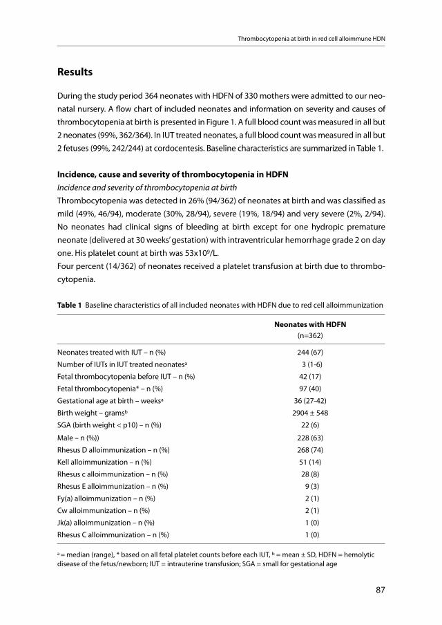
Thrombocytopenia at birth in red cell alloimmune HDN
87
Results
During the study period 364 neonates with HDFN of 330 mothers were admitted to our neo-natal nursery. A flow chart of included neonates and information on severity and causes of thrombocytopenia at birth is presented in Figure 1. A full blood count was measured in all but 2 neonates (99%, 362/364). In IUT treated neonates, a full blood count was measured in all but 2 fetuses (99%, 242/244) at cordocentesis. Baseline characteristics are summarized in Table 1.
Incidence, cause and severity of thrombocytopenia in HDFNIncidence and severity of thrombocytopenia at birthThrombocytopenia was detected in 26% (94/362) of neonates at birth and was classified as mild (49%, 46/94), moderate (30%, 28/94), severe (19%, 18/94) and very severe (2%, 2/94). No neonates had clinical signs of bleeding at birth except for one hydropic premature neonate (delivered at 30 weeks’ gestation) with intraventricular hemorrhage grade 2 on day one. His platelet count at birth was 53x109/L. Four percent (14/362) of neonates received a platelet transfusion at birth due to thrombo-cytopenia.
Table 1 Baseline characteristics of all included neonates with HDFN due to red cell alloimmunization
Neonates with HDFN (n=362)
Neonates treated with IUT – n (%) 244 (67)
Number of IUTs in IUT treated neonatesa 3 (1-6)
Fetal thrombocytopenia before IUT – n (%) 42 (17)
Fetal thrombocytopenia* – n (%) 97 (40)
Gestational age at birth – weeksa 36 (27-42)
Birth weight – gramsb 2904 ± 548
SGA (birth weight < p10) – n (%) 22 (6)
Male – n (%)) 228 (63)
Rhesus D alloimmunization – n (%) 268 (74)
Kell alloimmunization – n (%) 51 (14)
Rhesus c alloimmunization – n (%) 28 (8)
Rhesus E alloimmunization – n (%) 9 (3)
Fy(a) alloimmunization – n (%) 2 (1)
Cw alloimmunization – n (%) 2 (1)
Jk(a) alloimmunization – n (%) 1 (0)
Rhesus C alloimmunization – n (%) 1 (0)
a = median (range), * based on all fetal platelet counts before each IUT, b = mean ± SD, HDFN = hemolytic disease of the fetus/newborn; IUT = intrauterine transfusion; SGA = small for gestational age
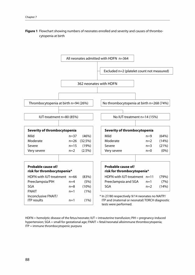
Chapter 7
88
* In 27/80 respectively 9/14 neonates no NAITP/ITP and (maternal or neonatal) TORCH diagnostic tests were performed.
All neonates admitted with HDFN n=364
362 neonates with HDFN
Thrombocytopenia at birth n=94 (26%) No thrombocytopenia at birth n=268 (74%)
IUT-treatment n=80 (85%) No IUT-treatment n=14 (15%)
Severity of thrombocytopenia
Mild n=37 (46%)Moderate n=26 (32.5%)Severe n=15 (19%)Very severe n=2 (2.5%)
Probable cause of/risk for thrombocytopenia*
HDFN with IUT-treatment n=66 (83%)Preeclampsia/PIH n=4 (5%)SGA n=8 (10%)FNAIT n=1 (1%)Inconclusive FNAIT/ITP results n=1 (1%)
Probable cause of/risk for thrombocytopenia*
HDFN with IUT-treatment n=11 (79%)Preeclampsia and SGA n=1 (7%)SGA n=2 (14%)
Severity of thrombocytopenia
Mild n=9 (64%)Moderate n=2 (14%)Severe n=3 (21%)Very severe n=0 (0%)
Excluded n=2 (platelet count not measured)
Figure 1 Flowchart showing numbers of neonates enrolled and severity and causes of thrombo-cytopenia at birth
HDFN = hemolytic disease of the fetus/neonate; IUT = intrauterine transfusion; PIH = pregnancy-induced hypertension; SGA = small for gestational age; FNAIT = fetal/neonatal alloimmune thrombocytopenia; ITP = immune thrombocytopenic purpura

Thrombocytopenia at birth in red cell alloimmune HDN
89
Fetal thrombocytopenia and IUT in neonates with thrombocytopenia at birthEighty of the 94 thrombocytopenic neonates at birth received at least one IUT (Figure 1). Thirty-one percent (24/78) and 95% (76/80) of IUT treated neonates with thrombocytope-nia at birth had fetal thrombocytopenia based on the first fetal platelet count (before the first IUT) and all fetal platelet counts, respectively.
Only one (non-hydropic) fetus received an intrauterine platelet transfusion at a platelet count of 27x109/L in addition to a single IUT at 33 weeks’ gestation. He was born after 35+4 weeks’ gestation with a birth weight of 2580 g (p25-p50) and Apgar scores of 4, 7 and 7 after 1, 5 and 10 min, respectively. Platelet count at birth was 22x109/L and he received one platelet transfusion on day one. Screening tests in this patient showed no evidence of FNAIT or TORCH congenital infection.
Diagnostic tests in neonates with thrombocytopenia at birthFifteen neonates with thrombocytopenia at birth were screened for FNAIT and one neonate had FNAIT coinciding with her Rhesus D HDFN. Maternal and/or neonatal serologic screen-ing tests for congenital TORCH infection were performed in 63% (59/94) of neonates with thrombocytopenia. All TORCH screening tests were negative. No cases of early onset neo-natal sepsis were detected.
Risk factors for thrombocytopenia at birthDetailed information on risk factors for thrombocytopenia at birth and blood results of neo-nates with and without thrombocytopenia at birth are summarized in Table 2.
Univariate analysisType of alloimmunization The incidence of thrombocytopenia at birth in neonates with Rhesus D, Kell, Rhesus c and other types of red cell alloimmunization was 26% (69/268), 24% (12/51), 36% (10/28) and 20% (3/15), respectively. Type of red blood cell alloimmunization was not associated with thrombocytopenia at birth (Table 2). The incidence of severe thrombocytopenia (platelet count ≤50x109/L) at birth was also not statistically different in neonates with Rhesus D, Kell, or Rhesus c compared to neonates without Rhesus D, Kell or Rhesus c respectively (p=0.672, p=0.434 and p=0.696, respectively).
Perinatal risk factors Several risk factors were associated with thrombocytopenia at birth including: treatment with IUT (OR 3.62, 95% CI 1.95-6.73, p<0.001), fetal hydrops (OR 2.97, 95% CI 1.58-5.58, p<0.001), PIH/preeclampsia (OR 7.36, 95% CI 1.40-38.6, p=0.015), lower gestational age at
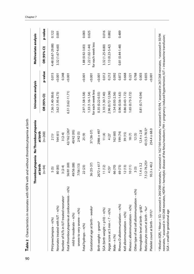
Chapter 7
90
Tabl
e 2
Cha
ract
eris
tics
in n
eona
tes
with
HD
FN w
ith a
nd w
ithou
t thr
ombo
cyto
peni
a at
birt
h
Thro
mbo
cyto
peni
a at
bir
th(n
=94)
No
Thro
mbo
cyto
peni
a at
bir
th(n
=268
)
Uni
vari
ate
anal
ysis
Mul
tiva
riat
e an
alys
is
OR
(95%
CI)
p-va
lue
OR
(95%
CI)
p-va
lue
PIH
/pre
ecla
mps
ia –
n(%
)5
(5)
2 (1
)c7.
36 (1
.40-
38.6
)0.
015
4.48
(0.6
7-29
.88)
0.12
2
Neo
nate
s tr
eate
d w
ith IU
T –
n(%
)80
(85)
164
(61)
3.62
(1.9
5-6.
73)
<0.0
013.
32 (1
.67-
6.60
)0.
001
Num
ber o
f IU
Ts in
IUT
trea
ted
neon
ates
a3
(2-4
)3
(2-4
)0.
348
Feta
l thr
ombo
cyto
peni
a at
cor
doce
ntes
is –
n(%
)56
/81
(69)
42/1
62 (2
6)d
6.51
(3.6
2-11
.71)
<0.0
01
mild
to m
oder
ate
– n(
%)
49/5
6 (8
8)40
/42
(95)
sev
ere
to v
ery
seve
re –
n(%
)7/
56 (1
3)2/
42 (5
)
Feta
l hyd
rops
– n
(%)
22 (2
3)25
(9)
2.97
(1.5
8-5.
58)
<0.0
011.
88 (0
.92-
3.83
)0.
083
Ges
tatio
nal a
ge a
t birt
h –
wee
ksa
36 (3
5-37
) 37
(36-
37)
1.33
(1.1
6-1.
54)
for e
ach
wee
k le
ss<0
.001
1.22
(1.0
2-1.
44)
for e
ach
wee
k le
ss0.
025
Birt
h w
eigh
t – g
ram
sb26
72 ±
617
2986
± 4
970.
35 (0
.22-
0.55
)<0
.001
SGA
(birt
h w
eigh
t < p
10) –
n(%
)11
(12)
11 (4
)3.
10 (1
.30-
7.40
)0.
012
3.32
(1.2
5-8.
80)
0.01
6
Apg
ar s
core
at 5
min
< 7
– n
(%)
4 (5
)e4
(2)f
2.96
(0.7
2-12
.06)
0.21
21.
13 (0
.23-
5.42
)0.
882
Mal
e –
n (%
)66
(70)
162
(60)
1.54
(0.9
3-2.
56)
0.09
2
Rhes
us D
allo
imm
uniz
atio
n –
n(%
)69
(73)
199
(74)
0.96
(0.5
6-1.
63)
0.87
20.
81 (0
.44-
1.48
)0.
489
Kell
allo
imm
uniz
atio
n –
n(%
)12
(13)
39 (1
5)0.
86 (0
.43-
1.72
)0.
668
Rhes
us c
allo
imm
uniz
atio
n –
n(%
)10
(11)
18 (7
)1.
65 (0
.73-
3.72
)0.
221
Oth
er ty
pe o
f red
cel
l allo
imm
uniz
atio
n –
n(%
)3
(3)
12 (5
)0.
768
Hem
oglo
bin
leve
l at b
irth
– g/
dLb
11.4
± 3
.2
12.4
± 2
.80.
81 (0
.71-
0.94
)0.
003
Retic
uloc
yte
coun
t at b
irth
– ‰
a7.
5 (2
-76.
25)g
43 (5
.5-7
8.5)
h0.
835
Plat
elet
cou
nt a
t birt
h –
109 /
Lb93
.3 ±
40.
225
4.4
± 68
.0<0
.001
a Med
ian
(IQR)
, b M
ean
± SD
, c as
sess
ed in
264
/268
neo
nate
s, d a
sses
sed
in 1
62/1
64 n
eona
tes,
e as
sess
ed in
93/
94, f a
sses
sed
in 2
67/2
68 n
eona
tes,
g as
sess
ed in
50/
94
neon
ates
, h ass
esse
d in
165
/268
neo
nate
s, H
DFN
= h
emol
ytic
dis
ease
of t
he fe
tus/
new
born
; PIH
= p
regn
ancy
indu
ced
hype
rten
sion
; IU
T =
intr
aute
rine
tran
sfus
ion;
SG
A =
sm
all f
or g
esta
tiona
l age
Thrombocytopenia at birth in red cell alloimmune HDN
91
birth (OR 1.33 for each week less, 95% CI 1.16-1.54, p<0.001) and SGA (OR 3.10, 95% CI 1.30-7.40, p=0.012). Only one mother had HELLP syndrome and one mother had diabetes (both neonates had normal platelet counts at birth).
Multivariate analysisOn multivariate analysis, the following risk factors were independently associated with throm-bocytopenia at birth: treatment with IUT, lower gestational age at birth and SGA (Table 2). Fetal hydrops was not significant at the 5% level, but the relatively low p-value (p=0.083) is suggestive of a possible independent association with thrombocytopenia at birth.Maternal diabetes and HELLP syndrome were excluded from multivariate analysis because of limited number of cases. As birth weight and gestational age at birth are closely related, birth weight was also excluded from multivariate analysis.
Discussion
This study demonstrates that thrombocytopenia at birth is common among neonates with HDFN due to red cell alloimmunization, occurring in 26% of neonates compared to 1-5% in the general population and 22-35% in the neonatal intensive care unit (NICU) population.13 Severe thrombocytopenia was present in 6% of all neonates with HDFN compared to 5-10% in the NICU population.
This is the first study describing the incidence of neonatal thrombocytopenia at birth in HDFN due to red cell alloimmunization. Koenig et al. described neonatal thrombocytope-nia in 11 of 20 (55%) neonates with Rhesus HDFN during admission.4 In this study, platelet count was not routinely measured at birth and in several cases only after exchange transfu-sion. Exchange transfusion is a known risk factor for thrombocytopenia, independently of red cell alloimmunization.5,14
We found a positive association between IUT treatment and thrombocytopenia at birth. The cause of this association is not clear and several mechanisms may play a role. Increased eryth-ropoiesis could theoretically lead to suppression of thrombopoiesis by the hematopoietic stem cells.1,4 However, since IUT is known to suppress erythropoiesis15, this theory only sup-ports fetal thrombocytopenia at the first IUT. Increased incidence of fetal thrombocytope-nia from 17% to 40% in fetuses treated with several IUTs may be explained by a decreased production, increased consumption, increased destruction, or dilution. In addition, IUT with packed red cells can cause dilution of platelets.16 However, it is unlikely that this effect is still present at the time of a consecutive IUT after two to three weeks.
Chapter 7
92
We found that type of red cell alloimmunization was not a risk factor for thrombocytope-nia at birth. In a previous study fetal thrombocytopenia (at first IUT) appeared to be less common in Kell HDFN than in Rhesus D HDFN.3 The discrepancy between the results may be due to several factors including methodological differences between the two studies. The higher rate of thrombocytopenia in Kell HDFN found in this study may be due to the higher number of IUTs in the Kell population.10
Prematurity and intrauterine growth restriction have previously been described as risk factors for early-onset (<72 hours) neonatal thrombocytopenia.7,8 In accordance, we dem-onstrated that lower gestational age at birth and SGA are independent risk factors for thrombocytopenia at birth in neonates with HDFN. In addition, we found that lower birth weight irrespective of gestational age is a risk factor for thrombocytopenia at birth in red cell alloimmunization.
Perinatal asphyxia (Apgar score <7), maternal PIH/preeclampsia and syndrome of HELLP have formerly been described as risk factors for thrombocytopenia at birth.7,17,18 In our study population perinatal asphyxia was not associated with thrombocytopenia at birth and the number of cases with PIH/preeclampsia was limited.Interestingly, one case of thrombocytopenia in this cohort was found to be due to FNAIT. Four other case reports of thrombocytopenia due to FNAIT have been described in fetuses/neonates with Rhesus hemolytic disease.19-22
Fortunately only one neonate had clinical sings of bleeding at birth (intraventricular hemor-rhage grade 2). Although this neonate was thrombocytopenic at birth, in this case other factors such as prematurity and hydrops could have contributed to this bleeding complica-tion. Moreover, the causal relation between thrombocytopenia and intraventricular hemor-rhage is controversial.23
The results of this study should be interpreted with care because of the retrospective study design. We have not systematically investigated all other possible causes of neonatal throm-bocytopenia such as maternal immune thrombocytopenic purpura, FNAIT and perinatal/neonatal infection. Hence the incidence of 26% of thrombocytopenia at birth due to red cell alloimmunization can be an overestimate. Finally, the number of spurious thrombocytope-nia because of clotted samples and platelet clumping is unclear.
In conclusion, this study shows that 26% of neonates with HDFN due to red cell alloim-munization have thrombocytopenia at birth. Risk for thrombocytopenia is independently associated with IUT treatment, SGA and lower gestational age at birth.
Thrombocytopenia at birth in red cell alloimmune HDN
93
References
1. Saade GR, Moise KJ, Jr., Copel JA, et al .: Fetal platelet counts correlate with the severity of the anemia in red-cell alloimmunization. Obstet Gynecol 1993; 82(6):987-991.
2. Van den Akker ES, de Haan TR, Lopriore E, et al.: Severe fetal thrombocytopenia in Rhesus D allo-immunized pregnancies. Am J Obstet Gynecol 2008; 199(4):387-4.
3. Van den Akker ES, Klumper FJ, Brand A, et al.: Kell alloimmunization in pregnancy: associated with fetal thrombocytopenia? Vox Sang 2008; 95(1):66-69.
4. Koenig JM, Christensen RD: Neutropenia and thrombocytopenia in infants with Rh hemolytic disease. J Pediatr 1989; 114(4 Pt 1):625-631.
5. Chadd MA, Gray OP, Hole DJ: Blood coagulation studies during exchange transfusion. J Obstet Gynaecol Br Commonw 1972; 79(4):373-376.
6. Wagner T, Bernaschek G, Geissler K: Inhibition of megakaryopoiesis by Kell-related antibodies. N Engl J Med 2000; 343(1):72.
7. Murray NA, Roberts IA: Circulating megakaryocytes and their progenitors in early thrombocytopenia in preterm neonates. Pediatr Res 1996; 40(1):112-119.
8. Watts T, Roberts I: Haematological abnormalities in the growth-restricted infant. Semin Neonatol 1999; 4(1):41-54.
9. Rath ME, Smits-Wintjens VE, Lindenburg I, et al.: Top-up transfusions in neonates with Rh hemolytic disease in relation to exchange transfusions. Vox Sang 2010; 99(1):65-70.
10. Rath ME, Smits-Wintjens VE, Lindenburg IT, et al .: Exchange transfusions and top-up transfusions in neo-nates with Kell haemolytic disease compared to Rh D haemolytic disease. Vox Sang 2010; 100(3):312-6.
11. Smits-Wintjens VE, Walther FJ, Rath ME, et al.: Intravenous immunoglobulin in neonates with rhesus hemolytic disease: a randomized controlled trial. Pediatrics 2011; 127(4):680-686.
12. Kloosterman GJ:.Intrauterine growth and intrauterine growth curves Ned Tijdschr Verloskd Gynaecol 1969; 69(5):349-365.
13. Roberts I, Stanworth S, Murray NA: Thrombocytopenia in the neonate. Blood Rev 2008; 22(4):173-186.14. Smits-Wintjens VE, Walther FJ, Lopriore E: Rhesus haemolytic disease of the newborn: Postnatal manage-
ment, associated morbidity and long-term outcome. Semin Fetal Neonatal Med 2008; 13(4):265-271.15. De Boer I, Zeestraten EC, Lopriore E, et al.: Pediatric outcome in Rhesus hemolytic disease treated with
and without intrauterine transfusion. Am J Obstet Gynecol 2008; 198(1):54. e1-54.e4.16. Viëtor HE, Klumper F, Meerman RJ, et al .: Intrauterine transfusions influence fetal leukocyte counts and
subsets. Prenat Diagn 1998; 18(4):325-331.17. Beiner ME, Simchen MJ, Sivan E, et al.: Risk factors for neonatal thrombocytopenia in preterm infants. Am
J Perinatol 2003; 20(1):49-54.18. Harms K, Rath W, Herting E, et al .: Maternal hemolysis, elevated liver enzymes, low platelet count, and
neonatal outcome. Am J Perinatol 1995; 12(1):1-6.19. Carbonne B, Chereau E, Larsen M, et al .: Concomitant fetal anemia and thrombocytopenia due to anti-D
and anti-HPA1a alloimmunization. Prenat Diagn 2005; 25(12):1172-1174.20. Klüter H, Germer U, Gortner L, et al.: Coincidence of neonatal alloimmune thrombocytopenia and mater-
nal anti-D immunization: case report. Br J Haematol 1998; 102(5):1383-1384.21. Schild RL, Hoch J, Plath H, et al .: Perinatal management of fetal hemolytic disease due to Rh incompat-
ibility combined with fetal alloimmune thrombocytopenia due to HPA-5b incompatibility. Ultrasound Obstet Gynecol 1999; 14(1):64-67.
22. Yeast JD, Plapp F: Fetal anemia as a response to prophylactic platelet transfusion in the management of alloimmune thrombocytopenia. Am J Obstet Gynecol 2003; 189(3):874-876.
23. Baer VL, Lambert DK, Henry E, et al.: Severe Thrombocytopenia in the NICU. Pediatrics 2009; 124(6):e1095-e1100.

94

8 Top-up transfusions in neonates with
Rhesus hemolytic disease in relation to exchange transfusions
Mirjam EA RathVivianne EHJ Smits-Wintjens
Irene TM LindenburgAnneke BrandDick Oepkes
Frans J WaltherEnrico Lopriore
Vox Sang 2010; 99(1):65-70
Chapter 8
96
Abstract
Objective: To study the effect of a restrictive guideline for exchange transfusion (ET) on the number of top-up transfusions in neonates with Rhesus hemolytic disease.
Study design: Retrospective study of all (near)-term neonates with Rhesus hemolytic disease admitted to our center between 2000 and 2008. In December 2005, policy changed from using liberal ET criteria to more restrictive ET criteria. We recorded the number of ETs and the number of top-up transfusions in the group of neonates before (group I, n =156) and after (group II, n =27) the guideline change.
Results: The percentage of neonates requiring an ET decreased from 66% (103/156) in group I to 26% (7/27) in group II (p < 0.01). The percentage of neonates receiving a top-up transfusion increased from 68% (105/154) in group I to 81% (22/27) in group II (p = 0.25). The median number of top-up transfusions increased from 1 (interquartile range 0-2) in group I to 2 (interquartile range 1-3) in group II (p = 0.01).
Conclusion: In this study, restrictive ET criteria in neonates with Rhesus hemolytic disease lead to a reduction of the rate of ET but an increase in the number of top-up transfusions for neonatal anemia.
Top-up transfusions in neonatal Rhesus hemolytic disease
97
Introduction
The mainstay of antenatal treatment for hemolytic disease of the fetus and newborn due to Rhesus alloimmunization (HDFN) is intrauterine transfusions (IUT) to treat severe fetal anemia. The mainstay of postnatal treatment for hemolytic disease of the newborn (HDN) secondary to Rhesus alloimmunization is (1) intensive phototherapy and exchange transfu-sion (ET) to treat hyperbilirubinemia and prevent kernicterus, and (2) top-up transfusions to treat neonatal anemia. Neonatal anemia secondary to Rhesus alloimmunization can be divided into two types: “hyporegenerative anemia” characterized by depressed erythropoi-esis and “late anemia of hemolytic disease” caused by persisting hemolysis by remaining antibodies.1 Both causes of anemia contribute to the necessity of top-up transfusions during the first months of life. The percentage of infants with HDN secondary to Rhesus alloimmu-nization requiring top-up transfusions for neonatal anemia varies from 27 to 83%.2,3
Several risk factors for neonatal anemia secondary to Rhesus alloimmunization have been reported, including IUT (due to suppression of fetal erythropoiesis)2 and severity of HDN.1,4,5 Use of ET during the neonatal period has been reported to protect against neo-natal anemia.3.In addition to removing excess bilirubin, ET also removes antibody-coated erythrocytes and maternal antibodies, hence reducing the risk for continuing hemolysis and neonatal anemia.3,6 However, the protective role of ET for neonatal anemia has only been demonstrated in one small study.3
In December 2005, we implemented a new guideline for the management of neonatal hyperbilirubinemia and HDN secondary to Rhesus alloimmunization based on the guide-lines from the American Academy of Pediatrics (AAP).7 The new guideline for ET was more restrictive than the previous ones and led to a more than 50% decrease in the rate of ET. Restrictive ET criteria may hypothetically lead to an increase in top-up transfusions ascribed to ongoing hemolysis due to antibody coated cells not being removed from the circulation. Whether the reduction in ET indeed resulted in an increased rate of top-up transfusions was not clear.
The aim of this study was to evaluate the effect of this new guideline on the number of top-up transfusions and determine if neonates with hemolytic disease of the newborn secondary to Rhesus alloimmunization treated with ET are less likely to develop neonatal anemia.
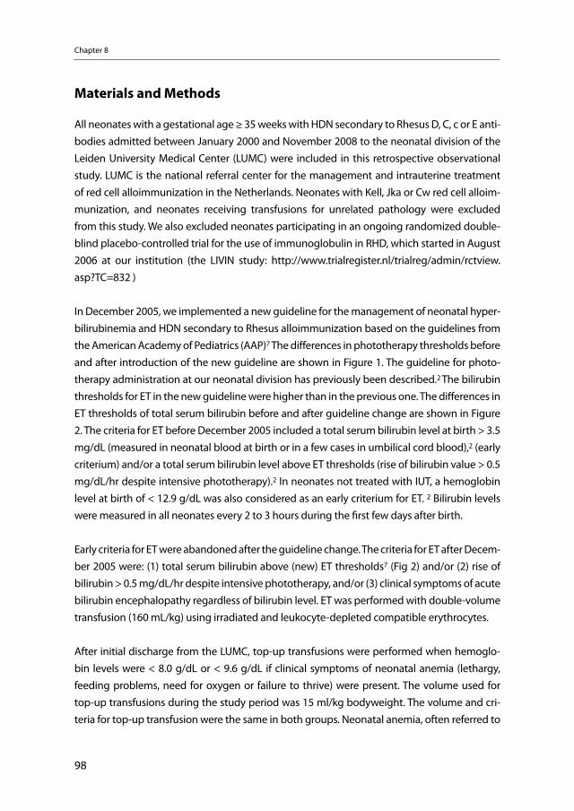
Chapter 8
98
Materials and Methods
All neonates with a gestational age ≥ 35 weeks with HDN secondary to Rhesus D, C, c or E anti-bodies admitted between January 2000 and November 2008 to the neonatal division of the Leiden University Medical Center (LUMC) were included in this retrospective observational study. LUMC is the national referral center for the management and intrauterine treatment of red cell alloimmunization in the Netherlands. Neonates with Kell, Jka or Cw red cell alloim-munization, and neonates receiving transfusions for unrelated pathology were excluded from this study. We also excluded neonates participating in an ongoing randomized double-blind placebo-controlled trial for the use of immunoglobulin in RHD, which started in August 2006 at our institution (the LIVIN study: http://www.trialregister.nl/trialreg/admin/rctview.asp?TC=832 )
In December 2005, we implemented a new guideline for the management of neonatal hyper-bilirubinemia and HDN secondary to Rhesus alloimmunization based on the guidelines from the American Academy of Pediatrics (AAP)7 The differences in phototherapy thresholds before and after introduction of the new guideline are shown in Figure 1. The guideline for photo-therapy administration at our neonatal division has previously been described.2 The bilirubin thresholds for ET in the new guideline were higher than in the previous one. The differences in ET thresholds of total serum bilirubin before and after guideline change are shown in Figure 2. The criteria for ET before December 2005 included a total serum bilirubin level at birth > 3.5 mg/dL (measured in neonatal blood at birth or in a few cases in umbilical cord blood),2 (early criterium) and/or a total serum bilirubin level above ET thresholds (rise of bilirubin value > 0.5 mg/dL/hr despite intensive phototherapy).2 In neonates not treated with IUT, a hemoglobin level at birth of < 12.9 g/dL was also considered as an early criterium for ET. 2 Bilirubin levels were measured in all neonates every 2 to 3 hours during the first few days after birth.
Early criteria for ET were abandoned after the guideline change. The criteria for ET after Decem-ber 2005 were: (1) total serum bilirubin above (new) ET thresholds7 (Fig 2) and/or (2) rise of bilirubin > 0.5 mg/dL/hr despite intensive phototherapy, and/or (3) clinical symptoms of acute bilirubin encephalopathy regardless of bilirubin level. ET was performed with double-volume transfusion (160 mL/kg) using irradiated and leukocyte-depleted compatible erythrocytes.
After initial discharge from the LUMC, top-up transfusions were performed when hemoglo-bin levels were < 8.0 g/dL or < 9.6 g/dL if clinical symptoms of neonatal anemia (lethargy, feeding problems, need for oxygen or failure to thrive) were present. The volume used for top-up transfusions during the study period was 15 ml/kg bodyweight. The volume and cri-teria for top-up transfusion were the same in both groups. Neonatal anemia, often referred to
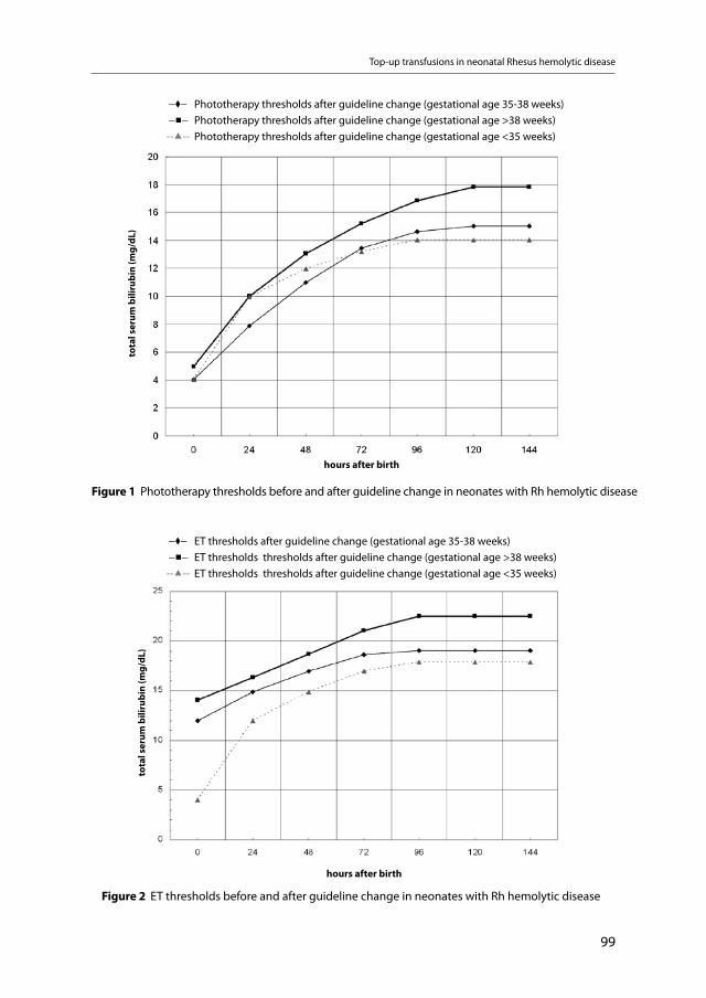
Top-up transfusions in neonatal Rhesus hemolytic disease
99
Figure 1 Phototherapy thresholds before and after guideline change in neonates with Rh hemolytic disease
total serum bilirubin (mg/dL)
_⧫_ ET thresholds after guideline change (gestational age 35-38 weeks) _◼_ ET thresholds thresholds after guideline change (gestational age >38 weeks)--▲-- ET thresholds thresholds after guideline change (gestational age <35 weeks)
Figure 2 ET thresholds before and after guideline change in neonates with Rh hemolytic disease
hours after birth
tota
l ser
um b
iliru
bin
(mg/
dL)
_⧫_ Phototherapy thresholds after guideline change (gestational age 35-38 weeks) _◼_ Phototherapy thresholds after guideline change (gestational age >38 weeks)--▲-- Phototherapy thresholds after guideline change (gestational age <35 weeks)
hours after birth
tota
l ser
um b
iliru
bin
(mg/
dL)
Chapter 8
100
as “late anemia” in previous studies3,5,8,9 was defined as a hemoglobin level below thresholds requiring a top-up transfusion during the first three months of life. Folic acid 50 mcg/day was administered orally during the first three months of life to all neonates with RHD. According to our management guidelines, neonates with RHD were not treated with erythropoietin.
We recorded the following obstetric and neonatal data: fetal hemoglobin at first IUT and number of IUTs, gestational age at birth, birth weight, hemoglobin level and reticulocyte count at birth, bilirubin level at birth, maximum bilirubin level during admission, duration of phototherapy (days), number of ETs required, number of top-up red blood cell transfu-sions received during the first 3 months of life and hemoglobin levels prior to the top-up transfusion. Obstetric data were gathered using a dedicated database for Rhesus compli-cated pregnancies in which data are prospectively collected. Neonatal data were collected using medical files. Data on top-up transfusions and hemoglobin levels prior to the top-up transfusion in infants who received follow-up outside the LUMC were gathered through cor-respondence with the local pediatrician or blood transfusion department.
Primary outcome was the number of top-up transfusions in the group of neonates admitted before (group I) and after (group II) the new guideline implementation in December 2005.Data are reported as means and standard deviations (SD) or as medians and interquartile range (IQR), as appropriate. Statistical analysis was performed using Student-t-test and Mann-Whitney test for continuous variables. Chi square and Fisher’s exact test were used for categorical variables, as appropriate. To assess the relationship between the number of ETs and the number of top-up transfusions, Spearman correlations were calculated. A p-value <0.05 was considered statistically significant. Statistical analysis was executed with SPSS 16.0 (SPSS Inc, Chicago, IL).
Results
During the study period 270 infants with hemolytic disease delivered ≥ 35 weeks’ gesta-tion were admitted to our neonatal division. Fifty neonates were excluded because of par-ticipation in a randomized double-blind placebo-controlled trial for the use of intravenous immunoglobulin. Thirty-six neonates were excluded due to hemolytic disease caused by Kell (n=34), Jka (n=1) or Cw (n=1). One neonate requiring major cardio-thoracic surgery for congenital heart disease was also excluded. A total of 183 patients were included in this study, of whom 156 in the group before (group I) and 27 in the group after (group II) the implementation of the new guideline in December 2005. Baseline characteristics in both groups are summarized in Table 1.
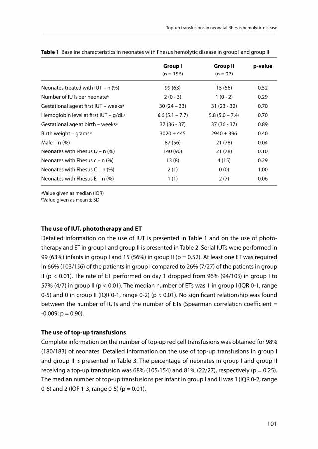
Top-up transfusions in neonatal Rhesus hemolytic disease
101
Table 1 Baseline characteristics in neonates with Rhesus hemolytic disease in group I and group II
Group I(n = 156)
Group II(n = 27)
p-value
Neonates treated with IUT – n (%) 99 (63) 15 (56) 0.52
Number of IUTs per neonatea 2 (0 - 3) 1 (0 - 2) 0.29
Gestational age at first IUT – weeksa 30 (24 – 33) 31 (23 - 32) 0.70
Hemoglobin level at first IUT – g/dLa 6.6 (5.1 – 7.7) 5.8 (5.0 – 7.4) 0.70
Gestational age at birth – weeksa 37 (36 - 37) 37 (36 - 37) 0.89
Birth weight – gramsb 3020 ± 445 2940 ± 396 0.40
Male – n (%) 87 (56) 21 (78) 0.04
Neonates with Rhesus D – n (%) 140 (90) 21 (78) 0.10
Neonates with Rhesus c – n (%) 13 (8) 4 (15) 0.29
Neonates with Rhesus C – n (%) 2 (1) 0 (0) 1.00
Neonates with Rhesus E – n (%) 1 (1) 2 (7) 0.06
aValue given as median (IQR)bValue given as mean ± SD
The use of IUT, phototherapy and ETDetailed information on the use of IUT is presented in Table 1 and on the use of photo-therapy and ET in group I and group II is presented in Table 2. Serial IUTs were performed in 99 (63%) infants in group I and 15 (56%) in group II (p = 0.52). At least one ET was required in 66% (103/156) of the patients in group I compared to 26% (7/27) of the patients in group II (p < 0.01). The rate of ET performed on day 1 dropped from 96% (94/103) in group I to 57% (4/7) in group II (p < 0.01). The median number of ETs was 1 in group I (IQR 0-1, range 0-5) and 0 in group II (IQR 0-1, range 0-2) (p < 0.01). No significant relationship was found between the number of IUTs and the number of ETs (Spearman correlation coefficient = -0.009; p = 0.90).
The use of top-up transfusionsComplete information on the number of top-up red cell transfusions was obtained for 98% (180/183) of neonates. Detailed information on the use of top-up transfusions in group I and group II is presented in Table 3. The percentage of neonates in group I and group II receiving a top-up transfusion was 68% (105/154) and 81% (22/27), respectively (p = 0.25). The median number of top-up transfusions per infant in group I and II was 1 (IQR 0-2, range 0-6) and 2 (IQR 1-3, range 0-5) (p = 0.01).
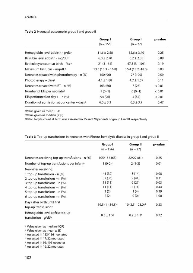
Chapter 8
102
Table 3 Top-up transfusions in neonates with Rhesus hemolytic disease in group I and group II
Group I(n = 156)
Group II(n = 27)
p-value
Neonates receiving top-up transfusions – n (%) 105/154 (68) 22/27 (81) 0.25
Number of top-up transfusions per infanta 1 (0-2)c 2 (1-3) 0.01
Neonates receiving: 1 top-up transfusion – n (%)2 top-up transfusions – n (%)3 top-up transfusions – n (%)4 top-up transfusions – n (%)5 top-up transfusions – n (%)6 top-up transfusions – n (%)
41 (39)37 (36)11 (11)11 (11)
2 (2)2 (2)
3 (14)9 (41)6 (27)3 (14)1 (4)0 (0)
0.080.310.030.440.391.00
Days after birth until first top-up transfusiona
19.5 (1 - 34.8)c 10 (2.5 – 23.0)d 0.23
Hemoglobin level at first top-up transfusion – g/dLb
8.3 ± 1.5e 8.2 ± 1.3f 0.72
a Value given as median (IQR) b Value given as mean ± SDc Assessed in 153/156 neonatesd Assessed in 17/22 neonatese Assessed in 95/105 neonatesf Assessed in 16/22 neonates
Table 2 Neonatal outcome in group I and group II
Group I(n = 156)
Group II(n = 27)
p-value
Hemoglobin level at birth - g/dLa 11.6 ± 2.58 12.6 ± 3.40 0.25
Bilirubin level at birth - mg/dLa 6.0 ± 2.70 6.2 ± 2.85 0.89
Reticulocyte count at birth – ‰b,c 21 (3 - 61) 47.5 (3 - 106) 0.19
Maximum bilirubin – mg/dLb 13.6 (10.3 – 16.8) 15.4 (13.2 -18.0) 0.03
Neonates treated with phototherapy – n (%) 150 (96) 27 (100) 0.59
Phototherapy – daysa 4.1 ± 1.88 4.7 ± 1.59 0.11
Neonates treated with ET – n (%) 103 (66) 7 (26) < 0.01
Number of ETs per neonateb 1 (0 -1) 0 (0 -1) < 0.01
ETs performed on day 1 – n (%) 94 (96) 4 (57) < 0.01
Duration of admission at our center – daysa 6.0 ± 3.3 6.3 ± 3.9 0.47
aValue given as mean ± SDbValue given as median (IQR)cReticulocyte count at birth was assessed in 75 and 20 patients of group I and II, respectively
Top-up transfusions in neonatal Rhesus hemolytic disease
103
Combining group I and II, we found a negative correlation between the number of ETs and the number of top-up blood transfusions (Spearman correlation coefficient -0.183; p = 0.01). This correlation between a lower number of ETs and a higher number of top-up transfusions was present mainly in the subgroup of neonates treated with IUT (n = 114) (Spearman cor-relation coefficient = -0.340; p < 0.01). In the subgroup of neonates without IUT (n = 69), the correlation between the number of ETs and the number of top-up blood transfusions was not significant (Spearman correlation coefficient = 0.011; p = 0.93). Of the 110 neonates that received an ET 73 neonates (66%) were treated with IUT.
Comment
This study shows that after implementation of a more restrictive guideline for ET in neonates with RHD, the rate of ETs decreased considerably from 66% to 26%. The reduction in ET rate was associated with a significant increase in the number of top-up transfusions for neona-tal anemia. Neonates in group II required twice more top-up transfusions than neonates in group I. Since the criteria for top-up transfusions remained unchanged before and after the ET guideline changes, our findings suggest that neonatal anemia in neonates with RHD is more likely to occur if restrictive ET criteria are used.
Our findings are in accordance with a previous study by al-Alaiyan et al. in a small group of 36 (near-) term neonates with RHD treated with IUT.3 Al-Alaiyan et al. reported a similar asso-ciation between ET rate and top-up transfusions.3 The risk of neonatal anemia in neonates treated with and without ET was 36% and 92%, respectively (p = 0.04).3
The reduced rate of top-up transfusions in infants treated with ET can be attributed to the removal of antibodies and IgG coated erythrocytes during ET, hence reducing the hemolytic process and the risk of neonatal anemia.3,6 However, the effect of ET on the antibody titer on the short term is limited. Since the antibodies are distributed in the intravascular as well as the extravascular fluid, ET reduces the antibody titer only by about 25 percent.4,10 The beneficial effect of ET lies in replacing nearly all of the Rhesus positive red cells by immunologically com-patible cells, hence surviving cells in the blood stream.4
In contrast with previous studies2,8 we found a significant negative correlation between the number of ETs and top-up transfusions. This negative correlation was found only in neonates treated with IUT, but was not found in neonates without IUT treatment. Since fetuses requir-ing IUT treatment are more severely affected by RHD than fetuses without IUT treatment, we speculate that the beneficial effect of ET in reducing the number of top-up transfusions occurs
Chapter 8
104
primarily in severely affected neonates with high titer antibodies. Washing out antibodies and replacing Rhesus positive cells through ET treatment may be particularly more effective in severely affected neonates.
The pathogenesis of neonatal or late anemia in RHD is not completely clarified and can be due to either depressed erythropoiesis (“hyporegenerative anemia”) or persisting reduction in half-life of the Rhesus positive erythrocytes caused by remaining antibodies (“late anemia of hemolytic disease”) if age-appropriate or elevated reticulocytes are present.11,12 “Hypore-generative anemia” occurs in particular after IUT due to suppression of the erythropoiesis.2,9 An alternative explanation for failing compensatory reticulocytosis is destruction of bone marrow precursors and reticulocytes by antibodies.4,13 Other contributing factors to neonatal anemia are reduced survival of transfused red blood cells,5 natural decline of the hemoglobin level toward the physiological nadir, and the increasing intravascular volume of the growing neonate.1 Finally, erythropoietin deficiency can be a possible contributing factor to neonatal anemia.12 Treatment with erythropoietin has been suggested to reduce the number of top-up transfusions, but the evidence to recommend routine use of erythropoietin is very limited.14
The data in this study should be interpreted with care due to the retrospective design of the study which may have led to a selection bias and influenced by changing transfusion attitudes over time. Although both groups were similar in terms of severity of fetal anemia, need of IUT, and hemoglobin levels at birth, it is conceivable that neonates in group II were more severely affected than neonates in group I, hence requiring more top-up transfusions.2 Importantly, the percentage of male infants in group II was higher than in group I. Male infants have a higher prevalence of HDN secondary to Rhesus alloimmunization,15 a higher neonatal death rate from kernicterus16 and appear to be more severely affected than females in terms of need for IUT, development of hydrops fetalis and perinatal mortality.17 A larger study is required to determine if this sex difference in baseline characteristic may have influenced our results.
In conclusion, this study shows that the number of ETs in neonates with HDN secondary to Rhesus alloimmunization decreased significantly after the introduction of restrictive ET crite-ria. Reduction of ET rate resulted in a doubling of the number of top-up transfusions. Restric-tive ET criteria in HDN secondary to Rhesus alloimmunization during the neonatal period may thus lead to an increased rate of neonatal anemia. Nevertheless, the risk of adverse events associated with ETs (in particular catheter-related complications) is high (7%)18 compared to the transfusion-related risks of blood transfusion in general (< 0.04%).19 A reduction in ETs, despite an increase in top-up transfusions may therefore be more beneficial for neonates with HDN secondary to Rhesus alloimmunization, although the long-term effects on neuro-developmental outcome requires longer follow-up.
Top-up transfusions in neonatal Rhesus hemolytic disease
105
References
1. Pessler F, Hart D: Hyporegenerative anemia associated with Rh hemolytic disease: treatment failure of recombinant erythropoietin. J Pediatr Hematol Oncol 2002; 24: 689-693.
2. De Boer, I, Zeestraten EC, Lopriore E, Van Kamp, I, Kanhai HH, Walther FJ: Pediatric outcome in Rhesus hemolytic disease treated with and without intrauterine transfusion. Am J Obstet Gynecol 2008; 198: 54. e1-54.e4.
3. Al-Alaiyan S, al OA: Late hyporegenerative anemia in neonates with rhesus hemolytic disease. J Perinat Med 1999; 27: 112-115.
4. Giblett ER, Varela JE, Finch CA: Damage of the bone marrow due to Rh antibody. Pediatrics 1956; 17: 37-44.
5. Koenig JM, Ashton RD, De Vore GR, Christensen RD: Late hyporegenerative anemia in Rh hemolytic disease. J Pediatr 1989; 115: 315-318.
6. Smits-Wintjens VEHJ, Walther FJ, Lopriore E: Rhesus haemolytic disease of the newborn: Postna-tal management, associated morbidity and long-term outcome. Semin Fetal Neonatal Med 2008; 13(4):265-71.
7. Management of hyperbilirubinemia in the newborn infant 35 or more weeks of gestation. Pediatrics 2004; 114: 297-316.
8. Ebbesen F: Late anaemia in infants with rhesus haemolytic disease treated with intensive photother-apy. Eur J Pediatr 1979; 130: 285-290.
9. Scaradavou A, Inglis S, Peterson P, Dunne J, Chervenak F, Bussel J: Suppression of erythropoiesis by intrauterine transfusions in hemolytic disease of the newborn: use of erythropoietin to treat the late anemia. J Pediatr 1993; 123: 279-284.
10. Diamond LK, Allen FH, Jr., Vann DD, Powers JR: Erythroblastosis fetalis. Pediatrics 1952; 10: 337-347.11. Hayde M, Widness JA, Pollak A, Kohlhauser-Vollmuth C, Vreman HJ, Stevenson DK: Rhesus isoimmuni-
zation: increased hemolysis during early infancy. Pediatr Res 1997; 41: 716-721.12. Millard DD, Gidding SS, Socol ML, MacGregor SN, Dooley SL, Ney JA, Stockman JA, III: Effects of intra-
vascular, intrauterine transfusion on prenatal and postnatal hemolysis and erythropoiesis in severe fetal isoimmunization. J Pediatr 1990; 117: 447-454.
13. Burk CD, Malatack JJ, Ramsey G: Misleading Rh phenotype and severe prolonged anemia in hemolytic disease of the newborn. Am J Dis Child 1987; 141: 712-713.
14. Ovali F, Samanci N, Dagoglu T: Management of late anemia in Rhesus hemolytic disease: use of recom-binant human erythropoietin (a pilot study). Pediatr Res 1996; 39: 831-834.
15. Woodrow JC, Donohoe WT: Rh-immunization by pregnancy: results of a survey and their relevance to prophylactic therapy. Br Med J 1968; 4: 139-144.
16. Walker W, Mollison PL: Haemolytic disease of the newborn; deaths in England and Wales during 1953 and 1955. Lancet 1957; 272: 1309-1314.
17. Ulm B, Svolba G, Ulm MR, Bernaschek G, Panzer S: Male fetuses are particularly affected by maternal alloimmunization to D antigen. Transfusion 1999; 39: 169-173.
18. Steiner LA, Bizzarro MJ, Ehrenkranz RA, Gallagher PG: A decline in the frequency of neonatal exchange transfusions and its effect on exchange-related morbidity and mortality. Pediatrics 2007; 120: 27-32.
19. Stainsby D, Jones H, Wells AW, Gibson B, Cohen H: Adverse outcomes of blood transfusion in children: analysis of UK reports to the serious hazards of transfusion scheme 1996-2005. Br J Haematol 2008; 141: 73-79.

106

9 Exchange transfusions and top-up transfusions
in neonates with Kell hemolytic disease compared to Rh D hemolytic disease
Mirjam EA RathVivianne EHJ Smits-Wintjens
Irene TM LindenburgAnneke Brand
Inge L van KampDick Oepkes
Frans J WaltherEnrico Lopriore
Vox Sang 2010; 100(3):312-316
Chapter 9
108
Abstract
Objective: To evaluate neonatal outcome in Kell hemolytic disease compared to Rh D hemolytic disease.
Study design: Retrospective study of all (near)-term neonates with Kell (n=34) and Rh D hemolytic disease (n=157) admitted to our center between January 2000 and December 2008. We recorded the need for exchange transfusion and top-up transfusions up to three months of age.
Results: Neonates in the Kell group required less days of phototherapy than neonates in the Rh D group (2.4 versus 4.1 days, respectively (p < 0.01)). The percentage of neonates requi-ring an exchange transfusion was lower in the Kell group than in the Rh D group (6% (2/34) and 62% (98/157), respectively (p < 0.01)). The percentage of neonates in the Kell group and Rh D group requiring a top-up transfusion was 62% (21/34) and 72% (113/157), respectively (p = 0.20). The median number of top-up transfusions per neonate in the Kell group and Rh D group was 1 (interquartile range (IQR) 0-2) and 2(IQR 0-2), respectively (p = 0.07).
Conclusion: Neonates with Kell hemolytic disease require less phototherapy and less exchange transfusions compared to neonates with Rh D hemolytic disease, but an equal number of top-up transfusions.
Neonatal outcome in Kell versus Rh D hemolytic disease
109
Introduction
Kell alloimmunization is second only to Rh D in causing antibody-mediated fetal anemia. Since the introduction of Rh D immunoprophylaxis, Kell antibodies account for 10% of antibody-mediated fetal anemia.1 After introduction of routine antibody-screening of all pregnant women in the Netherlands in 1998, perinatal survival of fetuses with Kell hemoly-tic disease of the neonate (HDN) treated with intrauterine transfusion (IUT) increased from 61% to 100%.2
In contrast to Rh D HDN, fetal anemia in Kell HDN is often more severe due to concomitant suppression of erythropoiesis rather than hemolysis of erythrocytes.3-5 Consequently, the immediate neonatal management in Kell HDN is different from Rh D HDN. A previous small study showed that neonates with Kell HDN have lower serum bilirubin levels and require less phototherapy and exchange transfusions (ETs) than neonates with Rh D hemolytic disease.6
In analogy with Rh D hemolytic disease, neonates with Kell HDN may require top-up trans-fusions for up to several months after birth.7 Whether the incidence and severity of neonatal anemia in Kell hemolytic disease differs from neonates with Rh D hemolytic disease, is not known. Only a limited number of studies (mostly case reports) have been published on the severity of anemia in the postnatal period.6, 8-10
The aim of this study was to evaluate neonatal and hematological outcome in a large series of neonates with Kell hemolytic disease compared to neonates with Rh D hemolytic disease.
Materials and Methods
All (near)-term neonates (gestational age ≥ 35 weeks) with hemolytic disease due to mater-nal Kell and Rh D alloimmunization, delivered between January 2000 and December 2008 at the Leiden University Medical Center (LUMC) were included in this retrospective observatio-nal study. Part of the neonates included in the Rh D group have previously been described in a different study.11 Our center is the national referral center for the management and intrau-terine treatment of red cell alloimmunization in the Netherlands. We excluded all neonates with other types of HDN and neonates participating in an ongoing randomized trial for the use of immunoglobulin in Rh D hemolytic disease, which started in August 2006 at our insti-tution (LIVIN study: http://www.trialregister.nl/trialreg/admin/rctview.asp?TC=832).
Chapter 9
110
The guideline for the application of phototherapy at our neonatal division has previously been described.11,12 The guidelines for ET used at our neonatal division were changed in December 2005. Before December 2005, criteria for ET included: (1) bilirubin level at birth > 3.5 mg/dL (so-called early criterion) and/or (2) total serum bilirubin level above ET thres-holds (rise of bilirubin value > 0.5 mg/dL/hr despite intensive phototherapy). In neonates not treated with IUT, a hemoglobin level at birth of < 12.9 g/dL was also considered as an early criterion for ET.12 In December 2005 a new guideline of the American Academy of Pediatrics (AAP) with higher bilirubin thresholds for phototherapy and ET was implemented by our department.13 The criteria for ET after December 2005 were: (1) total serum bilirubin above (higher) ET thresholds13 and/or (2) rise of bilirubin > 0.5 mg/dL/hr despite intensive phototherapy, and/or (3) clinical symptoms of acute bilirubin encephalopathy regardless of bilirubin level. ET was performed with double-volume transfusion (160 mL/kg) using irradi-ated and leukocyte-depleted compatible erythrocytes.
We recorded the following obstetric and neonatal data: fetal hemoglobin concentration at first IUT and number of IUTs, gestational age at birth, birth weight, hemoglobin level and reticulocyte count at birth, bilirubin level at birth, maximum bilirubin level during admis-sion, duration of phototherapy (days), number of ETs required, number of top-up red blood cell transfusions received during the first 3 months of life and hemoglobin levels prior to the top-up transfusion. Data on the number of top-up transfusions and hemoglobin levels prior to the top-up transfusion in infants who received follow-up outside the LUMC were collec-ted through correspondence with the local pediatrician or blood transfusion department. After discharge from our center, top-up transfusions were performed in referring hospitals when hemoglobin levels were < 8.0 g/dL, or < 9.6 g/dL if clinical symptoms of neonatal anemia were present (such as lethargy, feeding problems, need for oxygen or failure to thrive). Folic acid (50 mcg/day) was administered orally during the first three months of life to all neonates with hemolytic disease.
Primary outcome was the number of ETs and the number of top-up transfusions. Outcome was compared between neonates with Kell hemolytic disease (Kell group) and neonates with Rh D hemolytic disease (Rh D group).
Data are reported as means and standard deviations (SD) or as medians and interquartile range (IQR), as appropriate. Statistical analysis was performed using Student-t-test and Mann-Whitney test for continuous variables. Chi square and Fisher’s exact test were used for categorical variables, as appropriate. Linear regression analysis was performed using the Pearson Correlation coefficient. A p-value < 0.05 was considered statistically significant. Statistical analysis was executed with SPSS 16.0 (SPSS Inc, Chicago, IL, USA).
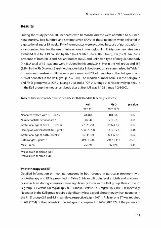
Neonatal outcome in Kell versus Rh D hemolytic disease
111
Results
During the study period, 309 neonates with hemolytic disease were admitted to our neo-natal nursery. Two hundred and seventy-seven (90%) of these neonates were delivered at a gestational age ≥ 35 weeks. Fifty-five neonates were excluded because of participation in a randomized trial for the use of intravenous immunoglobulin. Thirty-one neonates were excluded due to HDN caused by Rh c (n=17), Rh C (n=3), Rh E (n=3), Cw (n=2), Jka (n=1), presence of both Rh D and Kell antibodies (n=2), and unknown type of irregular antibody (n=3). A total of 191 patients were included in this study, 34 (18%) in the Kell group and 157 (82%) in the Rh D group. Baseline characteristics in both groups are summarized in Table 1. Intrauterine transfusions (IUTs) were performed in 82% of neonates in the Kell group and 66% of neonates in the Rh D group (p = 0.07). The median number of IUTs in the Kell group and Rh D group was 3 (IQR 2-4, range 0-5) and 2 (IQR 0-4, range 0-6) respectively (p = 0.01). In the Kell group the median antibody titer at first IUT was 1:128 (range 1:2-8000).
Table 1 Baseline characteristics in neonates with Kell and Rh D hemolytic disease
Kell(n = 34)
Rh D(n = 157)
p-value
Neonates treated with IUT – n (%) 28 (82) 104 (66) 0.07
Number of IUTs per neonate a 3 (2-4) 2 (0-3.5) 0.01
Gestational age at first IUT – weeks a 27 (23-29) 29 (24-33) 0.07
Hemoglobin level at first IUT – g/dL a 5.3 (3.5-7.3) 6.4 (5.0-7.4) 0.16
Gestational age at birth – weeks a 36 (36-37) 37 (36-37) 0.52
Birth weight – grams b 3190 ± 348 2947 ± 418 <0.01
Male – n (%) 25 (74) 92 (59) 0.11
a Value given as median (IQR)b Value given as mean ± SD
Phototherapy and ETDetailed information on neonatal outcome in both groups, in particular treatment with phototherapy and ET is presented in Table 2. Mean bilirubin level at birth and maximum bilirubin level during admission were significantly lower in the Kell group than in the Rh D group, 3.1 versus 6.0 mg/dL (p < 0.01) and 8.0 versus 14.3 mg/dL (p < 0.01), respectively. Neonates in the Kell group required significantly less days of phototherapy than neonates in the Rh D group (2.4 and 4.1 mean days, respectively, (p < 0.01)). At least one ET was required in 6% (2/34) of the patients in the Kell group compared to 62% (98/157) of the patients in
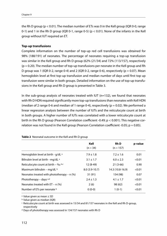
Chapter 9
112
the Rh D group (p < 0.01). The median number of ETs was 0 in the Kell group (IQR 0-0, range 0-1) and 1 in the Rh D group (IQR 0-1, range 0-5) (p < 0.01). None of the infants in the Kell group without IUT required an ET.
Top-up transfusionsComplete information on the number of top-up red cell transfusions was obtained for 98% (188/191) of neonates. The percentage of neonates requiring a top-up transfusion was similar in the Kell group and Rh D group (62% (21/34) and 72% (113/157), respectively (p = 0.20). The median number of top-up transfusions per neonate in the Kell group and Rh D group was 1 (IQR 0-2, range 0-4) and 2 (IQR 0-2, range 0-6), respectively (p = 0.07). Mean hemoglobin level at first top-up transfusion and median number of days until first top up transfusion were similar in both groups. Detailed information on the use of top-up transfu-sions in the Kell group and Rh D group is presented in Table 3.
In the sub-group analysis of neonates treated with IUT (n=132), we found that neonates with Rh D HDN required significantly more top-up transfusions than neonates with Kell HDN (median of 2 range 0-6 and median of 1 range 0-4), respectively (p = 0.02). We performed a linear regression analysis between the number of IUTs and the reticulocyte count at birth in both groups. A higher number of IUTs was correlated with a lower reticulocyte count at birth in the Rh D group (Pearson Correlation coefficient -0.49; p < 0.001). This negative cor-relation was not found in the Kell group (Pearson Correlation coefficient -0.05; p = 0.85).
Table 2 Neonatal outcome in the Kell and Rh D group
Kell(n = 34)
Rh D(n = 157)
p-value
Hemoglobin level at birth - g/dL a 7.9 ± 1.8 7.2 ± 1.6 0.01
Bilirubin level at birth - mg/dL a 3.1 ± 1.7 6.0 ± 2.3 <0.01
Reticulocyte count at birth – ‰ b,c 12 (8-49) 21 (3-66) 0.90
Maximum bilirubin – mg/dL b 8.0 (3.9-10.7) 14.3 (10.8-16.9) <0.01
Neonates treated with phototherapy – n (%) 31 (91) 154 (98) 0.07
Phototherapy – days a,d 2.4 ± 1.3 4.1 ± 1.7 <0.01
Neonates treated with ET – n (%) 2 (6) 98 (62) <0.01
Number of ETs per neonate b 0 (0-0) 1 (0-1) <0.01
a Value given as mean ± SDb Value given as median (IQR)c Reticulocyte count at birth was assessed in 15/34 and 81/157 neonates in the Kell and Rh D-group, respectivelyd Days of phototherapy was assessed in 134/157 neonates with Rh D
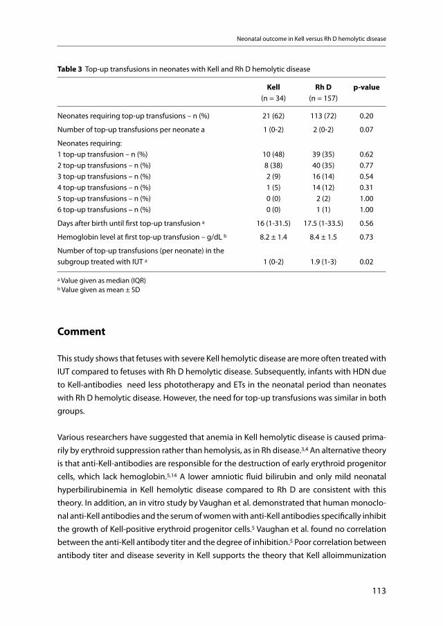
Neonatal outcome in Kell versus Rh D hemolytic disease
113
Comment
This study shows that fetuses with severe Kell hemolytic disease are more often treated with IUT compared to fetuses with Rh D hemolytic disease. Subsequently, infants with HDN due to Kell-antibodies need less phototherapy and ETs in the neonatal period than neonates with Rh D hemolytic disease. However, the need for top-up transfusions was similar in both groups.
Various researchers have suggested that anemia in Kell hemolytic disease is caused prima-rily by erythroid suppression rather than hemolysis, as in Rh disease.3,4 An alternative theory is that anti-Kell-antibodies are responsible for the destruction of early erythroid progenitor cells, which lack hemoglobin.5,14 A lower amniotic fluid bilirubin and only mild neonatal hyperbilirubinemia in Kell hemolytic disease compared to Rh D are consistent with this theory. In addition, an in vitro study by Vaughan et al. demonstrated that human monoclo-nal anti-Kell antibodies and the serum of women with anti-Kell antibodies specifically inhibit the growth of Kell-positive erythroid progenitor cells.5 Vaughan et al. found no correlation between the anti-Kell antibody titer and the degree of inhibition.5 Poor correlation between antibody titer and disease severity in Kell supports the theory that Kell alloimmunization
Table 3 Top-up transfusions in neonates with Kell and Rh D hemolytic disease
Kell(n = 34)
Rh D(n = 157)
p-value
Neonates requiring top-up transfusions – n (%) 21 (62) 113 (72) 0.20
Number of top-up transfusions per neonate a 1 (0-2) 2 (0-2) 0.07
Neonates requiring: 1 top-up transfusion – n (%)2 top-up transfusions – n (%)3 top-up transfusions – n (%)4 top-up transfusions – n (%)5 top-up transfusions – n (%)6 top-up transfusions – n (%)
10 (48)8 (38)2 (9)1 (5)0 (0)0 (0)
39 (35)40 (35)16 (14)14 (12)
2 (2)1 (1)
0.620.770.540.311.001.00
Days after birth until first top-up transfusion a 16 (1-31.5) 17.5 (1-33.5) 0.56
Hemoglobin level at first top-up transfusion – g/dL b 8.2 ± 1.4 8.4 ± 1.5 0.73
Number of top-up transfusions (per neonate) in the subgroup treated with IUT a 1 (0-2) 1.9 (1-3) 0.02
a Value given as median (IQR) b Value given as mean ± SD
Chapter 9
114
has a different pathogenesis than Rh alloimmunization. In a recent study of 43 pregnancies with Kell alloimmunization, we found that the vast majority of severely affected cases had antibody titers of 1:32 or more. Nevertheless, to be on the safe side, we recommended that all pregnancies with Kell titers of 1:2 or higher (and a proven Kell positive fetus) should be closely monitored.2
In accordance with previous studies from the group of Weiner4 and our group15 we found that antenatal course of fetuses with Kell alloimmunization is different from Rh D hemolytic disease. Fetuses with Kell alloimmunization have lower hemoglobin levels at first IUT and require more IUTs. Moreover, the first IUT was performed at an earlier gestational age than in Rh D fetuses.15 These findings underscore that fetal anemia is more severe in Kell sen-sitized fetuses than in Rh D sensitized fetuses. Weiner et al. also found a significant lower reticulocyte count, reflecting the destruction of Kell expressing erythroid progenitor cells in Kell hemolytic disease.4
In terms of neonatal management and outcome, this study shows that neonates with Kell hemolytic disease have milder hyperbilirubinemia, requiring less phototherapy and ETs than infants with Rh D hemolytic disease. In our study we found no relation between lack of IUT and number of ETs or top-up transfusions. Our findings are consistent with previous reports6,8,16-18 and reflect the observation that hemolysis of mature (hemoglobinised) ery-throcytes in Kell hemolytic disease is less than in Rh D hemolytic disease.3,4
Given the significantly higher number of IUTs in the Kell group, one could expect an increased incidence of postnatal anemia (and top-up transfusions). As shown in previous studies, repeated IUTs result in a decreased reticulocyte count, indicating a suppression of fetal erythropoiesis.12,19 In contrast, we found a trend towards less top-up transfusions in neonates with Kell hemolytic disease compared to neonates with Rh D hemolytic disease, however this difference was not significant (p = 0.07). This finding could support the fact that fetal and neonatal anemia due to Kell alloimmunization has a different pathogenesis than Rh alloimmunization. Larger studies are required to confirm these findings. In contrast to anti-D, most anti-Kell antibodies have a strong lytic potential which also affects red cell precursor cells,3,4,19 Vaughan and colleges found no correlation between the antibody titer and the degree of inhibition of Kell-positive erythroid progenitor cells. The titers of anti-Kell antibodies associated with fetal anemia are generally substantially lower compared to ten to 100 fold higher titers in case of Rh D hemolytic disease. Consequently anti-D antibodies may circulate in the newborn for a longer period after birth, whereas Kell antibodies disap-pear sooner, which may explain the more moderate late anemia in Kell hemolytic disease, despite concomitant suppression of the erythropoiesis.5
Neonatal outcome in Kell versus Rh D hemolytic disease
115
This is the first study comparing the degree of postnatal anemia in a relatively large number of infants with Kell- and Rh D hemolytic disease. Santiago et al. described three neonates with Kell HDN of whom only one required a top-up transfusion.17
Collinet et al. reported a case of severe fetal anemia due to Kell alloimmunization, which was postnatally treated with two top-up transfusions and recombinant erythropoietin and iron supplementation.7
The results of this study should be interpreted with care due to the small number of neo-nates with Kell hemolytic disease in this study which is inherent to the low incidence of this disease. Larger, multicenter studies are required to confirm our findings.
In conclusion, although neonates with Kell hemolytic disease require less phototherapy and exchange transfusions, the equal need for top-up transfusions justifies similar follow-up management as in Rh D hemolytic disease. Finally, because of the destruction of red cell precursor cells as well, treatment with erythropoietin may be more effective in neonates with Kell hemolytic disease than in neonates with Rh D hemolytic disease.9,10
References
1. Lee S, Russo D, Redman CM: The Kell blood group system: Kell and XK membrane proteins. Semin Hematol 2000; 37(2):113-121.
2. Kamphuis MM, Lindenburg I, Van Kamp IL, Meerman RH, Kanhai HH, Oepkes D: Implementation of routine screening for Kell antibodies: does it improve perinatal survival? Transfusion 2008; 48(5):953-957.
3. Vaughan JI, Warwick R, Letsky E, Nicolini U, Rodeck CH, Fisk NM: Erythropoietic suppression in fetal anemia because of Kell alloimmunization. Am J Obstet Gynecol 1994; 171(1):247-252.
4. Weiner CP, Widness JA: Decreased fetal erythropoiesis and hemolysis in Kell hemolytic anemia. Am J Obstet Gynecol 1996; 174(2):547-551.
5. Vaughan JI, Manning M, Warwick RM, Letsky EA, Murray NA, Roberts IA: Inhibition of erythroid proge-nitor cells by anti-Kell antibodies in fetal alloimmune anemia. N Engl J Med 1998; 338(12):798-803.
6. Babinszki A, Lapinski RH, Berkowitz RL: Prognostic factors and management in pregnancies complica-ted with severe kell alloimmunization: experiences of the last 13 years. Am J Perinatol 1998; 15(12):695-701.
7. Collinet P, Subtil D, Puech F, Vaast P: Successful treatment of extremely severe fetal anemia due to Kell alloimmunization. Obstet Gynecol 2002; 100(5 Pt 2):1102-1105.
8. Wenk RE, Goldstein P, Felix JK: Kell alloimmunization, hemolytic disease of the newborn, and perinatal management. Obstet Gynecol 1985; 66(4):473-476.
9. Dhodapkar KM, Blei F: Treatment of hemolytic disease of the newborn caused by anti-Kell antibody with recombinant erythropoietin. J Pediatr Hematol Oncol 2001; 23(1):69-70.
10. Manoura A, Korakaki E, Hatzidaki E, Saitakis E, Maraka S, Papamastoraki I, Matalliotakis E, Foundouli K, Giannakopoulou C: Use of recombinant erythropoietin for the management of severe hemolytic disease of the newborn of a K0 phenotype mother. Pediatr Hematol Oncol 2007; 24(1):69-73.
Chapter 9
116
11. Rath ME, Smits-Wintjens VE, Lindenburg I, Brand A, Oepkes D, Walther FJ, Lopriore E: Top-up transfusi-ons in neonates with Rh hemolytic disease in relation to exchange transfusions. Vox Sang 2010.
12. De Boer, I, Zeestraten EC, Lopriore E, Van K, I, Kanhai HH, Walther FJ: Pediatric outcome in Rhesus hemolytic disease treated with and without intrauterine transfusion. Am J Obstet Gynecol 2008; 198(1):54. e1-54.e4.
13. Management of hyperbilirubinemia in the newborn infant 35 or more weeks of gestation: Pediatrics 2004; 114(1):297-316.
14. Daniels G, Hadley A, Green CA: Causes of fetal anemia in hemolytic disease due to anti-K. Transfusion 2003; 43(1):115-116.
15. Van Kamp IL, Klumper FJ, Meerman RH, Oepkes D, Scherjon SA, Kanhai HH: Treatment of fetal anemia due to red-cell alloimmunization with intrauterine transfusions in the Netherlands, 1988-1999. Acta Obstet Gynecol Scand 2004; 83(8):731-737.
16. Bowman JM, Pollock JM, Manning FA, Harman CR, Menticoglou S: Maternal Kell blood group alloim-munization. Obstet Gynecol 1992; 79(2):239-244.
17. Santiago JC, Ramos-Corp, Oyonarte S, Montoya F: Current clinical management of anti-Kell alloim-munization in pregnancy. Eur J Obstet Gynecol Reprod Biol 2008; 136(2):151-154.
18. McKenna DS, Nagaraja HN, O’Shaughnessy R: Management of pregnancies complicated by anti-Kell isoimmunization. Obstet Gynecol 1999; 93(5 Pt 1):667-673.
19. Goodrum LA, Saade GR, Belfort MA, Carpenter RJ, Jr., Moise KJ, Jr.: The effect of intrauterine transfu-sion on fetal bilirubin in red cell alloimmunization. Obstet Gynecol 1997; 89(1):57-60.
10 Long-term neurodevelopmental outcome
after intrauterine transfusion for hemolytic disease of the fetus/newborn:
the LOTUS study
Irene TM LindenburgVivianne EHJ Smits-Wintjens
Jeanine M van KlinkEsther Verduin
Inge L van KampFrans J Walther
Henk SchonewilleIlias I Doxiadis
Humphrey H KanhaiJan M van Lith
Erik W van ZwetDick Oepkes
Anneke BrandEnrico Lopriore
(on behalf of the LOTUS study group)
Am J Obstet Gynecol 2011; Epub ahead of print
Chapter 10
118
Abstract
Objective: To determine the incidence and risk factors for neurodevelopmental impairment (NDI) in children with hemolytic disease of the fetus/newborn treated with intra uterine transfusion (IUT).
Study Design : Neurodevelopmental outcome in children at least 2 years of age was as-sessed using standardized tests, including the Bayley Scales of Infant Development, the Wechsler Preschool and Primary Scale of Intelligence and the Wechsler Intelligence Scale for Children, according to the children’s age. Primary outcome was the incidence of NDI defined as at least one of the following: cerebral palsy, severe developmental delay, bilateral deafness and/or blindness.
Results: A total of 291 children were evaluated at a median age of 8.2 years (range 2 to 17 years). Cerebral palsy was detected in six (2.1%) children, severe developmental delay in nine (3.1%) children and bilateral deafness in three (1.0%) children. The overall incidence of NDI was 4.8% (14/291). In a multivariate regression analysis including only pre-operative risk factors, severe hydrops was independently associated with NDI (OR 11.2, 95%, CI 1.7-92.7).
Conclusions: Incidence of NDI in children treated with IUT for fetal alloimmune anemia is low (4.8%). Prevention of fetal hydrops, the strongest pre-operative predictor for impaired neurodevelopment, by timely detection, referral and treatment may improve long-term outcome.
Long-term outcome after IUT: the LOTUS study
119
Introduction
Fetal and neonatal hemolytic disease results from maternal alloimmunization to red cell antigens, for which mother and fetus are incompatible. Maternal IgG antibodies pass the placenta into the fetal circulation and cause destruction of fetal red cells. The resulting pro-gressive fetal anemia leads, when untreated, to fetal hydrops and perinatal death.1
Before 1970, hemolytic disease due to antibodies against the Rhesus-D antigen was the most important cause of perinatal death.2 Several interventions have drastically reduced the incidence and severity of the disease, including postnatal and more recently antena-tal anti-D prophylaxis programs,3,4 improved diagnostic management and neonatal treat-ment.1,5-7 One of the major advances was the introduction in 1963 of intrauterine blood transfusions (IUTs),1 first performed by Liley using the intraperitoneal technique.8 In the 1980’s, this technique was replaced by the intravascular IUT.1 Nowadays, this treatment is the most successful procedure in fetal therapy, with perinatal survival rates exceeding 95% in experienced centers.1,7 However, one of the concerns of the more widespread and suc-cessful use of fetal therapy is that a decrease in perinatal mortality may lead to an increase of children with long-term handicaps. Only a few studies with small patient numbers have reported on long-term neurodevelopmental outcome after IUT, with an incidence of adverse outcome ranging from 4.5 to 12%.9-16 The aim of our study was to determine the incidence and risk factors for adverse neurodevelopmental outcome after IUT treatment in the largest cohort of children worldwide.
Methods
In 2008 we designed a large national cohort study to evaluate the long-term neurodevelop-mental outcome in children treated with IUT: the LOTUS study (LOng-Term follow-up after intra-Uterine transfusionS).17 All mothers with red cell alloimmunization treated with IUT between January 1st 1988 and January 1st 2008 at the Leiden University Medical Center and their children were invited to participate in this large follow-up study. For the purpose of this study we included all children of 2 to 17 years of age who had complete follow-up including a cognitive development test. Children with severe congenital anomalies and syndromal disorders were excluded. This study was approved by the ethics committee of the Leiden University Medical Center. Informed consent was obtained from all participating families. A limited outcome evaluation in a small part of our study group (11 children treated between 1991 and 1993) was described before.9 Primary outcome was a composite outcome termed neurodevelopmental impairment (NDI) defined as at least one of the following; cerebral
Chapter 10
120
palsy (CP), severe cognitive developmental delay (< –2 Standard Deviation (SD)), bilateral deafness requiring hearing amplification and/or bilateral blindness.
The Leiden University Medical Center serves as the single national reference center for the management of red cell alloimmunization in pregnancy in the Netherlands. IUTs are performed when signs of fetal anemia are detected on Doppler ultrasound examinations. Details on our management guidelines for alloimmunized pregnancies were previously described.18 Since the implementation of the IUT program using the ultrasound-guided intravascular transfusion technique at our center in 1987, all relevant perinatal data have prospectively been collected in a computerized database. Data included are: type of alloim-munization, gestational age at IUT, hemoglobin level, presence and severity of hydrops at the start of the intrauterine treatment, number of IUTs, gestational age at birth, gender, birth weight and neonatal outcome. Neonatal outcome data included: number of exchange transfusions due to severe hyperbilirubinemia, respiratory distress syndrome, necrotizing enterocolitis (classified according to Bell19), sepsis (defined as clinical symptoms of infection and a positive bacterial blood culture) and severe cerebral injury detected either on cranial ultrasound, Computed Tomography scan (CT) or Magnetic Resonance Imaging (MRI). Severe cerebral injury was defined as the presence of intraventricular hemorrhage ≥ grade 3 (classi-fied according to Volpe20), cystic periventricular leukomalacia ≥ grade 2 (classified according to de Vries21) and/or ventricular dilatation (defined according to Levene et al22). Other major cerebral abnormalities associated with adverse neurological outcome were also recorded and classified as severe cerebral lesions. We recorded the presence of perinatal asphyxia, defined as three or more of the following five criteria: non-reassuring cardiotocogram pat-terns, umbilical cord arterial pH < 7.10, Apgar score < 5 at 5 minutes after birth, failure of spontaneous breathing at 5 minutes after birth and onset of multiple organ failure.
Parental education was determined by the level of education of each parent individually. A score of 1 was given if the parent’s education was low, a score of 2 for an average educa-tional level, and a score of 3 for higher levels of education. Education scores of both parents were then added (score range from 2 to 6). Ethnicity was recorded as Caucasian or non-Caucasian. Children were considered to be Caucasian when one or both parent(s) were of Caucasian ethnicity.
Follow-upAll participating families visited our out-patients clinic from August 2008 to November 2010. At this visit, a physical and neurological examination according to Touwen23 and an assessment of cognitive development using standardized tests were performed.17 All chil-dren were assessed by one of the three investigators specialized in developmental assess-ment (IL, VS and EL).
Long-term outcome after IUT: the LOTUS study
121
Presence of CP was assessed according to the criteria of the European CP Network and clas-sified as diplegia, hemiplegia, quadriplegia, dyskinetic or mixed.24 Minor neurological dys-function (MND) was defined as a moderate abnormality of tone, posture, and movement leading to only minor functional impairment or minor developmental delay.23
Cognitive development in children aged 2 to 3 years was assessed according to the Dutch version of the Bayley Scales of Infant Development, 2nd edition (BSID-II).17 BSID-II scores provide a mental developmental index (MDI). Children between 3 and 7 years of age were tested with the Dutch version of the Wechsler Preschool and Primary Scale of Intelligence, 3rd edition (WPPSI-III-NL).17 Cognitive development in children between 7 and 17 years of age was assessed with the Dutch version of the Wechsler Intelligence Scale for Children, 3rd edition (WISC-III-NL).17 Both the WPPSI and the WISC provide a full scale IQ score. BSID-MDI, WPPSI and WISC scores follow a normal distribution curve with a mean score of 100. A score of 70-84 indicates mild delay (i.e. < -1 SD) and a score < 70 indicates severe delay (i.e. < -2 SD). A trained psychologist (JK), blinded to the antenatal course and neonatal outcome, performed the tests in all children.
Risk factorsPotential risk factors for NDI were investigated including severity of fetal anemia (actual hemoglobin level and Z-hemoglobin), presence and severity of fetal hydrops (classified according to van Kamp et al.25) at start of the intrauterine treatment, number of IUTs, ges-tational age at birth (divided in three groups: neonates born before 32 weeks’ gestation, between 32 and 35 weeks’ gestation and after 35 weeks’ gestation), severe neonatal mor-bidity and perinatal asphyxia. Standardized Z scores of hemoglobin (Z-hemoglobin) were defined as the number of standard deviations (SDs) that an actual value deviated from the normal mean for gestational age. Reference values for hemoglobin were derived from the literature.26 Severe neonatal morbidity was defined as the presence of one or more of the following: respiratory distress syndrome, necrotizing enterocolitis ≥ grade 2, sepsis and/or severe cerebral injury.
Statistical analysesWe used univariate logistic regressions to test the association between NDI and the poten-tial risk factors. We entered the risk factors into a multivariate logistic regression model and included additional potential confounders including gender, parental education and eth-nicity. Multiple logistic regression analysis was used to measure the independent effect of the potential risk factors for NDI. Results of logistic regression were considered significant at p-values < 0.05. We used the Pearson correlation test to calculate the correlation between hemoglobin at first IUT and IQ score. Analyses were performed using SPSS version 16 (SPSS Inc., Chicago, IL, USA).
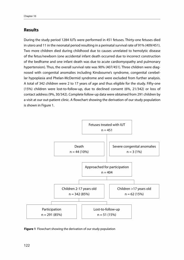
Chapter 10
122
Results
During the study period 1284 IUTs were performed in 451 fetuses. Thirty-one fetuses died in utero and 11 in the neonatal period resulting in a perinatal survival rate of 91% (409/451). Two more children died during childhood due to causes unrelated to hemolytic disease of the fetus/newborn (one accidental infant death occurred due to incorrect construction of the bedframe and one infant death was due to acute cardiomyopathy and pulmonary hypertension). Thus, the overall survival rate was 90% (407/451). Three children were diag-nosed with congenital anomalies including Kinsbourne’s syndrome, congenital cerebel-lar hypoplasia and Phelan-McDermid syndrome and were excluded from further analysis. A total of 342 children were 2 to 17 years of age and thus eligible for the study. Fifty-one (15%) children were lost-to-follow-up, due to declined consent (6%, 21/342) or loss of contact address (9%, 30/342). Complete follow-up data were obtained from 291 children by a visit at our out-patient clinic. A flowchart showing the derivation of our study population is shown in Figure 1.
Fetuses treated with IUTn = 451
Deathn = 44 (10%)
Severe congenital anomaliesn = 3 (1%)
Children 2-17 years oldn = 342 (85%)
Children >17 years oldn = 62 (15%)
Approached for participationn = 404
Participationn = 291 (85%)
Lost-to-follow-upn = 51 (15%)
Figure 1 Flowchart showing the derivation of our study population
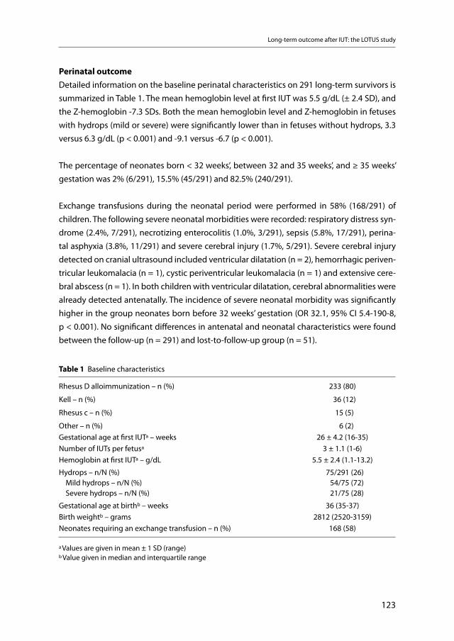
Long-term outcome after IUT: the LOTUS study
123
Perinatal outcomeDetailed information on the baseline perinatal characteristics on 291 long-term survivors is summarized in Table 1. The mean hemoglobin level at first IUT was 5.5 g/dL (± 2.4 SD), and the Z-hemoglobin -7.3 SDs. Both the mean hemoglobin level and Z-hemoglobin in fetuses with hydrops (mild or severe) were significantly lower than in fetuses without hydrops, 3.3 versus 6.3 g/dL (p < 0.001) and -9.1 versus -6.7 (p < 0.001).
The percentage of neonates born < 32 weeks’, between 32 and 35 weeks’, and ≥ 35 weeks’ gestation was 2% (6/291), 15.5% (45/291) and 82.5% (240/291).
Exchange transfusions during the neonatal period were performed in 58% (168/291) of children. The following severe neonatal morbidities were recorded: respiratory distress syn-drome (2.4%, 7/291), necrotizing enterocolitis (1.0%, 3/291), sepsis (5.8%, 17/291), perina-tal asphyxia (3.8%, 11/291) and severe cerebral injury (1.7%, 5/291). Severe cerebral injury detected on cranial ultrasound included ventricular dilatation (n = 2), hemorrhagic periven-tricular leukomalacia (n = 1), cystic periventricular leukomalacia (n = 1) and extensive cere-bral abscess (n = 1). In both children with ventricular dilatation, cerebral abnormalities were already detected antenatally. The incidence of severe neonatal morbidity was significantly higher in the group neonates born before 32 weeks’ gestation (OR 32.1, 95% CI 5.4-190-8, p < 0.001). No significant differences in antenatal and neonatal characteristics were found between the follow-up (n = 291) and lost-to-follow-up group (n = 51).
Table 1 Baseline characteristics
Rhesus D alloimmunization – n (%)
Kell – n (%)
Rhesus c – n (%)
Other – n (%)
233 (80)
36 (12)
15 (5)
6 (2)Gestational age at first IUTa – weeks 26 ± 4.2 (16-35)Number of IUTs per fetusa 3 ± 1.1 (1-6)Hemoglobin at first IUTa – g/dL 5.5 ± 2.4 (1.1-13.2)
Hydrops – n/N (%) Mild hydrops – n/N (%) Severe hydrops – n/N (%)
75/291 (26)54/75 (72)21/75 (28)
Gestational age at birthb – weeks 36 (35-37)Birth weightb – grams 2812 (2520-3159)Neonates requiring an exchange transfusion – n (%) 168 (58)
a Values are given in mean ± 1 SD (range)b Value given in median and interquartile range

Chapter 10
124
Long-term neurodevelopmental outcomeThe median age at follow-up was 8.2 years (range 2-17 years). The incidence of CP was 2.1% (6/291) (spastic quadriplegia: n = 3, spastic diplegia: n = 2, dyskinetic: n = 1). MND was recorded in 11.0% (32/291). None of the children had kernicterus. Nineteen children were evaluated using BSID-II tests, the average MDI score was 93 ± 14. A total of 89 children were tested using the WPPSI and 183 were tested using the WISC. The average full scale IQ in the WPPSI-group and WISC-group was 100 ± 14.8 and 101 ± 13.5, respectively. We found no cor-relation between hemoglobin level at first IUT and full scale IQ score (r = 0.1, p = 0.1) (Figure 2). Severe developmental delay (< -2 SD) was detected in 3.1% (9/291) of children. Moderate developmental delay (< -1 SD) was detected in 14.4% (42/291) of children. Bilateral deafness was present in three children (1.0%). None of the children had bilateral blindness. Table 2 summarizes the long-term neurodevelopmental outcome.
Figure 2. Relation between hemoglobin level at first IUT and full scale IQ score, in children
with (red dots) and without (blue dots) fetal hydrops.
IQ, intelligence quotient IUT, intrauterine transfusions
Q, intelligence quotient IUT, intrauterine transfusions
Figure 2 Relation between hemoglobin level at first IUT and full scale IQ score, in children with (red dots) and without (blue dots) fetal hydrops
Hemoglobin Level (g/dL)
Full
Scal
e IQ
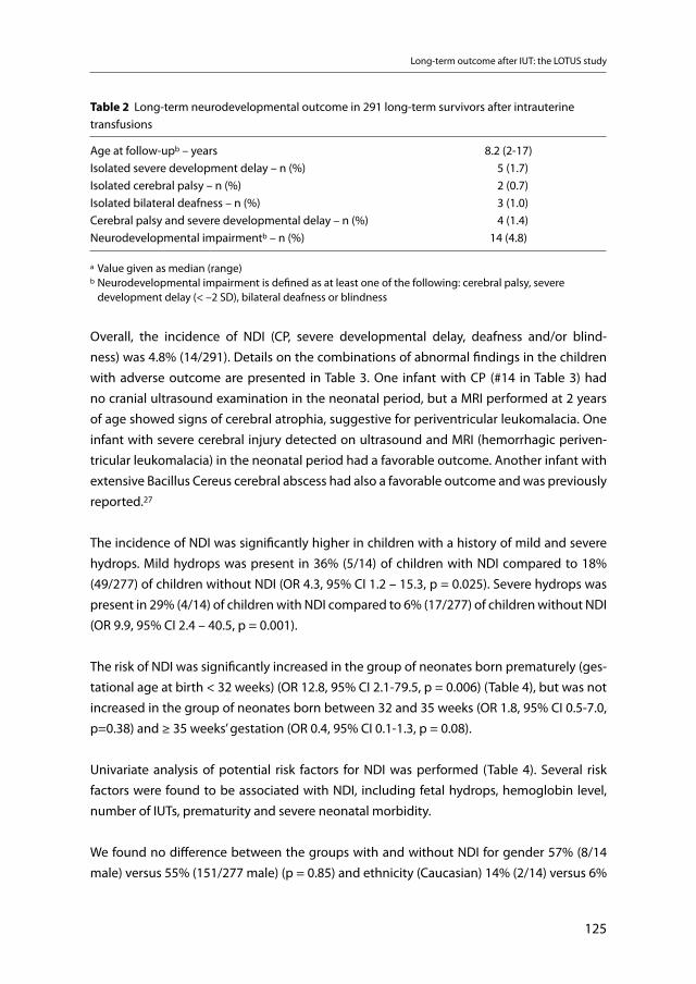
Long-term outcome after IUT: the LOTUS study
125
Overall, the incidence of NDI (CP, severe developmental delay, deafness and/or blind-ness) was 4.8% (14/291). Details on the combinations of abnormal findings in the children with adverse outcome are presented in Table 3. One infant with CP (#14 in Table 3) had no cranial ultrasound examination in the neonatal period, but a MRI performed at 2 years of age showed signs of cerebral atrophia, suggestive for periventricular leukomalacia. One infant with severe cerebral injury detected on ultrasound and MRI (hemorrhagic periven-tricular leukomalacia) in the neonatal period had a favorable outcome. Another infant with extensive Bacillus Cereus cerebral abscess had also a favorable outcome and was previously reported.27
The incidence of NDI was significantly higher in children with a history of mild and severe hydrops. Mild hydrops was present in 36% (5/14) of children with NDI compared to 18% (49/277) of children without NDI (OR 4.3, 95% CI 1.2 – 15.3, p = 0.025). Severe hydrops was present in 29% (4/14) of children with NDI compared to 6% (17/277) of children without NDI (OR 9.9, 95% CI 2.4 – 40.5, p = 0.001).
The risk of NDI was significantly increased in the group of neonates born prematurely (ges-tational age at birth < 32 weeks) (OR 12.8, 95% CI 2.1-79.5, p = 0.006) (Table 4), but was not increased in the group of neonates born between 32 and 35 weeks (OR 1.8, 95% CI 0.5-7.0, p=0.38) and ≥ 35 weeks’ gestation (OR 0.4, 95% CI 0.1-1.3, p = 0.08).
Univariate analysis of potential risk factors for NDI was performed (Table 4). Several risk factors were found to be associated with NDI, including fetal hydrops, hemoglobin level, number of IUTs, prematurity and severe neonatal morbidity.
We found no difference between the groups with and without NDI for gender 57% (8/14 male) versus 55% (151/277 male) (p = 0.85) and ethnicity (Caucasian) 14% (2/14) versus 6%
Table 2 Long-term neurodevelopmental outcome in 291 long-term survivors after intrauterine transfusions
Age at follow-upb – years 8.2 (2-17)Isolated severe development delay – n (%) 5 (1.7)Isolated cerebral palsy – n (%) 2 (0.7)Isolated bilateral deafness – n (%) 3 (1.0)Cerebral palsy and severe developmental delay – n (%) 4 (1.4)Neurodevelopmental impairmentb – n (%) 14 (4.8)
a Value given as median (range)b Neurodevelopmental impairment is defined as at least one of the following: cerebral palsy, severe development delay (< –2 SD), bilateral deafness or blindness
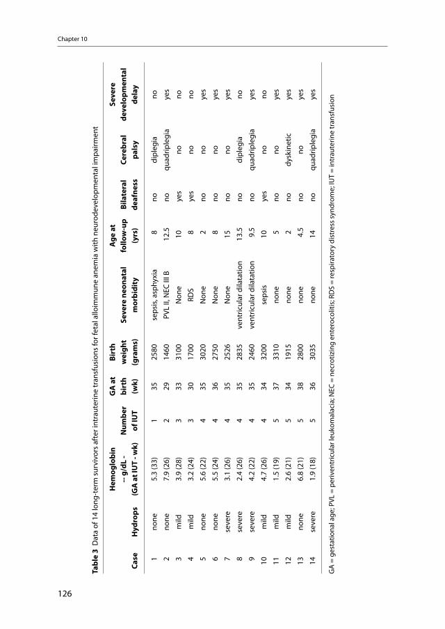
Chapter 10
126
Tabl
e 3
Dat
a of
14
long
-ter
m s
urvi
vors
aft
er in
trau
terin
e tr
ansf
usio
ns fo
r fet
al a
lloim
mun
e an
emia
with
neu
rode
velo
pmen
tal i
mpa
irmen
t
Case
H
ydro
ps
Hem
oglo
bin
-- g
/dL
- (G
A a
t IU
T - w
k)N
umbe
r of
IUT
GA
at
birt
h (w
k)
Birt
h w
eigh
t(g
ram
s)Se
vere
neo
nata
l m
orbi
dity
Age
at
follo
w-u
p(y
rs)
Bila
tera
l de
afne
ssCe
rebr
al
pals
y
Seve
re
deve
lopm
enta
l de
lay
1no
ne5.
3 (3
3)1
3525
80se
psis
, asp
hyxi
a 8
nodi
pleg
iano
2no
ne7.
9 (2
6)2
2914
60PV
L II,
NEC
III B
12.5
noqu
adrip
legi
aye
s
3m
ild3.
9 (2
8)3
3331
00N
one
10ye
sno
no
4m
ild3.
2 (2
4)3
3017
00RD
S 8
yes
nono
5no
ne5.
6 (2
2)4
3530
20N
one
2no
noye
s
6no
ne5.
5 (2
4)4
3627
50N
one
8no
noye
s
7se
vere
3.1
(26)
435
2526
Non
e15
nono
yes
8se
vere
2.4
(26)
435
2835
vent
ricul
ar d
ilata
tion
13.5
nodi
pleg
iano
9se
vere
4.2
(22)
435
2460
vent
ricul
ar d
ilata
tion
9.5
noqu
adrip
legi
aye
s
10m
ild4.
7 (2
6)4
3432
00se
psis
10ye
sno
no
11m
ild1.
5 (1
9)5
3733
10no
ne 5
nono
yes
12m
ild2.
6 (2
1)5
3419
15no
ne 2
nody
skin
etic
yes
13no
ne6.
8 (2
1)5
3828
00no
ne 4
.5no
noye
s
14se
vere
1.9
(18)
536
3035
none
14no
quad
riple
gia
yes
GA
= g
esta
tiona
l age
; PVL
= p
eriv
entr
icul
ar le
ukom
alac
ia; N
EC =
nec
rotiz
ing
ente
roco
litis
; RD
S =
resp
irato
ry d
istr
ess
synd
rom
e; IU
T =
intr
aute
rine
tran
sfus
ion
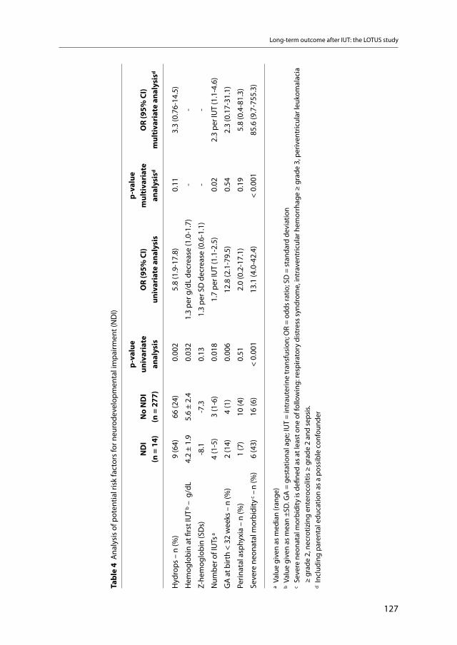
Long-term outcome after IUT: the LOTUS study
127
Tabl
e 4
Ana
lysi
s of
pot
entia
l ris
k fa
ctor
s fo
r neu
rode
velo
pmen
tal i
mpa
irmen
t (N
DI)
ND
I (n
= 1
4)N
o N
DI
(n =
277
)
p-va
lue
univ
aria
te
anal
ysis
OR
(95%
CI)
univ
aria
te a
naly
sis
p-va
lue
mul
tiva
riat
e an
alys
isd
OR
(95%
CI)
mul
tiva
riat
e an
alys
isd
Hyd
rops
– n
(%)
9 (6
4)66
(24)
0.0
02
5.8
(1.9
-17.
8) 0
.11
3.3
(0.7
6-14
.5)
Hem
oglo
bin
at fi
rst I
UT
b – g
/dL
4.2
± 1.
95.
6 ±
2.4
0.0
32
1.3
per g
/dL
decr
ease
(1.0
-1.7
) -
-
Z-he
mog
lobi
n (S
Ds)
-8
.1-7
.30.
131.
3 pe
r SD
dec
reas
e (0
.6-1
.1)
--
Num
ber o
f IU
Ts a
4 (1
-5)
3 (1
-6)
0.0
181.
7 pe
r IU
T (1
.1-2
.5)
0.0
22.
3 pe
r IU
T (1
.1-4
.6)
GA
at b
irth
< 32
wee
ks –
n (%
)2
(14)
4 (1
) 0
.006
12.8
(2.1
-79.
5) 0
.54
2.3
(0.1
7-31
.1)
Perin
atal
asp
hyxi
a –
n (%
)1
(7)
10 (4
)0.
51
2.0
(0.2
-17.
1) 0
.19
5.8
(0.4
-81.
3)
Seve
re n
eona
tal m
orbi
dity
c – n
(%)
6 (4
3)16
(6)
< 0.
001
13.1
(4.0
-42.
4)<
0.00
185
.6 (9
.7-7
55.3
)
a Va
lue
give
n as
med
ian
(ran
ge)
b Va
lue
give
n as
mea
n ±S
D. G
A =
ges
tatio
nal a
ge; I
UT
= in
trau
terin
e tr
ansf
usio
n; O
R =
odds
ratio
; SD
= s
tand
ard
devi
atio
nc
Seve
re n
eona
tal m
orbi
dity
is d
efine
d as
at l
east
one
of f
ollo
win
g: re
spira
tory
dis
tres
s sy
ndro
me,
intr
aven
tric
ular
hem
orrh
age
≥ gr
ade
3, p
eriv
entr
icul
ar le
ukom
alac
ia
≥
grad
e 2,
nec
rotiz
ing
ente
roco
litis
≥ g
rade
2 a
nd s
epsi
s.d
Incl
udin
g pa
rent
al e
duca
tion
as a
pos
sibl
e co
nfou
nder
Chapter 10
128
(18/277) (p = 0.24). Mean parental education was significantly lower in the NDI group com-pared to the no-NDI group 3.2 ± 1.1 vs. 4.2 ± 1.4, respectively (p = 0.016). Post-hoc analysis showed no difference in the incidence of exchange transfusion between the group with (57%, 8/14) and without NDI (58%, 160/277) (p = 0.96).
Potential risk factors and the possible confounder parental education were entered in a mul-tivariate logistic regression model to assess the independent association with NDI (Table 4). We excluded hemoglobin at first IUT from this multivariate analysis model, as this variable is strongly associated with the presence of hydrops and could possibly bias our results. In a multivariate regression analysis including prenatal and postnatal factors, the following risk factors were independently associated with NDI: number of performed IUTs (OR 2.3 per IUT, 95% CI 1.1-4.6, p = 0.02), severe neonatal morbidity (OR 85.6, 95% CI 9.7-755.3, p < 0.001) and parental education (OR 8.4, 95% CI 2.2-31.5, p = 0.002).
To determine the predictive role of prenatal risk factors, we entered the following factors in a separate multivariate regression model using only the following prenatal factors: mild hydrops, severe hydrops, level of hemoglobin at first IUT and number of IUTs. We found that only severe hydrops (OR 11.2, 95% CI 1.7-92.7, p = 0.011) was significantly independent associated with NDI.
Comment
This is the largest study to date on long-term neurodevelopmental outcome in children surviving a high-risk pregnancy thanks to invasive fetal therapy. The vast majority (over 95%) of children treated with IUT for severe fetal anemia had a normal neurodevelopmental outcome. The incidence of severe developmental delay (3.1%) was in line with the Dutch normative population (2.3%).28 In addition, the incidence of bilateral deafness in the general population was similar to what we found in our cohort.29 However, the rate of CP (2.1%) in our study was higher compared to the general population (0.7% at 32 to 36 weeks’ gesta-tion30 and 0.2% at 37 weeks’ gestation31).
A few small studies on the long-term neurodevelopmental outcome in children treated with IUT have been reported.9-16 The two largest studies to date reveal higher incidences of NDI when compared to our results, 10% (7/69) and 8% (3/38) respectively9,10 Differences in long-term outcome may be explained by methodological differences and heterogeneity between the studies.
Long-term outcome after IUT: the LOTUS study
129
Apart from the reassuring results valuable for counseling pregnant women with red cell alloimmunization, the importance of our analysis lies in the identification of potentially avoidable risk factors for adverse outcome. The current study shows a clear association with long-term impairment and the presence of hydrops and number of IUTs performed. Severe fetal hydrops was already known to be associated with increased perinatal mortality.25 The underlying mechanism causing cerebral damage and long-term NDI in hydropic and severely anemic fetuses is not yet known. Cerebral lesions may result from hypoxic injury related to severe anemia. Since short- and long-term outcome appears to be better in non-hydropic fetuses, clinicians should try to prevent or reduce the development of hydrops in fetuses at risk for fetal anemia. Interestingly, the actual hemoglobin concentration was more strongly associated with NDI than the hemoglobin Z-score. This concurs with the concept that tissue oxygenation depends more closely to the number of circulating red cells then on deviation of the hemoglobin level from the mean for gestational age. Whether more timely detection and treatment of fetal anemia, and prevention of hydrops improves outcome, and what degree of anemia actually requires transfusion needs further study.
Another risk factor for NDI was severe neonatal morbidity. As shown in our results, both the incidence of severe neonatal morbidity and the incidence of NDI were associated with the severity of prematurity. Severe prematurity is a well-known risk factor for neonatal morbid-ity, cerebral injury and long-term adverse outcome.32,33 We did not find a relation between NDI and exchange transfusions, which we interpret as confirmation of our relatively aggres-sive neonatal management protocol aimed at reducing the rate of severe hyperbilirubine-mia. None of the children had kernicterus.
Finally, parental education was independently associated with NDI. Socioeconomic status (SES) and parental educational level are well known determinants of child cognitive devel-opment.28,34-37 Both factors may influence child cognitive functioning for a variety of reasons, including reduced access to essential material resources (such as cognitively stimu-lating materials) and/or non-material resources (such as education, information and skills). Moreover, genetic conditions may account for up to 72% of the variance in intelligence.38 The two most important limitations of our study were the relatively incomplete follow-up and the lack of a control group. We were not able to trace 9% of children, mainly due to the long time-lap since IUT treatment. In addition, 6% of families declined to participate to the study. The risk for an adverse outcome has been shown to be higher in the lost-to-follow group as children at increased risk for severe neurodevelopmental compromise may not return for evaluation.39 Nevertheless, comparisons of antenatal and perinatal characteristics between the study group and the lost-to-follow-up showed no significant differences, sug-gesting that this type of bias was limited.
Chapter 10
130
Conclusions
The high rate of intact survival in this high-risk group of severely anemic fetuses confirms the success of this antenatal treatment. Although hemolytic disease of the fetus/newborn was the main cause of perinatal death for many years, the chance of successful recovery with adequate antenatal management can nowadays be considered as excellent. However, several factors were associated with increased risk for NDI including fetal hydrops, number of IUTs and severe neonatal morbidity. Future studies to reduce the incidence of these risk factors in children treated with IUT may help decrease the rate of adverse long-term outcome.
Acknowledgements
We wish to thank Jennie Verdoes for her dedicated work in approaching all families and coordinating all appointments for the follow-up assessments. We also thank all children and parents for participation in the LOTUS study.
Funding: The LOTUS study is funded by a grant of Sanquin (PPOC07-029) and the Fetal Maternal Research Foundation Leiden.
References
1. Moise KJ, Jr. Management of rhesus alloimmunization in pregnancy. Obstet Gynecol 2008;112:164-76.
2. Bennebroek GJ, Kanhai HH, Meerman RH, Ruys JH, Eernisse JG, Stroes TJ et al. Twenty-two years of intra-uterine intraperitoneal transfusions. Eur J Obstet Gynecol Reprod Biol 1989;33:71-77.
3. Bowman J. Thirty-five years of Rh prophylaxis. Transfusion 2003;43:1661-66. 4. Stockman JA, III. Overview of the state of the art of Rh disease: history, current clinical management,
and recent progress. J Pediatr Hematol Oncol 2001;23:385-93. 5. Oepkes D, Seaward PG, Vandenbussche FP, Windrim R, Kingdom J, Beyene J et al. Doppler ultrasonog-
raphy versus amniocentesis to predict fetal anemia. N Engl J Med 2006;355:156-64. 6. Mari G, Deter RL, Carpenter RL, Rahman F, Zimmerman R, Moise KJ, Jr. et al. Noninvasive diagnosis by
Doppler ultrasonography of fetal anemia due to maternal red-cell alloimmunization. Collaborative Group for Doppler Assessment of the Blood Velocity in Anemic Fetuses. N Engl J Med 2000;342:9-14.
7. Smits-Wintjens VE, Walther FJ, Lopriore E. Rhesus haemolytic disease of the newborn: Postnatal man-agement, associated morbidity and long-term outcome. Semin Fetal Neonatal Med 2008;13:265-71.
8. Liley AW. Iintrauterine transfusion of foetus in haemolytic disease. Br Med J 1963;2:1107-09. 9. Janssens HM, de Haan MJ, Van Kamp IL, Brand R, Kanhai HH, Veen S. Outcome for children treated with
fetal intravascular transfusions because of severe blood group antagonism. J Pediatr 1997;131:373-80.10. Doyle LW, Kelly EA, Rickards AL, Ford GW, Callanan C. Sensorineural outcome at 2 years for survivors
of erythroblastosis treated with fetal intravascular transfusions. Obstet Gynecol 1993;81:931-35.
Long-term outcome after IUT: the LOTUS study
131
11. Hudon L, Moise KJ, Jr., Hegemier SE, Hill RM, Moise AA, Smith EO et al. Long-term neurodevelopmen-tal outcome after intrauterine transfusion for the treatment of fetal hemolytic disease. Am J Obstet Gynecol 1998;179:858-63.
12. Harper DC, Swingle HM, Weiner CP, Bonthius DJ, Aylward GP, Widness JA. Long-term neurodevelop-mental outcome and brain volume after treatment for hydrops fetalis by in utero intravascular trans-fusion. Am J Obstet Gynecol 2006;195:192-200.
13. Grab D, Paulus WE, Bommer A, Buck G, Terinde R. Treatment of fetal erythroblastosis by intravascular transfusions: outcome at 6 years. Obstet Gynecol 1999;93:165-68.
14. Farrant B, Battin M, Roberts A. Outcome of infants receiving in-utero transfusions for haemolytic disease. N Z Med J 2001;114:400-03.
15. Weisz B, Rosenbaum O, Chayen B, Peltz R, Feldman B, Lipitz S. Outcome of severely anaemic fetuses treated by intrauterine transfusions. Arch Dis Child Fetal Neonatal Ed 2009;94:F201-F204.
16. Stewart G, Day RE, Del PC, Whittle MJ, Turner TL, Holland BM. Developmental outcome after intra-vascular intrauterine transfusion for rhesus haemolytic disease. Arch Dis Child Fetal Neonatal Ed 1994;70:F52-F53.
17. Verduin EP, Lindenburg IT, Smits-Wintjens VE, van Klink JM, Schonewille H, Van Kamp IL et al. LOng Term follow up after intra-Uterine transfusionS; the LOTUS study. BMC Pregnancy Childbirth 2010;10:77.
18. Van Kamp IL, Klumper FJ, Meerman RH, Oepkes D, Scherjon SA, Kanhai HH. Treatment of fetal anemia due to red-cell alloimmunization with intrauterine transfusions in the Netherlands, 1988-1999. Acta Obstet Gynecol Scand 2004;83:731-37.
19. Bell MJ, Ternberg JL, Feigin RD, Keating JP, Marshall R, Barton L et al. Neonatal necrotizing enterocoli-tis. Therapeutic decisions based upon clinical staging. Ann Surg 1978;187:1-7.
20. Volpe JJ. Intracranial hemorrhage: germinal matrix-intraventricular hemorrhage of the premature infant. In: Volpe JJ, editor. Neurology of the newborn. 4th Edition. Philadelphia: Saunders; 2001. p. 428-93.
21. De Vries LS, Eken P, Dubowitz LM. The spectrum of leukomalacia using cranial ultrasound. Behav Brain Res 1992;49:1-6.
22. Levene MI. Measurement of the growth of the lateral ventricles in preterm infants with real-time ultra-sound. Arch Dis Child 1981;56:900-04.
23. Touwen BC, Hempel MS, Westra LC. The development of crawling between 18 months and four years. Dev Med Child Neurol 1992;34:410-16.
24. Surveillance of cerebral palsy in Europe: a collaboration of cerebral palsy surveys and registers. Surveillance of Cerebral Palsy in Europe (SCPE). Dev Med Child Neurol 2000;42:816-24.
25. Van Kamp IL, Klumper FJ, Bakkum RS, Oepkes D, Meerman RH, Scherjon SA et al. The severity of immune fetal hydrops is predictive of fetal outcome after intrauterine treatment. Am J Obstet Gynecol 2001;185:668-73.
26. Nicolaides KH, Soothill PW, Clewell WH, Rodeck CH, Mibashan RS, Campbell S. Fetal haemoglobin measurement in the assessment of red cell isoimmunisation. Lancet 1988;14;1073-5.
27. Smits-Wintjens, VE, Steggerda, SJ, Oepkes, D, Van Kamp, IL, Kramer, CM, Walther, FJ et al. Bacillus cereus cerebral abscesses in a term neonate with rhesus hemolytic disease treated with exchange transfusion. J Pediatr Inf Dis 2010;5:277–80
28. Mazer P, Gischler SJ, Van der Cammen-van Zijp MH, Tibboel D, Bax NM, Ijsselstijn H et al. Early devel-opmental assessment of children with major non-cardiac congenital anomalies predicts development at the age of 5 years . Dev Med Child Neurol 2010;52:1154-59.
29. Korver AM, Konings S, Dekker FW, Beers M, Wever CC, Frijns JH et al. Newborn hearing screening vs later hearing screening and developmental outcomes in children with permanent childhood hearing impairment. JAMA 2010;304:1701-08.
30. Himpens E, Van den Broeck C, Oostra A, Calders P, Vanhaesebrouck P. Prevalence, type, distribution, and severity of cerebral palsy in relation to gestational age: a meta-analytic review. Dev Med Child Neurol 2008;50:334-40.
Chapter 10
132
31. Moster D, Wilcox AJ, Vollset SE, Markestad T, Lie RT. Cerebral palsy among term and postterm births. JAMA 2010;304:976-82.
32. Saigal S, Doyle LW. An overview of mortality and sequelae of preterm birth from infancy to adulthood. Lancet 2008;371:261-69.
33. Nongena P, Ederies A, Azzopardi DV, Edwards AD. Confidence in the prediction of neurodevelop-mental outcome by cranial ultrasound and MRI in preterm infants. Arch Dis Child Fetal Neonatal Ed 2010;95:F388-F390.
34. Weisglas-Kuperus N, Hille ET, Duivenvoorden HJ, Finken MJ, Wit JM, van BS et al. Intelligence of very preterm or very low birthweight infants in young adulthood. Arch Dis Child Fetal Neonatal Ed 2009;94:F196-F200.
35. Weisglas-Kuperus N, Baerts W, Smrkovsky M, Sauer PJ. Effects of biological and social factors on the cognitive development of very low birth weight children. Pediatrics 1993;92:658-65.
36. Verloove-Vanhorick SP, Verwey RA, Brand R, Gravenhorst JB, Keirse MJ, Ruys JH. Neonatal mortality risk in relation to gestational age and birthweight. Results of a national survey of preterm and very-low-birthweight infants in the Netherlands . Lancet 1986;1:55-57.
37. Landry SH, Denson SE, Swank PR. Effects of medical risk and socioeconomic status on the rate of change in cognitive and social development for low birth weight children. J Clin Exp Neuropsychol 1997;19:261-74.
38. Deary IJ, Spinath FM, Bates TC. Genetics of intelligence. Eur J Hum Genet 2006;14:690-700.39. Wolke D, Sohne B, Ohrt B, Riegel K. Follow-up of preterm children: important to document dropouts.
Lancet 1995;345:447.
11 General discussion and
future perspectives

134
General discussion and future perspectives
135
Discussion
Fetal and neonatal red cell alloimmune hemolytic disease results from alloimmunization to red cell antigens, for which mother and fetus are incompatible. Production of maternal IgG antibodies directed against the fetal red blood cells occurs when fetal red blood cells posi-tive for a certain antigen, pass into the blood circulation of a mother lacking that particular antigen. These maternal IgG antibodies may then cross the placenta into the fetal circula-tion and cause a wide scale of symptoms in the fetus, ranging from mild to severe hemolytic anemia and fetal hydrops.1 Maternal immunization to the Rh D-antigen is the most common cause of severe fetal and neonatal disease.2 However, more than 50 other (non-Rh D) red cell antigens have been reported to be associated with HDFN. Anti-Rh c and anti-Kell antibodies constitute the major causes of severe fetal and neonatal disease, whereas anti-Rh E, e, C, Cw and a few rare other antibodies are seen less frequently.2-4 Non-Rh D-immunizations mostly result from incompatible red blood cell transfusions, if not precautionary measures e.g. Kell-matched transfusions for female in (pre)fertile age are applied.2
In the Netherlands, the post-delivery Rh D prophylaxis program (introduced in 1969) resulted in a decline of new Rh D immunizations from 3.5% in 1969 to 0.6% in 1995.5 These rates were comparable with international studies on this subject.6 In the Netherlands, around 170 preg-nancies are affected each year with Rh D immunization and 380 with non-Rh D immunization. In approximately 30 of these cases severe fetal hemolytic disease will occur, requiring antena-tal treatment with IUT in the LUMC.
In this chapter we summarize the recent evidence and opinions on management and outcome of HDFN due to red cell alloimmunization and discuss future research perspectives.
Management HDN due to red cell alloimmunization can lead to severe hyperbilirubinemia, acute bilirubin encephalopathy and subsequently chronic bilirubin encephalopathy, also known as kernict-erus.7 Prevention of kernicterus is considered to be the primary goal of postnatal management of red cell alloimmune HDN.8 Treatment of hyperbilirubinemia consists primary of intensive phototherapy and ET.1 Phototherapy lowers serum bilirubin levels through photo-oxidation and converts bilirubin to a water-soluble substance.7 Phototherapy was first introduced in the late 1950s, when white light was the mainstay of treatment.9 Since then significant improve-ments have been made and it has become clear that the efficacy of phototherapy is dependent on a number of factors, including spectral quality of the delivered light, irradiance (intensity of light), surface area receiving phototherapy, distance from the light to the skin, skin pigmenta-tion, total serum bilirubin concentration at the start of phototherapy and duration of expo-
Chapter 11
136
sure.1,7,10,11 In HDN due to red cell alloimmunization, prompt and intensive phototherapy should be started immediately after birth (as bilirubin can rise sharply after birth), in order to reduce the need for ET.1 Intensive phototherapy implies the use of (1) emission of light in the blue-to-green range that overlaps the plasma bilirubin absorption spectrum (460-490 nm), (2) irradiance of at least 30 µW/cm2/nm and (3) illumination of maximal body surface (diaper should be removed).1,8,12 An exception on this intensive phototherapy regime is Kell alloimmunization, in which anemia is more prominent than hyperbilirubinemia. In Kell alloimmunization, anemia results mainly from reduced erythropoiesis by destruction of progenitor red blood cells rather than hemolysis of erythrocytes.13 Consequently, only minimal phototherapy is required, despite severe anemia. Studies reporting on adverse effects of phototherapy are limited. In neonates with cholestasis, phototherapy can cause the bronze baby syndrome, in which skin, urine and serum evolve a greyish-brown discolor-ation.7,14 The pathogenesis of this disorder is not fully understood, but it resolves spontane-ously when phototherapy is discontinued. Recent reports from Swedish research groups have suggested an association between phototherapy and type 1 diabetes and childhood asthma.15-17 The mechanism behind this association is unknown. However, effects of photo-therapy on the neonatal gut and gut immune response have been suggested. Other studies have been reported on blue light phototherapy as a risk factor for melanocytic nevus devel-opment.18,19 Additional prospective, multicenter studies are warranted to investigate the long-term adverse events of (intensive) phototherapy.
In case of failure of phototherapy, ET is used to remove bilirubin from the circulation. ET has the additional benefits of removing maternal antibodies (and consequently limiting further hemolysis) and correcting associated anemia.1 Another favourable effect of ET is a decrease in plasma ferritin and iron levels.20 ETs are performed with double volume transfusion (160 ml / kg) using irradiated, leucocyte-depleted compatible erythrocytes via an intravenous catheter, usually an umbilical vein. The rate of neonates with HDN requiring treatment with ET varies from 20 to more than 70%.21 In 2004 more restrictive ET guidelines were published by the American Academy of Pediatrics8 and which led to a decrease in the use of ET.22 This reduction in ET has led to an increased need of top-up transfusions due to ongoing hemo-lysis and remaining antibodies.22 Our center adopted the new guidelines in 2005 and the incidence of neonates with red cell alloimmunization requiring treatment with ET dropped significantly from almost 70% to less than 20% thereafter.23
After introduction in the late 1940s,24-26 neonatal treatment with ET became one of the most frequently performed neonatal procedures. However, ET remains a procedure with a significant risk of adverse effects. The current mortality rate is reported to be less than 2%, whereas rates of morbidity and ET-related adverse events can reach 74%.27-34 Reported
General discussion and future perspectives
137
adverse events include mainly catheter-related complications (malposition, sepsis), com-plications linked to the use of blood products (thromboembolization, graft versus host reactions, infection), metabolic derangements (acidosis, disturbance of serum levels of sodium, calcium, potassium and glucose) and cardio-respiratory reactions (including cardiac arrhythmias, cardiac arrest and apnea).27-34 Our study on morbidity after ET demon-strates that treatment with ET in neonates with HDN is associated with a 6-fold increased risk of sepsis (incidence 8% in the ET-group versus 1% in the no-ET-group, odds ratio (OR) 6.3, 95% confidence interval (CI) 1.7-22.9), a 25-fold increase in leukocytopenia (incidence 88% (versus 23%), OR 24.7, 95% CI 13.4-45.5), a 21-fold increase in severe thrombocyto-penia (incidence 63% (versus 8%), OR 21.4, 95% CI 11.5-39.7), a 29-fold increase in hypo-calcemia (incidence 22% (versus 1%), OR 29.1, 95% CI 6.8-124.5) and an increased risk of hypernatremia (incidence 8% (versus 0% in the no-ET-group)). Treatment with ET was not associated with neonatal death in our study population. The remarkably lower incidence of ET-related morbidity and mortality in our study compared to previous studies can be explained by methodological differences (different sizes of the various study cohorts) and differences in disease-severity between the studied cohorts (premature neonates in pre-vious studies versus (near) term-age neonates in our cohort).28,29,31 Another explanation could be that in the Netherlands treatment for intrauterine and postnatal red cell alloim-munization is centralized in one tertiary center. Subsequently almost all severely affected neonates with HDN due to red cell allo immunization are born and treated in our center. As a result, ET is a frequently performed and standardized procedure in our unit and part of routine practise. We speculate that in experienced hands severe permanent sequelae due to ET-procedures can be kept at a minimum. We therefore advocate a centralized manage-ment of neonatal red cell allo immunization.
Neonatal treatment with IVIg has been suggested as an alternative therapy for ET in HDN due to red cell alloimmunization.8 In many Western countries, including the Netherlands, IVIg is widely used.35 In a few small RCTs, IVIg reduced the need for ET and duration of phototherapy in neonates with red cell alloimmunization.36-39 However, these studies were restricted by several important methodological limitations.23 In 2002 a Cochrane review concluded that further well-designed trials are needed before routine use of IVIg can be recommended.40 In the last decade, two other study-groups performed a RCT on this topic, favouring the use of IVIg. However, these studies were flawed due to with important methodological restrictions related to unclear randomization and blinding procedures.41,42 In contrast, our double-blind, placebo-controlled RCT on the prophylactic use of IVIg in neonatal red cell alloimmuniza-tion demonstrated that IVIg does not reduce the need for ET nor the rates of other adverse neonatal outcomes.23 Recently, a research group from Brazil finalized a similar RCT and also found no difference between both groups on the rate of ET.43 A possible explanation for the
Chapter 11
138
lack of effect of IVIg in our study could be that treatment with intensive and prophylactic phototherapy, starting immediately after birth, reduces the risk of severe hyperbilirubine-mia.23 In view of the absence of beneficial effects and because of rare but potential adverse effects,44-46 we do not recommend the use of IVIg in HDN due to red cell alloimmunization.23 A new meta-analysis of all recently published RCTs is needed to determine the efficacy and safety of IVIg in neonatal red cell alloimmunization.
In the past, various other treatment strategies for hyperbilirubinemia in neonatal red cell alloimmunization have been investigated, including treatment with albumin, phenobarbi-tal, metalloporphyrines and clofibrate.47 Administration of albumin before ET might increase the efficacy of ET, because more bilirubin will be mobilized and excreted from tissue to blood.1,48 In 2009 Shahian et al. performed a RCT to determine the role of administration of intravenous albumin prior to ET in term, otherwise healthy neonates. They observed that infusion of 20% albumin one hour prior to ET significantly reduced the post-ET total serum bilirubin level and duration of phototherapy.48 However, evidence that albumin infu-sion increases long-term outcome in infants with red cell alloimmunization is not available and thus routine use of albumin is not recommended.1 Phenobarbital increases bilirubin uptake, conjugation and excretion1 and its potential effect on hyperbilirubinemia has been studied for decades.49-53 A recent retrospective study by Trevett et al. showed that antenatal maternal administration of phenobarbital significantly reduces the need for ET in neonates affected with HDN due to red cell alloimmunization. The incidence of ET in neonates with and without antenatal phenobarbital administration was 9% versus 52%, respectively (p < 0.01).54 Further study in a randomized controlled trial is necessary to confirm these results. Recentlt, Chawla et al. performed a meta-analysis to evaluate the role of phenobarbital in the management of unconjugated hyperbilirubinemia during the first two weeks of life in preterm neonates. The authors reported that phenobarbital reduces peak serum bilirubin, duration and need of phototherapy and need of ET in preterm very low birth weight neo-nates. These impressive findings warrant further studies to evaluate adverse effects and neurodevelopmental outcome.55 Metalloporphyrins, synthetic heme analogs, are competi-tive inhibitors of heme oxygenase, the rate-limiting enzyme in bilirubin production.1,56 Their use has been proposed as an alternative strategy for treating severe hyperbilirubinemia by preventing the formation of bilirubin.56 However, a recent Cochrane review suggests that placebo-controlled RCTs are required to report on outcomes such as severe hyperbiliru-binemia, kernicterus, ET and long-term neurodevelopmental impairment.52,56-58 Finally, a few studies report beneficial effects of clofibrate on hyperbilirubinemia. This drug activates peroxisome proliferator-activated receptors and increases bilirubin conjugation and excre-tion. One single dose of clofibrate has been reported to be effective, safe and cost-effective in view of reducing duration of admission.59 However, long-term clofibrate treatment has
General discussion and future perspectives
139
been associated with serious adverse effects and therefore more research is needed to clarify its safety.47
Short-term outcome In the past, various postnatal complications in neonatal red cell alloimmunization have been reported, including hematological complications (anemia, thrombocytopenia and leucopenia) and cholestatic liver disease.1,20
AnemiaAnemia in red cell alloimmunization results from hemolysis of fetal red blood cells by mater-nal IgG antibodies. Maternal antibodies usually persist in the infants circulation for several months after birth, causing prolonged hemolysis. Anemia in HDN due to red cell alloimmuni-zation can be divided into early onset anemia (within 7 days after birth), caused by antibody dependent hemolysis of red blood cells, and late onset anemia (from 1 week until 3 months after birth). Late onset anemia may be secondary to either ineffective erythropoiesis due to suppressed bone marrow (‘late hyporegenerative anemia’) and/or persistent hemolysis (‘late anemia of hemolytic disease’).20,60 Late anemia is a common problem in infants with red cell alloimmunization and we therefore advocate that a full work-up, including invasive diagnostic tests to exclude other causes of anemia is generally not necessary. Treatment of anemia exists of top-up transfusions, which can be necessary up to the third month of life. Infants must therefore be checked for the rate of hemoglobin fall once a week until three months of age. Approximately 80% of infants treated with IUT require at least one top-up transfusion for late anemia in the first three months of life, compared to around 65% of infants without IUT.1,22,60 International guidelines for top-up transfusions in the first months of life including transfu-sion triggers are not available. In our center transfusion triggers include: haemoglobin level < 8 g/dL (5 mmol/L) or < 9.6 g/dL (6 mmol/L) when clinical symptoms of anemia are present (need of extra oxygen, poor feeding, tachycardia, tachypnea).1,20 Generally, top-up transfu-sions given to neonates with HDN consist of 10-20 mL/kg irradiated, ABO/Rh type-specific and antigen-negative red blood cells.20
Erythropoietin (EPO) can be used to prevent late anemia and reduces the need for top-up transfusions. However, there is insufficient evidence to recommend routine use of EPO in HDN due to red cell alloimmunization.61-69 Larger RCTs are needed to study this topic.
Various supplements, including folic acid and iron could theoretically support erythropoiesis. However, evidence on optimal dosage and side effects is lacking. Nevertheless, in our center we routinely administer folic acid 0.05 mg/day orally to infants with HDN during the first three months of life.1,20 Iron supplementation is sporadically used to support erythropoiesis in
Chapter 11
140
anemic neonates with HDN. However, the vast majority of neonates usually do not lack iron, due to multiple intrauterine and/or postnatal transfusions. With each top-up transfusion, iron is transfused as well. Iron overload can cause damage to the liver, heart and other organs.70 Therefore iron supplementation should be withheld, especially in transfused infants. More studies are needed to define indications for chelation therapy, which is sporadically used to treat iron overload in HDN.71
ThrombocytopeniaLimited studies have shown that fetuses with red cell alloimmunization are at increased risk of thrombocytopenia.72-74 We investigated this topic in our study population and found that 26% of neonates with red cell alloimmunization had thrombocytopenia (<150 x 109 / L) at birth.75 Thrombocytopenia at birth was independently associated with IUT treatment, small for date and lower gestational age at birth. Etiologic factors contributing to thrombo-cytopenia in red cell alloimmunization include decreased production, increased destruc-tion and dilution.20 Moreover, thrombocytopenia is a well known complication of ET due to platelet-poor blood and/or catheter-related thrombosis.76-78
LeucopeniaOnly scarce data are available on leucopenia in neonatal red cell alloimmunization. It appears that the incidence of neutropenia increases if HDN is more severe, but little is known about incidence and morbidity this complication deserves further scrunity.79,80
Liver diseaseCholestatic liver disease may occur in HDN due to red cell alloimmunization and has been associated with iron overload due to multiple IUTs.81-84 However, data on incidence and severity is limited and little is known about pathogenesis, risk factors, neonatal manage-ment and outcome. In our study on this topic we found that cholestasis occurs in 13% of neonates with red cell alloimmunization and is independently associated with IUT treat-ment and Rh D type of alloimmunization. Extensive investigations were performed to rule out other causes of cholestasis, but all tests were normal. Cholestasis resolved spontane-ously within 1 week to 3 months after birth in almost half of the patients. One patient was treated with iron chelation therapy due to a prolonged and severe course of hyperferritine-mia. We suggest that a full work-up to exclude other causes of cholestasis in a child with red cell alloimmunization treated with at least one IUT, is not necessary, provided that no other factors are involved and monitoring of ferritin, liver enzymes and conjugated bilirubin levels is guaranteed during the first 3 months of life.
General discussion and future perspectives
141
Long-term outcomeBefore the LOTUS study, only a few small studies have reported on the long-term neurode-velopmental outcome after IUT with incidences of adverse outcome ranging from 4.5 to 12%.85-92 In the LOTUS study, a large national cohort study designed to evaluate long-term neurodevelopmental outcome in children treated with IUT for red cell alloimmunization at our center, we examined 291 children at a median age of 8.2 years. The overall incidence of neurodevelopmental impairment (NDI) was low, 4.8% (including cerebral palsy, severe developmental delay and bilateral deafness). Several factors were associated with increased risk for NDI, including fetal hydrops, number of IUTs and severe neonatal morbidity. The high rate of intact survival proves the success of antenatal IUT-treatment. Whether reducing the incidence of risk factors (in particular severe hydrops) will also reduce the incidence of long-term neurodevelopmental outcome needs to be investigated in future studies.
Future perspectives
During the last decades, a significant evolution in prenatal and postnatal care strategies for patients with red cell alloimmunization has occurred. New management options have led to a remarkable decrease in perinatal mortality and morbidity. However, several questions are still unanswered. This paragraph focuses on future research perspectives.
Neonatal management• Prospective, multicenter trials are warranted to investigate the long-term adverse events
of (intensive) phototherapy, including bronze baby syndrome, asthma, type 1 diabetes and melanocytic nevus development.
• Prospective, double-blinded RCTs are necessary to evaluate benefits, adverse effects and neurodevelopmental outcome of administration of albumin, phenobarbital, metallopor-phyrines and clofibrate in neonatal red cell alloimmunization.
• A new meta-analysis of all recently published RCTs on IVIg is needed to definitively estab-lish the efficacy and safety of IVIg in neonatal red cell alloimmunization.
• Larger well-designed trials are needed to recommend on the use of EPO in neonates with HDFN to reduce the number of top-up transfusions.
• Studies on the use of folic acid are needed to determine if and in which dosage this therapy could be beneficial (although this is of minor importance compared to the issues mentioned here above).
Chapter 11
142
Short-term outcome• More studies are needed to determine the incidence and risk factors of iron overload in
infants with HDN treated with and without IUT and to define indications for chelation therapy in infants with red cell alloimmune hemolytic disease.
• Large prospective follow-up studies are required to determine the exact course of cholestasis in neonates with red cell alloimmune hemolytic disease.
• Further research is required to study the prevalence and clinical significance of neutrope-nia and thrombocytopenia in relation to red cell alloimmunization and ET.
Long-term outcome•Further studies are warranted to reduce the incidence of risk factors, including fetal
hydrops, associated with adverse long-term outcome in children treated with IUT. •More research is required to determine the effect of factors such as phototherapy, ET, IVIg
and iron overload on the immune system and the risk of diabetes, allergy and asthma.
References
1. Smits-Wintjens VE, Walther FJ, Lopriore E. Rhesus haemolytic disease of the newborn: Postnatal manage-ment, associated morbidity and long-term outcome. Semin Fetal Neonatal Med . 2008;13:265-271.
2. Moise KJ. Fetal anemia due to non-Rhesus-D red-cell alloimmunization. Semin Fetal Neonatal Med . 2008;13:207-214.
3. Koelewijn JM, Vrijkotte TG, van der Schoot CE, Bonsel GJ, de HM. Effect of screening for red cell antibod-ies, other than anti-D, to detect hemolytic disease of the fetus and newborn: a population study in the Netherlands. Transfusion . 2008;48:941-952.
4. Moran P, Robson SC, Reid MM. Anti-E in pregnancy. BJOG . 2000;107:1436-1438. 5. van Dijk BA, Hirasing RA, Overbeeke MA. Hemolytic disease of the newborn and irregular blood group
antibodies in the Netherlands: prevalence and morbidity. Ned Tijdschr Geneeskd . 1999;143:1465-1469. 6. Urbaniak SJ. The scientific basis of antenatal prophylaxis. Br J Obstet Gynaecol . 1998;105 Suppl 18:11-18. 7. Maisels MJ, McDonagh AF. Phototherapy for neonatal jaundice. N Engl J Med . 2008;358:920-928. 8. Management of hyperbilirubinemia in the newborn infant 35 or more weeks of gestation. Pediatrics .
2004;114:297-316. 9. Cremer RJ, Perryman PW, Richards DH. Influence of light on the hyperbilirubinaemia of infants. Lancet .
1958;1:1094-1097.10. Murray NA, Roberts IA. Haemolytic disease of the newborn. Arch Dis Child Fetal Neonatal Ed . 2007;92:F83-
F88.11. Roberts IA. The changing face of haemolytic disease of the newborn. Early Hum Dev . 2008;84:515-523.12. Phototherapy to Prevent Severe Neonatal Hyperbilirubinemia in the Newborn Infant 35 or More Weeks
of Gestation. Pediatrics . 2011;128:e1046-e1052.13. Rath ME, Smits-Wintjens VE, Lindenburg IT, Brand A, Van Kamp IL, Oepkes D, et al. Exchange transfusions
and top-up transfusions in neonates with Kell haemolytic disease compared to Rh D haemolytic disease. Vox Sang . 2010;100(3):312-316.
14. Kopelman AE, Brown RS, Odell GB. The “bronze” baby syndrome: a complication of phototherapy. J Pediatr . 1972;81:466-472.
General discussion and future perspectives
143
15. Dahlquist G, Kallen B. Indications that phototherapy is a risk factor for insulin-dependent diabetes. Diabetes Care . 2003;26:247-248.
16. Aspberg S, Dahlquist G, Kahan T, Kallen B. Is neonatal phototherapy associated with an increased risk for hospitalized childhood bronchial asthma? Pediatr Allergy Immunol . 2007;18:313-319.
17. Aspberg S, Dahlquist G, Kahan T, Kallen B. Confirmed association between neonatal phototherapy or neonatal icterus and risk of childhood asthma. Pediatr Allergy Immunol . 2010;21:e733-e739.
18. Brewster DH, Tucker JS, Fleming M, Morris C, Stockton DL, Lloyd DJ, et al. Risk of skin cancer after neona-tal phototherapy: retrospective cohort study. Arch Dis Child . 2010;95:826-831.
19. Csoma Z, Toth-Molnar E, Balogh K, Polyanka H, Orvos H, Ocsai H, et al. Neonatal blue light phototherapy and melanocytic nevi: a twin study. Pediatrics . 2011;128:e856-e864.
20. Rath ME, Smits-Wintjens VE, Walther FJ, Lopriore E. Hematological morbidity and management in neo-nates with hemolytic disease due to red cell alloimmunization. Early Hum Dev . 2011;87:583-588.
21. De Boer I, Zeestraten EC, Lopriore E, Van K, I, Kanhai HH, Walther FJ. Pediatric outcome in Rhesus hemolytic disease treated with and without intrauterine transfusion. Am J Obstet Gynecol . 2008;198:54. e1-54.e4.
22. Rath ME, Smits-Wintjens VE, Lindenburg I, Brand A, Oepkes D, Walther FJ, et al. Top-up transfusions in neonates with Rh hemolytic disease in relation to exchange transfusions. Vox Sang . 2010;99(1):65-70.
23. Smits-Wintjens VE, Walther FJ, Rath ME, Lindenburg IT, te Pas AB, Kramer CM, et al. Intravenous immu-noglobulin in neonates with rhesus hemolytic disease: a randomized controlled trial. Pediatrics . 2011;127:680-686.
24. Wallerstein H. Treatment of severe erythroblastosis by simultaneous removal and replacement of the blood of the newborn infant. Science . 1946;103:583.
25. Wiener AS, Wexler IB, Grundfast TH. Therapy of erythroblastosis fetalis with exchange transfusion. Bull N Y Acad Med . 1947;23:207-220.
26. Diamond LK, Allen FH, Jr., Thomas WO, Jr. Erythroblastosis fetalis. VII. Treatment with exchange transfu-sion. N Engl J Med . 1951;244:39-49.
27. Jackson JC. Adverse events associated with exchange transfusion in healthy and ill newborns. Pediatrics . 1997;99:E7.
28. Patra K, Storfer-Isser A, Siner B, Moore J, Hack M. Adverse events associated with neonatal exchange transfusion in the 1990s. J Pediatr . 2004;144:626-631.
29. Steiner LA, Bizzarro MJ, Ehrenkranz RA, Gallagher PG. A decline in the frequency of neonatal exchange transfusions and its effect on exchange-related morbidity and mortality. Pediatrics . 2007;120:27-32.
30. Badiee Z. Exchange transfusion in neonatal hyperbilirubinaemia: experience in Isfahan, Iran. Singapore Med J . 2007;48:421-423.
31. Keenan WJ, Novak KK, Sutherland JM, Bryla DA, Fetterly KL. Morbidity and mortality associated with exchange transfusion. Pediatrics . 1985;75:417-421.
32. Ip S, Chung M, Kulig J, O’Brien R, Sege R, Glicken S, et al. An evidence-based review of important issues concerning neonatal hyperbilirubinemia. Pediatrics . 2004;114:e130-e153.
33. Hosseinpour SS, Gharehbaghi MM. Exchange transfusion in severe hyperbilirubinemia: an experience in northwest Iran. Turk J Pediatr . 2010;52:367-371.
34. Davutoglu M, Garipardic M, Guler E, Karabiber H, Erhan D. The etiology of severe neonatal hyperbiliru-binemia and complications of exchange transfusion. Turk J Pediatr . 2010;52:163-166.
35. New HV, Stanworth SJ, Engelfriet CP, Reesink HW, McQuilten ZK, Savoia HF, et al. Neonatal transfusions. Vox Sang . 2009;96:62-85.
36. Rubo J, Albrecht K, Lasch P, Laufkotter E, Leititis J, Marsan D, et al. High-dose intravenous immune globu-lin therapy for hyperbilirubinemia caused by Rh hemolytic disease. J Pediatr . 1992;121:93-97.
37. Gottstein R, Cooke RW. Systematic review of intravenous immunoglobulin in haemolytic disease of the newborn. Arch Dis Child Fetal Neonatal Ed . 2003;88:F6-10.
38. Alpay F, Sarici SU, Okutan V, Erdem G, Ozcan O, Gokcay E. High-dose intravenous immunoglobulin therapy in neonatal immune haemolytic jaundice. Acta Paediatr . 1999;88:216-219.
Chapter 11
144
39. Dagoglu T, Ovali F, Samanci N, Bengisu E. High-dose intravenous immunoglobulin therapy for rhesus haemolytic disease. J Int Med Res . 1995;23:264-271.
40. Alcock GS, Liley H. Immunoglobulin infusion for isoimmune haemolytic jaundice in neonates. Cochrane Database Syst Rev . 2002;CD003313.
41. Nasseri F, Mamouri GA, Babaei H. Intravenous immunoglobulin in ABO and Rh hemolytic diseases of newborn. Saudi Med J . 2006;27:1827-1830.
42. Elalfy MS, Elbarbary NS, Abaza HW. Early intravenous immunoglobin (two-dose regimen) in the man-agement of severe Rh hemolytic disease of newborn-a prospective randomized controlled trial. Eur J Pediatr . 2011;170:461-467.
43. Santos MC, Sa CA, Gomes SC, Camacho LA, Moreira ME. High-dose intravenous immunoglobulin therapy for hyperbilirubinemia due Rh hemolytic disease: a randomized clinical trial. Pediatric Academic Societies-annual meeting-Vancouver 2010 [E-PAS2010:2851.333], 143. 2010.
44. Walsh S, Molloy EJ. Towards evidence based medicine for paediatricians. Is intravenous immunoglobulin superior to exchange transfusion in the management of hyperbilirubinaemia in term neonates? Arch Dis Child . 2009;94:739-741.
45. Kumar A, Teuber SS, Gershwin ME. Intravenous immunoglobulin: striving for appropriate use. Int Arch Allergy Immunol . 2006;140:185-198.
46. Figueras-Aloy J, Rodriguez-Miguelez JM, Iriondo-Sanz M, Salvia-Roiges MD, Botet-Mussons F, Carbonell-Estrany X. Intravenous immunoglobulin and necrotizing enterocolitis in newborns with hemolytic disease. Pediatrics . 2010;125:139-144.
47. Cuperus FJ, Hafkamp AM, Hulzebos CV, Verkade HJ. Pharmacological therapies for unconjugated hyper-bilirubinemia. Curr Pharm Des . 2009;15:2927-2938.
48. Shahian M, Moslehi MA. Effect of albumin administration prior to exchange transfusion in term neo-nates with hyperbilirubinemia-a randomized controlled trial. Indian Pediatr . 2010;47:241-244.
49. Boreus LO, Jalling B, Wallin A. Plasma concentrations of phenobarbital in mother and child after com-bined prenatal and postnatal administration for prophylaxis of hyperbilirubinemia. J Pediatr . 1978;93:695-698.
50. Trolle D. Decrease of total serum-bilirubin concentration in newborn infants after phenobarbitone treat-ment. Lancet . 1968;2:705-708.
51. Trolle D. Decrease in the mortality rates for low-birth-weight infants after phenobarbitone treatment. Acta Obstet Gynecol Scand . 1976;55:13-20.
52. Greenough A. Rhesus disease: postnatal management and outcome. Eur J Pediatr . 1999;158:689-693.53. Valdes OS, Maurer HM, Shumway CN, Draper DA, Hossaini AA. Controlled clinical trial of phenobarbital
and-or light in reducing neonatal hyperbilirubinemia in a predominantly Negro population. J Pediatr . 1971;79:1015-1017.
54. Trevett TN, Jr., Dorman K, Lamvu G, Moise KJ, Jr. Antenatal maternal administration of phenobarbital for the prevention of exchange transfusion in neonates with hemolytic disease of the fetus and newborn. Am J Obstet Gynecol . 2005;192:478-482.
55. Chawla D, Parmar V. Phenobarbitone for prevention and treatment of unconjugated hyperbilirubinemia in preterm neonates: a systematic review and meta-analysis. Indian Pediatr . 2010;47:401-407.
56. Stevenson DK, Wong RJ. Metalloporphyrins in the management of neonatal hyperbilirubinemia. Semin Fetal Neonatal Med . 2010;15:164-168.
57. Hansen TW. Recent advances in the pharmacotherapy for hyperbilirubinaemia in the neonate. Expert Opin Pharmacother . 2003;4:1939-1948.
58. Suresh GK, Martin CL, Soll RF. Metalloporphyrins for treatment of unconjugated hyperbilirubinemia in neonates. Cochrane Database Syst Rev . 2003;CD004207.
59. Fallah R, Islami Z, Lotfi SR. Single Dose of 50 mg/kg Clofibrate in Jaundice of Healthy Term Neonates: Randomised Clinical Trial of Efficacy and Safety. Indian J Pediatr . 2011.
60. Al-Alaiyan S, al OA. Late hyporegenerative anemia in neonates with rhesus hemolytic disease. J Perinat Med . 1999;27:112-115.
General discussion and future perspectives
145
61. Aher S, Ohlsson A. Late erythropoietin for preventing red blood cell transfusion in preterm and/or low birth weight infants. Cochrane Database Syst Rev . 2006;3:CD004868.
62. Aher SM, Ohlsson A. Early versus late erythropoietin for preventing red blood cell transfusion in preterm and/or low birth weight infants. Cochrane Database Syst Rev . 2006;3:CD004865.
63. Ohlsson A, Aher SM. Early erythropoietin for preventing red blood cell transfusion in preterm and/or low birth weight infants. Cochrane Database Syst Rev . 2006;3:CD004863.
64. Ovali F, Samanci N, Dagoglu T. Management of late anemia in Rhesus hemolytic disease: use of recom-binant human erythropoietin (a pilot study). Pediatr Res . 1996;39:831-834.
65. Dhodapkar KM, Blei F. Treatment of hemolytic disease of the newborn caused by anti-Kell antibody with recombinant erythropoietin. J Pediatr Hematol Oncol . 2001;23:69-70.
66. Manoura A, Korakaki E, Hatzidaki E, Saitakis E, Maraka S, Papamastoraki I, et al. Use of recombinant eryth-ropoietin for the management of severe hemolytic disease of the newborn of a K0 phenotype mother. Pediatr Hematol Oncol . 2007;24:69-73.
67. Pessler F, Hart D. Hyporegenerative anemia associated with Rh hemolytic disease: treatment failure of recombinant erythropoietin. J Pediatr Hematol Oncol . 2002;24:689-693.
68. Zuppa AA, Alighieri G, Calabrese V, Visintini F, Cota F, Carducci C, et al. Recombinant human erythropoi-etin in the prevention of late anemia in intrauterine transfused neonates with Rh-isoimmunization. J Pediatr Hematol Oncol . 2010;32:e95-101.
69. Zuppa AA, Maragliano G, Scapillati ME, Florio MG, Girlando P, Noia G, et al. Recombinant erythropoietin in the prevention of late anaemia in intrauterine transfused neonates with Rh-haemolytic disease. Fetal Diagn Ther . 1999;14:270-274.
70. Siddappa AM, Rao R, Long JD, Widness JA, Georgieff MK. The assessment of newborn iron stores at birth: a review of the literature and standards for ferritin concentrations. Neonatology . 2007;92:73-82.
71. Yilmaz S, Duman N, Ozer E, Kavas N, Oren H, Demircioglu F, et al. A case of rhesus hemolytic disease with hemophagocytosis and severe iron overload due to multiple transfusions. J Pediatr Hematol Oncol . 2006;28:290-292.
72. Saade GR, Moise KJ, Jr., Copel JA, Belfort MA, Carpenter RJ, Jr. Fetal platelet counts correlate with the severity of the anemia in red-cell alloimmunization. Obstet Gynecol . 1993;82:987-991.
73. Van den Akker ES, de Haan TR, Lopriore E, Brand A, Kanhai HH, Oepkes D. Severe fetal thrombocytopenia in Rhesus D alloimmunized pregnancies. Am J Obstet Gynecol . 2008;199:387-4.
74. Van den Akker ES, Klumper FJ, Brand A, Kanhai HH, Oepkes D. Kell alloimmunization in pregnancy: asso-ciated with fetal thrombocytopenia? Vox Sang . 2008;95:66-69.
75. Rath ME, Smits-Wintjens VE, Oepkes D, van Zwet EW, Van Kamp IL, Brand A, et al. Thrombocytopenia at birth in neonates with red cell alloimmune haemolytic disease. Vox Sang . 2011; Epub ahead of print.
76. Petaja J, Johansson C, Andersson S, Heikinheimo M. Neonatal exchange transfusion with heparinised whole blood or citrated composite blood: a prospective study. Eur J Pediatr . 2000;159:552-553.
77. Gharehbaghi MM, Hosseinpour SS. Exchange transfusion in neonatal hyperbilirubinaemia: a compari-son between citrated whole blood and reconstituted blood. Singapore Med J . 2010;51:641-644.
78. Samsom JF, Groenendijk MG, van der Lei J, Okken A. Exchange transfusion in the neonate, a comparison between citrate-, heparinized- and reconstituted whole blood. Eur J Haematol . 1991;47:153-154.
79. Koenig JM, Christensen RD. Neutropenia and thrombocytopenia in infants with Rh hemolytic disease. J Pediatr . 1989;114:625-631.
80. Segal N, Leibovitz E, Juster-Reicher A, Even-Tov S, Mogilner B, Barak Y. Neutropenia complicating Rh-hydrops fetalis: the effect of treatment with recombinant human granulocyte colony-stimulating factor (rhG-CSF). Pediatr Hematol Oncol . 1998;15:193-197.
81. Lasker MR, Eddleman K, Toor AH. Neonatal hepatitis and excessive hepatic iron deposition following intrauterine blood transfusion. Am J Perinatol . 1995;12:14-17.
82. Aygun C, Tekinalp G, Gurgey A. Increased fetal iron load in rhesus hemolytic disease. Pediatr Hematol Oncol . 2004;21:329-333.
83. Berger HM, Lindeman JH, van Zoeren-Grobben D, Houdkamp E, Schrijver J, Kanhai HH. Iron overload, free radical damage, and rhesus haemolytic disease. Lancet . 1990;335:933-936.
Chapter 11
146
84. Nasrat HA, Nicolini U, Nicolaidis P, Letsky EA, Gau G, Rodeck CH. The effect of intrauterine intravascular blood transfusion on iron metabolism in fetuses with Rh alloimmunization. Obstet Gynecol . 1991;77:558-562.
85. Janssens HM, de Haan MJ, Van Kamp IL, Brand R, Kanhai HH, Veen S. Outcome for children treated with fetal intravascular transfusions because of severe blood group antagonism. J Pediatr . 1997;131:373-380.
86. Doyle LW, Kelly EA, Rickards AL, Ford GW, Callanan C. Sensorineural outcome at 2 years for survivors of erythroblastosis treated with fetal intravascular transfusions. Obstet Gynecol . 1993;81:931-935.
87. Hudon L, Moise KJ, Jr., Hegemier SE, Hill RM, Moise AA, Smith EO, et al. Long-term neurodevelopmental outcome after intrauterine transfusion for the treatment of fetal hemolytic disease. Am J Obstet Gynecol . 1998;179:858-863.
88. Harper DC, Swingle HM, Weiner CP, Bonthius DJ, Aylward GP, Widness JA. Long-term neurodevelopmen-tal outcome and brain volume after treatment for hydrops fetalis by in utero intravascular transfusion. Am J Obstet Gynecol . 2006;195:192-200.
89. Grab D, Paulus WE, Bommer A, Buck G, Terinde R. Treatment of fetal erythroblastosis by intravascular transfusions: outcome at 6 years. Obstet Gynecol . 1999;93:165-168.
90. Farrant B, Battin M, Roberts A. Outcome of infants receiving in-utero transfusions for haemolytic disease. N Z Med J . 2001;114:400-403.
91. Weisz B, Rosenbaum O, Chayen B, Peltz R, Feldman B, Lipitz S. Outcome of severely anaemic fetuses treated by intrauterine transfusions. Arch Dis Child Fetal Neonatal Ed . 2009;94:F201-F204.
92. Stewart G, Day RE, Del PC, Whittle MJ, Turner TL, Holland BM. Developmental outcome after intravascu-lar intrauterine transfusion for rhesus haemolytic disease. Arch Dis Child Fetal Neonatal Ed . 1994;70:F52-F53.

12 Summary

148
Summary
149
Rhesus hemolytic disease of the neonate (RHDN) results from alloimmunization to red cell antigens, for which mother and fetus are incompatible. In RHDN, maternal immuno-globulin (IgG) antibodies cross the placenta and cause destruction of fetal red blood cells. RHDN may lead to excessive hyperbilirubinemia and prolonged fetal and neonatal anemia. Unconjugated bilirubin may pass through the blood-brain-barrier and lead to permanent brain damage due to kernicterus. Traditional neonatal treatment of RHDN consists of inten-sive phototherapy and exchange transfusion (ET).
In this thesis, several studies on neonatal red cell alloimmune hemolytic disease are pre-sented, including various management options, associated complications and co-morbidi-ties and the short-term and long-term outcome of children with RHDN. In Chapter 2 an overview of the literature is presented. This review focuses on the manage-ment of neonatal and pediatric complications associated with Rhesus hemolytic disease, discusses postnatal treatment options and summarizes the results of studies on short-term and long-term outcome.
In Chapter 3 we present the results of a randomized double-blind, placebo-controlled trial, to test whether the prophylactic use of IVIg reduces the need for ETs in neonates with Rhesus hemolytic disease (HDN): the LIVIN study. ET is an invasive, high-risk procedure asso-ciated with a significant rate of adverse effects. To avoid ET, international guidelines recom-mend the use of intravenous immunoglobulin (IVIg) in neonates with Rhesus hemolytic disease in case of failure of phototherapy. However, recommendations for the routine use of IVIg are controversial because of the small number of RCTs reported on this topic and the methodological limitations of these studies. In the LIVIN study we found no difference in the rate of ETs between the IVIg and placebo groups (17% versus 15%), nor in duration of phototherapy (4.7 versus 5.1 days), maximum bilirubin levels (14.8 versus 14.1 mg/dL) and proportion of neonates requiring top-up red cell transfusions in the first three months of life (83% versus 87%). Our findings do not support the use of IVIg in neonates with Rhesus hemolytic disease. In view of the absence of beneficial effects, the use of IVIg for this indica-tion should be discouraged.
In Chapter 4 we report a term neonate with RHDN treated with an ET through an umbilical venous catheter who developed brain abscesses due to a Bacillus cereus sepsis. This severe complication has not previously been reported. We discuss possible causes for this severe infection, discuss the possible association with ET treatment and provide suggestions for prevention.
Chapter 12
150
In Chapter 5 we present the results of a study on complications related to ET. As previ-ously stated, ET is a high-risk invasive procedure requiring the use of central lines. Reported ET-related adverse events include mainly catheter-related complications, metabolic derangements, hematologic complications and cardio-respiratory reactions. To investigate morbidity and mortality rates associated with ET in our unit, we studied a large series of neonates with RHDN admitted to our center. We recorded the number and rate of com-plications during admission in the group of neonates treated with and without ET. A total of 347 infants with red cell alloimmune hemolytic disease were included, 39% was treated with at least one ET during admission (ET-group) and 61% did not require ET (no-ET-group). Comparison between the ET-group and no-ET-group showed that ET treatment was inde-pendently associated with: proven sepsis (8% versus 1% respectively), leukocytopenia (88% versus 23%), severe thrombocytopenia (platelet count < 50 x 109/L) (63% versus 8%), hypocalcemia (22% versus 1%) and hypernatremia (8% versus 0%). Neonatal death did not occur in the group treated with ET. We conclude that in experienced hands severe perma-nent morbidity and mortality rates due to ET-procedures can be reduced to a minimum.
Chapter 6 focuses on cholestasis, a frequently observed neonatal disorder associated with red cell alloimmunization. Etiology of cholestatic liver disease in neonates with RHDN has been associated with iron overload due to (multiple) IUT(s). Data on the incidence and sever-ity of cholestasis in neonates with HDN due to red cell alloimmunization is scarce, and little is known about pathogenesis, risk factors, neonatal management and outcome. We retro-spectively studied a large group of 313 infants with red cell alloimmune hemolytic disease treated with or without IUT, admitted to our center. We found that cholestasis occurred in 13% of these infants and was indepently associated with IUT treatment and Rhesus D type of alloimmunization. Although cholestasis is mild and transient in most cases, a few neonates have severe cholestatic liver disease with protracted course and require intensive treatment and in one case chelation therapy was needed. We therefore conclude that larger follow-up studies are required to determine the exact course and etiology of cholestasis in infants with red cell alloimmune hemolytic disease.
In Chapter 7 we describe the occurrence of thrombocytopenia at birth, another frequently noticed disorder associated with RHDN. Limited studies have shown that fetuses with red cell alloimmunization are at increased risk of thrombocytopenia. However, incidence and severity of and risk factors for thrombocytopenia at birth in neonates with red cell alloim-munization is unclear. Therefore we retrospectively investigated the platelet count at birth in 362 neonates with red cell alloimmunization admitted to our center. We determined the incidence of thrombocytopenia (platelet count < 150x109/L) and severe thrombocytopenia (platelet count < 50x109/L) and evaluated risk factors for thrombocytopenia. We found that
Summary
151
thrombocytopenia was present in 26% of included neonates at birth. Severe thrombocy-topenia was found in 6% of neonates. Only one neonate with thrombocytopenia had clini-cal sings of bleeding at birth (intraventricular hemorrhage grade 2). Although this neonate was thrombocytopenic at birth, other factors such as prematurity and hydrops could have contributed to this bleeding complication. We found that three risk factors were indepen-dently associated with thrombocytopenia at birth: treatment with IUT, small for gestational age (SGA) and lower gestational age at birth.
In Chapter 8 we studied the effect of a restrictive guideline for ET on the number of top-up transfusions (red blood cell transfusions) in neonates with RHDN in the first three months of life. In December 2005 we changed our ET policy (according to the recommendations of the American Academy of Pediatrics) from using liberal ET criteria to more restrictive ET criteria. In this study we included 183 (near)-term neonates with RHDN admitted to our center. We recorded the number of ETs and the number of top-up transfusions in the group of neonates before (group I, n = 156) and after (group II, n = 27) the guideline change. The percentage of neonates requiring an ET decreased significantly from 66% in group I to 26% in group II. The percentage of neonates receiving a top-up transfusion increased from 68% in group I to 81% in group II. We conclude that restrictive ET criteria in neonates with RHDN lead to a reduction of the rate of ET but an increase in the number of top-up transfusions for neonatal anemia.
The aim of the study described in Chapter 9 was to evaluate neonatal and hematological outcome in a large series of neonates with Kell HDN compared to neonates with Rhesus D HDN. Kell type of red cell alloimmunization is second only to Rhesus D in causing antibody-mediated fetal anemia and accounts for 10% of all antibody-mediated fetal anemias. In con-trast to Rhesus D HDN, fetal anemia in Kell HDN is primarily due to concomitant suppression of erythropoiesis rather than hemolysis of erythrocytes and is thus associated with milder hyperbilirubinemia. Consequently, the immediate neonatal management in Kell HDN is dif-ferent from Rhesus D HDN and is mainly based on top-up transfusions rather than photo-therapy or ET. In this study, we included 191 neonates and found that fetuses with severe Kell HDN were more often treated with IUT than fetuses with Rhesus D HDN (82% versus 66% respectively). Infants with HDN due to Kell-antibodies needed less phototherapy (2.4 versus 4.1 days) and ETs (6% versus 62%) in the neonatal period than neonates with Rh D hemolytic disease. However, the need for top-up transfusions was similar in both groups (62% versus 72%), justifying similar follow-up management as in Rhesus D hemolytic disease.
The long-term neurodevelopmental outcome in children with alloimmune hemolytic disease of the fetus/newborn treated with IUT is presented in Chapter 10. Nowadays, treat-
Chapter 12
152
ment with IUT is the most successful procedure in fetal therapy, with perinatal survival rates exceeding 95% in experienced centers. However, one of the concerns of the more widespread and successful use of fetal therapy is that a decrease in perinatal mortality may lead to an increase of children with long-term handicaps. Only a few studies with small patient numbers have reported on long-term neurodevelopmental outcome after IUT. Therefore the LOTUS study was designed. The aim of this study was to determine the inci-dence and risk factors for adverse neurodevelopmental outcome after IUT treatment in the largest cohort of children worldwide. Neurodevelopmental outcome in children at least 2 years of age was assessed using standardized tests, including the Bayley Scales of Infant Development, the Wechsler Preschool and Primary Scale of Intelligence and the Wechsler Intelligence Scale for Children, according to the children’s age. Primary outcome was the incidence of neurodevelopmental impairment (NDI) defined as at least one of the follow-ing: cerebral palsy, severe developmental delay, bilateral deafness and/or blindness. A total of 291 children were evaluated at a median age of 8.2 years (range 2 to 17 years). Cerebral palsy was detected in 2.1% of children, severe developmental delay in 3.1% of children and bilateral deafness in 1.0% of children. The overall incidence of NDI was 4.8%. We also found that severe hydrops was independently associated with NDI. We concluded that prevention of fetal hydrops by timely detection, referral and treatment may further improve long-term outcome.
In conclusion, perinatal morbidity and mortality rates in red cell alloimmunization decreased remarkably during the last 50 years due to the significant evolution in prenatal and postna-tal care strategies. However, several questions still remain unanswered and provide a basis for future research.

13 Samenvatting

154
Samenvatting
155
Tijdens de zwangerschap kan zogenaamde zwangerschapsimmunisatie ontstaan. Hierbij maakt de moeder antistoffen tegen een stof (bloedgroepantigeen) die niet op haar eigen rode bloedcellen (erytrocyten) aanwezig is, maar wel op die van de foetus. Als deze anti-stoffen van de moeder (zogenaamde immunoglobulinen type IgG) de placenta kunnen passeren en in de bloedsomloop van de foetus komen, kunnen ze het bloed van de foetus afbreken waardoor deze bloedarmoede (anemie) krijgt. De afbraak van erytrocyten wordt hemolyse genoemd en het ziektebeeld heet derhalve hemolytische ziekte van de foetus en pasgeborene (HZFP). Het sterkste bloedgroepantigeen is het Rhesus D-antigeen en om die reden wordt HZFP ook wel Rhesus (hemolytische) ziekte genoemd.
HZFP kan bij de foetus en de pasgeborene (neonaat) leiden tot ernstige bloedarmoede door sterk verhoogde bloedafbraak. Bij de afbraak van rode bloedcellen komt bilirubine vrij in de bloedbaan, een stof die een gele verkleuring van de huid geeft. Ongebonden (onge-conjugeerd) bilirubine kan bij pasgeborenen de bloed-hersen-barrière passeren,zich afzet-ten in het hersenweefsel en tot ernstige hersenschade leiden. Dit beeld wordt kernicterus genoemd. Het kind kan blijvende neurologische verschijnselen en gehoorverlies hieraan overhouden of zelfs hierdoor overlijden. Als HZFP adequaat behandeld wordt, kan hersen-schade voorkomen worden. Na de geboorte bestaat de traditionele behandeling van HZFP uit intensieve fototherapie (lichttherapie) en wisseltransfusie. Bij fototherapie wordt het ongeconjugeerde bilirubine onder invloed van licht omgezet in een wateroplosbare stof die het lichaam kan verlaten. Fototherapie bij HZFP dient direct na de geboorte gestart te worden, omdat het bilirubinegehalte snel kan stijgen. Dit houdt in dat een zo groot moge-lijk huidoppervlak door fototherapielampen beschenen moet worden: het kind ligt zonder kleding en luier op een biliblanket (lichtmatje) en wordt beschenen door maximaal 4 spots. Als ondanks intensieve fototherapie het bilirubinegehalte verder doorstijgt, wordt een wis-seltransfusie verricht. Met een wisseltransfusie wordt het bloed van het kind in kleine porties gewisseld voor bloed van een donor. Meestal wordt gewisseld met een volume bloed dat tweemaal zo groot is als het circulerend bloedvolume van de baby. Dit gebeurt via een katheter die aangelegd wordt in de navelstrengader. Met behulp van een wisseltransfusie worden het bilirubine en de hemolytische antistoffen verwijderd en kan tevens een eventu-ele bloedarmoede gecorrigeerd worden.
In dit proefschrift worden verschillende studies over HZFP gepresenteerd, waaronder studies over verschillende (andere) behandelingsmogelijkheden, bijkomende complicaties en comorbiditeit en korte en lange termijn uitkomsten van kinderen met HZFP.
Chapter 13
156
Hoofdstuk 2 bevat een samenvatting van de literatuur en een overzicht van de behan-deling van neonatale en pediatrische complicaties die geassocieerd zijn met HZFP. Ook worden de verschillende behandelingsopties besproken en de resultaten van studies over korte en lange termijn uitkomst.
In Hoofdstuk 3 presenteren we de resultaten van een gerandomiseerde, dubbel-blinde, placebo-gecontroleerde studie die we uitgevoerd hebben op de afdeling neonatologie van het LUMC naar het effect van het preventief geven van intraveneus immunoglobuline (IVIg) direct na de geboorte. Het doel van deze studie (LIVIN studie) was om te onderzoeken of het gebruik van IVIg de behoefte aan wisseltransfusies zou doen afnemen bij pasgebore-nen met HZFP. Een wisseltransfusie is een invasieve, risicovolle procedure met een aanzien-lijke kans op complicaties. Complicaties die kunnen optreden zijn met name gerelateerd aan problemen die kunnen ontstaan met een katheter in de navelstrengader, zoals infec-ties, stolsels en bloedingen. Om wisseltransfusies te voorkomen, wordt in internationale richtlijnen geadviseerd om IVIg te gebruiken bij pasgeborenen met HZFP als fototherapie faalt. Aanbevelingen voor het routinematig gebruik van IVIg zijn echter controversieel vanwege het kleine aantal studies dat bekend is over dit onderwerp en de methodolo-gische beperkingen van deze studies. In de LIVIN studie vonden we geen verschil in het percentage wisseltransfusies tussen de IVIg- en de placebogroep (17% versus 15%), noch in de duur van de fototherapie (4,7 versus 5,1 dagen), maximum bilirubine (14,8 versus 14,1 mg/dL) en het percentage pasgeborenen dat een zogenaamde top-up transfusie met rode bloedcellen in de eerste drie maanden na de geboorte nodig had (83% versus 87%). Bovenstaande resultaten bieden daarom geen ondersteuning voor het gebruik van IVIg bij neonaten met Rhesus hemolytische ziekte. Met het oog op het ontbreken van posi-tieve effecten, moet het gebruik van IVIg voor deze indicatie dan ook worden ontmoedigd.
In Hoofdstuk 4 rapporteren we een à terme pasgeborene met Rhesus hemolytische ziekte die werd behandeld met een wisseltransfusie via een katheter in de navelstrengader. Dit kind ontwikkelde hersenabcessen als gevolg van een Bacillus cereus sepsis. Deze ernstige complicatie is niet eerder beschreven. We bespreken mogelijke oorzaken voor deze infectie, het mogelijke verband met de wisseltransfusie en suggesties voor preventie.
In Hoofdstuk 5 bespreken we de resultaten van een studie over complicaties gerelateerd aan wisseltransfusies. Zoals eerder vermeld, is een wisseltransfusie een risicovolle, inva-sieve procedure waarbij gebruik gemaakt wordt van centrale lijnen in de bloedbaan (bij-voorbeeld in een navelstrengader). Eerder gerapporteerde bijwerkingen van wisseltrans-fusies omvatten hoofdzakelijk katheter-gerelateerde complicaties, metabole stoornissen (van elektrolieten), hematologische complicaties en cardio-respiratoire problemen. Om te
Samenvatting
157
onderzoeken wat het ziekte- en sterftepercentage ten gevolge van wisseltransfusies was op onze afdeling, hebben we een grote serie pasgeborenen met Rhesus hemolytische ziekte bestudeerd, die opgenomen waren geweest op onze afdeling. We registreerden het aantal complicaties tijdens opname in de groep van pasgeborenen behandeld met en zonder wisseltransfusie. Van totaal 347 kinderen met HZFP die geanalyseerd werden, werd 39% behandeld met ten minste één wisseltransfusie tijdens opname (ET-groep) en in 61% werd geen wisseltransfusie gegeven (niet-ET-groep). Vergelijking tussen de ET-groep en niet-ET-groep toonde aan dat behandeling met wisseltransfusie onafhankelijk geas-socieerd was met: bewezen sepsis (respectievelijk 8% versus 1%), leukopenie (te kort aan witte bloedcellen) (88% versus 23%), ernstige trombocytopenie (te kort aan bloedplaat-jes) (trombocytenaantal < 50 x 109/L) (63% versus 8%), hypocalciëmie (te laag calcium-gehalte in het bloed) (22% versus 1%) en hypernatriëmie (te hoog natriumgehalte in het bloed) (8% versus 0%). Dit waren allemaal voorbijgaande problemen. Neonatale sterfte kwam niet voor in de groep die behandeld werd met wisseltransfusie. Concluderend kunnen we stellen dat in ervaren handen ernstige blijvende morbiditeit en mortali-teit ten gevolge van wisseltransfusie-procedures tot een minimum kan worden beperkt.
Hoofdstuk 6 bespreekt het probleem van cholestase, een regelmatig waargenomen com-plicatie van Rhesus hemolytische ziekte. Cholestase is een leverziekte waarbij er een te hoge concentratie geconjugeerd (gebonden) bilirubine in de bloedbaan is. Etiologie van cho-lestatische leverziekte bij neonaten met HZFP wordt in verband gebracht met ijzerstape-ling als gevolg van rode bloedceltransfusies aan de foetus in de baarmoeder (intra-uteriene transfusies, IUT). Gegevens over de incidentie en de ernst van cholestase bij pasgeborenen met HZFP zijn schaars en er is weinig bekend over pathogenese, risicofactoren, neonatale behandeling en uitkomst. We bestudeerden retrospectief een grote groep van 313 kin-deren met Rhesus hemolytische ziekte die behandeld waren met of zonder IUT en na de geboorte opgenomen op onze afdeling. We zagen dat cholestase voorkwam bij 13% van de pasgeborenen en dat het onafhankelijk geassocieerd was met behandeling met IUT en met het hebben van Rhesus D-type alloimmunisatie. Hoewel de cholestase meestal mild en van voorbijgaande aard was, waren er enkele pasgeborenen met een ernstige cholestatische leverziekte met langdurig beloop, waarbij intensieve behandeling en in één geval ontijze-ringstherapie nodig was. We concluderen daarom dat grotere follow-up studies zijn nodig om het exacte beloop en de etiologie van cholestase vast te stellen bij kinderen met HZFP.
In Hoofdstuk 7 beschrijven we een andere veel voorkomende complicatie van HZFP, namelijk trombocytopenie bij de geboorte. Beperkte studies hebben aangetoond dat foetussen met Rhesus hemolytische ziekte een verhoogd risico hebben op trombocy to-penie. Echter, de incidentie, ernst en risicofactoren van trombocytopenie bij de geboorte
Chapter 13
158
zijn onduidelijk. Daarom hebben we retrospectief 362 neonaten met HZFP onderzocht, die opgenomen waren geweest op onze afdeling. We hebben bloedplaatjes gemeten bij de geboorte en de incidentie van trombocytopenie (< 150x109/L), ernstige trombocyto-penie (< 50x109/L) en risicofactoren voor trombocytopenie geëvalueerd. Wij zagen dat trombocytopenie voorkwam in 26% van de pasgeborenen met HZFP bij de geboorte. Ernstige trombocytopenie werd gevonden in 6% van de pasgeborenen. Slechts één pas-geborene met trombocytopenie had klinische symptomen van een hersenbloeding bij de geboorte (intraventriculaire bloeding graad 2). Hoewel deze neonaat trombocy-topenie had bij de geboorte, hebben zeer waarschijnlijk ook andere factoren, zoals pre-maturiteit en hydrops bijgedragen aan het krijgen van een bloedingscomplicatie. We vonden dat drie risicofactoren onafhankelijk van elkaar geassocieerd waren met trom-bocytopenie bij de geboorte, namelijk behandeling met IUT, dysmaturiteit (te klein voor de zwangerschapsduur) en prematuriteit (lagere zwangerschapsduur bij de geboorte).
In Hoofdstuk 8 bestudeerden we het effect van een restrictieve richtlijn voor het geven van wisseltransfusies op het aantal top-up transfusies (rode bloedceltransfusies) bij neo-naten met HZFP in de eerste drie maanden na de geboorte. In december 2005 veran-derden we ons wisseltransfusie-beleid (volgens de aanbevelingen van de American Academy of Pediatrics) van het gebruik van liberale wisseltransfusie-criteria naar meer restrictieve wisseltransfusie-criteria. In deze studie hebben we 183 (bijna) à terme neo-naten met HZFP geïncludeerd, allen opgenomen op onze afdeling. We registreerden het aantal wisseltransfusies en het aantal top-up transfusies in de groep van pasgeborenen vóór (groep I, n = 156) en na (groep II, n = 27) de wijziging van de richtlijn. Het percen-tage van pasgeborenen bij wie een wisseltransfusie werd gegeven, daalde aanzienlijk van 66% in groep I naar 26% in groep II. Het percentage dat een top-up transfusie kreeg, was gestegen van 68% in groep I tot 81% in groep II. We concludeerden dat meer res-trictieve wisseltransfusie-criteria bij neonaten met HZFP leiden tot een vermindering van het aantal wisseltransfusies, maar een toename van het aantal top-up transfusies.
Het doel van de studie beschreven in Hoofdstuk 9 was om neonatale uitkomsten en hema-tologische complicaties te evalueren in een grote serie van neonaten met Kell hemolytische ziekte vergeleken met neonaten met Rhesus D hemolytische ziekte. Kell alloimmunisatie speelt na Rhesus D de belangrijkste rol in het ontstaan van door antistof veroorzaakte foetale bloedarmoede. Het mechanisme van het ontstaan van foetale bloedarmoede bij Kell hemo-lytische ziekte is anders dan bij Rhesus D hemolytische ziekte. Bij Kell hemolytische ziekte ligt de nadruk niet zozeer op de afbraak (hemolyse) van de rode bloed cellen maar op de aanmaak van nieuwe rode bloed cellen. De aanmaak is namelijk sterk onderdrukt en kan leiden tot ern-stige foetale bloedarmoede. Daardoor is de neonatale behandeling van pasgeborenen met
Samenvatting
159
Kell hemolytische ziekte anders dan bij Rhesus D hemolytische ziekte. Doordat de hemolyse minder ernstig verloopt, hebben neonaten met Kell hemolytische ziekte ook minder last van hyperbilirubinemie. Wel hebben ze, net als bij Rhesus D hemolytische ziekte, last van bloedarmoede en moeten regelmatig met een top-up transfusie behandeld worden. In deze studie hebben we 191 pasgeborenen geïncludeerd en we zagen dat neonaten met Kell-antistoffen minder fototherapie nodig hadden (2,4 versus 4,1 dagen) en minder wisseltrans-fusies (6% versus 62%) vergeleken met neonaten met Rhesus D hemolytische ziekte. Echter, de behoefte aan top-up transfusies was vergelijkbaar in beide groepen (62% versus 72%).
De lange termijn neurologische uitkomsten bij kinderen met HZFP, die behandeld zijn met IUT, worden gepresenteerd in Hoofdstuk 10. De behandeling met IUT is tegenwoordig één van de meest succesvolle procedures in de foetale therapie, met een perinatale overleving van meer dan 95% in ervaren centra. Echter, één van de zorgen omtrent het succesvolle gebruik van foetale therapie is dat een daling van de perinatale sterfte kan leiden tot een toename van kinderen met lange termijn handicaps en/of ontwikkelingsachterstand. Slechts een paar studies met kleine aantallen patiënten hebben in het verleden gekeken naar lange termijn neurologische uitkomsten na IUT. Daarom is de LOTUS studie opgezet. Het doel van deze studie was om de incidentie en risicofactoren van afwijkende psychomotorische ontwikkeling na behandeling met IUT te bepalen. Psychomotorische uitkomst bij kinderen van tenminste 2 jaar oud werd beoordeeld met behulp van gestandaardiseerde tests, waar-onder de ‘Bayley Scales of Infant Development’, de ‘Wechsler Preschool and Primary Scale of Intelligence’ en de ‘Wechsler Intelligence Scale for Children’, afhankelijk van de leeftijd van het kind. Primaire uitkomstmaat was de incidentie van afwijkende psychomotorische ontwikke-ling, gedefinieerd als tenminste één van de volgende: spasticiteit, ernstige vertraging in de ontwikkeling en bilaterale doofheid en/of blindheid. In totaal werden 291 kinderen onder-zocht met een mediane leeftijd van 8,2 jaar, variërend van 2 tot 17 jaar. Spasticiteit kwam in 2,1% van de kinderen voor, ernstige ontwikkelingsachterstand in 3,1% en bilaterale doofheid in 1,0%. De totale incidentie van ernstige afwijkende neurologische ontwikkeling was 4,8%. We vonden ook dat ernstige hydrops onafhankelijk geassocieerd was met afwijkende psycho-motorische ontwikkeling. We concludeerden dat preventie van foetale hydrops door tijdige detectie, verwijzing en behandeling, de lange termijn uitkomst nog verder kan verbeteren. Concluderend kunnen we stellen dat perinatale morbiditeit en mortaliteit van rode bloed-cel alloimmunisatie sterk is afgenomen de laatste 50 jaren als gevolg van een significante vooruitgang in pre- en postnatale zorg en behandeling. Echter, een aantal vragen omtrent de behandeling en complicaties van HZFP blijft nog onbeantwoord en dit vormt de basis voor het doen van toekomstig onderzoek.

160

List of abbreviations
Authors and affiliations
Curriculum Vitae
Publications
Dankwoord

162
163
List of abbreviations
AAP American Academy of Pediatrics
BSID Bayley Scales of Infant Development
CI Confidence interval
CP Cerebral palsy
EPO Erythropoietin
ET(s) Exchange transfusion(s)
Hb Hemoglobulin
HDFN Hemolytic disease of the fetus/newborn
HDN Hemolytic disease of the neonate/newborn
IgG Immunoglobulin
IQR Interquartile range
IUT(s) Intrauterine transfusion(s)
IVIg Intravenous immunoglobulin
LIVIN Leiden’s IVIg trial in Rhesus disease of the Neonate
LOTUS LOng-Term follow-up after intraUterine transfusionS
LUMC Leiden University Medical Center
MDI Mental developmental index
MND Minor neurological dysfunction
NDI Neurodevelopmental impairment
OR Odds ratio
PT Phototherapy
RCT Randomized controlled trial
Rh Rhesus
Rh D Rhesus D
RHDN Rhesus hemolytic disease of the neonate/newborn
SD Standard deviation
WISC Dutch version of Wechsler Intelligence Scale for Children
WPPSI Dutch version of Wechsler Preschool and Primary Scale of Intelligence

164
165
Authors and affiliations
From the Division of Neonatology, Department of Pediatrics,Leiden University Medical Center, Leiden:Vivianne Smits-Wintjens, Mirjam Rath, Sylke Steggerda, Arjan te Pas, Frans Walther, Enrico Lopriore
From the Division of Fetal Medicine, Department of Obstetrics,Leiden University Medical Center, Leiden:Irene Lindenburg, Dick Oepkes, Inge van Kamp, Jan van Lith, Humphrey Kanhai
From the Department of Immuno-Hematology and Blood Transfusion,Leiden University Medical Center, Leiden:Anneke Brand, Esther Verduin, Henk Schonewille, Ilias Doxiadis
From the Department of Biostatistics, Leiden University Medical Center, Leiden:Erik van Zwet
From the Department of Medical Psychology, Leiden University Medical Center, Leiden:Jeanine van Klink
From Sanquin Blood Supply Foundation, Amsterdam:Christine Kramer

166

167
Curriculum Vitae
Vivianne Smits-WintjensGeboren op 17 februari 1972 te Maastricht
1984 - 1991 Gymnasium bèta, Sint Maartenscollege, Maastricht
1991 - 1998 Studie Geneeskunde, Rijksuniversiteit Groningen
1995 Wetenschapsstage afdeling Neonatologie, Landeskrankenhaus, Graz
1998 - 2000 AGNIO Kindergeneeskunde, Isala Klinieken, Zwolle
2000 - 2001 AGNIO Kinder-IC, Leids Universitair Medisch Centrum, Leiden
2001 - 2005 Opleiding Kindergeneeskunde, Leids Universitair Medisch Centrum, Leiden (Prof. dr. J.M. Wit) Reinier de Graaf Gasthuis, Delft (dr. N. van der Lely)
2005 Chef de Clinique afdeling Kindergeneeskunde,
Reinier de Graaf Gasthuis, Delft
2006 - 2009 Fellowship Neonatologie, Leids Universitair Medisch Centrum, Leiden
(Prof. dr. F.J. Walther)
2006 - 2011 Promotieonderzoek (o.a. LIVIN studie), afdelingen Neonatologie en
Verloskunde, Leids Universitair Medisch Centrum, Leiden
Vanaf 2009 Staflid Neonatologie, Leids Universitair Medisch Centrum, Leiden

168
169
Publications
1. Smits-Wintjens VEHJ, Rath MEA, Lindenburg ITM, Oepkes D, van Zwet EW, Walther FJ, Lopriore E. Cholestasis in neonates with red cell alloimmune hemolytic disease: inci-dence, risk factors and outcome.Neonatology; In press
2. Lindenburg IT, Smits-Wintjens VE, van Klink JM, Verduin E, van Kamp IL, Walther FJ, Schonewille H, Doxiadis II, Kanhai HH, van Lith JM, van Zwet EW, Oepkes D, Brand A, Lopriore E; LOTUS study group. Long-term neurodevelopmental outcome after intrau-terine transfusion for hemolytic disease of the fetus/newborn: the LOTUS study.Am J Obstet Gynecol 2011; Epub ahead of print.
3. Steggerda SJ, de Bruïne FT, Smits-Wintjens VE, Walther FJ, van Wezel-Meijler G. Ultrasound detection of posterior fossa abnormalities in full-term neonates.Early Hum Dev 2011; Epub ahead of print.
4. Rath ME, Smits-Wintjens VE, Oepkes D, van Zwet EW, van Kamp IL, Brand A, Walther FJ, Lopriore E. Thrombocytopenia at birth in neonates with red cell alloimmune haemo-lytic disease.Vox Sang 2011; Epub ahead of print.
5. Rath ME, Smits-Wintjens VE, Walther FJ, Lopriore E. Hematological morbidity and man-agement in neonates with hemolytic disease due to red cell alloimmunization.Early Hum Dev 2011; 87(9):583-8.
6. Smits-Wintjens VE, Walther FJ, Rath ME, Lindenburg IT, te Pas AB, Kramer CM, Oepkes D, Brand A, Lopriore E. Intravenous immunoglobulin in neonates with rhesus hemolytic disease: a randomized controlled trial.Pediatrics 2011; 127(4):680-6.
7. Rath ME, Smits-Wintjens VE, Lindenburg IT, Brand A, van Kamp IL, Oepkes D, Walther FJ, Lopriore E. Exchange transfusions and top-up transfusions in neonates with Kell haemolytic disease compared to Rh D haemolytic disease.Vox Sang 2011;100(3):312-6.
170
8. Verduin EP, Lindenburg IT, Smits-Wintjens VE, van Klink JM, Schonewille H, van Kamp IL, Oepkes D, Walther FJ, Kanhai HH, Doxiadis II, Lopriore E, Brand A. Long-Term follow up after intra-Uterine transfusionS; the LOTUS study.BMC Pregnancy Childbirth 2010; 10:77.
9. Van der Lugt NM, Smits-Wintjens VE, van Zwieten PH, Walther FJ.Short and long term outcome of neonatal hyperglycemia in very preterm infants: a retrospective follow-up study.BMC Pediatr 2010;10:52.
10. Verheij GH, Te Pas AB, Witlox RS, Smits-Wintjens VE, Walther FJ, Lopriore E. Poor accuracy of methods currently used to determine umbilical catheter insertion length.Int J Pediatr 2010;873167.
11. Rath ME, Smits-Wintjens VE, Lindenburg IT, Brand A, Oepkes D, Walther FJ, Lopriore E. Top-up transfusions in neonates with Rh hemolytic disease in relation to exchange transfusions.Vox Sang 2010;99(1):65-70.
12. Smits-Wintjens VEHJ, Steggerda SJ, Oepkes D, van Kamp IL, Kramer CM, Walther FJ, Lopriore E. Bacillus cereus cerebral abscesses in a term neonate with Rhesus haemolytic disease treated with exchange transfusion.J Pediatr Inf Dis 2010; 5(3):277-280.
13. Verheij G, Smits-Wintjens V, Rozendaal L, Blom N, Walther F, Lopriore E. Cardiac arrhyth-mias associated with umbilical venous catheterisation in neonates.BMJ Case Rep 2009; bcr04.2009.1778.
14 . Smits-Wintjens VE, Walther FJ, Lopriore E. Rhesus haemolytic disease of the newborn: Postnatal management, associated morbidity and long-term outcome.Semin Fetal Neonatal Med 2008 Aug;13(4):265-71.
15. Smits-Wintjens VE, Zwart P, Brand PL. Underlying cow’s milk protein intolerance in ex-cessively crying infants; desirable and undesirable effects of an elimination diet.Ned Tijdschr Geneeskd 2000;144(48):2285-7.

171
Veel dank aan...
Mika en alle andere participerende kinderen en hun ouders
en iedereen die op een positieve manier heeft bijdragen om dit proefschrift tot iets moois te maken.

172
...sjoen op tied veerdig!

Vivianne Smits - Wintjens
Neonatal management and outcome in red cell alloimmunization
Neonatal m
anagement and outcom
e in red cell alloimm
unization Vivianne Smits - W
intjens