Embed Size (px)
Citation preview
Redefining the Identification of Knops Blood Group
Alloimmunization
Jennifer O’Connor
BloodCenter of Wisconsin
Abstract
BACKGROUND
Antibodies directed against antigens in the Knops blood group system can be difficult to identify
due to variable weak reactivity with most red cells tested. Although generally thought to be
clinically insignificant, these antibodies need to be identified correctly, because they can mimic
other antibodies. Therefore, algorithms that include new techniques in addition to serologic
hemagglutination, are important to ensure proper identification and patient safety.
STUDY DESIGN AND METHODS
Three different approaches to Knops antibody identification were explored. First, donors were
genotyped using real-time PCR to create a selected cell panel capable of definitively ruling-in the
specific antibody and ruling-out all other Knops antibodies. This same methodology was
explored to genotype individuals with Knops antibodies. Finally, soluble complement receptor 1
(sCR1) substance was utilized to neutralize Knops antibodies.
RESULTS
A panel of donors was created capable of ruling in Anti-Kna, Anti-McCa, Anti-Sl1, or Anti-
KCAM while ruling-out other Knops antibodies. Additional donors would need to be screened if
the goal is to rule out all routinely tested clinically significant antibodies. Genotyping of patients
with previously identified Knops antibodies confirmed several antibodies, but two donors with
identified Anti-Kna tested Kn(a+b-). Finally, at high concentrations sCR1 was capable of
neutralizing Knops antibodies.
CONCLUSION

A three-pronged approach utilizing these three methods would be effective to identify Knops
antibodies. First, the sCR1 substance could be used to pinpoint specificity to the Knops blood
group system, followed by running the selected cell panel to determine specific identification.
Finally, patient genotyping would confirm specificity.
Introduction
The Knops blood group system is localized to the transmembrane protein complement
receptor 1 (CR1/CD35), whose main function is the regulation of the complement system
through the binding of complement components 3b and 4b in immune complexes. Four
structurally different types of CR1 have been identified ranging from 190kDa to 280kDa
resulting from deletions and duplications. 1 The most common form is a 220kDa protein, which
contains the Knops blood group system antigens.2
Antibodies directed against antigens in the Knops blood group system were included in
the now obsolete group referred to as high-titer, low-avidity antibodies. Although generally
thought to be clinically insignificant, alloantibodies formed against Knops blood group system
antigens can be difficult to identify due to variable weak reactivity with most red cells.
Therefore, they can mask the presence of other clinically significant antibodies, cause difficulty
in identification of other alloantibodies, and can mimic clinically significant antibodies, such as
anti-Yta, -Lub, -Vel, and -Ge.
The polymorphisms that cause all nine known Knops blood group system antigens have
been identified in either exon 26 or exon 29 of the gene that encodes for CR1/CD35. 2-6. Using
this information, assay have been created for genotyping eight Knops antigens. If enough donors
were genotyped to find those negative for high prevalence Knops blood group system antigens, a
“Knops panel” could be created to help identify these antibodies.
Additionally, the use of patient genotyping for Knops blood group antigens was explored
due to difficulties in phenotyping patients. Commercially-available antisera are not available to
phenotype individuals for Knops antigens. As a result, the red cells used as selected cells to help
identify these antibodies are suspect since they are typically typed using somewhat unpredictable
patient antibodies previously identified. Therefore, all current identification of these antibodies
relies on the proper identification of previous antibodies.
Finally, although neutralization using plasma (as is done with Chido/Rodgers antibodies),
is not possible for Knops blood group system antibodies due to very low concentrations of CR1
in plasma, Moulds and Rowe have shown that use of recombinant DNA technology can create
soluble CR1 (sCR1) substance, which is capable of neutralizing these antibodies.7 Due to the
high prevalence of many of the Knops antigens that antibodies are formed against, neutralization
allows easier identification of underlying antibodies and aids in confirmation of Knop blood
group specificity.
Due to the difficulty of identifying these antibodies, some immunohematology reference
labs do not definitively identify these antibodies, but rather titrate and report them as high-titer,
low avidity antibodies. However, the use of other techniques in addition to traditional serologic
antibody identification could be beneficial. In this study, three techniques were explored, which
included patient genotyping, antibody neutralization, and the creation of a panel of selected
donors negative for the high prevalence Knops antigens. Our goal was to find the best way to
utilize these techniques to more accurately and efficiently identify Knops blood group system
antibodies to improve patient safety.
Materials and Methods
Real-time PCR to Genotype Patients with Knops Antibodies
Patients and donors with previously identified Knops blood group system antibodies were
obtained from the immunohematology reference lab, including 4 donors with anti-Kna, one
patient with anti-McCa, and 2 patients with anti-Sla. DNA from these patients/donors was
isolated using a QIAamp DNA Mini Kit (Qiagen). The DNA was then genotyped for the antigen
that corresponded to the alloantibody that they had presumably developed to confirm or refute
proper identification. Those donors whose genotype did not support the antibody identified by
serology were then further genotyped for other antigens in the Knops blood group system.
Genotyping was performed using assays designed in-house or by Life Technologies, Inc.
(LTI) to detect the SNPs that cause the alleles of interest (Table 1). The initial assay for Yka
genotyping failed due to non-specific reactions resulting from base changes adjacent to the Yka
SNP and therefore was not included in this study.
All assays were designed to detect nucleotide changes using a 5’ nuclease hydrolysis
SNP genotyping assay on a LightCycler480 (Roche Applied Bioscience, Indianapolis,
IN) real-time PCR instrument. Each assay used 12.5 µL of LTI’s ready-master mix, 1 µL of
DNA, 10.25 µL water, and 1.25 µL of the pre-formulated assay mix specific for the SNP of
interest which included: 2 amplification primers flanking exon 29 and two fluorogenic probes.
One probe is 5’ labeled with VIC dye and detects one allele; the other is 5’ labeled with FAM
dye and detects the other allele. Each probe is labeled at the 3’ end with a non-fluorescent
quencher. FAM/VIC ratios were calculated to determine the donors’ zygosity for the allele of
interest.
Use of Soluble CR1 to Neutralize Knops Antibodies
Soluble CR1 substance was created in the research laboratory by transfecting Chinese
hamster ovarian cells with recombinant DNA. It was purified using a heparin column. The final
product was then electrophoresed using a Phastgel system and stained with Coomassie to ensure
that a 220kDa band was present indicating that the process was successful and the purification
left the intended product behind (Figure 1). Pool3 and Pool4 were shown to have a concentration
of approximately 1mg/mL.
Once purified, the sCR1 substance (pool3 and pool4) was diluted 1:100 in 6% albumin
and doubling dilutions were created up to 1:51200. One drop of each of these dilutions was then
mixed with two drops of patient’s serum that contained a strongly reactive (2+) Anti-Kna. A
dilution control was run simultaneously by adding 1 drop of 6% albumin to 2 drops of the same
serum. They were incubated at room temperature for 15 minutes and then one drop of a 2-5%
cell suspension of a cell, known to show strong positive reactivity with Anti-Kna, was added and
incubated at 37°C for 30 minutes. The cells were then washed four times and polyspecific AHG
was added. Although noticeable reduction in antibody strength was noted with some of the lesser
dilutions, none of the dilutions resulted in complete neutralization. As a result, the procedure was
repeated starting with a 1:10 dilution and performing a doubling dilution up to 1:160 with pool3,
because it was the pool with the slightly higher concentration of sCR1 substance. The procedure
and control remained the same, but varied incubation times (15 and 30 minutes) and
temperatures (25°C and 37°C) were attempted to see if it helped neutralization.
Real-time PCR to Genotype Donors
DNA samples used were obtained from blood donors previously isolated for routine
donor genotyping. DNA isolations were from Caucasian donors with a history or at least three
previous donations over the last three years, or any African American, Hispanic and Asian
donor. Each week, a total of 180 donors DNA isolations were performed, which consisted of
mostly African American donors and other ethnicities on one plate and Caucasians on another.
Genotyping was performed using the same methods/assays mentioned above. When running
Knops genotyping to identify those negative for the high prevalence Knops antigens, donor
plates were preferentially selected to maximize the greatest number of rare negative genotypes.
For example, when running a Kn(a/b) assay, donors were chosen to maximize the number of
Caucasian donors genotyped, since the Knb/Knb genotype is more prevalent in Caucasians.
After preferential genotyping to find the donors that were negative for the high
prevalence antigens (i.e. Kn(a-), McC(a-), Sl1-, Sl3-, and KCAM-), these donors were then
genotyped for four other Knops antigens in order to obtain a Knops genotype for these donors.
Results of routine blood group antigen genotyping were then reviewed to determine the donors’
red cell antigen profiles for common antigens. Antigen profiles of all of these donors were then
evaluated to select the best donors to make a red cell panel capable of ruling out underlying
clinically significant alloantibodies in the presence of a Knops antibody.
Results
When donors/patients with previously identified Kna antibodies were genotyped, only
two of the four confirmed negative for Kna. While the one patient with anti-McCa and two with
anti-Sla confirmed negative for their respective antigens. Due to the unexpected results obtained
using the Kn(a/b) assay, known Knb/Knb and Kna/Knb samples were obtained (a gift from Dr. J
Moulds) to evaluate whether the assay gave expected results. All controls performed as expected.
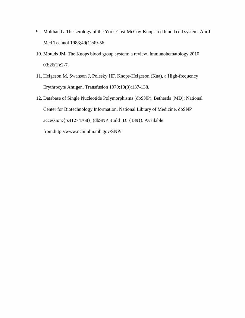
As previously mentioned, when testing sCR1 substance at dilutions greater than 1:100 a
decrease in reactivity was noted, but the reactions remained positive. Serologic results of 1:10
through 1:160 with varying incubation conditions (Table 2) show that only the 1:10 dilution of
the sCR1 substance was able to neutralize the Anti-Kna when incubated for thirty minutes at
room temperature. Unfortunately, not enough sCR1 substance remained after testing to do any
further testing at a 1:10 dilution to determine if the neutralization was specific and would not
neutralize other common antibodies.
The number of donors genotyped using each assay, as well as the number of rare
homozygous negative donors and those heterozygous for the genes, are summarized in Table 3.
Based on these results, we were able to calculate the observed allele frequency, which were used
to determine if the populations were in Hardy-Weinberg equilibrium by determining the
expected number of each genotype and using a Chi-square test to determine a p-value. The p-
values for McCa/McCb, Sl1/Sl2, and KCAM were less than 0.05 meaning the null hypothesis can
be rejected, and they are not in Hardy-Weinberg equilibrium (Table 4).
Of particular interest is that out of all the donors run using the Kn(a/b) assay, only 3
donors were Knb/Knb, 2 of which were already known to have anti-Kna. Therefore, at random
only of 1 out of 849 donors was negative for Kna. Due to these unexpected results, self-declared
ethnicities of the donors genotyped were analyzed to try to determine a cause for this
discrepancy (Table 5). Based on this data, it would appear that the frequency of Knb/Knb in our
Caucasian donor population 1 in 528 or approximately 0.19%.
With the donors screened, we were able to create a panel that would definitively identify
any of the five Knops antibodies that were included in this study (Figure 2). Two additional A
cells were included as extra panel cells due to their ability to help rule out certain other clinically
significant antibodies.
Discussion
Although serologic phenotyping with licensed reagents is a regulatory requirement for
donor unit labeling, genotyping has revolutionized blood banking. It has allowed for widespread
screening of donors for antigens, which allows donor centers to have a large stock of “pre-
screened” units to select for serological phenotyping.8 In addition, as shown in this study, it is
also possible to utilize genotyping to assist in the identification of antibodies for which we don’t
have very effective typing sera. To alleviate difficulties surrounding properly identifying Knops
antibodies, we integrated the use of patient genotyping, sCR1 substance neutralization, and a
Knops selected cell panel.
Serologically Misidentified Antibodies
Out of the seven patients/donors with identified Knops blood group system antibodies,
two with serologically identified anti-Kna genotyped Kna/Kna, which likely indicates that the
antibodies were misidentified. Upon further genotyping, one of these donors was negative for the
KCAM while the other was positive for all the high prevalence Knops antigens tested. Therefore,
anti-KCAM should be a suspect for the first donor and additional testing should be performed. If
we are to believe the second donor’s antibody was in the Knops system, there is the possibility
they may have an anti-Yka, because genotyping was not completed for Yka as the assay was still
under development at the time this study.
Unfortunately, as a result of the difficulty in identification of Knops blood group system
antibodies, it is a good possibility some of them are misidentified. This difficulty stems from the
variable weak strength of positive reactions and a lack of commercial antisera or quality patient
antibodies to use to type patients. However, misidentification within the Knops blood group
system (i.e. identifying an anti-KCAM as an anti-Kna) will not typically adversely affect the
patient since neither antibody is considered clinically significant,10 but it is of the utmost
importance to differentiate them from other clinically significant antibodies that they may mimic.
sCR1 Substance Benefits
A widely used technique to help identify Knops blood group system antibodies is the use
of DTT/AET to destroy the corresponding antigens to help confirm the identity of the antibody
and allow for the identification of underlying antibodies. However, if sCR1 substance was used
in conjunction, it would allow for a more definitive confirmation of these antibody
identifications and also allow for the exclusion of other antibodies whose antigens are sensitive
to DTT/AET, such as antibodies directed against Kell blood group system antigens. Although we
were able to effectively neutralize a Knops blood group system antibody, there was insufficient
quantities of the sCR1 substance to do additional testing to prove that it only neutralizes Knops
antibodies. In order for this procedure to be feasible, production of the sCR1 substance would
need to be scaled-up to produce a large enough quantity to perform validation and to support the
workload of suspected Knops antibodies.
Donor Genotype Anomalies
After genotyping over 2600 donors, two interesting topics for further research were
identified. As previously mentioned, the frequency of Knb/Knb donors in our Caucasian donor
population seems to be much lower than currently published values of one to two percent 9, 10.
However, when originally describing the first anti-Kna, Helgeson et al. published values very
similar to our own with 0.2% Knb/Knb donors.11 Additionally, our data is supported by allele
frequency information found in National Center for Biotechnology Information’s SNP database,
which cites two studies in which they found the allele frequencies for Kna and Knb to be 0.976
and 0.023 respectively in one study and 0.974 and 0.026 in the other.12 This discrepancy between
reported antigen and allele frequency may be due to the inaccuracy of phenotyping especially in
those patients with low copy numbers of the CR1 molecule who can falsely type Kn(a-) or due to
the use of patient antibodies as typing sera.
Additionally, all Knb/Knb, McCb/McCb and Sl2/Sl2 donors that we identified were also
KCAM-. As far as we are aware, this relationship has yet to be reported. This observation is
likely due to the closeness of these SNPs in exon 29 which results in recombination being very
rare. Out of these antigens the farthest SNP from the KCAM SNP is that which encodes for
Kna/Knb, which is only 54 amino acids away. However, this finding is worthy of additional
research. Finally, the McCa/McCb, Sl1/Sl2, and KCAM frequency results were not in Hardy-
Weinberg equilibrium, which may be due to bias in the sample tested. One such source of bias
was that many additional samples were tested for these antigens due to their negative results for
another Knops blood group system antigen and preferential selection of donors to test based on
reported race.
Creating Knops Antigen Negative Selected Cell Panel
With the possibility of screening large number of donors for Knops blood group system
antigen, it is possible to find enough antigen negative donors to create a Knops antigen negative
selected cell panel. However, if the goal was to be able to rule out all other clinically significant
antibodies in the presence of said antibodies many additional donors would need to be screened
due to the prevalence of these negative phenotypes in certain races and the prevalence of other
significant antigens in those races (i.e. McC(a-) is found mostly in blacks; double dose Fyb is rare
in blacks). For example, our panel is unable to rule out anti-Jkb in the presence of Anti-Kna.
Using the prevalence of the Kna in our donor population and published Jkb frequencies, it can be
determined that in order to find Jkb/Jkb & Knb/Knb cell we would need to genotype
approximately 2300 more Caucasian donors. In addition, donors included in the panel would
need zygosity testing run for the D antigen and genotyping assays would need to be analyzed to
determine if the black donors carried the Fy allele. By doing so, we would be able to determine if
the red cells were single dose or double dose for D and Fya and Fyb antigens.
Creation of such a panel of cells to help with the identification of Knops antibodies could
be invaluable in definitively ruling out other antibodies. In larger reference labs that routinely
identify Knops blood group system antibodies, this panel could be a supplemental panel prepared
weekly and run only when other testing or patient history indicates that the antibody being tested
is likely in the Knops system. However, even in smaller reference labs, this type of panel could
be extremely beneficial and worthwhile. After donors are identified, units or large samples of
blood could be drawn and frozen in small aliquots. Then whenever a Knops antibody is
suspected a set of these aliquots that makes up the panel could be thawed and used to confirm the
identity and rule out other clinically significant antibodies.
Conclusion
Once all three methods (Knops panel, sCR1 substance, and patient genotyping) are
validated and implemented, identification of Knops antibodies will be significantly improved
(Figure 3). If a technologist suspects a sample has a Knops antibody, the sample can be tested
using the sCR1 substance to prove it is directed against that system. Then, the sample could be
run against the Knops antigen negative selected cell panel to determine the exact specificity and
help rule out other clinically significant antibodies. Finally, in order to prove the specificity
identified is correct, the patient’s sample could be genotyped for the corresponding antigen to
prove the patient is antigen negative. However, if the patient is positive for the corresponding
antigen, sequencing exons 26 and 29 of the CR1/CD35 gene could help classify these Knops
related antibodies. This algorithm for identification is much improved versus traditional
serologic methods, because it lessens the doubt and guesswork that currently is involved in
identification of Knops antibodies. As a result, more immunohematology reference labs may
choose to do full identification of “HTLA” antibodies. Using these methodologies, technologists’
insecurity when identifying these antibodies can be alleviated providing the patient with the
safest transfusion possible.
References
1. Hourcade D, Miesner DR, Bee C, Zeldes W, Atkinson JP. Duplication and divergence of
the amino-terminal coding region of the complement receptor 1 (CR1) gene. An example
of concerted (horizontal) evolution within a gene. Journal of Biological Chemistry 1990
January 15;265(2):974-980.
2. Moulds JM, Thomas BJ, Doumbo O, Diallo DA, Lyke KE, Plowe CV, et al.
Identification of the Kna/Knb polymorphism and a method for Knops genotyping.
Transfusion 2004 02;44(2):164-169.
3. Moulds JM, Zimmerman PA, Doumbo OK, Kassambara L, Sagara I, Diallo DA, et al.
Molecular identification of Knops blood group polymorphisms found in long
homologous region D of complement receptor 1. Blood 2001;97(9):2879-2885.
4. Moulds JM, Zimmerman PA, Diallo OK, Atkinson JP, Krych-Goldberg M, Hourcade
DE, et al. Expansion of the Knops blood group system and subdivision of Sl[sub a].
Transfusion 2002 02;42(2):251-256.
5. Veldhuisen B, Ligthart PC, Vidarsson G, Roels I, Folman CC, van dS, et al. Molecular
analysis of the York antigen of the Knops blood group system. Transfusion 2011
07;51(7):1389-1396.
6. Moulds JM, Pierce S, Peck KB, Tulley ML, Doumbo O, Moulds JJ. KAM: a new allele
in the Knops Blood Group System (abstract). Transfusion 2005;45(Supplement s3):27a.
7. Moulds JM, Rowe KE. Neutralization of Knops system antibodies using soluble
complement receptor 1. Transfusion 1996 06;36(6):517.
8. Denomme GA, Johnson ST, Pietz BC. Mass-scale red cell genotyping of blood donors.
Transfusion Apheresis Sci 2011 2;44(1):93-99.
9. Molthan L. The serology of the York-Cost-McCoy-Knops red blood cell system. Am J
Med Technol 1983;49(1):49-56.
10. Moulds JM. The Knops blood group system: a review. Immunohematology 2010
03;26(1):2-7.
11. Helgeson M, Swanson J, Polesky HF. Knops-Helgeson (Kna), a High-frequency
Erythrocyte Antigen. Transfusion 1970;10(3):137-138.
12. Database of Single Nucleotide Polymorphisms (dbSNP). Bethesda (MD): National
Center for Biotechnology Information, National Library of Medicine. dbSNP
accession:{rs41274768}, (dbSNP Build ID: {139}). Available
from:http://www.ncbi.nlm.nih.gov/SNP/

TABLE 1. SNP position and Amino Acid Change for Knops Antigens
Antigen Position Amino Acid Change
Kn(a/b) CR1 4681G>A V1561M
McC(a/b) CR1 4786A>G K1590E
Sl1/Sl2 CR1 4801A>G R1601G
Sl3+/Sl3- CR1 4828T>A/4801A S1610T/R1601
KCAM+/KCAM- CR1 4843A>G I1615V
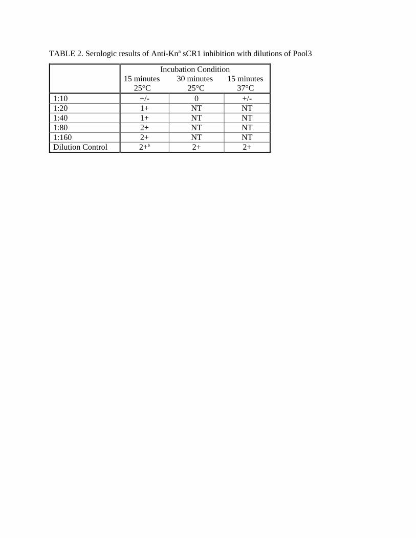
TABLE 2. Serologic results of Anti-Kna sCR1 inhibition with dilutions of Pool3
Incubation Condition
15 minutes 30 minutes 15 minutes
25°C 25°C 37°C
1:10 +/- 0 +/-
1:20 1+ NT NT
1:40 1+ NT NT
1:80 2+ NT NT
1:160 2+ NT NT
Dilution Control 2+s 2+ 2+
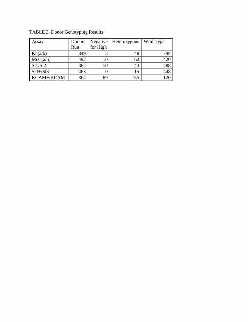
TABLE 3. Donor Genotyping Results
Assay Donors
Run
Negative
for High
Heterozygous Wild Type
Kn(a/b) 849 3 48 798
McC(a/b) 492 10 62 420
Sl1/Sl2 382 50 43 288
Sl3+/Sl3- 463 0 15 448
KCAM+/KCAM- 364 89 155 120
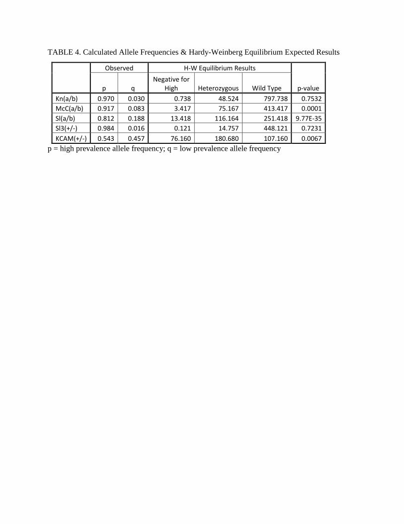
TABLE 4. Calculated Allele Frequencies & Hardy-Weinberg Equilibrium Expected Results
Observed H-W Equilibrium Results
p-value p q Negative for
High Heterozygous Wild Type
Kn(a/b) 0.970 0.030 0.738 48.524 797.738 0.7532
McC(a/b) 0.917 0.083 3.417 75.167 413.417 0.0001
Sl(a/b) 0.812 0.188 13.418 116.164 251.418 9.77E-35
Sl3(+/-) 0.984 0.016 0.121 14.757 448.121 0.7231
KCAM(+/-) 0.543 0.457 76.160 180.680 107.160 0.0067
p = high prevalence allele frequency; q = low prevalence allele frequency
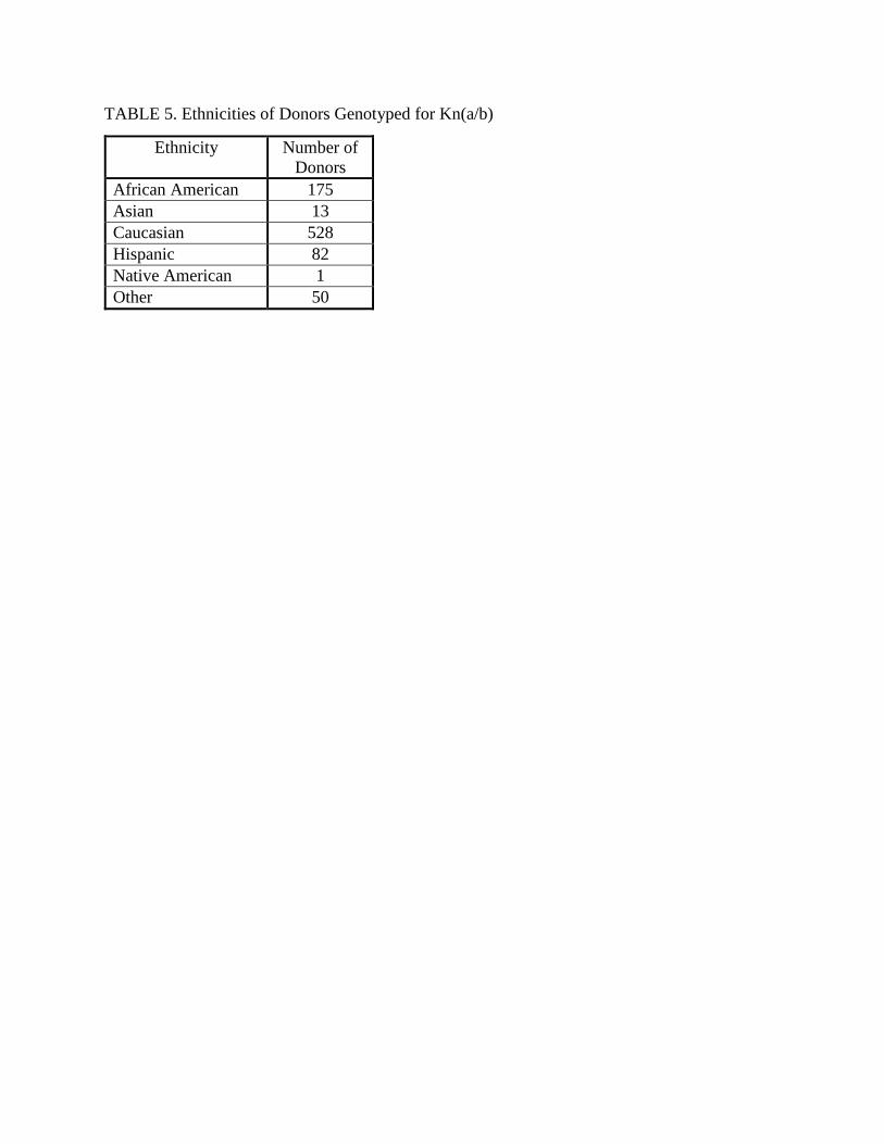
TABLE 5. Ethnicities of Donors Genotyped for Kn(a/b)
Ethnicity Number of
Donors
African American 175
Asian 13
Caucasian 528
Hispanic 82
Native American 1
Other 50
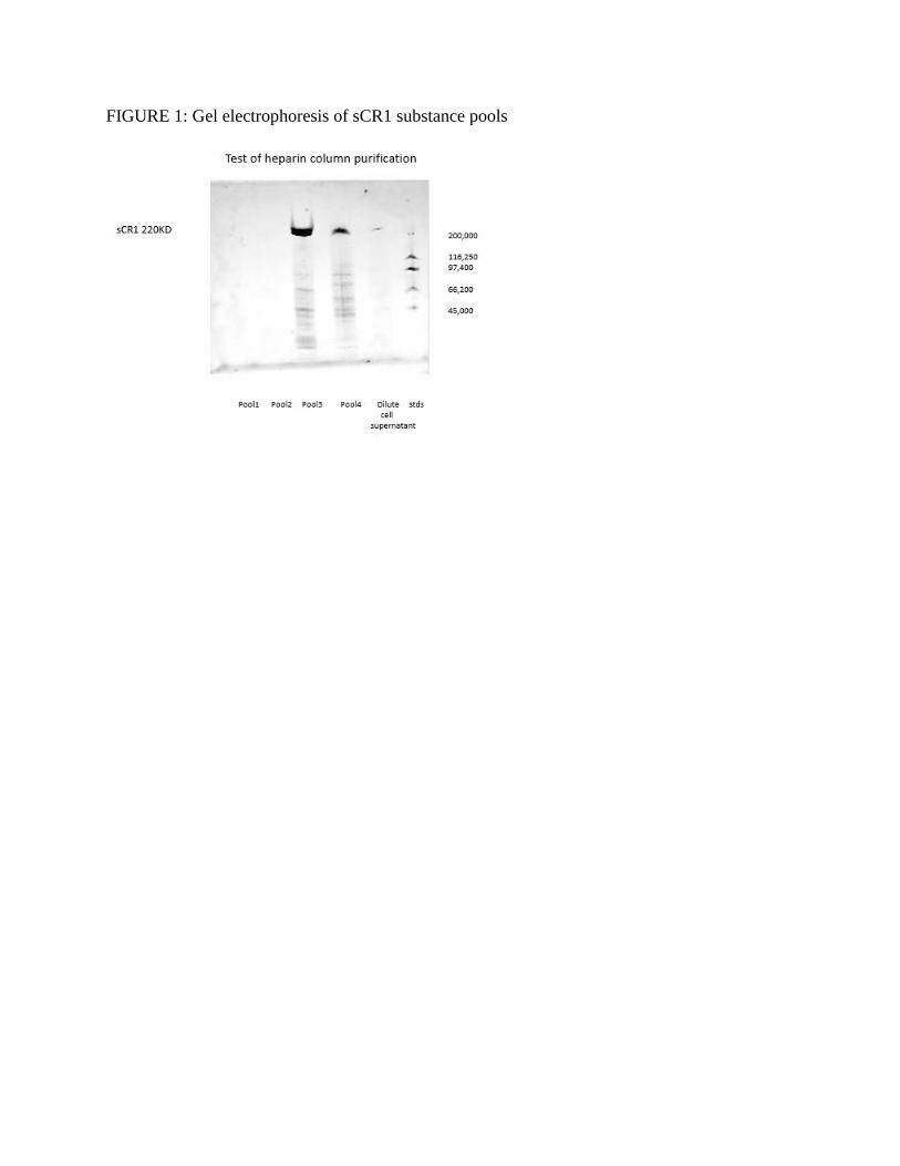
FIGURE 1: Gel electrophoresis of sCR1 substance pools
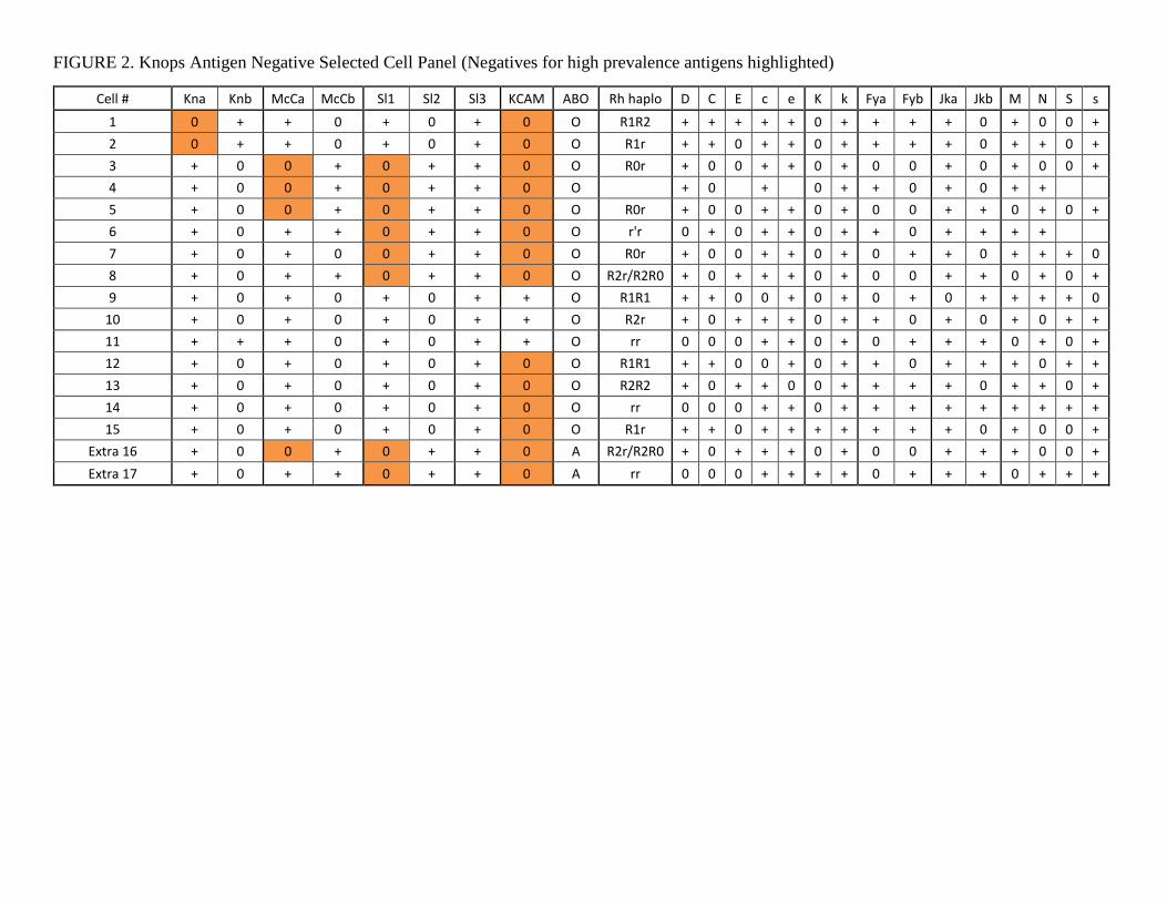
FIGURE 2. Knops Antigen Negative Selected Cell Panel (Negatives for high prevalence antigens highlighted)
Cell # Kna Knb McCa McCb Sl1 Sl2 Sl3 KCAM ABO Rh haplo D C E c e K k Fya Fyb Jka Jkb M N S s
1 0 + + 0 + 0 + 0 O R1R2 + + + + + 0 + + + + 0 + 0 0 +
2 0 + + 0 + 0 + 0 O R1r + + 0 + + 0 + + + + 0 + + 0 +
3 + 0 0 + 0 + + 0 O R0r + 0 0 + + 0 + 0 0 + 0 + 0 0 +
4 + 0 0 + 0 + + 0 O + 0 + 0 + + 0 + 0 + +
5 + 0 0 + 0 + + 0 O R0r + 0 0 + + 0 + 0 0 + + 0 + 0 +
6 + 0 + + 0 + + 0 O r'r 0 + 0 + + 0 + + 0 + + + +
7 + 0 + 0 0 + + 0 O R0r + 0 0 + + 0 + 0 + + 0 + + + 0
8 + 0 + + 0 + + 0 O R2r/R2R0 + 0 + + + 0 + 0 0 + + 0 + 0 +
9 + 0 + 0 + 0 + + O R1R1 + + 0 0 + 0 + 0 + 0 + + + + 0
10 + 0 + 0 + 0 + + O R2r + 0 + + + 0 + + 0 + 0 + 0 + +
11 + + + 0 + 0 + + O rr 0 0 0 + + 0 + 0 + + + 0 + 0 +
12 + 0 + 0 + 0 + 0 O R1R1 + + 0 0 + 0 + + 0 + + + 0 + +
13 + 0 + 0 + 0 + 0 O R2R2 + 0 + + 0 0 + + + + 0 + + 0 +
14 + 0 + 0 + 0 + 0 O rr 0 0 0 + + 0 + + + + + + + + +
15 + 0 + 0 + 0 + 0 O R1r + + 0 + + + + + + + 0 + 0 0 +
Extra 16 + 0 0 + 0 + + 0 A R2r/R2R0 + 0 + + + 0 + 0 0 + + + 0 0 +
Extra 17 + 0 + + 0 + + 0 A rr 0 0 0 + + + + 0 + + + 0 + + +
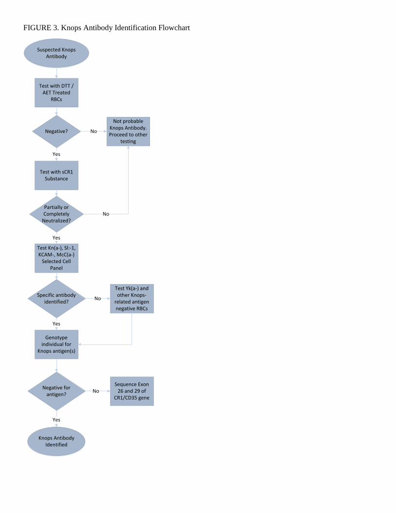
FIGURE 3. Knops Antibody Identification Flowchart
Suspected Knops Antibody
Test with DTT / AET Treated
RBCs
Negative?
Not probable Knops Antibody. Proceed to other
testing
Test with sCR1 Substance
Yes
Partially or Completely
Neutralized?No
Test Kn(a-), Sl:-1, KCAM-, McC(a-)
Selected Cell Panel
Yes
Specific antibody identified?
Test Yk(a-) and other Knops-
related antigen negative RBCs
No
No
Genotype individual for
Knops antigen(s)
Yes
Negative forantigen?
No
Yes
Knops Antibody Identified
Sequence Exon 26 and 29 of
CR1/CD35 gene