Embed Size (px)
Citation preview

Neural Control of the Splanchnic Circulation
in AngII-salt Hypertension
A DISSERTATION
SUBMITTED TO THE FACULTY OF
UNIVERSITY OF MINNESOTA
BY
Marcos Takuya Kuroki
IN PARTIAL FULFILLMENT OF THE REQUIREMENTS
FOR THE DEGREE OF
DOCTOR OF PHILOSOPHY
John W. Osborn, PhD
Advisor
May, 2014

© Marcos Takuya Kuroki, 2014

i
Acknowledgements
First and foremost, I would like to thank my advisor, Dr. John
Osborn, for his continuous support throughout my training. I truly
appreciate his dedication toward his students, his generosity with his
time and willingness to provide whatever laboratory resources were
needed for his student’s success. His optimism, no matter what the
circumstances, kept me on track, and proved to be a key driving force
towards the completion of this thesis.
I would also like to thank the members of my thesis committee, Dr.
Virginia Seybold, Dr. William Engeland, and Dr. Alessandro
Bartolomucci for their feedback and suggestions. I would also like to
thank the late Dr. Zofia Zukowska for her support and encouragement in
the early phase of my training.
I extend my gratitude to my mentor in Japan, Dr. Kenju Miki, who
agreed to train me on the art of peripheral nerve recording in conscious
rats, and helped me apply his technique to perform splanchnic nerve
recordings in the conscious rat.
My friends Adam and Moony deserve a special thank you for their
encouragement and providing an outsider’s perspective to my work. I
would like to thank my colleagues in lab, Britta, Jason, Dusty, and Pilar
for their intellectual and technical contributions to my work.
Finally, I would not have been able to complete my graduate training
without the daily support from my wife, Sheila, and lifelong support from
my parents Tetsuya and Kaori, my sister, Mai, and my grandparents,
Shizuya and Yu Kuroki, and Shojiro and Sachiko Matsuo.

ii
Dedication
To my family, for laying the foundations.

iii
Abstract
Sympathetic nervous system (SNS) activity is elevated in some forms
of essential hypertension. What causes sympathetic tone to be elevated,
and how it mediates hypertension, however, is unclear. Angiotensin II
(AngII) and a high dietary salt appears to be involved since in rats,
chronic peripheral infusion of AngII induces a form of hypertension that
is accompanied by increased indices of peripheral sympathetic tone
selectively when they are fed a high sodium diet. Studies in this model
have shown that contrary to prevailing views, peripheral sympathetic
tone was diminished to the kidneys, but instead, suggested that it may be
elevated selectively to the splanchnic vascular bed. Based on these
findings, the initial aim of this thesis was to characterize, in conscious
rats, the local hemodynamics within the splanchnic vascular bed and the
role of the SNS in mediating changes in splanchnic vascular
hemodynamics during AngII-salt hypertension. Studies were carried out
to test the hypothesis that in addition to sympathetically mediated
increases in splanchnic venous tone, AngII-salt hypertension was
mediated by enhanced sympathetic vasoconstriction of splanchnic
arterioles occurring through its peripheral sympathetic nerve supply.
Splanchnic vascular resistance was found to be elevated in AngII-salt
hypertensive rats; however, these hemodynamic changes occurred even
after removal of direct sympathetic innervation to the splanchnic vascular
bed by surgical denervation (celiac ganglionectomy). Furthermore, unlike
previously shown, celiac ganglionectomy did not result in lowering of
blood pressure during AngII-salt hypertension. Thus, contrary to the
original hypothesis, changes in direct sympathetic input to the splanchnic
vasculature did not mediate AngII-salt hypertension. Additional studies

iv
in this thesis found that part of the problem with this inconsistent
finding may be related to the technique commonly used to generate the
model. Furthermore, studies in this thesis found, using chronic
pharmacological adrenergic blockade, that the contribution of the SNS in
AngII-salt hypertension may have been overestimated. Thus, the
combined findings in this thesis and prior studies suggest that a fraction
of AngII-salt hypertension is mediated by enhanced peripheral
sympathetic tone, not through direct vasoconstrictive input to the
splanchnic vasculature, but possibly via its influence on other non-renal
splanchnic organs.

v
Table of Contents
Acknowledgements ........................................................................ i
Dedication .................................................................................... ii
Abstract ....................................................................................... iii
Table of Contents ........................................................................ v
List of Tables ............................................................................ viii
List of Figures ............................................................................ ix
Chapter 1: Introduction ..................................................................... 1
1.1 Rationale ...................................................................................... 2
1.2 AngII-induced hypertension and the AngII-salt model of
experimental hypertension ........................................................... 6
1.3 Focus and organization of thesis ................................................ 10
1.4 Figures ....................................................................................... 13
Chapter 2: Time-dependent changes in autonomic control of
splanchnic vascular resistance and heart rate in ANG II-salt
hypertension ........................................................................................ 16
Chapter Overview .................................................................................. 17
2.1 Introduction ............................................................................... 18
2.2 Materials and Methods .............................................................. 21
2.3 Results ....................................................................................... 25
2.4 Discussion .................................................................................. 29
2.5 Figures ....................................................................................... 39

vi
Chapter 3: Effect of Celiac Ganglionectomy on Splanchnic
Hemodynamics in AngII-Salt Hypertensive Rats ....................... 46
Chapter Overview .................................................................................. 47
3.1 Introduction ............................................................................... 48
3.2 Methods ..................................................................................... 50
3.3 Results ....................................................................................... 57
3.4 Discussion .................................................................................. 60
3.5 Figures and Table ...................................................................... 70
Chapter 4: Effect of chronic α1/2β1-adrenergic receptor blockade
on the development of AngII-Salt Hypertension ........................ 83
Chapter Overview .................................................................................. 84
4.1 Introduction ............................................................................... 85
4.2 Methods ..................................................................................... 88
4.3 Results ....................................................................................... 96
4.4 Discussion ................................................................................ 101
4.5 Figures and Table .................................................................... 110
Chapter 5: Conclusion .................................................................... 125
5.1 Summary of main findings ....................................................... 127
5.2 Implications of combined findings ............................................ 131
5.3 Figures and Table .................................................................... 135
Bibliography ...................................................................................... 138

vii
Appendix 1: Comparison of arterial pressure and plasma AngII
responses to three methods of subcutaneous AngII
administration ................................................................................... 154
Chapter Overview ................................................................................ 155
6.1 Introduction ............................................................................. 156
6.2 Methods ................................................................................... 160
6.3 Results ..................................................................................... 168
6.4 Discussion ................................................................................ 171
6.5 Figures ..................................................................................... 178

viii
List of Tables
Chapter 3
Table 3.1. Baseline parameters ......................................................... 71
Chapter 4
Table 4.1. Change in body weight in α1/2β1-AR antagonist treated and
vehicle treated rats .................................................................. 113
Table 4.2. Level of α1-AR blockade during chronic α1/2β1-AR
antagonist treatment ................................................................ 117

ix
List of Figures
Chapter 1
Figure 1.1. Central Hypothesis ......................................................... 14
Chapter 2
Figure 2.1. Original tracing for typical AP, MBF, MVR, and HR
response to ganglionic blockade ................................................. 40
Figure 2.2. Change in MAP, MBF, MVR, and HR during 2wk AngII
infusion in high and low salt rats ............................................... 42
Figure 2.3. MAP, MVR, and HR response to acute ganglionic
blockade during AngII induced HTN in high and low salt rats . 44
Chapter 3
Figure 3.1. Tissue NE content .......................................................... 73
Figure 3.2. Daily food/water intake, changes in body WT during
AngII-salt hypertension .............................................................. 75
Figure 3.3. Representative AP, MBF Waveform .............................. 77
Figure 3.4. 24hr hemodynamic profile of MAP, MVR, MBF, and HR
during AngII-salt hypertension .................................................. 79
Figure 3.5. Changes in responsiveness of HR, MAP and MVR to acute
ganglionic blockade during AngII-salt hypertension .................. 81
Chapter 4
Figure 4.1. 24hr Food and Water Intake in α1/2β1-AR antagonist
treated and vehicle treated rats ............................................... 111
Figure 4.2. Blood pressure response to acute phenylephrine injection
in α1/2β1-AR antagonist treated and vehicle treated rats ......... 115

x
Figure 4.3. Effect of α1/2β1-AR blockade on MAP and HR during
AngII-induced hypertension in rats fed a 2% or 0.1% NaCl diet
................................................................................................. 119
Figure 4.4. Effect of dietary salt on MAP and HR during AngII-
induced hypertension in α1/2β1-AR antagonist treated and vehicle
treated rats .............................................................................. 121
Figure 4.5. Estimated time course and magnitude of the neurogenic
component of AngII-salt hypertension ..................................... 123
Chapter 5
Figure 5.1. Revised Central Hypothesis .......................................... 136
Appendix 1
Figure 6.1. Spontaneous drop in MAP during AngII-salt hypertension
using Alzet pumps ................................................................... 179
Figure 6.2. Description of study protocol ....................................... 181
Figure 6.3. Changes in plasma AngII levels in response to
physiological salt loading, water deprivation, and pharmacological
salt depletion ............................................................................ 183
Figure 6.4. Differences in MAP profile of AngII-salt hypertension
generated using Alzet, Harvard or iPrecio pumps ................... 185
Figure 6.5. Changes in plasma AngII levels during AngII-salt
hypertension generated using Alzet, Harvard or iPrecio pumps187

1
Chapter 1
Introduction
1 Chapter 1: Introduction 1 Chapter 1 1 Chapter 1

2
1.1 Rationale
The renewed interest of targeting the sympathetic nervous system as a treatment for hypertension
It’s been known through observational studies that the risk of death
attributable to ischemic heart disease and stroke is related linearly to
the level of blood pressure starting from a systolic blood pressure (SBP)
of 115mmHg and a diastolic blood pressure (DBP) of 75mmHg (70).
Because of this wide range, the definition of a high blood pressure as a
“disease” is somewhat arbitrary. However, the public health burden of
suboptimal blood pressure is clear; it shortens an individual’s life
expectancy by as much as 5 years (43), and it has been reported to be
the number one attributable risk of death throughout the world (21).
The current cutoff for classifying blood pressure levels as
“hypertensive” is based on observational data showing that adults at
low-risk of developing cardiovascular diseases can benefit from blood
pressure lowering interventions to a target SBP < 140mmHg and DBP
of < 90 mmHg (113). Under this definition, 50 million or more
Americans is afflicted by the disease, with 2009 to 2010 prevalence
estimated at 30.5% among men and 28.5% among women in the United
States (48). Worldwide, it is estimated that 972 million adults have
hypertension, with the number predicted to rise to a total of 1.56 billion
by 2025 (59). Given its current trend, hypertension is projected to be
the single most important risk factor of cardiovascular diseases by the
year 2020 (59).
Despite improvements in pharmacological therapy seen in the past
half-century and their efficacy in lowering blood pressure in many cases
of hypertension, only 40% and 56% of hypertensive men and women,

3
respectively, have their blood pressure effectively controlled in the
United States (48). Although factors such as patient compliance may
play a role to this relatively poor control rate (20), this is also likely
because the current understanding on the etiology of hypertension is
incomplete as multiple physiological abnormalities driven by interactions
between genetic, behavioral and environmental factors can cause
hypertension (16).
Although once controversial, there is now indisputable evidence that
increased sympathetic nervous system activity (SNA) is one such
physiological abnormality that plays a key role in the etiology of human
hypertension (35). However, the clinical use of sympathetic nervous
system (SNS) targeting therapies have been in decline, and has been the
“forgotten pathway” in the treatment of hypertension (36). This is
mostly because current pharmacotherapy not only block sympathetic
control of arterial pressure, but many other functions as well, resulting
in unwanted side effects and reduced patient compliance.
This view has recently changed since the exciting recent
demonstration in human patients of long-term antihypertensive
responses to a novel device-based method of selective renal denervation
(37, 105), fueling renewed interest in therapies targeting SNS in
hypertension. In order to understand the physiological impact, efficacy,
and potential benefit of such therapies to a wide range of hypertensive
patients, however, would require a better understanding of how elevated
SNA contributes to hypertension, specifically, by elucidating the
principal effector organs of elevated SNA and the specific patterns of
sympathetic outflow that result in the altered hemodynamic state.

4
Sympathetic target organs other than the kidneys may play an important role in hypertension
It has long been thought that the principal effector organ of elevated
SNA in neurogenic forms of hypertension is the kidney. Activation of
sympathetic efferents to the kidney would favor salt and water
retention, leading to expansion of blood volume and increase in blood
pressure. Over time, autoregulatory changes in the peripheral
vasculature would occur to counteract tissue overperfusion, resulting in
elevated total peripheral resistance (TPR), a hallmark hemodynamic
change in hypertension (72). Indeed, the rationale for targeting the renal
nerves in recent clinical trials was largely based on this theory. This
hypothesis initially appeared to be supported by the fact that arterial
pressure is reduced for 2 years following a single renal denervation
procedure (37, 105). However, in one case report in which SNA to
skeletal muscle (MSNA) was measured before and after renal
denervation, it was found to be reduced from 56 bursts/min before the
procedure to 41 bursts/min 1 month later and 19 burst/min 1 year after
the procedure (94). In addition, it has been shown in a recent case series
of 35 patients with resistant hypertension that reductions in MSNA after
renal denervation is more pronounced when measured at the single-unit
level, compared to multi-unit MSNA (51). This observation, coupled
with the fact that the magnitude of the decrease in whole body
norepinephrine spillover following renal denervation cannot be explained
by loss of renal efferent activity alone, suggests that the procedure
decreases SNA to non-renal vascular beds as well (94).
The mechanism by which renal denervation in humans lead to a
reduction in peripheral sympathetic nerve activity is currently unknown.

5
One possibility that has been proposed is that renal denervation results
in the destruction of renal afferent nerves which drive
sympathoexcitation to other cardiovascular organs (94). This raises the
possibility that part or majority of the antihypertensive response to
renal denervation could be secondary to withdrawal of sympathetic tone
to non-renal vascular bed.
Studies in an experimental model of hypertension suggest the role of the splanchnic vascular bed as an important non-renal sympathetic target organ in hypertension
Through experiments using the AngII-salt model of hypertension in
rats, a neurogenic model for human hypertension, our group has recently
uncovered the potential critical role for the sympathetic regulation of
splanchnic vascular bed in the development of hypertension (64).
Previous reports in humans support this finding and the possible role for
the splanchnic vasculature in the development of hypertension. First,
surgical splanchnicectomy was an effective treatment for hypertension
prior to the advent of pharmacotherapy (100); and secondly, vascular
resistance has been reported to be elevated in the hepatosplanchnic
circulation before any other vascular bed in humans with borderline
hypertension (102). Thus, elucidating the contribution of sympathetic
control of the splanchnic vasculature in the development of experimental
AngII-salt hypertension may provide further insights into the role of
neural control of non-renal vascular beds in hypertension, and discovery
of novel targeted therapies.

6
1.2 AngII-induced hypertension and the AngII-salt model of experimental hypertension
(NOTE: The first two sections has been previously published in a
review article I have coauthored with John W. Osborn, PhD and
Gregory D. Fink, PhD (86))
AngII-induced activation of the sympathetic nervous system is dependent on salt intake
Hypertension caused by infusion of AngII in animals involves
multiple control systems whose influence on arterial pressure is
dependent on the dose of AngII as well as the presence of other factors
such as salt intake (85, 87). Doses of AngII that increase arterial
pressure slowly over a course of days to weeks produce what is
commonly referred to as the “slow pressor AngII” model. It is thought
that the hypertension is mediated, at least in part, by an elevated level
of sympathetic nerve activity (SNA) (9, 38, 39). It has long been known
that the severity of AngII-induced hypertension is directly dependent on
the prevailing level of salt intake; and more recent studies suggest that
the level of sympathoactivation is as well (63, 64). Thus, it is important
to keep in mind that neurogenic mechanisms may not play an equally
important role in “AngII-induced” hypertension in animals subjected to a
normal or low salt intake as they would in those subjected to a high salt
intake (“AngII-salt” hypertension).
The salt-sensitive nature of AngII-induced hypertension often has
been ignored in the literature, and this could account for the
contradictory conclusions about the role of the sympathetic nervous
system in the model. Two different methods for assessing the role of the

7
sympathetic nervous system in AngII-induced hypertension have
commonly been employed: 1) changes in the depressor response to
ganglionic blockade, and 2) changes in tissue or plasma norepinephrine
(NE) concentration and turnover. These indices serve as an indicator for
AngII induced changes in peripheral SNA, which can be generated at
any level of the neuraxis. Changes in responses to ganglion blockade
suggest changes in SNA effects on arterial pressure. However, it is
important to note that changes in plasma NE and NE turnover do not
necessarily reflect changes in SNA that directly affect arterial pressure.
Our group and others have reported a 5-7 day delayed increase in the
acute depressor response to ganglionic blockade in AngII-induced (13)
and AngII-salt (63) models a finding consistent with the hypothesis that
AngII hypertension is due, in part, to delayed activation of peripheral
sympathetic outflow. In contrast, tissue NE has been reported to be
regionally decreased in AngII-induced rats (65) and plasma NE
unchanged in AngII-induced rabbits (11). A factor that may explain
these disparate findings is that the level of sympathetic outflow
measured by these indices during AngII-induced hypertension is highly
dependent on the level of dietary salt intake. Our studies have
demonstrated that both an increase in the response to ganglionic
blockade and a parallel increase in whole body NE spillover is present in
rats on a high but not a normal salt diet. These increases are not
observed until 5-7 days of AngII administration (63, 64). These findings
suggest that administration of AngII when combined with a high salt
diet leads to a delayed enhancement of peripheral sympathetic outflow
that contributes to, but does not exclusively cause, the associated
hypertension.

8
The splanchnic vascular bed is the critical neural target in AngII-salt hypertension
Another potential explanation for the disparate findings between
laboratories regarding the contribution of the sympathetic nervous
system to AngII-induced hypertension is a focus on the kidney as the
most important sympathetic effector organ in long-term blood pressure
regulation. This long-standing view stems from the kidney’s role in
regulation of blood volume, which has been hypothesized to be directly
linked to the long-term control of arterial pressure (49). The concept is
supported by reports that renal denervation prevents some forms of
experimental neurogenic hypertension (29, 56) as well as by recent
studies showing that renal denervation results in sustained decreases in
arterial pressure in humans with drug-resistant hypertension (95).
However, it is important to note that these studies have not
demonstrated that renal denervation decreases arterial pressure
secondary to loss of efferent neural control of kidney function and
subsequent changes in blood volume. To the contrary, there is a building
consensus that the response of arterial pressure to renal denervation is
due to destruction of sensory fibers from the kidney resulting in
decreased SNA to other vascular beds such as skeletal muscle, as was
recently reported in humans (94).
In regard to the contribution of renal nerves to AngII-induced
hypertension specifically, a number of studies have consistently found
that renal SNA is decreased in this model, irrespective of salt intake.
Indirect assessment of renal SNA in dogs suggested that it was
decreased in AngII-induced hypertension (73), a finding that was later
confirmed in rabbits in the first study to record SNA directly over a

9
period of weeks (4). We have recently reported similar results using
direct long-term recording of renal SNA in AngII-salt rats (115). In
addition, renal denervation does not prevent AngII-salt hypertension in
the rat (64) or AngII-induced hypertension in the rabbit (13). Although
these observations have been used as an argument against the role of the
sympathetic nervous system in AngII-induced hypertension (82), this
view assumes that the kidney is the only neural target that can result in
hypertension and disregards the contribution of changes in sympathetic
nerve activity to non-renal vascular beds to the pathogenesis of
neurogenic hypertension.
We have addressed this issue by utilizing a number of indirect and
direct methods to assess the relative importance of SNA to renal and
non-renal vascular beds in AngII-salt hypertension. Based on direct
long-term recording of lumbar SNA and hind limb norepinephrine
spillover (61, 115), as well as lumbar sympathectomy (Fink, unpublished
observation), we conclude that SNA to skeletal muscle does not
contribute to AngII-salt hypertension. On the other hand, in contrast to
the finding that renal denervation has no effect on this model,
denervation of the splanchnic vascular bed by celiac ganglionectomy
(CGX) markedly attenuates the neurogenic phase of AngII-salt
hypertension (64). This finding is consistent with an earlier study in
which direct recording of splanchnic SNA revealed it was increased in
AngII-induced hypertensive rats compared to normotensive controls
(76). Collectively these studies demonstrate that SNA is differentially
regulated in AngII-salt rats and, more importantly, suggest that the
splanchnic vascular bed is the primary target of the sympathetic nervous
system in this model of hypertension.

10
Hemodynamic mechanism by which an enhanced sympathetic vasomotor tone to splanchnic vascular bed contributes to AngII-salt hypertension
The hemodynamic mechanism by which an increase in sympathetic
vasomotor tone to the splanchnic vascular bed can lead to hypertension
is partially uncovered and has been largely attributed to a reduction of
vascular capacitance secondary to venoconstriction at the splanchnic
vascular bed (62, 64). Decrease in venous capacitance would lead to a
shift of blood volume from the venous to the less compliant arterial
compartment of the circulation, resulting in a rise in arterial blood
pressure (40). It remains currently unknown, however, whether elevated
SNA to the splanchnic vascular bed also mediates constriction of
splanchnic resistance arteries. It has been shown in a select cohort of
human prehypertensives that splanchnic vascular resistance is elevated
(102). Given anatomical evidences that the majority of postganglionic
nerves in the celiac-superior mesenteric ganglia dually innervate both
veins and arteries (53), it is very likely that changes in SNA to the
splanchnic vascular bed will result in changes both to veins and arteries.
Thus, it is hypothesized that hypertension in this model is caused by a
concerted action of reduced vascular capacitance and elevated total
peripheral resistance due to sympathetically mediated constriction of the
splanchnic vascular bed.
1.3 Focus and organization of thesis
The goal of this thesis was to further clarify the role of sympathetic
vasomotor tone to the splanchnic vascular bed in the rat model of
AngII-salt hypertension. This work was motivated by 3 main prior

11
findings discussed above: 1) vascular capacitance, a measure of systemic
venous compliance of which the majority is determined by splanchnic
venous tone, was decreased during AngII-salt hypertension, 2) reduction
in vascular capacitance was reversible by ganglionic blockade, and 3)
sympathetic denervation of splanchnic organs by celiac ganglionectomy
(CGx) attenuated AngII-salt hypertension and prevented the reduction
in vascular capacitance. These 3 findings, and other related work
supporting the sympathoexcitatory role of AngII, led to the working
hypothesis that AngII-salt hypertension in the rat is mediated, in part,
by an increased sympathetic vasomotor tone to the splanchnic vascular
bed, which elevates pressure by reducing vascular capacitance and
increasing total peripheral resistance. A schematic view of this
hypothesis is illustrated in Figure 1.1.
The major limitation of the previous work was that the conclusions
were mainly based on measures of whole body cardiovascular parameters
coupled with targeted denervation to infer the role of sympathetic
vasomotor tone to the splanchnic vascular bed. To overcome this
limitation, I devised a surgical technique for continuous monitoring of
superior mesenteric artery blood flow, in addition to arterial pressure, in
conscious, freely moving animals. This allowed for the monitoring of
hemodynamic changes specifically at the splanchnic vascular bed and
calculation of mesenteric vascular resistance, a direct index of splanchnic
arteriolar tone.
The work in this thesis was organized into 3 main chapters and a
supporting chapter in the form of an appendix. In Chapter 2, I
determined whether changes consistent with the hypothesis occur to
splanchnic vascular resistance during AngII-salt hypertension. In

12
Chapter 3, I assessed whether changes in splanchnic vascular resistance
were determined by sympathetic input to splanchnic vascular bed.
Based on findings in Chapter 2 and 3, I reassessed the contribution of
global sympathetic tone to the development of AngII-salt hypertension
in Chapter 4. Finally, in Chapter 5, I provide a summary of all findings
and implications to the original hypothesis.
In Chapter 2, the AngII-salt model was generated by subcutaneous
infusion of AngII using an implantable osmotic minipump. Observations
in Chapter 2 warranted an optimization to the method of AngII delivery
for improving the stability of the model. The results from this study are
presented in Appendix 1. Studies in Chapters 3 and 4 were performed
using the optimized method of AngII delivery based on results presented
in Appendix 1.

13
1.4 Figures

14
Figure 1.1. Central Hypothesis
Diagram depicting the central and peripheral neural pathways and
major sympathetic end organs thought to be involved in the neurogenic
mechanism of AngII-salt hypertension. We hypothesized that AngII and
salt stimulate sympathetic premotor neurons in the brain leading to
activation of peripheral sympathetic pathways in a site-specific manner.
Prior studies have shown that renal and lumbar sympathetic nerve
activities are decreased or unchanged, respectively, during AngII-salt
hypertension. Additionally, cardiac denervation by stellate
ganglionectomy has no effect on AngII-salt hypertension. These results
suggest that sympathetic modulation of cardiac and renal function, and
skeletal muscle vascular tone plays little or no role in the neurogenic
mechanism of AngII-salt hypertension (see text). Indirect evidence
suggests that splanchnic sympathetic nerves may be preferentially
activated during AngII-salt hypertension, causing a reduction in
splanchnic vascular capacitance, which increases effective circulating
blood volume. Additionally, an increase in sympathetic outflow to the
splanchnic vascular bed is also thought to increase splanchnic vascular
resistance. The central hypothesis of this thesis is that these combined,
sympathetically mediated changes in splanchnic vascular capacitance
and resistance are the primary neurogenic mechanisms responsible for
the sustained increase in arterial pressure during AngII-salt
hypertension.

15
Figure 1.1

16
Chapter 2
Time-dependent changes in autonomic control of splanchnic vascular resistance and heart rate
in ANG II-salt hypertension
Marcos T. Kuroki, Pilar A. Guzman, Gregory D. Fink, and John W. Osborn
American journal of physiology. Heart and circulatory physiology 302: H763--H769, 2012
2 Chapter 2: Time-dependent changes in autonomic control of splanchnic vascular resistance and heart rate in ANG II-salt
hypertension 2 Chapter 2 2 Chapter 2

17
Chapter Overview
Previous studies suggest that AngII-induced hypertension in rats fed
a high salt diet (AngII-salt hypertension) has a neurogenic component
dependent on an enhanced sympathetic tone to the splanchnic veins,
and independent from changes in sympathetic nerve activity to the
kidney or hind limb. The purpose of this study was to extend these
findings and test whether altered autonomic control of splanchnic
resistance arteries and the heart also contributes to the neurogenic
component. Mean arterial pressure (MAP), heart rate (HR), superior
mesenteric artery blood flow, and mesenteric vascular resistance (MVR)
were measured during 4 control days , 14 days of AngII delivered
subcutaneously (150ng/kg/min), and 4 days of recovery in conscious rats
fed a high salt (HS; 2% NaCl) or low salt (LS; 0.1% NaCl) diet.
Autonomic effects on MAP, HR and MVR was assessed by acute
ganglionic blockade with hexamethonium (20mg/kg IV) on day 3 of
control, days 1, 3, 5, 7, 10, and 13 of AngII, and day 4 of recovery. MVR
increased during AngII infusion in HS and LS rats, but remained
elevated only in HS rats. Additionally, the MVR response to
hexamethonium was enhanced on days 10 and 13 of AngII selectively in
HS rats. Compared to LS rats, heart rate in HS rats was higher during
the 2nd week of AngII, and its response to hexamethonium was greater
on days 7, 10 and 13 of AngII. These results suggest that AngII-salt
hypertension is associated with delayed changes in autonomic control of
splanchnic resistance arteries and the heart.

18
2.1 Introduction
Under certain conditions, hypertension resulting from systemic
administration of angiotensin-II (AngII-induced hypertension) is
exacerbated by activation of the sympathetic nervous system (SNS).
Our group and others have shown that salt intake is one such condition
(79, 87).In rats fed a relatively high salt diet (2% NaCl), the level of
blood pressure achieved in AngII-induced hypertension is significantly
higher than in rats fed a normal salt diet (0.4% NaCl); this is associated
with an increase in whole body norepinephrine (NE) spillover (63) and
enhanced mean arterial pressure (MAP) responses to ganglionic blockade
(62, 64). In contrast, these measures of whole body sympathetic tone in
rats fed a normal salt diet remain near control levels.
Despite increased “whole body” sympathetic tone in AngII-salt (i.e.
those fed a high salt diet) hypertensive rats, we recently reported that
sympathetic nerve activity (SNA) to the kidney and hind limb were
reduced or unchanged, respectively (115). Suppression of renal SNA has
also been directly measured during AngII-induced hypertension in
rabbits (4) and indirectly in dogs (17), suggesting that this suppression
is not a salt dependent effect, per se, but rather a baroreceptor mediated
phenomenon. Indeed, the chronic AngII-induced decrease in renal SNA
is not observed in sinoaortic denervated animals (3, 75). Additionally,
AngII-induced hypertension is unaffected by sinoaortic denervation,
suggesting that baroreflex mediated effects on renal SNA are not critical
to the development of hypertension. Further evidence that renal SNA
does not contribute to AngII-induced hypertension is that renal
denervation has no effect on the final level of AngII-salt hypertension in
rats (64), as well as AngII-induced hypertension in rabbits (13).

19
Combined, these latter results have been the major argument against
the importance of the SNS in this form of hypertension (73).
In contrast to changes in sympathetic control to the kidney,
relatively little attention had been given to a possible role for elevated
SNA to non-renal vascular beds in the pathogenesis of AngII-induced
hypertension. Recent studies by King and Fink suggest that the SNS
contributes to AngII-salt hypertension via an influence to the splanchnic
vascular bed. Consistent with prior studies in AngII-salt hypertensive
dogs (118), mean circulatory filling pressure (MCFP) was found to be
elevated in AngII-salt hypertensive rats (62). Since the increase in
MCFP was not associated with increased blood volume, this finding
suggests that venomotor tone is elevated in AngII-salt rats.
Furthermore, the elevated MCFP was sensitive to ganglionic blockade
and prevented by splanchnic sympathectomy via celiac ganglionectomy.
More importantly, this latter procedure attenuated AngII-salt
hypertension to levels similar to those observed in AngII-induced
hypertension in rats fed a normal salt diet (64).These findings suggest
that the increase in MCFP during AngII-salt hypertension is secondary
to sympathetically mediated venoconstriction in the splanchnic vascular
bed causing a reduction in splanchnic vascular capacitance. Based on
these findings, it has been proposed that the neurogenic reduction in
splanchnic vascular capacitance contributes to higher levels of AngII-
induced hypertension in high salt rats by redistributing blood volume
from the venous to the arterial circulation (40).
Functional consequences of enhanced splanchnic SNA, however, are
not restricted to veins. A question that remains unanswered is whether
sympathetic vasoconstriction to splanchnic resistance arteries also is

20
enhanced during AngII-salt hypertension. This seems likely since
labeling studies indicate that the majority of neurons in prevertebral
ganglia (a major source of splanchnic sympathetic input) dually
innervate arteries and veins (53). Thus, the proposed increase in
sympathetic tone to the splanchnic vascular bed in AngII-salt
hypertensive rats may exert its impact on blood pressure via enhanced
constriction of splanchnic resistance arteries, as well as veins.
We addressed this question in the present study by measuring
arterial pressure (AP) and splanchnic blood flow continuously in
conscious unrestrained rats before, during and after AngII
administration in rats on a low or high salt diet. We hypothesized that
AngII-induced increases in splanchnic vascular resistance, as calculated
from measures of AP and splanchnic blood flow, would be greater in rats
consuming a high salt diet compared to rats on a low salt diet.
Moreover, we predicted that the neurogenic contribution to splanchnic
vascular resistance during AngII administration, as assessed by the acute
splanchnic vasodilation during ganglionic blockade, would be greater in
high salt rats compared to low salt rats.

21
2.2 Materials and Methods
2.2.1 Animal Subjects
Male Sprague-Dawley rats (Charles River Laboratories International,
Inc., Wilmington, MA) weighing ~ 270 to 370g (312±4g) were used for
these experiments. Animal care and experimentation were performed in
accordance with the National Institutes of Health Animals Use and Care
Guideline based on a protocol submitted to, and approved by, the
University of Minnesota Institutional Animal Care and Use Committee.
2.2.2 Animal Instrumentation and Care
Rats were housed in a temperature controlled environment with 12hr
light-dark cycle and acclimatized to a high salt (2.0% NaCl) or low salt
(0.1% NaCl) diet (Research Diets, Inc., New Brunswick, NJ) for at least
7 days. On the day of surgery, rats were atropinized (0.2mg/kg, i.p.,
Baxter International, Inc., Deerfield, IL) and anesthetized with
isoflurane (2% mixture in 100% O2, Baxter International, Inc.).
Gentamicin (0.05ml, i.m., Hospira, Inc., Lake Forest, IL) was given pre-
surgically for antimicrobial prophylaxis. Surgery was performed using
aseptic techniques. An arterial pressure telemeter (TA11PA-C40, Data
Sciences International (DSI), Saint Paul, MN) was implanted as
previously described (110). A venous catheter made from a 7cm segment
of Silastic® tubing (508-001, 0.3mm I.D., 0.64mm O.D., Dow Corning,
Corp., Midland, MI) attached to a 75cm Tygon S-54HL medical catheter
(AAQ04103, 0.60mm I.D., 1.52mm O.D., Saint-Gobain Performance
Plastics, Corp., Akron, OH) was implanted into the inferior vena cava
via the left femoral vein. A 1mm Transonic flow probe (MC-1PRS-JS,

22
Transonic Systems, Inc., Ithaca, NY) was placed on the superior
mesenteric artery (SMA) approached retroperitoneally though an
incision at the left flank. The venous catheter and the flow probe cable
were tunneled subcutaneously and exteriorized through an incision made
at the level of the scapulae. A tether anchor made from a circular piece
of surgical polyester mesh (PETKM14002, Textile Development
Associates, Inc., Surgical Mesh Division, Brookfield, CN) attached to a
silicone rubber catheter, (51135K78, McMaster-Carr, Co., Elmhurst, IL)
was sutured to the skin at the incision over the scapulae. A 38cm
stainless steel spring (Exacto Spring, Corp., Grafton, WI) was threaded
halfway into the silicone portion of the tether anchor. The venous
catheter and flow probe cables were externalized through this spring.
Rats were then housed individually in a custom made Plexiglass®
cylindrical cage and tethered via the spring attached to an electrical
swivel (SVL6C, Kent Scientific, Corp., Torrington, CT) suspended
above the cage. At least 10 days were given for recovery. During this
time, food and water intake were monitored along with signs of
appropriate recovery from surgery. A combination of ampicillin
(50mg/kg sid, i.v., Sandoz International, GmbH, Holzkirchen,
Germany), tobramycin (3mg/kg sid, i.v., Teva Pharmaceuticals USA,
Irvine, CA), and buprenorphine (0.05mg/kg bid, i.v., Reckitt Benckiser
Pharmaceuticals, Inc., Richmond, VA) was given for antimicrobial
prophylaxis and analgesia during the first 3 days of recovery.
2.2.3 Experimental Protocol
The experimental protocol was conducted in 2 groups: high salt (HS;
N=10) and low salt (LS; N=10) rats. AP and superior mesenteric artery
23
blood flow (MBF) were continuously measured throughout the 22 day
protocol. At the end of a 4 day control period, AngII was administered
for 14 days at a rate of 150ng/kg/min using ALZET osmotic pumps
(2ML2, DURECT, Corp., Cupertino, CA) implanted subcutaneously. On
the morning after the 14th day of AngII, rats were anesthetized with
isoflurane, the minipump was removed, and animals were returned to
their cage for an additional 4 days of measurements (recovery period).
Assessment of autonomic control of MAP, heart rate (HR), and
mesenteric vascular resistance (MVR) during AngII infusion was
performed by ganglionic blockade on days 1, 3, 5, 7, 10, and 13 of AngII,
and compared to values obtained on the 3rd day of control and 4th day
of recovery. Ganglionic blockade was achieved by intravenous
administration of hexamethonium (H0879, Sigma-Aldrich, Co., St.
Louis, MO) at a dose of 20mg/kg (93). Animals were weighed in the
morning on the day of the experiment and injections were performed in
the afternoon between 3 and 6 PM. At least 30 min. after the injection,
the i.v. catheter was flushed with saline containing 50U/mL heparin
(Sagent Pharmaceuticals, Inc., Schaumburg, IL).
2.2.4 Data Acquisition and Analysis
The AP and MBF signals were collected continuously at 500Hz using
DSI software (Dataquest ART v.4.0 Platinum, DSI). The AP signal was
acquired using a wireless receiver (RPC-1, DSI), and the MBF signal
was acquired using a dual channel flow meter (T206, Transonic Systems,
Inc.) connected to an analogue to digital converter box (C11V, DSI).
MAP, HR, and mean MBF was calculated on-line from consecutive 10s
segments of the pulsatile waveform and stored to disk. These data were

24
imported into MATLAB (v.R2009b, The Mathworks, Inc., Natick, MA)
for calculating MVR, averaging daily hemodynamic values, and
analyzing the ganglionic blockade data. MVR was calculated off-line
from the MAP and mean MBF data as MAP/MBF. 12hr averages of
MAP, HR, MBF, and MVR was calculated starting from the beginning
of the dark cycle on day 0 of the protocol after removal of the 4hr
segments immediately following ganglionic blockade. To determine the
response to ganglionic blockade, data was first smoothed with a 3rd
order median filter. The peak/trough response of MAP, HR or MVR
was determined on this smoothed dataset and subtracted from the
average of the 10min baseline period (see Figure 1).
2.2.5 Statistical analysis
Variables in HS and LS rats were analyzed by factorial ANOVA. The
effect of dietary salt intake and AngII administration, on the 12hr mean
hemodynamic values for each light-dark cycle, and response to
ganglionic blockade was analyzed by two-way repeated measures
ANOVA using SigmaStat (v.3.5, Systat Software, Inc., San Jose, CA). A
significant interaction or main effect was further analyzed by post-hoc
multiple comparisons with respect to the control samples for each factor
(low salt for diet, and control day 3 for day of protocol) using the Holm-
Sidak method. A p < 0.05 was considered statistically significant.

25
2.3 Results
Figure 1 shows a typical trace of the pulsatile AP and MBF
waveform from a single HS rat taken during control. MBF data was of
high fidelity (as shown by the pulsatile flow profile at each cardiac
cycle), and allowed for determination of absolute flow rate to the
mesenteric vascular bed throughout the entire 22 days of the protocol.
Also shown in the figure are the 10s mean AP, MBF, and HR traces
calculated from the waveform data, and the MVR trace calculated from
the 10s mean AP and MBF data points. These 10s mean data were used
to determine the responses to ganglionic blockade and changes in 12hr
hemodynamics before, during, and after AngII treatment in HS and LS
rats.
2.3.1 Effect of AngII on 12hr hemodynamics in rats
fed a high or low salt diet
Figure 2 shows the daily 12hr average MAP, MBF, MVR and HR for
the entire 22 days of the protocol. Rats were allowed to recover from
surgery (typically 10 days) until a distinct circadian rhythm was
observed in every variable during baseline. There were no differences in
the 12hr MAP, MBF, MVR or HR during the 4 day control period
between HS and LS rats. All variables recovered to control levels
following 14 days of AngII. There were no differences in the 12hr
hemodynamic values during the 4 day recovery period between HS and
LS rats except for the nighttime MAP on the second day of recovery.
MAP was identical in both groups during the first 24hr of AngII
administration. However, whereas MAP reached a plateau in LS rats by

26
the first day of AngII infusion, MAP did not reach steady state until the
second week of AngII in HS rats. The steady state level of MAP
(averaged over days 10 thru 13 of AngII) was 162±2mmHg and
119±2mmHg for HS and LS rats, respectively, during nighttime, and
149±2mmHg and 117±2mmHg for HS and LS rats, respectively, during
daytime.
MBF decreased transiently during the initial stage of AngII
infusion in both HS and LS rats and this decrease was similar in
magnitude. Similarly, MVR increased to the same magnitude during this
time in both groups. Following the acute drop, MBF gradually returned
towards control levels in both HS and LS rats. Although there were no
between group differences in MBF on a day to day basis throughout the
protocol, the recovery of MBF, compared to the within group control
levels, took 5 days in HS rats compared to 2 days in LS rats.
Changes in MVR following the peak at day 1 of AngII deviated
significantly between HS and LS rats. In HS rats, higher MVR levels
persisted until removal of AngII, while in LS rats, MVR sharply dropped
following the day 1 peak and gradually returned toward control levels.
MVR in LS rats during AngII infusion was no longer statistically
significant from control after day 6 of AngII. MVR averaged over days
10 thru 13 of AngII was 14.3±0.7mmHg*min/mL and
9.5±0.4mmHg*min/mL in HS and LS rats, respectively, during
nighttime, and 14.9±0.7mmHg*min/mL and 10.5±0.5mmHg*min/mL in
HS and LS rats, respectively, during daytime. Removal of the minipump
on day 14 of AngII caused an acute drop in MVR toward control levels
in HS rats.

27
Administration of AngII resulted in an initial bradycardic response
that was more pronounced in HS than LS rats. There was a clear trough
for this response in HS rats at the end of day 4 of AngII but then HR
gradually rose toward control levels. In contrast, HR in LS rats
remained at levels slightly below control thru day 11 of AngII. HR, both
nighttime and daytime, on days 9, 10, 11, and 12 of AngII was higher in
HS rats compared to LS rats, although these levels in HS rats were not
statistically distinguishable from their own control.
2.3.2 Effect of AngII on the hemodynamic responses
to acute ganglionic blockade in rats fed a high or low
salt diet
Figure 3 summarizes the results from the ganglionic blockade
experiments and shows the change in the contribution of autonomic tone
to basal hemodynamics before, during and after AngII administration.
During the control period there was no difference in the depressor
response to hexamethonium between HS and LS rats. However, the
depressor response to hexamethonium in HS rats increased starting at
day 3 of AngII with this trend continuing until day 13 of AngII. In
contrast, the depressor response to hexamethonium in LS rats was
indistinguishable from control levels throughout the protocol. The
depressor response to hexamethonium returned to control levels after
removal of the minipumps in both HS and LS rats.
The MVR responses to hexamethonium showed a pattern similar
to those observed with MAP in that responses increased during AngII
administration in HS salt rats but not in LS rats. Specifically, there
were no differences during the control period between HS and LS rats.

28
There were no between group differences in the MVR response to
hexamethonium during the first week of AngII except for day 3 of AngII
where the response was significantly higher in HS rats. By days 10 and
13 of AngII, the MVR response to hexamethonium was significantly
higher in HS compared to LS rats. The response of MVR to
hexamethonium returned to control levels by the 4th day of the recovery
period and was not different between groups.
HR dropped in response to hexamethonium in both groups to a
similar degree during control, and this was not different between groups.
However, on day 3 of AngII, the HR response to hexamethonium
switched from a bradycardia to a tachycardia in HS rats in contrast to
LS rats, which did not change. As observed in the responses of MAP
and MVR, the HR response to hexamethonium during the second week
of AngII was significantly higher in HS rats compared to LS rats. In LS
rats, the HR response to hexamethonium tended to gradually decrease in
magnitude over time, however, these changes were not statistically
different from control levels. The HR responses to hexamethonium in HS
rats were greater than those measured in LS rats on days 7, 10, and 13
of AngII, however, these values were not statistically different from the
within group control period. Finally, the HR response to
hexamethonium measured on the 4th day of the recovery period was
similar between HS and LS rats, and not different from their respective
within group control periods.

29
2.4 Discussion
Rats subjected to the AngII-salt protocol, where they are fed a high
salt diet during continuous infusion of AngII, have been shown to
develop hypertension by multiple mechanisms including a delayed
increase in “whole body” sympathetic activity (62, 63). Consistent with
these previous findings, AngII-induced hypertension in our current study
was significantly higher in HS compared to LS rats, and there was a
delayed increase in the depressor response to hexamethonium in HS rats
but not LS rats (62). Previous findings that celiac ganglionectomy
attenuates the development of AngII-salt hypertension suggested that
the splanchnic vascular bed is a significant contributor to the neurogenic
component of hypertension in this model (64). Furthermore, direct
measurement of renal and lumbar SNA, which were previously found to
be slightly below or unchanged from baseline (115), suggested that the
sympathoexcitatory response in the AngII-salt model is a regionally
localized phenomenon. Although SNA to the splanchnic vascular bed has
not been directly measured in AngII-salt rats, the evidence reported here
supports a role for splanchnic sympathetic nerves in mediating
peripheral hemodynamic changes that ultimately contribute to the
severity of hypertension in this model.
To date, the only finding that provides a mechanistic insight into the
hemodynamic effect of a localized increase in sympathetic tone to the
splanchnic vascular bed during AngII-salt hypertension has been the
observation that MCFP is elevated in this model, which has been shown
to be progressively more sensitive to ganglionic blockade and dependent
on an intact sympathetic innervation to the splanchnic vascular bed (62,
64). Our present study extends these findings by showing that

30
continuous infusion of AngII results in a sustained increase in MVR
associated with an elevated MVR response to ganglionic blockade in HS
but not LS rats (62). This new finding adds further support to the
overall hypothesis that the higher level of dietary salt contributes to the
severity of AngII-induced hypertension in HS rats by increasing
sympathetic vasoconstrictor tone to the splanchnic vascular bed. This
appears to contribute to the hypertension by increasing splanchnic
vascular resistance and reducing splanchnic vascular capacitance, and
through their expected overall impact on total peripheral resistance
(TPR) and effective circulating blood volume. These combined effects
would elevate arterial pressure by effectively translocating blood from
the high compliant venous compartment to the less compliant arterial
compartment as suggested in a recent review (40).
The increase in sympathetic tone during AngII-salt hypertension is
delayed, as suggested by the timing at which measurable changes in
whole body NE spillover occur (week 2 of AngII) (63), but more
importantly, its contribution to arterial pressure is progressive, as
suggested by the time course in which the depressor response to
ganglionic blockade increases, starting at around day 3 of AngII. The
current study suggests that the neurogenic influence on the splanchnic
vasculature also occurs in a time-dependent manner. The contribution
of neurogenic tone to MCFP (which is largely determined by splanchnic
venous capacitance) was increased as early as one day after initiating
the infusion of AngII (62). In the present study, although the magnitude
of mesenteric vasodilation during ganglionic blockade was increased 3
days after the start of AngII infusion, it was not consistently elevated
until the 10th day of AngII. This difference in the timing of the onset of

31
sustained neurogenically mediated increases in MCFP (day 1 of AngII)
and MVR (day 10 of AngII) may be the result of a gradually increasing
peripheral sympathetic tone in AngII-salt rats. For instance, the
threshold for a functional response to increased sympathetic tone may
be different in splanchnic veins and arteries. It has been reported that
the frequency-response curve to nerve stimulation is shifted to the left
(i.e. lower frequency) for splanchnic veins compared to splanchnic
arteries (33). Thus, it is possible that splanchnic SNA increases
gradually during AngII infusion, and that earlier and lower frequencies
of sympathetic nerve discharge during AngII infusion in HS rats affect
mostly the splanchnic veins, while later and higher frequencies of
sympathetic discharge affects both veins and arteries. Thus a progressive
increase in the levels of SNA and differential responses of splanchnic
resistance and capacitance vessels may be the underlying mechanism for
the progressive rise in MAP seen in HS rats treated with AngII. It is
important to note, however, that there are several other possibilities to
explain this observation, including changes in arterial reactivity to NE
and release of vasodilators, which may counter the early constrictor
effects of increased SNA to splanchnic resistance arteries. Further
studies are necessary to establish more certainly the mechanism behind
the progressive change in the sympathetic contribution to arterial
pressure during AngII-salt hypertension.
In addition to changes in sympathetic vasoconstrictor tone in the
splanchnic vascular bed, non-neurogenic vasoconstrictor mechanisms to
the splanchnic and potentially other vascular beds appear to play a role
in AngII-induced hypertension. For instance, the MVR peak on day 1 of
AngII, and the early differences in MVR between HS and LS rats

32
starting at day 2 cannot be explained by an elevated sympathetic
vasoconstrictor tone since MVR responses to ganglionic blockade were
not consistently elevated until day 10 of AngII. The initial increase in
MVR in both HS and LS rats may be a result of direct vasoconstrictor
effects of AngII; the early differences in MVR between HS and LS rats
may reflect differences in the compensatory responses triggered in
response to the initial vasoconstriction and rise in MAP, or altered
reactivity of mesenteric arteries to AngII. It has been shown that high
salt intake can increase the reactivity of cremasteric arterioles to AngII
(44). Whether similar dietary salt-dependent sensitization occurs in
mesenteric arterioles is unknown, but the initial increase in MVR in
response to AngII infusion was similar between HS and LS rats
suggesting that any effect of sensitization to AngII mediated
vasoconstriction was small. Furthermore, the sustained increase in MAP
in LS rats during AngII infusion despite a return of MVR to control
levels indicates that there is a persistent non-neurogenic vasoconstrictor
stimulus to non-splanchnic vascular beds. The effects of AngII on the
renal vascular bed are well known and likely play a prominent role in
the non-neurogenic mechanisms underlying AngII-induced hypertension.
In this study, we also assessed whether autonomic tone to the heart
increases during AngII-salt hypertension. Although HR was higher and
the bradycardic response to ganglionic blockade more prominent in HS
compared to LS rats during the second week of AngII infusion, these
levels in HS rats were not statistically higher compared to their own
control levels. Similarly, the HR response to ganglionic blockade in LS
rats during the second week of AngII infusion were not statistically
different compared to control, making it difficult to draw conclusions

33
from the between group differences found in this study. Nevertheless, it
is possible that the difference in baseline HR between HS and LS rats
during the second week of AngII is due to an inappropriate balance of
sympathetic and parasympathetic cardiac autonomic activity. Although
further studies are necessary to determine the role of cardiac
sympathetic nerves in AngII-salt hypertension, preliminary findings in
cardiac denervated rats and rats chronically treated with atenolol
suggest that changes in sympathetic tone to the heart play little or no
functional role in the neurogenic component of AngII-salt hypertension
(52). It is clear however, that baseline HR is initially suppressed during
AngII-induced hypertension, as shown by the tachycardic response to
ganglionic blockade on day 3 of AngII in HS rats. This initial decrease in
sympathetic tone (and/or increase in parasympathetic tone) to the heart
is likely baroreflex mediated, which is consistent with previous studies
showing that renal SNA is initially suppressed during AngII-induced
hypertension in a baroreflex dependent manner (3, 75). Suppression of
sympathetic activity to the heart and renal SNA is not seen with
sympathetic effects on splanchnic resistance arteries and lumbar SNA
(115), suggesting that there is a region-specific difference in the degree
of baroreceptor inhibition of sympathetic tone in response to the initial
phase of AngII hypertension. This hypothesis of minimal baroreflex
control of SNA to the splanchnic vascular bed relative to neural control
of the heart, kidney or hind limb skeletal muscle needs to be tested in
further studies.

34
2.4.1 Strengths and Limitations of the Study
To our knowledge, this study is the first to employ direct continuous
long term recording of superior mesenteric artery blood flow and AP in
conscious unrestrained animals before, during and after the induction of
any model of experimental hypertension. The combination of this
approach, with intermittent assessment of neural control of splanchnic
hemodynamics using ganglionic blockade, has generated novel results
regarding the role of neural control of this vascular bed in the
pathogenesis of AngII-salt hypertension. The strength of this approach is
the ability to assess neural control of a specific vascular bed over a long
period of time using a repeated measures experimental design. Although
we did not directly measure SNA to the splanchnic vascular bed, a key
variable of interest was calculated, i.e. mesenteric vascular resistance.
Ganglionic blockade has been a standard technique for determining
the contribution of the SNS to AP and regional hemodynamics, and it
has been shown that the peak response is relatively unaffected by
compensatory release of hormones (e.g. vasopressin) in response to the
rapid hypotensive effect (93). Nevertheless, results must be interpreted
with caution (81). Although we feel that an increase in SNA to the
splanchnic vascular bed is the most likely explanations for the obtained
results, other possibilities exist. One is that the response to ganglionic
blockade is a reflection of a generalized increase in sensitivity to
vasoconstrictors due to “vascular amplifier” effect secondary to AngII-
induced vascular hypertrophy (47). Another possibility is a withdrawal
of myogenic tone in response to a drop in pressure secondary to
withdrawal of sympathetic tone elsewhere. Finally, AngII may amplify
nerve transmission at the sympathetic ganglia, post-ganglionic pre-

35
synaptic nerve terminal and vascular neuroeffector junction (90), which
could result in sympathetic nerve dependent changes in the splanchnic
vascular bed without an actual increase in nerve activity. Thus, further
experiments, such as those measuring MVR in celiac ganglionectomized
rats and directly recording splanchnic SNA, are needed to determine
whether enhanced sympathetic vasomotor nerve activity to the
splanchnic vascular bed was responsible for the observed changes in
MVR.
However, several studies argue against these alternate possibilities.
For example, it has been shown that prevention of vascular hypertrophy
does not affect AngII-induced hypertension, suggesting that AngII-
induced hypertension is not solely due to development of vascular
amplifiers (32). Second, since sympathetic nerves to two major vascular
beds, the hind limb and kidney, do not play a role in AngII-salt
hypertension (64, 115), the depressor response to ganglionic blockade is
most likely due to inhibition of vasomotor tone to the splanchnic
vascular bed. Taken together these findings strongly support the idea
that decrease in MVR following ganglionic blockade in AngII-salt rats is
due to a decrease in sympathetic tone rather than autoregulatory
responses secondary to withdrawal of sympathetic tone to other vascular
beds.
Although the AngII-induced hypertension model has been a popular
model for the study of both neurogenic and non-neurogenic mechanisms
of hypertension, the protocols employed in the literature are highly
variable both between species and within species. Importantly, the
mechanisms that participate and predominate in the hypertension
generated by infusion of AngII appears to be highly dependent on the
36
administered dose (97) and the level of dietary salt (79). Although our
protocol generates a model of salt sensitive hypertension dependent on a
neurogenic mechanism at high levels of salt intake, others, such as that
of Luft and coworkers, have reported a significant neurogenic component
in rats given a higher dose of AngII but fed a normal salt diet (76). It is
possible that higher levels of AngII would offset the threshold for dietary
salt levels required to activate the neurogenic response. Despite these
differences, it appears that the splanchnic sympathetic nerves play a
prominent role when a neurogenic mechanism is activated during AngII-
induced hypertension as shown by our studies and studies by Luft and
colleagues which reported increases in directly measured splanchnic SNA
in their model of AngII-induced hypertension (76).
2.4.2 Perspectives
What is the clinical relevance of a delayed change in autonomic tone
in the AngII-salt model of hypertension? In our view, it demonstrates
that there is a clear role for the autonomic nervous system during
established phase of salt sensitive forms of hypertension. Although
several mechanisms contribute to hypertension in the AngII-salt model,
the neurogenic component activated not only exacerbates the
hypertension, it also likely participates in inducing end organ damage
and augmenting cardiovascular risk in general. Furthermore, our studies
highlight the fact that neurogenic mechanisms underlying some forms of
hypertension may not be limited to effects on the kidneys alone. Our
results in fact indicate that changes in sympathetic control of splanchnic
vascular resistance and capacitance dominate the neurogenic component
of AngII-salt hypertension (40). Whether the splanchnic vascular bed is

37
an important sympathetic target in other salt-sensitive forms of
hypertension is yet to be determined. A better understanding of the
contribution of organ specific changes in sympathetic nerve activity to
neurogenic forms of hypertension could lead to development of new
targeted approaches for the treatment of hypertension in the human
population, such as those recently demonstrated using catheter based
renal denervation (37).
38
Grants
Funded by a National Heart, Lung, and Blood Institute grant (R01
HL076312) awarded to the Neurogenic Cardiovascular Diseases
Consortium. MTK is supported by an AHA Predoctoral Fellowship
award (11PRE7810000).
Disclosures
None

39
2.5 Figures

40
Figure 2.1. Original tracing for typical AP, MBF, MVR, and HR response to ganglionic blockade
Representative traces for the responses of AP, MBF, MVR and HR
to ganglionic blockade in a single rat during control. AP and MBF
signal was sampled and saved to disk continuously at 500Hz (dark grey).
Mean AP, MBF and HR was calculated and saved every 10sec during
acquisition (light grey for AP and MBF, dark grey for HR). MVR was
calculated from the 10sec mean AP and MBF data as AP/MBF. The
peak response of AP, MVR and HR after injection of hexamethonium
was determined with respect to baseline (average of 10min immediately
preceding injection).

41
Figure 2.1

42
Figure 2.2. Change in MAP, MBF, MVR, and HR during 2wk AngII infusion in high and low salt rats
12hr average HR, MAP, MBF and MVR for HS (n=10), and LS
(n=10) rats during 4 days of control, 2 weeks of AngII infusion, and 4
days of recovery following cessation of AngII infusion. AngII was given
via osmotic minipumps implanted subcutaneously after the control
period, and removed after 14 days. Changes in MBF are comparable
between HS and LS rats. ( , denote within group statistical
significance compared to daytime or nighttime values from day 4 of
control at an α < 0.05 for HS or LS rats, respectively. Statistical
significance for nighttime or daytime values is denoted by , or ,
respectively, which are placed on the top or bottom corner at the right
side of each graph. , denote between group statistical significance
at an α < 0.05 for nighttime or daytime values, respectively, compared
to values from LS rats used as control.)
N D

43
Figure 2.2

44
Figure 2.3. MAP, MVR, and HR response to acute ganglionic blockade during AngII induced HTN in
high and low salt rats
HR, MAP, and MVR response to acute ganglionic blockade for HS
(n=10) and LS (n=10) rats on control day 3, AngII days 1, 3, 5, 7, 10,
and 13, and recovery day 4. ∆HR, MAP, and MVR were calculated by
subtracting the peak/trough values after ganglionic blockade from the
10min. baseline period immediately preceding the injection. denote
within group statistical significance compared to day 3 of control at an α
< 0.05 for HS rats denote between group statistical significance at an
α < 0.05 compared to values from LS rats used as control. )

45
Figure 2.3

46
Chapter 3
Effect of Celiac Ganglionectomy on Splanchnic Hemodynamics in AngII-Salt Hypertensive Rats
3 Chapter 3: Effect of Celiac Ganglionectomy on Splanchnic Hemodynamics in AngII-Salt Hypertensive Rats
3 Chapter 3 3 Chapter 3

47
Chapter Overview
A previous study showed that celiac ganglionectomy (CGx)
attenuates AngII-salt hypertension by preventing a sympathetic
mediated reduction in splanchnic vascular capacitance. We have
subsequently shown that splanchnic vascular resistance is elevated
during AngII-salt hypertension; a response that was reversible by acute
ganglionic blockade. We hypothesized that CGx attenuates AngII-salt
hypertension by preventing a sympathetically mediated rise in
splanchnic vascular resistance. In the present study we measured mean
arterial pressure (MAP), heart rate (HR), superior mesenteric artery
blood flow (MBF), and mesenteric vascular resistance (MVR) in Sham
and CGx treated rats fed a 2% NaCl diet subjected to 14 days of AngII
infusion. Autonomic effects on MAP, MVR, and HR was assessed by
ganglionic blockade (hexamethonium) during control and days 1, 3, 5, 7,
10, and 13 of AngII. CGx rats had a lower baseline MAP and MVR, and
higher HR and MBF compared to Sham. However, CGx had no effect on
the rise in MAP during AngII infusion or the acute depressor response
to hexamethonium. MVR rose in both Sham and CGx rats during AngII
infusion. MVR stabilized at a slightly lower level in CGx rats, in part
due to lower control levels, but this had no effect on the MAP response.
CGx had no effect on the acute response of MVR to hexamethonium.
These results suggest that changes in MVR during AngII-salt
hypertension are primary mediated by non-neurogenic mechanisms, and
that previously reported effect of CGx may not be due to sympathetic
denervation of the splanchnic vasculature alone.

48
3.1 Introduction
Hypertension resulting from systemic administration of angiotensin-II
(AngII) in conjunction with a high salt diet, i.e. AngII-salt hypertension,
is associated with an increase in whole body norepinephrine (NE)
spillover (63) and a progressive rise in the hypotensive response to acute
ganglionic blockade (62, 64, 66). Combined, these results suggest that
AngII-salt hypertension is mediated, in part, by an increase in peripheral
sympathetic tone. However, direct recording of sympathetic nerve
activity (115) in conscious rats has shown that renal and lumbar
sympathetic nerve activity (SNA) is depressed or unchanged,
respectively, during AngII-salt hypertension, suggesting that the
peripheral target of enhanced sympathetic tone is neither the kidneys
nor skeletal muscle.
Neurogenic constriction of capacitance vessels has been proposed as a
potential mechanism contributing to AngII-salt hypertension (40). It has
been shown that mean circulatory filling pressure (MCFP), a function of
total blood volume and systemic vascular capacitance, is elevated in this
model and reversible by acute ganglionic blockade. Since there are no
measurable changes in total blood volume during AngII-salt
hypertension, it has been hypothesized that the increase in MCFP is
mediated by enhanced sympathetic tone to capacitance vessels,
primarily in the splanchnic vascular bed (62). Consistent with this
hypothesis, King and Fink have shown that surgical sympathectomy of
the splanchnic vascular bed by celiac ganglionectomy (CGx) not only
prevents the rise in MCFP in AngII-salt rats, but significantly
attenuates hypertension in this model (64). These findings provided
further support for the hypothesis that AngII-salt hypertension is

49
mediated, in part, by enhanced sympathetic tone to the splanchnic
vascular bed.
We recently measured arterial pressure and superior mesenteric
artery blood flow (MBF) continuously, before and during AngII-induced
hypertension in rats consuming a high salt diet. Consistent with the
original hypothesis, mesenteric vascular resistance (MVR) and mean
arterial pressure (MAP) increased in parallel in AngII-salt rats, and
both responses were acutely reversible by ganglionic blockade (66). This
provided further evidence, using a more localized index of splanchnic
vascular tone in conscious freely moving animals, that sympathetic tone
is increased to the splanchnic resistance vessels of AngII salt rats.
Our recent finding that the chronic increase in MVR in AngII-salt
rats can be acutely reversed by ganglionic blockade is consistent with
the hypothesis that AngII-salt hypertension is driven by sympathetic
input to the splanchnic vascular bed. If this is true, we predicted that
chronic sympathetic denervation of the splanchnic vascular bed, by
CGx, should prevent the increase in MVR in AngII-salt rats. The
present study was designed to test this hypothesis.

50
3.2 Methods
Experiments were performed in conscious, chronically instrumented
rats in accordance with NIH guidelines. All experimental procedures
were conducted after approval and under supervision by the institutional
animal care and use committee of the University of Minnesota.
3.2.1 Animal use and care
28 male Sprague-Dawley rats from Charles River Laboratories
(Wilmington, MA) weighing 226-275g were used in this study. Upon
arrival, rats were switched to a 2% NaCl diet (Research Diets Inc., New
Brunswick, NJ) and housed 2 per cage in a 12-12hr light-dark cycled
room (8:30/20:30 cycle). Distilled water was available ad-libidum.
Animals were allowed to acclimate 7-10 days prior to surgery.
3.2.2 Surgical procedures
Average rat weight at surgery was 326±11g and 329±11g for the
celiac ganglionectomy (CGx) and Sham groups, respectively. Surgery
was performed under isoflurane anesthesia (2.5% isoflurane, Baxter
International, Inc. in 100% O2 delivered via a nose cone at 1mL/min
flow rate). After induction, rats were given atropine (0.2mg/kg, i.p.,
Baxter International, Inc., Deerfield, IL) for reduction of salivary and
bronchial secretions, preoperative antibiotic prophylaxis (gentamicin,
0.05mL, i.m., Hospira, Inc., Lake Forest, IL), preoperative pain relief
(ketoprofen, 5mg/kg, s.c., Fort Dodge Animal Health, Inc., Overland
Park, KS) and placed on a heated surgical bench. Surgical instruments
were heat sterilized and all implants were cold sterilized overnight in a

51
solution of glutaraldehyde (Cidex Plus, Johnson & Johnson Services,
Inc., New Brunswick, NJ).
Rats were instrumented with a DSI pressure transmitter (TA11PA-
C40, Data Sciences International (DSI), Saint Paul, MN) for
measurement of arterial pressure (AP), a 1mm Transonic flow probe
(MC-1PRS-JS, Transonic Systems, Inc., Ithaca, NY) for measurement of
superior mesenteric artery blood flow (MBF), a subcutaneous catheter
for AngII delivery, and a femoral venous catheter. Implantation of the
pressure transmitter, superior mesenteric artery flow probe and femoral
venous catheter was performed as described previously (66). The
subcutaneous catheter (23g, 0.02” I.D. x 0.06” O.D. Tygon Micro-Bore
tubing S-54-HL, Saint-Gobain Performance Plastics, Corp., Akron, OH)
used in this experiment for delivery of AngII via an external infusion
pump was tunneled through a small dorsal skin incision. The tip of the
catheter was positioned over the animal’s lower right flank. The catheter
and flow probe cable were exteriorized through a skin incision over the
scapulae, anchored to the underlying subcutaneous tissue using a
circular surgical polyester mesh (PETKM14002, Textile Development
Associates, Inc., Surgical Mesh Division, Brookfield, CN) which was
implanted subcutaneously and sutured to the skin upon closure of the
incision.
CGx was performed in half of the rats as described by (64). After a
midline laparotomy, the intestines were exteriorized onto a wet gauze
and the stomach and spleen were retracted rostrally, without
exteriorizing, to expose the great vessels and the root of the superior
mesenteric artery. The superior mesenteric and celiac ganglia were
removed along with the connective tissue and nerve fibers comprising

52
the celiac plexus. Care was taken to keep the lymphatic vessel coursing
along the superior mesenteric artery and abdominal aorta intact. The
other half of rats were subject to sham surgery in which the celiac and
superior mesenteric ganglia were exposed, but kept intact by dissecting
the overlying peritoneal membrane.
Rats were given 10-14 days to recover from surgery. Ketoprofen
(5mg/kg, s.c., Fort Dodge Animal Health, Inc., Overland Park, KS) was
administered daily for 3 days post surgery for pain management. Fluid
intake was monitored closely during recovery and supplemental fluid
was given (6-8mL isotonic saline, s.c. bolus, B. Braun Medical, Inc.,
Irvine, CA) through the subcutaneous catheter as needed.
3.2.3 Tethering and Housing
The distal ends of the catheters and probe cable were tunneled
though a stainless steel spring (Exacto Spring, Corp., Grafton, WI) used
for tethering the rat to swivel. The end of the spring in contact with the
rat was fitted with a 1” piece of Tygon tubing (3/16” I.D. x 5/16” O.D.
Tygon S-50-HL Saint-Gobain Performance Plastics, Corp., Akron, OH)
to cushion its contact with the skin. Unlike the previously described
technique where the spring was affixed to the animal using a
conventional subcutaneous button tether with the spring passing
through the skin, the spring was allowed to sit freely over the skin. The
catheters and cables were attached to the spring by placing a piece of
tape over where they exited the spring. This simplified tethering
technique, adapted from that used at Dr. Kenju Miki’s laboratory (Nara
Women’s University), improved healing of the exit wound and improved

53
reliability of tethering compared to our previously described method
(66).
Instrumented rats were housed throughout the study in a standard
box cage (Allentown, Inc., Allentown, NJ) with an electrical swivel
(SVL6C, Kent Scientific, Corp., Torrington, CT) mounted above the
cage for the flow probe connection. The subcutaneous catheter for AngII
infusion was attached to a single channel hydraulic swivel (Model
375/22PS, Instech Laboratories, Inc., Plymouth Meeting, PA) mounted
above the electric swivel.
3.2.4 Experimental protocol
The experimental protocol consisted of 4 days of control followed by
a 14 day AngII infusion period. AngII (A9525, Sigma-Aldrich, Co.,
LLC., St. Louis, MO) was administered at a dose of 150ng/kg/min at a
volume flow rate of 5uL/hr through the subcutaneous catheter attached
to an external Harvard infusion pump (Model 935, Harvard Apparatus,
South. Natick, MA). This infusion rate was chosen to match that in
Alzet osmotic minipumps (2ML2, DURECT, Corp., Cupertino, CA)
used in our previous study (66). Rats were given distilled water and a
pelleted high salt diet (2.0% NaCl added rodent diet, Research Diets,
New Brunswick, NJ) ad libitum throughout the experiment. Isotonic
saline was infused through the subcutaneous catheter during the control
period. The femoral venous catheter was flushed every other day with
50U/mL heparinized saline (Sagent Pharmaceuticals, Inc., Schaumburg,
IL). An amount of AngII infusate needed for the length of the protocol
was prepared in isotonic saline on the first day of infusion and used
throughout the 14days of infusion.

54
Sympathetic tone was assessed by measuring the responses of mean
arterial pressure (MAP), heart rate (HR), and mesenteric vascular
resistance (MVR) to acute ganglionic blockade with hexamethonium
(20mg/kg, i.v., H0879, Sigma-Aldrich, Co., St. Louis, MO) on day 3 of
the control period and days 1,3,5,7,10, and 13 of AngII infusion. The
dose of hexamethonium was adjusted based on the body weight
measured on the day of experiment.
3.2.5 Confirmation of successful denervation
Sympathetic denervation of the splanchnic organs following celiac
ganglionectomy was determined post-mortem by measurement of
norepinephrine (NE) content in the spleen. NE content was also
measured in both kidneys to assess the effect of CGx on renal
sympathetic innervation. Tissues were explanted on the 15th day of
AngII infusion. Explanted tissues were rinsed in isotonic saline and
gently blotted for removal of excess blood, stored in 1.5mL
microcentrifuge tubes and flash frozen in liquid nitrogen. Tissues were
stored in -80°C until analytic determination of NE content using high-
performance liquid chromatography with electrochemical detection (64).
3.2.6 Data collection
AP and MBF were recorded continuously throughout the experiment
at a 500Hz sampling rate using acquisition software from DSI
(DataQuest ART Acquisition). The acquisition hardware for AP and
MBF has been described in detail previously (66). MAP, HR and mean
MBF were calculated and stored every 10s using the built-in online
analysis routine in the acquisition software.

55
3.2.7 Data analysis and Statistics
10s interval data for MAP, HR, and mesenteric blood flow (MBF)
was averaged over 24hrs through the 19 days of the protocol using
analysis software from DSI (Dataquest ART Analysis) and exported to
JMP software (version 10, SAS Institute, Inc., Cary, NC). MVR was
calculated as MAP/MBF from the 12hr averaged values. Baseline values
for food intake, water intake, MAP, MBF and MVR during control were
obtained from the average value of the 4 control days.
The acute response to ganglionic blockade was analyzed using
LabChart Pro (ADInstruments, Dunedin, New Zealand) software after
exporting DSI arterial pressure and blood flow waveform data to open
source EDF file format using custom written software in C++ using
reference code provided by DSI (ART Reader 4.0, available from DSI
upon request). MAP, HR, MBF, and MVR were calculated from the
pulsatile data on a beat-by-beat basis for determining the peak changes
following ganglionic blockade. The 10min baseline value immediately
prior to injection was subtracted from this value and used as index to
assess changes in peripheral sympathetic tone.
3.2.8 Statistics
Baseline BW, food intake, water intake, MAP, MBF and MVR
during control were compared between Sham and CGx groups by
multiple unpaired t-tests. Between and within group differences in the
response of the various parameters measured throughout the AngII-salt
protocol were tested for statistical significance using repeated-measures
analysis of variance (ANOVA) followed by a Holm-Sidak multiple

56
comparisons test using SigmaPlot (version 11, Systat Software, Inc.,
Richmond, CA)
57
3.3 Results
Of the 28 rats used in the study, 8 were excluded from the final
analysis due to lack or loss of superior mesenteric artery blood flow
(SMABF) signal after flow probe implantation. The remaining 20 rats
consisted of 10 subjected to CGx and 10 Sham treated rats. Successful
sympathetic denervation of splanchnic organs by CGx was confirmed by
measurement of tissue NE content in the duodenum and spleen. Results
are shown in Figure 1 along with tissue NE content in the left kidney to
assess the degree of renal involvement in CGx. Results are consistent
with previously published data and confirms that CGx resulted in
effective splanchnic sympathectomy with relative renal sparing.
3.3.1 Effect of CGx on baseline parameters
Shown in Table 1 are comparisons between Sham and CGx rats for
variables measured throughout the study. There were no differences
between groups for body weight, food or water intake. As seen in
previous studies (42, 64, 110), CGx rats had a lower MAP and slightly
higher HR during the baseline period. This was associated with elevated
mesenteric blood flow and reduced mesenteric vascular resistance in
CGx rats.
3.3.2 Effect of CGx on the splanchnic hemodynamic
profile of AngII-salt hypertension
There were no appreciable differences in body weight, food intake,
and water intake between CGx and Sham treated rats during baseline
and throughout the 14 day AngII infusion period (Figure 2). A

58
representative example of the arterial pressure and mesenteric blood
flow (MBF) signals obtained over a 60-minute period in conscious freely
moving Sham and CGx rats is shown in Figure 3 (bottom panel).
Examination of a 3 second window from this recording period reveals
the stability and high fidelity of these recordings. These signals were
used to derive 24-hour averages of mean arterial pressure (MAP), MBF,
mesenteric vascular resistance (MVR) and heart rate (HR) for both
experimental groups.
As shown in Figure 4, AngII infusion caused a gradual and
continuous rise in MAP, accompanied by a rise in MVR with no change
in MBF, in both groups. HR decreased during the first week of AngII
infusion in both groups and appeared to stabilize at this lower level in
Sham rats, in contrast to a gradual recovery toward baseline in CGx
treated rats. There were no statistically significant differences in the
changes to MAP and HR during AngII infusion between CGx and Sham
treated rats.
Differences in MVR between CGx and Sham treated rats were not
statistically significant during days 1 through 8 of AngII. However,
MVR in CGx treated rats stabilized at statistically lower values
compared to Sham on days 9 and 11 through 14 of AngII. There was a
corresponding, statistically significant difference in MBF between CGx
and Sham treated rats on days 8 through 14 of AngII.
3.3.3 Effect of CGx on responses of MAP, MVR and
HR to acute ganglionic blockade
The contribution of sympathetic activity to MAP, MVR and HR in
Sham and CGx rats was assessed by measuring the response of these

59
variables to acute ganglionic blockade (see Figure 5). As we have
previously reported (66), the depressor effect of ganglionic blockade
gradually increased during the AngII infusion period in Sham rats in
that the depressor response was statistically greater on days 3, 5, 7, 10
and 13 of AngII compared to control, Surprisingly, the response was
similar in CGx rats and there were no statistically significant differences
between groups at any time during the protocol.
The mesenteric vasodilatory response to ganglionic blockade tended
to increase during AngII infusion in both Sham and CGx treated groups
(Figure 5; middle panel). These responses were not statistically different
from baseline however except for day 13 of AngII in CGx treated rats.
There were no statistically significant between group differences at any
of the tested time points.
The bradycardic response to ganglionic blockade appeared to be
reduced during AngII days 5 and 7 in Sham and CGx treated rats,
respectively. This change, however, was not statistically significant.
There were no statistically significant between group differences in the
bradycardic response to ganglionic blockade at any of the tested time
points.

60
3.4 Discussion
The aim of this study was to extend previous findings generated by
the collaboration between our two laboratories (J.W.O and G.D.F.) and
further test the hypothesis that the neurogenic component of AngII-salt
hypertension is primarily mediated by an increase in neurogenic tone to
the splanchnic vascular bed. Our previous studies have shown that
AngII-salt hypertension is accompanied by a reduction in systemic
vascular capacitance, which is reversible by ganglionic blockade and
prevented by sympathectomy of the splanchnic vascular bed by CGx.
Most importantly, CGx attenuated AngII-salt hypertension to levels
seen in AngII-hypertensive rats fed a normal NaCl diet, a model thought
to have a minimal neurogenic component (63). We have recently shown
by direct continuous measurement of superior mesenteric artery blood
flow in conscious rats that AngII-salt hypertension is accompanied by a
sustained increase in mesenteric vascular resistance (MVR), and this
response is acutely reversible by ganglionic blockade (66). This finding is
consistent with our initial hypothesis, and provided a more direct index
of splanchnic vascular hemodynamics in AngII-salt hypertensive rats.
However, this finding does not exclude the possibility that non-
neurogenic mechanisms also contribute to increased MVR in AngII-salt
hypertension. Therefore, the present study was designed to test the
hypothesis that CGx would attenuate or abolish the increase in MVR
observed during the development of AngII-salt hypertension.
There were three main findings in this study. First, CGx chronically
lowered baseline MVR in normotensive rats. Second, continuous AngII
administration for 2 weeks caused a similar increase in MVR in Sham
and CGx rats. Finally, CGx had no effect on the development of AngII-

61
salt hypertension. The interpretation and significance of these findings
are discussed below.
3.4.1 CGx chronically decreases mesenteric vascular
resistance and arterial pressure in normotensive rats
The contribution of organ specific basal sympathetic tone to long-
term control of arterial pressure under normal conditions has not been
extensively studied with the exception of the kidney. Specifically, renal
denervation has been shown to cause a sustained reduction in arterial
pressure in normotensive Sprague-Dawley rats (55, 57). This response is
not due to reductions in renin release or sodium balance and has been
hypothesized to be secondary to a chronic reduction in renal vascular
resistance (57). Although this remains a controversial topic (30), it has
been shown that basal renal SNA in normal rats contributes to renal
blood flow fluctuation during normal behavior (116). Malpas et.al. have
shown that in rabbits, renal denervation chronically lowers renal
vascular resistance (77, 78). More recently, a study in pigs has shown
that catheter based radiofrequency ablation of renal nerves results in a
sustained decrease in renal vascular resistance and increased renal blood
flow one month post ablation, suggesting that tonic sympathetic input is
an important contributor to basal levels of renal vascular tone (109).
We observed in the present study that denervation of the splanchnic
vascular bed by CGx also results in sustained hypotension, similar in
magnitude to renal denervation (55, 57). Although similar observation of
a chronic hypotensive effect of CGx has been reported by others (64,
110), the mechanisms mediating this response are not clear. It has been
suggested that the acute hypotensive effect of CGx in anesthetized

62
animals is primarily due to an increase in portal venous capacitance,
which decreases venous return, cardiac output, and consequently arterial
pressure (45). The mechanisms mediating the chronic hypotensive effect
of CGx in conscious freely moving animals, however, appear to be driven
by a different process. Total vascular capacitance, which is
predominantly determined by splanchnic venous tone (33, 41), is similar
in Sham and CGx rats (64). This finding suggests that either basal
sympathetic activity to splanchnic veins is very low or adaptations occur
to restore vascular capacitance following CGx (64). In contrast, we
found in this study that MVR was chronically decreased in CGx rats
suggesting that sustained vasodilatation of the splanchnic resistance
vessels is partially responsible for the chronic hypotensive effect of CGx.
Taken together, the studies discussed above suggest that sympathetic
activity is an important long-term controller of basal renal and
mesenteric vascular resistance and arterial pressure under normal
conditions.
3.4.2 Effect of CGx on mesenteric vascular resistance
in AngII-salt hypertensive rats
Although basal MVR was chronically decreased under basal
conditions in CGx rats, the mesenteric vasoconstrictor response to AngII
administration was similar to Sham control rats. Absolute values for
MVR were lower in CGx compared to Sham rats on days 9 and 11-14 of
AngII, in part because of a lower basal level compared to Sham rats.
Despite these differences in absolute level of MVR between Sham and
CGx rats, the final level of arterial pressure after 14 days of AngII was
the same in both groups.

63
This unexpected finding suggests that the increase in MVR during
AngII administration is driven by non-neurogenic mechanisms. One
possibility is that MVR increases in response to elevated perfusion
pressure of the splanchnic vascular bed. Such pressure sensitive
mechanisms may include changes in the myogenic response of mesenteric
arterioles (104), impairment of flow-mediated vasodilation (1, 103), or
an increase in AngII receptor density on mesenteric arterioles due to
exposure to high perfusion pressure (71) leading to an increase in direct
AngII vasoconstrictor tone.
Our hypothesis that increased MVR in AngII-salt rats is
sympathetically mediated was based on our recent finding that the
mesenteric vasodilatory response to ganglionic blockade increases
statistically over the 14 day period of AngII administration in intact rats
(66). We observed the same trend in Sham rats in the present study
although it was not statistically significant. More importantly, in the
present study, this response was the same in CGx and Sham rats. One
explanation for this finding is that the MVR response to ganglionic
blockade is a secondary autoregulatory response to decreased perfusion
pressure. The mechanism mediating the MVR response to ganglionic
blockade in CGx rats remains to be determined. Overall, these findings
suggest that AngII mediated changes in splanchnic arteriolar
hemodynamics assessed by measurement of superior mesenteric artery
blood flow are predominantly mediated by non-neurogenic mechanisms.

64
3.4.3 Effect of CGx on the development of AngII-salt
HTN: Reconciliation with previous studies
We have previously reported that CGx attenuates AngII-salt
hypertension (64). In the present study, however, neither the slow rising
pressor response to AngII administration, or final level of arterial
pressure, was affected by CGx. Furthermore, the gradual increase in the
depressor response to ganglionic blockade over the 2 week period of
AngII administration, an indirect indicator of “whole body” sympathetic
tone, was unaffected by CGx. These findings are inconsistent with the
hypothesis that the neurogenic component of AngII-salt hypertension is
mediated by increased sympathetic tone to the splanchnic vascular bed.
Failure of CGx to attenuate AngII-salt hypertension occurred despite
successful splanchnic sympathectomy, as evidenced by the baseline
hemodynamic profile of CGx rats (i.e., decreased MAP and MVR) and
significantly lower tissue NE content in the duodenum and spleen. The
response of tissue NE content in this study is similar to those reported
by others from our group (42, 58, 64, 110).
There are at least three explanations for this unexpected finding.
First, differences in the method of AngII delivery between studies may
have impacted the overall characteristics of the model. In our previous
studies (62, 64) AngII was delivered subcutaneously using Alzet osmotic
minipumps. Although this method of delivery is standard for AngII-
induced rodent models, our experience with unpredictability of pump
performance led us to switch to using an external syringe pump. This
decision was influenced by two factors. First, since animals needed to be
tethered for externalization of the flow probe cable, exteriorizing a
65
subcutaneous catheter was straightforward and would not complicate
the preparation. Second, we reasoned that use of an external syringe
pump, which was carefully calibrated and monitored daily to insure
stable long-term delivery of AngII, would be superior to the Alzet
minipumps. In a recent preliminary study we discovered that, despite
using the same volume flow rate and AngII infusate concentration as our
previous studies, Alzet pumps result in a marked spike in plasma AngII
on Day 3 of infusion compared to a traditional external syringe pump
(67). The relevance of this finding is that Johnson and colleagues have
recently reported that a brief period of AngII administration amplifies
the hypertensive response to subsequent AngII infusion and this
response is most likely to be neurally mediated (114). This would be
consistent with the idea that the “spike” in plasma AngII observed with
Alzet minipumps delivery may “prime” the neurogenic component of
AngII-salt hypertension. Whether or not differences in the plasma AngII
profile between these two methods of AngII administration is sufficient
to explain the discrepancy between the present study and our previous
reports remains to be established.
A second explanation is that the extent of denervation affecting the
kidney was different between this study and the previous study by King
and colleagues (64). As expected, tissue NE content was decreased in
CGx rats in both studies in splanchnic organs. In the current study,
renal NE content was not affected in CGx rats indicating innervation of
the kidneys was not affected to a significant extent. This contrasts with
the statistically significant decrease in NE content of the left kidney in
CGx rats in the study of King et al. (64). Since the surgical approach
for CGx exposes the left peritoneal cavity by exteriorizing the gut

66
content to the left surgical field, it is possible that in the study of King
et al CGx not only removed the celiac plexus (celiac and superior
mesenteric ganglia), but also extended to renal ganglia and possibly as
far as the left suprarenal ganglion (101), thus affecting the innervation
to the left kidney to a greater extent than the right. Although renal
denervation alone did not affect AngII-salt hypertension in that study
(64), it is conceivable that CGX resulted in denervation of the
splanchnic vascular bed and sympathetic input to the kidneys resulting
in an additive blood pressure lowering effect. We have recently reported
such an additive effect of renal denervation and CGx on blood pressure
in the Dahl-S rat model of hypertension (42).
A third possibility is suggested by the recent hypothesis that
sympathetically mediated release of endogenous ouabain from the
adrenal cortex plays a role in salt sensitive hypertension by amplifying
sympathetic input to the vasculature (8). Indeed, we came to this same
conclusion to explain our recent studies on neural mechanisms mediating
increased arterial pressure in rats during 48hrs of water deprivation
(110). Although the pressor response to water deprivation is
sympathetically driven, it is not affected by targeted sympathetic
ablation of the kidneys, splanchnic vascular bed or hind limb (110).
However, adrenalectomy, but not adrenal demedullation, attenuated this
pressor response in the same time frame as chronic adrenergic receptor
blockade. We hypothesized that neural control of adrenal cortical release
of endogenous ouabain may be the mechanism mediating this response
(110). Similar to the AngII-salt model (87), 48 hours of water
deprivation is believed to drive sympathetic activity via a central
osmotic stimulus and elevated plasma AngII levels (110). This link raises

67
the possibility that the hypotensive response of AngII-salt rats to CGx
reported by King and Fink, but not in this study, may have been the
result of denervation of the adrenal cortex (i.e. endogenous ouabain
release) rather than the splanchnic vasculature per se. The possibility
that endogenous ouabain release mediate the hemodynamic changes
observed in this model is supported by early experiments in dogs, which
have shown an increase in MVR, and decrease in vascular capacitance in
response to exogenously administered ouabain (46). This interesting
hypothesis remains to be tested.
3.4.4 Perspectives
Our laboratories (J.W.O. and G.D.F.) have collaborated over a
number of years to investigate the role of regional sympathetic activity
in the rat model of AngII-salt hypertension (64, 66, 85, 86, 87, 88, 108,
115). To enable comparison of results between laboratories, we have
used a standardized protocol and same sources for rats, specialized diets,
and AngII. In addition, both laboratories have employed the same
method of subcutaneous AngII delivery using implantable osmotic
minipumps. Although this method is convenient, our experience is that
it suffers from unpredictability of pump performance. In this study,
AngII was delivered subcutaneously using an external syringe pump
using the identical AngII infusate concentration and volume flow rate as
our previous studies. We reasoned that this subtle difference in AngII
delivery would not impact the model.
Contrary to our hypothesis, and a previous paper from our group,
CGx had no impact on the response of MVR or arterial pressure to
AngII administration in rats consuming a high salt diet. One possibility

68
for the difference between studies is the method of AngII administration.
It has been shown that the mechanism and severity of AngII-induced
hypertension is sensitive to dose and level of dietary salt intake (26, 79,
97), and prior physiological stimuli that may transiently elevate plasma
AngII levels (114). Another possibility is subtle differences between
laboratories for the method of CGx and target organs affected.
Although our study presents the challenge of reconciling results
between two laboratories that have worked closely together using a
standardize protocol, it also highlights the much greater challenge of
comparing studies in the literature from different laboratories using
differences species of study and vastly different protocols of AngII
induced hypertension (e.g. salt diet, dose of AngII, route and method of
AngII delivery). A continued collaboration between our laboratories to
reconcile the effects of CGx on AngII-salt hypertension are most likely
lead to new discoveries regarding both neural and non-neural
mechanisms of this experimental model of hypertension.

69
Grants
Funded by a National Heart, Lung, and Blood Institute grant (R01
HL076312) awarded to the Neurogenic Cardiovascular Diseases
Consortium. MTK is supported by an AHA Predoctoral Fellowship
award (11PRE7810000).
Disclosures
None

70
3.5 Figures and Table

71
Table 3.1. Baseline parameters
Baseline parameters in Sham and CGx rats during control period.
Values for food intake, water intake, heart rate (HR), mean arterial
pressure (MAP), mesenteric blood flow (MBF), and mesenteric vascular
resistance (MVR) are averages of 24hr values from the 4 control days.
(∗) denote significant (p < 0.001) between group differences.

72
Table 3.1
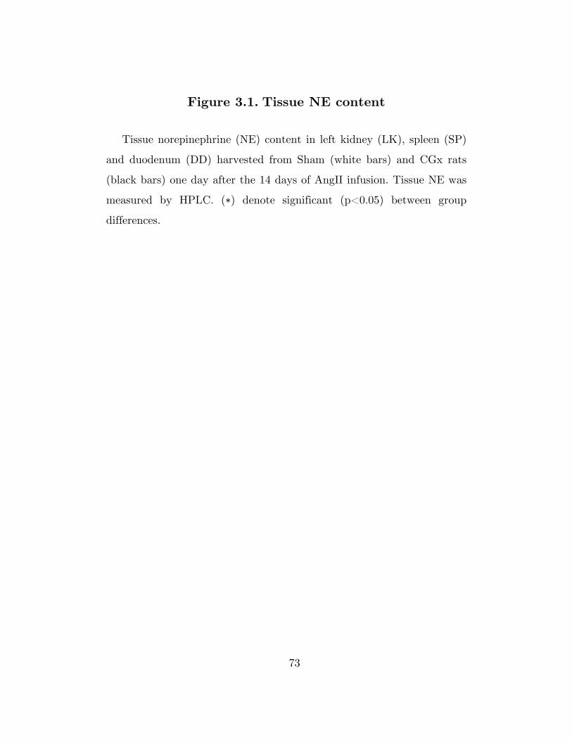
73
Figure 3.1. Tissue NE content
Tissue norepinephrine (NE) content in left kidney (LK), spleen (SP)
and duodenum (DD) harvested from Sham (white bars) and CGx rats
(black bars) one day after the 14 days of AngII infusion. Tissue NE was
measured by HPLC. (∗) denote significant (p<0.05) between group
differences.

74
Figure 3.1

75
Figure 3.2. Daily food/water intake, changes in body WT during AngII-salt hypertension
Changes in body weight and daily food and water intake during
control and AngII-salt hypertension in Sham (unfilled bar and circles)
and CGx (filled bar and circles) rats. “C” and “A” prefixes on x-axis
denote control and AngII days, respectively. (†) and (‡) denote
significant within group difference compared to day 3 of control in Sham
and CGx rats, respectively. Error bars are S.E.M.

76
Figure 3.2
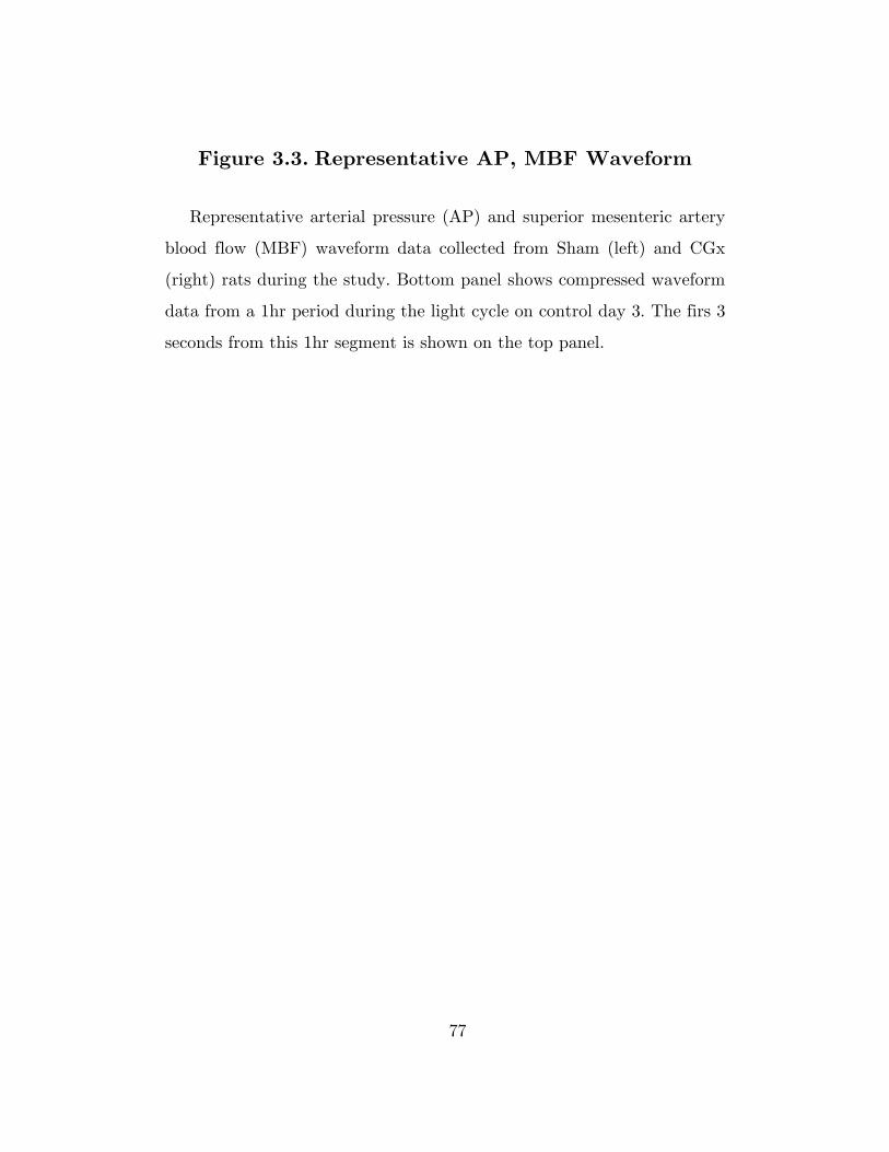
77
Figure 3.3. Representative AP, MBF Waveform
Representative arterial pressure (AP) and superior mesenteric artery
blood flow (MBF) waveform data collected from Sham (left) and CGx
(right) rats during the study. Bottom panel shows compressed waveform
data from a 1hr period during the light cycle on control day 3. The firs 3
seconds from this 1hr segment is shown on the top panel.

78
Figure 3.3

79
Figure 3.4. 24hr hemodynamic profile of MAP, MVR, MBF, and HR during AngII-salt hypertension
24hr average mean arterial pressure (MAP), mesenteric vascular
resistance (MVR), superior mesenteric artery blood flow (MBF), and
heart rate (HR) during control and AngII-salt hypertension in Sham
(unfilled circles) and CGx (filled circles) rats. “C” and “A” prefixes on x-
axis denote control and AngII days, respectively. (†) and (‡) denote
significant within group difference compared to day 3 of control in Sham
and CGx rats, respectively. (∗) denote significant (p<0.05) between
group differences. Error bars are S.E.M.

80
Figure 3.4

81
Figure 3.5. Changes in responsiveness of HR, MAP and MVR to acute ganglionic blockade during AngII-
salt hypertension
Peak changes in mean arterial pressure (MAP), mesenteric vascular
resistance (MVR), and heart rate (HR) after ganglionic blockade
(hexamethonium, 20mg/kg, i.v.). Values represent absolute differences
from a 10min baseline immediately preceding the injection. Injections
were performed on control day 3 and days 1, 3, 5, 7, 10, and 13 of
AngII. Unfilled and filled bars represent responses in Sham and CGx
rats, respectively. “C” and “A” prefixes on x-axis denote control and
AngII days, respectively. (†) and (‡) denote significant within group
difference compared to day 3 of control in Sham and CGx rats,
respectively. Error bars are S.E.M.

82
Figure 3.5

83
Chapter 4
Effect of chronic α1/2β1-adrenergic receptor
blockade on the development of AngII-Salt Hypertension
4 Chapter 4: Effect of chronic α1/2β1-adrenergic receptor blockade on the development of AngII-Salt Hypertension
4 Chapter 4 4 Chapter 4

84
Chapter Overview
The contribution of global sympathetic pressor tone in
angiotensin-II salt (AngII-salt) hypertension has been inferred mostly
from repeated measurements of whole body norepinephrine (NE)
spillover and the depressor response to acute ganglionic blockade.
Complementary evidence using chronic sympathetic blockade has been
lacking for the AngII-salt model in the rat. To address this issue, we
tested the effect of chronic, global sympathetic blockade on AngII-salt
hypertension by continuous infusion of phentolamine and atenolol, a
non-selective α1/2 and selective β1-adrenergic receptor antagonist,
respectively. We found that α1/2β1-adrenergic receptor blockade
attenuates AngII-induced hypertension in rats fed a high (2%) salt
(NaCl) diet (AngII-salt hypertension) but not those in a low (0.1%)
NaCl diet. Global sympathetic pressor tone accounted for approximately
27% of the total change in pressure in AngII-salt hypertensive rats, as
estimated by the differences in pressure between chronic α1/2β1-
adrenergic receptor blocked and vehicle treated rats. In addition,
although the salt-dependent differences in the severity of AngII-induced
hypertension was blunted by chronic α1/2β1-adrenergic receptor blockade,
it was not completely blocked, suggesting that an increase in peripheral
adrenergic tone plays some role, but is not the major determinant of the
salt dependent effects on the severity of AngII-induced hypertension. We
conclude that global sympathetic pressor tone contributes to AngII-salt
hypertension, but to an extent that is significantly smaller than what
has been previously assumed by acute ganglionic blockade.

85
4.1 Introduction
Hypertension that results from chronic administration of AngII
(AngII-induced hypertension) is thought to be mediated, in part, by
neurogenic mechanisms (86, 87). Several line of evidence supports this
neurogenic hypothesis, including a progressively higher depressor
response to ganglionic blockade (13, 62, 66, 79), prevention of this form
of hypertension by neonatal sympathectomy (26, 98), and attenuation of
hypertension by chronic adrenergic blockade (76). However, other
studies employing similar techniques have found conflicting results (65,
83). These inconsistencies can be attributed in part, to the dependence
of the model on the level of dietary salt intake (87). The severity of
AngII-induced hypertension in dogs (24), rats (62, 63, 64, 66), and mice
(22) has been shown to increase progressively with higher levels of
dietary salt intake. In both rats, and rabbits, it has been shown that the
depressor response to ganglionic blockade is elevated during AngII-
induced hypertension selectively in animals with a high salt intake
(AngII-salt hypertension) (62, 64, 66, 79). Furthermore, our group has
shown that NE spillover is elevated in high salt diet rats, but not those
fed a normal salt diet (63).
Although global indices of sympathetic tone were highly indicative of
a rise in adrenergic activity and enhanced sympathetic vasomotor tone
during AngII-salt hypertension, attempts at directly measuring increases
in sympathetic nerve activity or identifying a critical peripheral
sympathetic target organ has been mostly unsuccessful (85). Consistent
with other studies in AngII-induced hypertension, it has been shown
that renal nerves have little contribution to the rise in pressure during
AngII-salt hypertension, both on the basis of denervation (13, 64), and

86
direct nerve recording experiments (4, 115). Direct measurement of
lumbar sympathetic nerve activity has been shown to remain unchanged
during AngII-salt hypertension (115), and correspondingly, hind limb
NE spillover was shown to be unchanged (61). Preliminary results from
our lab have shown that sympathetic input to the heart has no
contribution to AngII-salt hypertension based on findings from cardiac
denervation and chronic β1-adrenergic receptor (β1-AR) blockade
experiments (52). The peripheral sympathetic target organ responsible
for the neurogenic mechanism underlying AngII-salt hypertension was
thought to have been finally identified when it was shown that AngII-
salt hypertension is significantly attenuated by celiac ganglionectomy
(CGx) (64). Most recently, however, we found conflicting results
showing that CGx does not attenuate AngII-salt hypertension (see
results in Chapter 3).
The expected contribution of sympathetic pressor tone on AngII-salt
hypertension has largely been based on the magnitude drop in pressure
following acute ganglionic blockade. Although this technique has been
validate by our lab and others as an indicator of sympathetic pressor
tone in the rat, repeated measures obtained under chronic experimental
settings may not necessarily reflect the response when the sympathetic
nervous system is inhibited in the chronic setting (23, 93).
Complementary techniques for addressing this issues, such as continuous
administration of adrenergic blocking agents or neonatal
sympathectomy, have been used to investigate the sympathetic
contribution to AngII-induced hypertension, but with mixed results as
alluded to earlier (26, 76, 98). To our knowledge, there have been no

87
studies assessing the effect of chronic sympathetic blockade specifically
during AngII-salt hypertension.
The objective of this study was to reassess the contribution of global
sympathetic pressor tone during AngII-salt hypertension by chronic
continuous administration of phentolamine, a competitive, non-specific
α-adrenergic antagonist, and atenolol, a specific β1-AR antagonist.
Phentolamine was chosen over a more selective α-adrenergic antagonist
to account for α2-adrenergic receptor (α2-AR) mediated vasoconstrictor
effects in veins (28). Atenolol was added to the infusate primarily to
counteract β1-AR mediated increases in plasma renin activity (84)
expected due to increases in plasma catecholamines following non-
selective α-adrenergic blockade (92). We compared the effect of α1/2β1-
adrenergic receptor (α1/2β1-AR) blockade during AngII-induced
hypertension in rats fed a high (2%) salt (NaCl) diet (AngII-salt
hypertension), which is hypothesized to be highly dependent on a
sympathetic pressor tone, and a low (0.1%) NaCl diet, in which the
sympathetic component is thought to be less or absent.

88
4.2 Methods
Experiments were performed in conscious, chronically instrumented
rats in accordance with NIH guidelines. All acute and chronic
experimental procedures were conducted after approval and under
supervision by the institutional animal care and use committee of the
University of Minnesota.
4.2.1 Animal use and care
Male Sprague-Dawley rats from Charles River Laboratories
(Wilmington, MA) weighing 200-225g upon transfer to our facility were
used in this study. Upon arrival, rats were switched to a high (2%) or
low (0.1%) salt (NaCl) diet (Research Diets, Inc., New Brunswick, NJ).
Animals were housed 2 per cage in a 12-12hr light-dark cycled room
(8:30/20:30 cycle). Distilled water was available ad-libidum. Animals
were allowed to acclimate for at least 1 week prior to surgery.
4.2.2 Surgical procedure
Surgery was performed under isoflurane anesthesia (2.5% isoflurane
in 100% O2 delivered via a nose cone at 1mL/min flow rate). After
induction, rats were given atropine (0.2mg/kg, i.p., Baxter International,
Inc., Deerfield, IL) for reduction of salivary and bronchial secretions,
preoperative antibiotic prophylaxis (gentamicin, 0.05mL, i.m., Hospira,
Inc., Lake Forest, IL), preoperative pain relief (ketoprofen, 5mg/kg, s.c.,
Fort Dodge Animal Health, Inc., Overland Park, KS) and placed on a
heated surgical bench. Surgical instruments were heat sterilized and all
implanted instruments were cold sterilized overnight in a solution of

89
glutaraldehyde (Cidex Plus, Johnson & Johnson Services, Inc., New
Brunswick, NJ).
The left external jugular vein was cannulated via an incision slightly
above the clavicles along the mid-clavicular line. A custom made jugular
venous catheter was advanced 3 cm towards the right atrium. This was
later used for acute injections of phenylephrine to test for the efficacy of
α1-adrenergic receptor (α1-AR) blockade. The detailed surgical procedure
and method of fabrication of the jugular venous catheter has been
described previously (see Appendix 1). A custom made femoral venous
catheter was advanced into the lower abdominal vena cava through the
left femoral vein as described previously (66). A DSI pressure
transmitter (TA11PA-C40, Data Sciences International (DSI), Saint
Paul, MN) was implanted intraperitoneally through a midline
laparotomy, and its catheter was inserted into the left femoral artery for
measurement of arterial pressure as described previously (66). Finally, a
catheter was implanted subcutaneously, through a dorsal skin incision
on the right upper back of the animal, which was later used for chronic
administration of AngII using an external syringe pump (model
935, Harvard Apparatus, South. Natick, MA) as previously described
(see Appendix 1).
All three catheters were exteriorized through a skin incision over the
scapulae, anchored to the underlying subcutaneous tissue using a
circular surgical polyester mesh (PETKM14002, Textile Development
Associates, Inc., Surgical Mesh Division, Brookfield, CN) which was
implanted subcutaneously and sutured to the skin upon closure of the
incision (see Chapter 3). The exteriorized catheters were threaded
through a stainless steel spring used for tethering the rat to a dual

90
channel hydraulic swivel (model 375/D/22, Instech Laboratories, Inc.,
Plymouth Meeting, PA) mounted above their cage. The free end of the
subcutaneous catheter and the femoral venous catheter were each
connected to a port on the swivel.
Rats were given a minimum of 7 days to recover from surgery.
Ketoprofen (5mg/kg, s.c., Fort Dodge Animal Health, Inc., Overland
Park, KS) was administered daily for 3 days post surgery for pain
management. The femoral and jugular venous catheters were flushed
every 4 days with 50U/mL heparinated saline (Hospira, Inc., Lake
Forest, IL) to maintain patency between uses.
4.2.3 Experimental protocol
The study consisted of two factors, dietary salt (NaCl) and drug
treatment, each with two levels; high (2%) and low (0.1%) NaCl for
“dietary NaCl”, and vehicle and α1/2β1-AR antagonist for “drug
treatment”. α1/2β1-AR blockade was achieved by administration of
phentolamine and atenolol (see below for dose). Experiments with all
four groups were conducted in parallel. The experimental protocol
consisted of 7 days of control followed by 14 days of AngII, and a 3 day
recovery period. Administration of vehicle or an α1/2β1-AR antagonist
solution was started on day 0 of control and continued throughout the
duration of the protocol. Additionally, physiological saline (B. Braun
Medical, Inc., Irvine, CA) was administered continuously through the
subcutaneous catheter starting on day 0 of control to maintain patency
until switched to AngII on the 8th day of the protocol (AngII day 0).
Vehicle or a α1/2β1-AR antagonist solution was administered through
the femoral venous catheter using an external syringe pump (model 975,

91
Harvard Apparatus) set to deliver the solutions at a rate of 2.13mL/day
using a 3mL syringe (Monoject, Tyco Healthcare Group, Mansfield,
MA). The 3mL syringe was coupled to a 0.2μm syringe filter (Acrodisc,
Pall Corporation, Ann Arbor, MI) and connected to a dual channel
hydraulic swivel using a 23g Tygon tubing (3/16” I.D. x 5/16” O.D.
Tygon S-50-HL Saint-Gobain Performance Plastics, Corp., Akron, OH).
The α1/2β1-AR antagonist was prepared fresh, daily, from stock solutions
(see section below).
We monitored the efficacy of α1-AR blockade in α1/2β1-AR antagonist
treated rats by measuring the pressor response to an acute injection of
phenylephrine (10ug/kg, i.v. via jugular venous catheter). The pressor
response to phenylephrine was tested on day 0 of control prior to
starting the α1/2β1-AR antagonist or vehicle infusion, days 1 and 7 of
control, and on the last day of recovery in all experimental groups.
Vehicle or AngII was infused through the subcutaneous catheter
using an external syringe pump (model 935, Harvard Apparatus)
calibrated to an infusion rate of 5uL/hr using a 1mL syringe (Monoject,
Tyco Healthcare Group). The syringe was coupled to a port on the dual
channel hydraulic swivel using 23g Tygon tubing. On the 8th day of the
protocol (day 0 of AngII), the syringe containing physiological saline was
switched to one filled with AngII infusate. The subcutaneous catheter
was primed with AngII based on the estimated catheter dead space
(approximately 0.2mL) prior to restarting the infusion.
Food intake and water intake was measured daily during the
protocol. Body weights were measured periodically, and dose
adjustments for the α1/2β1-AR antagonist solution were made weekly on
days 7 of control, and days 7 and 14 of AngII based on weight. Dose
92
adjustment was performed only if there was an increase in weight;
otherwise the dose was kept unchanged.
4.2.4 Pharmacological agents
The α1/2β1-AR antagonist solution consisted of an aqueous mixture of
phentolamine (methanesulfonate salt; P302560, Toronto Research
Chemicals, Ontario, Canada) and atenolol (A-7655, Sigma-Aldrich, Co.,
LLC., St. Louis, MO) containing citric acid (C0759, Sigma-Aldrich, Co.)
and sodium citrate (S4641, Sigma-Aldrich, Co.) as buffering agents. A
stock solution of phentolamine was prepared in sterile water at a
concentration of 50mg/mL. As atenolol is poorly soluble in water, a
custom recipe was formulated to prepare a 20mg/mL stock solution. It
was found that atenolol at the required concentration would solubilize in
a solution of 50 parts water and 3 parts stock citrate buffer with
vigorous sonication. The stock citrate buffer solution consisted of 5.5
parts citric acid and 3 parts sodium citrate in sterile water (final
concentration of approximately 0.07M citric acid and 0.026M sodium
citrate).
The stock solutions of phentolamine and atenolol were prepared fresh
weekly. The phentolamine stock solution was stored at -80°C. The two
drugs were mixed daily to prepare an infusate calculated to administer
phentolamine at a rate of 3mg/kg/hr and atenolol at a rate of
2mg/kg/hr. The delivery rate of atenolol was based on a previously
published study from our lab (57, 110). The delivery rate of
phentolamine was based on previously reported doses (7, 106) and
further titrated during a preliminary study to result in complete
blockade of the pressor response to phenylephrine after a one day

93
infusion of phentolamine. The resulting mixture had a pH of 6.4. The
osmolality of the final solution was measured using an osmometer
(model 3320, Advanced Instruments, Inc., Norwood, MA) and corrected
to 290mOsms/kg using a 200mg/mL hypertonic saline solution. The
vehicle solution was prepared from the stock citrate buffer diluted to the
same concentration as the drug mixture (approximately 16.6% v/v). The
pH was adjusted to 6.4 using sodium hydroxide, and osmolality was
corrected as described above.
The AngII infusate was prepared by diluting AngII (A9525, Sigma-
Aldrich) in physiological saline to result in a delivery rate of
150ng/kg/min at an infusion rate of 5μL/hr. A working solution of
phenylephrine was prepared in physiological saline at a concentration of
50μg/mL.
4.2.5 Data collection
Arterial pressure (AP) was collected continuously throughout the
experiment at a 500Hz sampling rate using acquisition software from
DSI (DataQuest ART Acquisition). The acquisition hardware has been
described in detail previously (66). MAP, and HR were calculated and
stored every 10s using the built-in online analysis routine in the
acquisition software.
4.2.6 Data analysis and Statistics
Time dependent changes in MAP and HR was quantified using 10s
interval data averaged over 24hrs using analysis software from DSI
(Dataquest ART Analysis). Both original values and changes from
baseline (ΔMAP and ΔHR) were used for analysis. Baseline MAP and

94
HR was calculated from the average of 7 days of control, and compared
between groups by a t-test. ΔMAP and ΔHR for each animal were
calculated by subtracting the baseline value from each data point, and
used for determining the effect of chronic α1/2β1 receptor blockade on
AngII-induced hypertension for each level of dietary salt. The effect of
α1/2β1 receptor antagonist or vehicle treatment on the dietary salt
dependent differences in the magnitude of AngII-induced hypertension
was determined from the original MAP and HR values. Between and
within group differences in MAP, ΔMAP, HR, and ΔHR during AngII
infusion was tested for statistical significance by 2-way repeated-
measures ANOVA followed by a Holm-Sidak multiple comparisons test
when appropriate.
The pressor response to acute phenylephrine injection was analyzed
using LabChart Pro (version 7, ADInstruments, Dunedin, New Zealand)
and Matlab (version R2012a, Mathworks, Inc.). MAP data was
extracted from the 500Hz signal and averaged over XXXs intervals using
LabChart Pro. Quantification of the pressor response was carried out in
Matlab. The average MAP from a 5 min. period immediately before the
injection was subtracted from each trace to account for baseline MAP
differences between vehicle and α1/2β1 receptor antagonist treated rats.
The pressor response was quantified as an area under the curve (AUC)
calculated from the MAP trace starting at the beginning of PE injection
to 5min after the injection. The extent of α1 receptor blockade resulting
from α1/2β1 receptor antagonist infusion for each animal was determined
by calculating the AUC ratio using the AUC from the PE response prior
to vehicle or antagonist infusion (on control day 0) as denominator.
Between group differences in AUC and within group changes in the

95
AUC ratio were tested for statistical significance by 2-way repeated-
measures ANOVA followed by a Holm-Sidak multiple comparisons test
when appropriate.
Data were tabulated and graphed using JMP software (version 10,
SAS Institute, Inc., Cary, NC). Statistical analysis was performed in
SigmaPlot (version 11, Systat Software, Inc., Richmond, CA). All values
are shown as mean ± S.E.M unless otherwise noted.
96
4.3 Results
4.3.1 Effect of chronic α1/2β1-AR blockade on 24hr
food, water intake and body weight
Food intake was transiently lower in α1/2β1-AR antagonist treated
rats, especially in the low salt group, as shown in Figure 4.1. In the low
salt group, rats treated with α1/2β1-AR antagonist had lower food intake
compared to those receiving vehicle solution during most of control
through day 5 of AngII. After day 5 of AngII, food intake was, for the
most part, comparable between vehicle and α1/2β1-AR antagonist treated
rats. In the high salt group, food intake were similar between vehicle
and α1/2β1-AR antagonist treated rats except for day 1 of control and
days 2 and 3 of AngII.
Water intake was for the most part unaffected by α1/2β1-AR
blockade. Water intake in the low salt group was lower in α1/2β1-AR
antagonist treated rats on day 1 of control, but otherwise the same as in
vehicle treated rats throughout the protocol. In the high salt group,
water intake was lower in α1/2β1-AR antagonist treated compared to
vehicle treated rats on day 1 of control, and days 2 and 3 of AngII;
corresponding to the same days that food intake was lower in the α1/2β1-
AR antagonist treated rats.
Body weights were comparable across all 4 groups of rats until day 7
of control (Table 4.1). From control day 7 onwards, vehicle treated rats
in the low salt group showed greater weight gain compared to α1/2β1-AR
antagonist treated rats fed a low salt diet. The weight gain observed in
vehicle treated rats fed a low salt diet was also greater compared to
those seen in vehicle treated rats fed a high salt diet. The weight

97
difference between high and low salt rats in the vehicle treated group
was statistically significant on days 7 and 14 of AngII. There were no
significant weight differences between the remaining three groups of rats
at any point during the protocol.
4.3.2 Effectiveness of α1-AR blockade during chronic
α1/2β1-AR antagonist treatment
The acute injection of 10ug/kg phenylephrine during control day
0, prior to starting the infusion of α1/2β1-AR antagonist or vehicle,
resulted in a peak pressor response of approximately 50mmHg (Figure
4.1). Infusion of α1/2β1-AR antagonist blocked 87% and 93% of this α1-
AR mediated pressor response after one day of treatment in high and
low salt rats, respectively (Table 4.2). Prolonged treatment with α1/2β1-
AR antagonist resulted in a gradual loss in the efficacy of α1-AR
blockade. This was more significant in the high salt group where the
efficacy of α1-AR blockade dropped to 55% after 26 days of α1/2β1-AR
antagonist treatment. This drop in the efficacy of blockade in α1/2β1-AR
antagonist treated rats was accompanied by a 113% increase in the
pressor response to PE in vehicle treated rats fed a high salt diet. In the
low salt group, the efficacy of the α1-AR blockade was 83% by the end
of the protocol. There was no statistically significant change in the
pressor response to the PE in the vehicle treated rats as was observed in
the high salt group.

98
4.3.3 Effect of chronic α1/2β1-AR blockade on AngII-
induced hypertension in high and low salt rats
The effect of α1/2β1-AR blockade on AngII-induced hypertension in
high and low salt rats was compared with their respective vehicle
treated control after subtraction of the baseline differences in MAP and
HR (Figure 4.2). Baseline MAP and HR was calculated from the average
24hr MAP during the 7 day control period. The baseline MAP in the
α1/2β1-AR antagonist treated group was 87.6±1.4 and 87.7±1.4 mmHg in
high and low salt rats, respectively. The baseline MAP in vehicle treated
rats was 106.3±2.1 and 102.0±1.5mmHg in high and low salt rats,
respectively. The difference in baseline MAP between blocked versus
vehicle treated groups was 18.7 and 14.3 mmHg in high and low salt
rats, respectively. Baseline HR in the α1/2β1-AR antagonist treated group
was 353.4±6.1 and 346.2±4.1 BPM in high and low salt rats,
respectively. These HR values were lower by 76.2 and 94.0 BPM
compared to high and low salt rats in the vehicle treated group, which
were 429.6±5.5 and 440.2±7.6 BPM, respectively.
In high salt rats, the initial change in MAP during the first 4 days of
AngII was similar between α1/2β1-AR antagonist treated and vehicle
treated groups. In high salt rats, the rise in MAP on the first day of
AngII was 21.7±3.0mmHg and 22.0±2.4mmHg in α1/2β1-AR antagonist
treated and vehicle treated groups, respectively. In low salt rats, the rise
in MAP on the first day of AngII was 8.7±2.0mmHg and 9.1±2.1mmHg
in α1/2β1-AR antagonist treated and vehicle treated groups, respectively.
The response of HR to the initial changes in MAP during AngII
infusion was also minimally affected by α1/2β1-AR blockade in high salt

99
rats. The peak drop from baseline HR was observed on the 4th day of
AngII in both α1/2β1-AR antagonist treated and vehicle treated groups.
In low salt rats, the initial and overall change in HR was more gradual
without any notable troughs.
α1/2β1-AR blockade blunted the AngII-induced rise in MAP, with
respect to baseline, from days 5 to 14 of AngII in high salt rats. The
final change in MAP from baseline, averaged over days 11-14 of AngII
when responses appeared to have plateaued, was 42.2±1.5mmHg and
58.2±1.5mmHg from baseline in α1/2β1-AR blockade and vehicle treated
high salt rats, respectively. This 16mmHg difference between the two
groups represented 27% of the total rise in MAP seen in the vehicle
group. In contrast to these effects in high salt rats, α1/2β1-AR blockade
in low salt rats had no effect on the level of AngII-induced rise in MAP
from baseline.
High salt plus vehicle treated rats had a pronounced rise in HR
following the trough on the 4th day of AngII. Absolute HR on days 7 to
14 of AngII in high salt plus vehicle treated rats was back to baseline
HR despite the persistently elevated MAP. α1/2β1-AR blockade
significantly blunted this response. In low salt rats, α1/2β1-AR blockade
did not result in a consistent difference in the change in HR observed
during AngII-infusion.
4.3.4 Effect of chronic α1/2β1-AR blockade on the
difference in MAP between high and low salt rats
during control and AngII-salt hypertension
There were no differences in MAP between high and low salt rats in
the vehicle or α1/2β1-AR antagonist treated groups during control. AngII

100
infusion resulted in an immediate difference in MAP between high and
low salt rats on day 1 of AngII. This between salt level differences in
MAP appeared to be slowly amplified through the 14 days of AngII
infusion in the vehicle treated group. The final between high and low
salt group differences in MAP averaged over days 11 to 14 of AngII in
the vehicle treated group was 36mmHg. A slowly increasing between salt
level differences in MAP remained after α1/2β1-AR blockade. The final
between high and low salt group differences in MAP averaged over days
11 to 14 of AngII in the α1/2β1-AR blockade group was 21mmHg.

101
4.4 Discussion
The increasing depressor response to acute ganglionic blockade
observed during AngII-salt hypertension has been used as evidence
suggesting a dominant role of the sympathetic nervous system in the
pathogenesis of AngII-salt hypertension (62, 64, 66). This observation
led to multiple experiments designed to identify the regional
sympathetic pathways contributing to the neurogenic mechanism of the
model. Many of these experiments, however, have been unsuccessful or
inconsistent at identifying the sympathetic pathway(s) underlying the
hypothesized neurogenic mechanism of AngII-salt hypertension. Studies
from our lab and others have so far demonstrated, via chronic
denervation experiments, that cardiac sympathetic nerves and renal
sympathetic nerves do not play a significant role during AngII-salt
hypertension (52, 64). We have also shown that lumbar sympathetic
nerves are not activated during AngII-salt hypertension, and hence
concluded that lumbar sympathetic nerves do not play an active role in
AngII-salt hypertension (61, 115). Although celiac ganglionectomy was
shown to have a significant anti-hypertensive effect during AngII-salt
hypertension in one study (64), a follow up study in our laboratory
showed no effect of celiac ganglionectomy (see Chapter 3). These mixed
results have called into question whether neurogenic mechanisms are
indeed a major component of AngII-salt hypertension as suggested by
acute ganglionic blockade. To address this issue, we re-assessed the
sympathetic contribution in AngII-salt hypertension by way of chronic,
global sympathetic blockade by continuous administration of
phentolamine and atenolol, a non-selective α1/2 and selective β1-AR
antagonist, respectively.

102
The main finding in this study was that chronic α1/2β1-AR blockade
attenuated AngII-salt hypertension. Despite the negative results so far
from experiments utilizing peripheral denervation for targeted
sympathetic blockade, this study confirms the presence of a sympathetic
contribution to the model, and supports the results from acute
ganglionic blockade. It also complements the results showing elevated
norepinephrine spillover during AngII-salt hypertension (63), and
together suggests that elevated α1/2β1-AR signaling due to enhanced
norepinephrine release from sympathetic nerve terminals contributes to
AngII-salt hypertension.
Another important finding in this study was that α1/2β1-AR blockade
attenuated AngII-induced hypertension in rats fed a high salt diet (i.e.
AngII-salt hypertension) but not those in a low salt diet. This finding
was also consistent with results from our prior studies using acute
ganglionic blockade (62, 66) and further supports the idea that elevated
dietary salt intake and circulating plasma AngII act in a synergistic
manner to elevate peripheral sympathetic outflow during AngII-salt
hypertension. The mechanism by which circulating AngII and dietary
salt leads to enhanced sympathetic outflow is currently not well
understood. We have previously proposed that the integration of the
two signals occur centrally via circumventricular organs that ultimately
lead to increased output from brainstem sympathetic premotor neurons
(86, 87), but further studies are necessary to elucidate the exact
mechanisms involved in this interaction.
4.4.1 Mechanism of blood pressure lowering effect of
chronic α1/2β1-AR antagonist treatment during AngII-

103
salt hypertension: α versus β adrenergic receptor
mediated effects
Blockade of α1-AR mediated arteriolar vasoconstriction likely played
a major role in the blood pressure lowering effect of chronic α1/2β1-AR
antagonist treatment during AngII-salt hypertension. Since
phentolamine is a non-specific α-AR antagonist, its inhibitory effects on
post-synaptic α2-AR also likely played a role in the observed effects.
Although the cardiovascular effects of α2-AR are often associated with
its effect presynaptically, post-synaptic α2-ARs are known to exert a
significant vasoconstrictive effect, especially in veins (5, 91). Although
blockade of α2-AR alone would result in peripheral sympathoactivation
with a concomitant rise in arterial pressure, it has been shown that in
the setting of concomitant α1-AR blockade, α2-AR antagonists have an
additional blood pressure lowering effect (69, 74). This is thought to be
secondary to venodilation, which would raise systemic vascular
capacitance and lower effective circulating blood volume (28, 50, 106). It
has been shown previously that vascular capacitance is reduced during
AngII-salt hypertension, which appears to be sympathetically mediated
based on its response to acute ganglionic blockade (62). Thus, reduction
in total peripheral vascular resistance and blockade of sympathetically
mediated changes in vascular capacitance likely contributed to the blood
pressure lowering effect of α1/2β1-AR antagonist treatment during
AngII-salt hypertension.
In addition to α receptor blockade with phentolamine, atenolol, a
selective β1-AR antagonist, was also added to the adrenergic antagonist
infusate in order to prevent β1 mediated renin release from

104
juxtaglomerular cells (84) in response to elevated plasma catecholamine
levels expected from chronic α1/2-AR blockade (92). This combined use
of α/β antagonists raises the question whether the effect seen in this
experiment were due primarily from α1/2 or β1 receptor antagonism.
Consistent with cardiac β1-AR blockade, the blood pressure lowering
effect of chronic α1/2β1-AR antagonist treatment was accompanied by
significantly lower heart rates at baseline and during AngII infusion. In
addition to its effect on baseline HR, β1-AR blockade in high salt rats
significantly attenuated the rise in HR seen in vehicle treated rats
during the second week of AngII. Thus, it is possible that the blood
pressure lowering effect seen in AngII-salt hypertensive rats treated with
α1/2β1-AR antagonist was partially due to suppression of this HR
response. Although the hypotensive effect of β1-AR blockade in normal
rats is well established, the mechanism of this effect is currently not
fully understood. It has been shown, however, that its effect on blocking
sympathetic input to the heart alone does not account for the blood
pressure lowering effect of β1-AR blockade (117) in normal rats. The
effect of β1-AR blockade on renin release could have played some role,
but it has also been shown in renal denervated rats that this mechanism
does not fully account for the effect on basal blood pressure either (57).
Thus, it has been hypothesized that β1-AR blockade exerts its
hypotensive effect by reducing central sympathetic outflow through its
activity on central β1-AR (117).
Although the present study cannot exclude the possibility that β1-AR
played a major role in the blood pressure lowering of AngII-salt
hypertensive rats, preliminary studies in our lab have so far shown that
β1-AR blockade alone is unable to attenuate AngII-salt hypertension

105
(52). Furthermore, a study in cardiac denervated rats by stellate
ganglionectomy has shown that interruption of sympathetic tone to the
heart has no significant effects on the normal progression of AngII-salt
hypertension (52).
4.4.2 Chronic α1/2β1-AR blockade explains only a
fraction of the total effect seen by acute ganglionic
blockade in prior studies
Previous studies from our lab showed an approximate 40mmHg
increase in the peak depressor response to ganglionic blockade at 2
weeks of AngII-salt hypertension1, which suggests that approximately
60-80% of the total rise in pressure seen during AngII-salt hypertension
(usually about 50-60mmHg) is sympathetically mediated (66). In
contrast, α1/2β1-AR blockade only reduced AngII-salt hypertension by
27%, suggesting a much smaller sympathetic contribution to the blood
pressure rise seen in the model. Although this suggests that acute
ganglionic blockade may have overestimated the sympathetic component
of AngII-salt hypertension, the lower estimate in the present study were
likely due to limitations to the method employed in this study. First,
there was a gradual decrease in α1 receptor blockade efficacy during the
protocol, and second, only noradrenergic mediated sympathetic effects
on blood pressure were addressed by this study.
The decreasing α1 blockade efficiency we observed throughout the 3
week protocol is likely one reason by which chronic α1/2/β1-AR blockade
1 Estimate based on peak depressor response of ~80mmHg at 2 weeks of AngII-salt
hypertension, and ~40mmHg at baseline.
106
underestimates the underlying sympathetic contribution during AngII-
salt hypertension. Since the dosage for the α1/2β1-AR antagonist was
adjusted for body weight every 7 days, decreased effective drug
concentration due to changes in body weight was unlikely to have
played any major role in this reduction in α1 receptor blockade efficacy.
Increases in α1-AR expression have been shown to occur in rats after
chronic treatment with prazosin (119), hence changes in α1-AR
expression level in response to chronic phentolamine treatment likely
contributed to the increased pressor response to phenylephrine (i.e. the
decrease in α1-AR blockade efficacy). Interestingly, the development of
tolerance was more pronounced in rats treated with a high salt
compared to low salt rats. In addition, a 113% increase in the pressor
response to acute phenylephrine injection was seen in vehicle treated
rats fed a high salt diet, suggesting that there is an underlying increase
in phenylephrine sensitivity specifically in high salt rats treated with
AngII. This could also explain the decrease in α1-AR blockade efficacy in
high salt rats treated with phentolamine. The mechanism and precise
time course of the change in phenylephrine sensitivity, presumably
secondary to an increase in α1-AR expression, and whether these changes
occur during AngII-salt hypertension remains to be addressed in a future
study.
Another possible explanation why chronic α1/2β1-AR blockade may
underestimate the total sympathetic contribution to AngII-salt
hypertension is that it does not account for the post junctional actions
of other sympathetic cotransmitters, specifically, ATP and NPY (89). It
has been shown, however, that ATP mediated vasoconstriction via P2X1
receptor is attenuated in isolated renal afferent arterioles from rats made

107
hypertensive by AngII-infusion and fed an 8% NaCl diet (54), suggesting
that P2X1 mediated vasoconstriction may not play a significant role in
the sympathetic component of AngII-salt hypertension. On the other
hand, multiple studies have shown increases in tissue and plasma levels
of NPY as well as elevated vascular responsiveness to NPY in various
animal models of hypertension (112). Although results from studies
looking at the effect of pharmacological blockade of Y1 receptors have so
far been negative in the SHR (120) or mixed in Goldblatt hypertensive
rats (80, 96), it may be possible that NPY signaling is elevated in AngII-
salt hypertension. Future studies could address whether Y1 receptor
mediated vasoconstriction account for the greater fall in pressure seen
by acute ganglionic blockade in AngII-salt hypertensive rats, compared
to blood pressure lowering effect of chronic α1/2β1-AR blockade alone.
4.4.3 The salt sensitive component of AngII-salt
hypertension is only partially mediated by α1/2β1-AR
dependent neurogenic mechanisms
Salt sensitivity of MAP during chronic administration of AngII has
been thought to be primarily due to inappropriately high levels of SNA
for a given level of salt intake (10, 12). Consistent with this idea, α1/2β1-
AR antagonist treatment reduced the incremental difference in pressure
between high and low salt rats after 2 weeks of AngII infusion (i.e. the
salt sensitivity of AngII-induced hypertension). However, chronic α1/2β1-
AR antagonist treatment did not eliminate the salt-sensitivity of AngII-
induced hypertension, suggesting that a significant portion is driven by
non-α1/2β1-AR mediated mechanisms. As discussed previously, the
remaining salt-sensitive component of pressure in AngII-infused rats

108
after α1/2β1-AR antagonist treatment group could still have a neurogenic
component that is non-adrenergic. There are, however, non-neurogenic
mechanisms that likely contribute to the remaining salt-sensitivity.
These salt-dependent non-neurogenic mechanisms include, but are not
limited to, AngII mediated changes in renal diuretic/natriuretic function
(25, 72), high-salt diet mediated changes in vascular reactivity to
circulating AngII (27), and changes in the pressor effects of other
circulating hormones, such as endothelin and its actions on ET-A
receptors (2).
4.4.4 Summary and Perspectives
This study confirms the role of sympathetic mechanisms, specifically,
a combined α1/2β1-AR mediated mechanisms, to the hypertension
resulting from chronic AngII infusion, and its selective effect in rats fed
a high NaCl diet. In Figure 4.5, a schematized plot of AngII-induced
hypertension in high and low salt rats summarizes our main findings. A
question that remains unanswered is whether any specific regional
sympathetic pathway contributes to the observed effect. The
ineffectiveness of peripheral sympathectomy approaches at recapitulating
the findings from this and other studies utilizing global sympathetic
blockade, so far, suggests that targets other than cardiac, renal,
splanchnic and skeletal muscle vascular beds are involved in the
neurogenic mechanism of AngII-salt hypertension. One possibility, based
on similar findings by Veitenheimer et.al. in the 48hr water deprivation
model of acute neurogenic hypertension (110), is the adrenal gland, and
the role of adrenal cortical hormones in mediating global increases in
sympathetic nervous system activity. An alternative possibility,

109
proposed in the same study by Veitenheimer et.al., however, is that the
sympathetic nervous system is activated in these settings to maintain a
certain arterial pressure set point, and is able to adapt to compensate
for a loss of sympathetic activity to any single vascular bed. Thus in this
latter scenario, only global sympathetic blockade would be able to fully
block the neurogenic component of the hypertensive response. Further
studies are needed to test whether any of these possibilities hold true in
the AngII-salt model of hypertension.
Although it has been suggested that the mechanism of AngII-induced
hypertension, in the later phases, is almost exclusively neurogenic (6),
we found that the overall contribution of sympathetic tone to the final
level of pressure at 2 weeks of AngII-salt hypertension is considerably
smaller than estimated by ganglionic blockade when tested using chronic
α1/2β1-AR blockade. The discrepancy in results obtained from ganglionic
blockade and chronic α1/2β1-AR blockade may have been due to the
effect of tolerance to chronic pharmacologic treatment, and non-
adrenergic sympathetic cotransmitters that were not tested in this
study. However, our results also suggest that a significant component of
hypertension during the later phase of AngII infusion may be mediated
by non-neurogenic mechanisms, and that data from ganglionic blockade
experiments may have overestimated the overall neurogenic contribution
to the model.

110
4.5 Figures and Table

111
Figure 4.1. 24hr Food and Water Intake in α1/2β1-AR antagonist treated and vehicle treated rats
24hr food and water intake in α1/2β1-AR antagonist treated (light
grey) and vehicle treated rats (black) fed a high (2%) or low (0.1%) salt
(NaCl) diet. Measurements were carried out once a day during the light
phase of the dark-light cycle. Values represent group mean±S.E.M. (†)
and (‡) denote significant (p<0.05) within group difference compared to
day 7 of control in vehicle and α1/2β1-adrenergic antagonist treated rats,
respectively. (∗) denote significant (p<0.05) between group differences n
= 7 for both α1/2β1-AR antagonist treated and vehicle treated groups fed
a 2% NaCl diet; n = 6 for the α1/2β1-AR antagonist treated group fed a
0.1% NaCl diet; n = 8 for the vehicle treated group fed a 0.1% NaCl
diet.

112
Figure 4.1

113
Table 4.1. Change in body weight in α1/2β1-AR antagonist treated and vehicle treated rats
Body weight prior to initial surgery, beginning of protocol, and
weekly changes during chronic α1/2β1-AR antagonist or vehicle
treatment, and 2 weeks of AngII administration. Weights are reported in
grams. Values represent mean±S.E.M. n = 7 for both α1/2β1-AR
antagonist treated and vehicle treated groups fed a 2% NaCl diet; n = 6
for the α1/2β1-AR antagonist treated group fed a 0.1% NaCl diet; n = 8
for the vehicle treated group fed a 0.1% NaCl diet.

114
Table 4.1

115
Figure 4.2. Blood pressure response to acute phenylephrine injection in α1/2β1-AR antagonist
treated and vehicle treated rats
The degree of α1-AR blockade in rats chronically treated with an
α1/2β1-AR antagonist was tested on days 1 and 7 of control and 3rd day
after cessation of AngII infusion (Recovery; “R”) by measuring the
arterial pressure response to an acute injection of phenylephrine (PE; 10
μg/kg, i.v.). The tracing shown under “C00” was obtained prior to
starting the vehicle or α1/2β1-AR antagonist infusion. The tracings show
the group averaged change in arterial pressure in response to
phenylephrine in vehicle (light grey) and α1/2β1-AR antagonist treated
(black) rats fed a 2% (top panel) or 0.1% NaCl (bottom panel) diet. The
shaded areas represent the S.E.M. The baseline arterial pressure
calculated from the average of the 5min period immediately preceding
the PE injection was subtracted from each tracing to account for
differences in MAP between vehicle and α1/2β1-AR antagonist treated
rats. n = 7 for both α1/2β1-AR antagonist treated and vehicle treated
groups fed a 2% NaCl diet; n = 6 for the α1/2β1-AR antagonist treated
group fed a 0.1% NaCl diet; n = 8 for the vehicle treated group fed a
0.1% NaCl diet.

116
Figure 4.2

117
Table 4.2. Level of α1-AR blockade during chronic α1/2β1-AR antagonist treatment
The degree of α1-AR blockade in rats chronically treated with an
α1/2β1-AR antagonist was tested on days 1 and 7 of control and 3rd day
after cessation of AngII infusion (Recovery; “R”) by measuring the
arterial pressure response to an acute injection of phenylephrine (PE; 10
μg/kg, i.v.). The degree of α1-AR blockade was quantified by calculating
the area under the curve (AUC) from the arterial pressure response
tracing during the first 5 min after injection of PE. The baseline arterial
pressure calculated from the average of the 5min period immediately
preceding the PE injection was subtracted from each tracing prior to
calculating the AUC. The values in parenthesis represent the AUC as a
fraction of the PE response obtained prior to vehicle or α1/2β1-AR
antagonist infusion (“C00”). (†) and (‡) denote significant (p<0.05)
within group difference compared to baseline in vehicle and α1/2β1-AR
antagonist treated rats, respectively. (∗) denote significant (p<0.05)
between group differences within each dietary salt level. Values are
reported in arbitrary units (a.u.), and represent mean±S.E.M. n = 7 for
both α1/2β1-AR antagonist treated and vehicle treated groups fed a 2%
NaCl diet; n = 6 for the α1/2β1-AR antagonist treated group fed a 0.1%
NaCl diet; n = 8 for the vehicle treated group fed a 0.1% NaCl diet.

118
Table 4.2

119
Figure 4.3. Effect of α1/2β1-AR blockade on MAP and HR during AngII-induced hypertension in rats fed a
2% or 0.1% NaCl diet
24hr average mean arterial pressure (MAP), and heart rate (HR)
during control and 2 weeks of AngII-infusion followed by 3 days of
recovery in vehicle (filled circles, black line) and α1/2β1-adrenergic
antagonist treated (unfilled circles, grey line) rats. Data for rats fed a
low (0.1%) salt (NaCl) diet are plotted on the left panel. Right panel
shows data for rats fed a high (2%) NaCl diet. Values are shown after
baseline subtraction. “C”, “A”, and “R” prefixes on x-axis denote control,
AngII days, and recovery, respectively. (†) and (‡) denote significant
(p<0.05) within group difference compared to baseline in vehicle and
α1/2β1-adrenergic antagonist treated rats, respectively. (∗) denote
significant (p<0.05) between group differences. Values are mean±S.E.M.
n = 7 for both α1/2β1-AR antagonist treated and vehicle treated groups
fed a 2% NaCl diet; n = 6 for the α1/2β1-AR antagonist treated group
fed a 0.1% NaCl diet; n = 8 for the vehicle treated group fed a 0.1%
NaCl diet.

120
Figure 4.3

121
Figure 4.4. Effect of dietary salt on MAP and HR during AngII-induced hypertension in α1/2β1-AR
antagonist treated and vehicle treated rats
24hr average mean arterial pressure (MAP), and heart rate (HR)
during control and 2 weeks of AngII-infusion followed by 3 days of
recovery in rats fed a high (2%) salt (NaCl; black circles, black line) or
low (0.1%) NaCl diet (grey circles, grey line). Data for α1/2β1-AR
antagonist treated rats are plotted on the left panel. Right panel shows
data for vehicle treated rats. “C”, “A”, and “R” prefixes on x-axis denote
control, AngII days, and recovery, respectively. (∗) denote significant
(p<0.05) between 2% NaCl and 0.1% NaCl group differences. Values are
mean±S.E.M. n = 7 for both α1/2β1-AR antagonist treated and vehicle
treated groups fed a 2% NaCl diet; n = 6 for the α1/2β1-AR antagonist
treated group fed a 0.1% NaCl diet; n = 8 for the vehicle treated group
fed a 0.1% NaCl diet.

122
Figure 4.4

123
Figure 4.5. Estimated time course and magnitude of the neurogenic component of AngII-salt hypertension
Summary of main findings from this study depicted as schematized
blood pressure response during AngII-salt hypertension. The neurogenic
component of AngII-salt hypertension at 2 weeks is estimated at
approximately 27% (rounded to 30%), and appears to be activated in a
delayed fashion, approximately 4-5 days after AngII-infusion. At least
40% of the blood pressure response in AngII-salt hypertension is non-
neurogenic. The mechanism responsible for the remaining 30% of AngII-
salt hypertension is likely mediated by salt-sensitive non-neurogenic
mechanisms, or possibly other sympathetic mechanisms mediated by
non-adrenergic cotransmitters.

124
Figure 4.5

125
Chapter 5
Conclusion
5 Chapter 5: Conclusion 5 Chapter 5

126
The aim of this thesis was to determine whether changes in
sympathetic vasomotor tone to the splanchnic vascular bed was a
contributing mechanism to the neurogenic component of AngII-salt
hypertension. The question to be answered was: “Is there evidence for
regional hemodynamic changes that support the role of a targeted
increase in sympathetic vasomotor tone to the splanchnic vascular bed?”
Based on prior findings (62, 64, 76) , and then available anatomical
basis for sympathetic innervation of splanchnic arteries and veins (53),
we hypothesized that the neurogenic mechanism contributing to AngII-
salt hypertension was mediated by an increase in sympathetic vasomotor
tone to both arteries and veins, causing a rise in total peripheral
resistance and a reduction in vascular capacitance (40, 86). If true, the
hypothesis would predict that there would be measurable changes in
mesenteric vascular resistance, a variable that could be measured in
conscious rats by measuring local blood flow at the superior mesenteric
artery. Therefore, studies in this thesis were conducted using a technique
developed for measuring superior mesenteric artery blood flow
continuously, in conscious, freely moving tethered rats. The main
findings of this thesis, along with results from2 supporting studies are
summarized below.

127
5.1 Summary of main findings
5.1.1 The sustained increase in splanchnic vascular
resistance observed during AngII-salt hypertension is
not mediated by an increase in sympathetic
vasomotor tone (Chapters 2 & 3)
In Chapter 2, I found that AngII-salt hypertension, but not AngII-
induced hypertension in low salt rats was associated with a sustained
rise in mesenteric vascular resistance (MVR). Furthermore, the rise in
MVR observed in AngII-salt hypertensive rats was also associated with
a gradually increasing vasodilatory response to acute ganglionic
blockade. These two evidences were consistent with the predicted
hemodynamic changes and responsiveness to ganglionic blockade based
on the original hypothesis. However, the responsiveness of MVR to
ganglionic blockade alone was not conclusive evidence that the rise in
MVR was due to increases in sympathetic vasomotor tone. An indirect
mechanism involving withdrawal of myogenic tone in response to the
depressor response to ganglionic blockade was equally likely. The study
in Chapter 3 was designed to directly address the limitation in Chapter
2 by measuring changes in mesenteric vascular resistance in rats with
intact or devoid of sympathetic innervation to the splanchnic vascular
bed. This was achieved by surgical stripping of the celiac-superior
mesenteric ganglion plexus (celiac ganglionectomy; CGx) as had been
previously described (64).
Results from Chapter 3 showed that increases in MVR during AngII-
salt hypertension occurred in both CGx and Sham rats, suggesting that
the rise in MVR was not dependent on an intact sympathetic

128
innervation to the splanchnic vascular bed. Another findings in this
study was that the trend for a gradually increasing vasodilatory response
to ganglionic blockade was present in both CGx and the surgical sham
group, suggesting that the vasodilatory response to ganglionic blockade
was not an index of sympathetic vasomotor tone in the splanchnic
vascular bed.
The findings from the above two studies leads to a conclusion that:
“The sustained increase in splanchnic vascular resistance observed
during AngII-salt hypertension is not mediated by an increase in
sympathetic vasomotor tone.”
5.1.2 Celiac ganglionectomy does not attenuate AngII-
salt hypertension (Chapter 3)
Another major unexpected finding from the study in Chapter 3 was
that CGx did not attenuate AngII-salt hypertension as previously
reported (64). One likely explanation for the discrepant findings is the
possibility for a difference in the extent of organ involvement of CGx
between the study in this thesis and that in the previous work. As was
shown by tissue norepinephrine (NE) content measurements, the renal
involvement of CGx in the previous work was greater compared to CGx
performed in the study presented in this thesis and works by others in
our laboratory (42, 110) . The interpretation and possible mechanisms
for the discrepant finding is revisited in a section below.
5.1.3 Chronic α1/2β1-adrenergic receptor shows a
much smaller role for sympathetic pressor tone during

129
AngII-salt hypertension than predicted by acute
ganglionic blockade (Chapter 4)
The combined evidence from renal denervation (no effect) (64), renal
sympathetic nerve recording (decreased) (115), lumbar sympathetic
nerve recording (unchanged) (115), hind limb norepinephrine spillover
(unchanged) (61), stellate ganglionectomy (no effect) (52), chronic β1-
adrenergic receptor blockade (no effect) (52) and the inconsistent
findings with celiac ganglionectomy from Chapter 3, raised the question
whether a sympathetic pressor tone had a significant contribution in
AngII-salt hypertension. Although the role for a sympathetic pressor
tone had been assessed by acute ganglionic blockade, thus far, no
chronic peripheral sympathoinhibitory intervention had shown a role for
the importance of sympathetic pressor tone to any particular vascular
bed in the development and maintenance of our model of AngII-salt
hypertension. In Chapter 4, I reassessed the role of global sympathetic
pressor tone in the development of AngII-salt hypertension by means of
a chronic infusion of a combined α1/2β1-adrenergic receptor antagonist
(Phentolamine and Atenolol). I found that α1/2β1-adrenergic receptor
mediated mechanisms contribute approximately 27% of the final level of
pressure in AngII-salt hypertensive rats, far less than what could be
inferred from acute ganglionic blockade studies (62, 66).

130
5.1.4 AngII-salt hypertension generated by Alzet
osmotic minipumps can spontaneously lose its
hypertensive phenotype due to pump dependent
mechanisms (Appendix 1)
In addition to the findings discussed above, one important issue
surfaced at the conclusion of the studies described in Chapter 2. Almost
half of the rats subjected to the AngII-salt hypertension protocol failed
to develop a sustained hypertension after implantation of the Alzet
pump. This response was unpredictable, uncorrelated to predicted pump
infusion rate (based on post-explantation infusate volume), and equally
affected rats fed a high or a low salt diet. The spontaneous drops in
pressure in a subset of rats was problematic because it would have made
results in studies testing blood pressure lowering treatments difficult to
interpret. Based on preliminary findings from our laboratory showing
good repeatability of AngII-salt hypertension using a recently introduced
implantable mechanical pump (iPrecio) (107), we hypothesized that the
problems observed with Alzet rats were due to failure of Alzet pumps to
maintain a constant level of plasma AngII, which would not be observed
in mechanical infusion devices such as iPrecio or external syringe pumps.
A study was conducted to compare changes in mean arterial pressure
and plasma AngII during AngII-salt hypertension generated by Alzet,
iPrecio, or an external syringe pump (Harvard). This study is presented
in Appendix 1, and showed that AngII-salt hypertension was generated
more consistently using mechanical infusion devices (both iPrecio and
Harvard pumps) compared to Alzet osmotic minipumps. Following these
findings, AngII-salt hypertension in Chapters 3 and 4 were generated

131
using an external infusion pump connected to a subcutaneously
implanted catheter. Unlike our original hypothesis, however, we did not
find that differences in the arterial pressure profile observed between
pumps were correlated with significant differences in plasma AngII
levels. Thus, further studies may be needed to address the pump
dependent differences in the arterial pressure response during AngII-salt
hypertension.
5.2 Implications of combined findings
5.2.1 The neurogenic mechanism of AngII-salt
hypertension is mediated by a sympathetic drive
outside of the splanchnic vascular bed
The study by our group (64) that formed the basis for our initial
hypothesis (86) suggested that sympathetic vasomotor tone to the
splanchnic vascular bed was elevated by combining a measure of global
index of venous tone with a surgical technique that, among other
splanchnic organs, removed the sympathetic innervation to the
splanchnic vascular bed. The results from Chapters 2 & 3, however,
suggest that direct sympathetic innervation to the splanchnic
vasculature is not necessary for the increases in splanchnic vasomotor
tone, and that the increases can be mediated solely by non-neurogenic
mechanisms. Although peripheral denervation techniques and both
direct and indirect assessment of sympathetic tone to other peripheral
vascular beds, combined with the negative findings from Chapter 3
suggest that peripheral sympathetic tone may not be a significant
contributor to AngII-salt hypertension, global indices of sympathetic

132
tone, combined with results from Chapter 4 suggest that peripheral
sympathetic activity is enhanced and contribute partially to the overall
level of pressure in the model.
How can these apparently discrepant findings be reconciled? As
previously discussed, there were slight differences in the tissue NE
content profile of CGx treated rats from the study presented in this
thesis and the prior study, which suggests the possibility that CGx on
the prior study was more extensive, involving extrinsic innervations to
the kidney from renal ganglia along rostral segments of the various
branches of the renal nerve, as well as from the suprarenal ganglion.
Thus, it may be possible that the hypotensive response observed in the
previous work was a result from unintended damages to the
aforementioned structures, creating a condition where renal and
splanchnic organs were dually denervated. This dual denervation has
been shown to have more than an additive hypotensive effect by a study
in Dahl-S rats (42). However, in this study, both RDNx and CGx were
individually shown to attenuate hypertension in Dahl-S rats, making
this an unlikely explanation in the AngII-salt model.
Another organ that could have been affected by a more extensive
CGx, which could provide a unifying hypothesis to the overall findings,
would be the adrenal cortex (see Figure 5.1), if damages extended to the
suprarenal ganglia. This possibility is suggested by the known
anatomical innervation of the adrenal cortex from postganglionic
neurons in the suprarenal ganglia (34), and a recent hypothesis that
sympathetically mediated release of endogenous ouabain from the
adrenal cortex plays a role in salt sensitive hypertension by both direct
and indirect vasoconstrictor actions on the peripheral vasculature (8).

133
Findings from a related study in our lab, the pressor response to 48hr
water deprivation (110), also supports this possibility as discussed in
Chapter 2. Future studies in the AngII-salt model employing
adrenalectomy and splanchnicectomy of the adrenal cortex will likely
shed light to the current outstanding questions.
5.2.2 The method for generating AngII-induced
hypertension by the subcutaneous route must be
reconsidered for better reproducibility between and
within laboratories
Although the AngII-induced model of hypertension generated by
peripheral administration of AngII is a convenient model of experimental
hypertension, it suffers from one glaring weakness: between and within
laboratory and experimenter variability in the resulting blood pressure
phenotype. The prevailing level of dietary salt and dose of AngII
administration are two known factors that can contribute to this
variability. It has also been speculated whether the model generated by
peripheral administration using intravenous or subcutaneous routes are
comparable models on the basis of the 10-20 fold differences in the dose
used to generate the model (15). My experience with the AngII-salt
model of hypertension using subcutaneously implanted osmotic
minipumps and results from Appendix 1 suggests that the osmotic
minipumps may be another source of variability to the model. These
sources of variability make it difficult to compare results between
studies and deter progress in uncovering the neurogenic basis of AngII-
induced hypertension, a model based largely on a study by Dickinson

134
and Lawrence reported 50 years ago (31). Future investigations may
benefit by adopting mechanical infusion devices for generating AngII-
induced hypertension.

135
5.3 Figures and Table

136
Figure 5.1. Revised Central Hypothesis
Diagram depicting the possible role of the adrenal gland in the
neurogenic mechanism of AngII-salt hypertension. One possible
explanation for the discrepancy between the previously reported blood
pressure lowering effect of celiac ganglionectomy from the one presented
in this thesis is that there were differences in the extent of organ
denervation resulting from the surgical ganglionectomy. Norepinephrine
spillover data from the kidneys suggest that nerve pathways rostral to
the celiac-superior mesenteric ganglia were affected in the prior study. A
rostral structure other than the renal nerve that could have been
affected is the suprarenal ganglion, which contains sympathetic post-
ganglionic neurons that modulate adrenal cortical activity. Recent
studies have highlighted the role of adrenal cortical hormones, with a
focus on endogenous ouabain, in the pathogenesis of other neurogenic,
salt-sensitive models of hypertension. We propose the possibility that
the neurogenic mechanism of AngII-salt hypertension may involve
increases in sympathetic input to the adrenal cortex leading to increased
release of adrenal cortical hormones that may in turn act directly on
cardiovascular end-organs, or modulate the sympathetic control of these
organs which ultimately may lead to a sustained increase in blood
pressure. Further studies are needed to test this hypothesis.

137
Figure 5.1

138
Bibliography
1. Arenas IA, Xu Y, Davidge ST. Age-associated impairment in vasorelaxation to fluid shear stress in the female vasculature is improved by TNF-alpha antagonism. American journal of physiology. Heart and circulatory physiology 290: H1259-63, 2006.
2. Ballew JR, Fink GD. Role of ETA receptors in experimental ANG II-induced hypertension in rats. American journal of physiology. Regulatory, integrative and comparative physiology 281: R150--R154, 2001.
3. Barrett CJ, Guild S, Ramchandra R, Malpas SC. Baroreceptor denervation prevents sympathoinhibition during angiotensin II-induced hypertension. Hypertension 46: 168--172, 2005.
4. Barrett CJ, Ramchandra R, Guild S, Lala A, Budgett DM, Malpas SC. What sets the long-term level of renal sympathetic nerve activity: a role for angiotensin II and baroreflexes? Circulation research 92: 1330--6, 2003.
5. Bentley GA, Widdop RE. Postjunctional α2-adrenoceptors mediate venoconstriction in the hindquarters circulation of anaesthetized cats. British journal of pharmacology 92: 121--128, 1987.
6. Blaine EH, Cunningham JT, Hasser EM, Dale WE, Li Q, Sullivan M. Angiotensin hypertension. Clinical and experimental pharmacology & physiology. Supplement 25: S16-20, 1998.
7. Blantz RC, Tucker BJ, Gushwa LC, Peterson OW, Wilson CB. Glomerular immune injury in the rat: The influence of angiotensin II and alpha-adrenergic inhibitors. Kidney international 20: 452 - 461, 1981.
8. Blaustein MP, Leenen FH, Chen L, Golovina VA, Hamlyn JM, Pallone TL, Van Huysse JW, Zhang

139
J, Wier WG. How NaCl raises blood pressure: a new paradigm for the pathogenesis of salt-dependent hypertension. American journal of physiology. Heart and circulatory physiology 302: H1031--H1049, 2012.
9. Brody MJ, Haywood JR, Touw KB. Neural mechanisms in hypertension. Annual review of physiology 42: 441--453, 1980.
10. Brooks VL. Interactions between angiotensin II and the sympathetic nervous system in the long-term control of arterial pressure. Clinical and experimental pharmacology & physiology 24: 83-90, 1997.
11. Brooks VL, Hatton DC. Chronic ANG II infusion and reflex control of norepinephrine and corticosterone in conscious rabbits. American journal of physiology. Regulatory, integrative and comparative physiology 272: R487-96, 1997.
12. Brooks VL, Scrogin KE, McKeogh DF. The interaction of angiotensin II and osmolality in the generation of sympathetic tone during changes in dietary salt intake. An hypothesis. Annals of the New York Academy of Sciences 940: 380-94, 2001.
13. Burke SL, Evans RG, Moretti J, Head GA. Levels of renal and extrarenal sympathetic drive in angiotensin II-induced hypertension. Hypertension 51: 878--883, 2008.
14. Camara AK, Osborn JL. Alpha-adrenergic systems mediate chronic central AII hypertension in rats fed high sodium chloride diet from weaning. Journal of the autonomic nervous system 76: 28-34, 1999.
15. Campbell DJ. Do intravenous and subcutaneous angiotensin II administration increase blood pressure by different mechanisms? Clinical and experimental pharmacology & physiology (March 30, 2013). doi:/10.1111/1440-1681.12085.

140
16. Carretero OA, Oparil S. Essential hypertension. Part I: definition and etiology. Circulation 101: 329-35, 2000.
17. Carroll RG, Lohmeier TE, Brown AJ. Chronic angiotensin II infusion decreases renal norepinephrine overflow in conscious dogs. Hypertension 6: 675-81, 1984.
18. Cassis LA, Marshall DE, Fettinger MJ, Rosenbluth B, Lodder RA. Mechanisms contributing to angiotensin II regulation of body weight. American journal of physiology. Endocrinology and metabolism 274: E867-76, 1998.
19. Charles CJ, Espiner EA, Richards AM, Sybertz EJ. Endopeptidase inhibition in angiotensin-induced hypertension. Effect of SCH 39370 in sheep. Hypertension 26: 89--94, 1995.
20. Chobanian AV. Shattuck Lecture. The hypertension paradox--more uncontrolled disease despite improved therapy. The New England journal of medicine 361: 878--87, 2009.
21. Chobanian AV, Bakris GL, Black HR, Cushman WC, Green LA, Izzo JLJ, Jones DW, Materson BJ, Oparil S, Wright JTJ, Roccella EJ. Seventh report of the Joint National Committee on Prevention, Detection, Evaluation, and Treatment of High Blood Pressure. Hypertension 42: 1206--1252, 2003.
22. Cholewa BC, Meister CJ, Mattson DL. Importance of the renin-angiotensin system in the regulation of arterial blood pressure in conscious mice and rats. Acta physiologica Scandinavica 183: 309-20, 2005.
23. Collister JP, Osborn JW. The chronic infusion of hexamethonium and phenylephrine to effectively clamp sympathetic vasomotor tone. A novel approach. Journal of pharmacological and toxicological methods 42: 135 - 147, 1999.

141
24. Cowley AW, McCaa RE. Acute and chronic dose-response relationships for angiotensin, aldosterone, and arterial pressure at varying levels of sodium intake. Circulation research 39: 788-97, 1976.
25. Cowley AWJ. Long-term control of arterial blood pressure. Physiological reviews 72: 231--300, 1992.
26. Csiky B, Simon G. Effect of neonatal sympathectomy on development of angiotensin II-induced hypertension. American journal of physiology. Heart and circulatory physiology 272: H648-56, 1997.
27. Csiky B, Simon G. Synergistic vascular effects of dietary sodium supplementation and angiotensin II administration. American journal of physiology. Heart and circulatory physiology 273: H1275-82, 1997.
28. D'Oyley HM, Pang CC. Effects of alpha 1- and alpha 2-adrenoceptor antagonists on venous tone in conscious rats. European journal of pharmacology 182: 283 - 290, 1990.
29. DiBona GF. Neural control of renal function: cardiovascular implications. Hypertension 13: 539--548, 1989.
30. DiBona GF, Sawin LL. Effect of renal denervation on dynamic autoregulation of renal blood flow. American journal of physiology. Renal physiology 286: F1209-18, 2004.
31. Dickinson C, Lawrence JR. A slowly developing pressor response to small concentrations of angiotensin. Its bearing on the pathogenesis of chronic renal hypertension. Lancet 1: 1354-6, 1963.
32. Dilley RJ, Nataatmadja MI. Heparin inhibits mesenteric vascular hypertrophy in angiotensin II-infusion hypertension in rats. Cardiovascular research 38: 247--255, 1998.
33. Donald DE. Splanchnic circulation. In: Handbook Of Physiology. Peripheral Circulation And Organ Blood Flow, edited by

142
Shepherd J, Abboud F, and Geiger S. Bethesda, MD: Am. Phys. Soc., 1983, p. 219--240.
34. Engeland WC. Functional innervation of the adrenal cortex by the splanchnic nerve. Hormone and metabolic research 30: 311-4, 1998.
35. Esler M. The sympathetic system and hypertension. American journal of hypertension 13: 99S-105S, 2000.
36. Esler M. The 2009 Carl Ludwig Lecture: Pathophysiology of the human sympathetic nervous system in cardiovascular diseases: the transition from mechanisms to medical management. Journal of applied physiology (Bethesda, Md.: 1985) 108: 227--237, 2010.
37. Esler MD, Krum H, Sobotka PA, Schlaich MP, Schmieder RE, Böhm M. Renal sympathetic denervation in patients with treatment-resistant hypertension (The Symplicity HTN-2 Trial): a randomised controlled trial. Lancet 376: 1903--1909, 2010.
38. Ferrario CM. Neurogenic actions of angiotensin II. Hypertension 5: V73-9, 1983.
39. Fink GD. Long-term sympatho-excitatory effect of angiotensin II: a mechanism of spontaneous and renovascular hypertension. Clinical and experimental pharmacology & physiology 24: 91--95, 1997.
40. Fink GD. Arthur C. Corcoran Memorial Lecture. Sympathetic activity, vascular capacitance, and long-term regulation of arterial pressure. Hypertension 53: 307--312, 2009.
41. Fink GD, Osborn JW. The splanchnic circulation. In: Primer On The Autonomic Nervous System, edited by Biaggioni I and Robertson D. Amsterdam: Elsevier, 2012, p. 211--213.

143
42. Foss J, Fink G, Osborn J. Reversal of genetic salt-sensitive hypertension by targeted sympathetic ablation. Hypertension 61: 806--811, 2013.
43. Franco OH, Peeters A, Bonneux L, de Laet C. Blood pressure in adulthood and life expectancy with cardiovascular disease in men and women: life course analysis. Hypertension 46: 280-6, 2005.
44. Frisbee JC, Falck JR, Lombard JH. Contribution of cytochrome P-450 omega-hydroxylase to altered arteriolar reactivity with high-salt diet and hypertension. American journal of physiology. Heart and circulatory physiology 278: H1517--26, 2000.
45. Fujita Y. Splanchnic circulation following coeliac plexus block. Acta anaesthesiologica Scandinavica 32: 323--327, 1988.
46. Goldman MR, Wolk SW, Rutlen DL, Powell WJ Jr. Effect of ouabain on total vascular capacity in the dog. The Journal of clinical investigation 69: 175-84, 1982.
47. Griffin SA, Brown WC, MacPherson F, McGrath JC, Wilson VG, Korsgaard N, Mulvany MJ, Lever AF. Angiotensin II causes vascular hypertrophy in part by a non-pressor mechanism. Hypertension 17: 626-35, 1991.
48. Guo F, He D, Zhang W, Walton RG. Trends in prevalence, awareness, management, and control of hypertension among United States adults, 1999 to 2010. Journal of the American College of Cardiology 60: 599-606, 2012.
49. Guyton AC, Coleman TG, Cowley AW, Manning RD, Norman RA, Ferguson JD. Brief reviews: A systems analysis approach to understanding Long-Range arterial blood pressure control and hypertension. Circulation Research 35: 159--176, 1974.

144
50. Hainsworth R. Vascular capacitance: its control and importance. Reviews of physiology, biochemistry and pharmacology 105: 101--173, 1986.
51. Hering D, Lambert EA, Marusic P, Walton AS, Krum H, Lambert GW, Esler MD, Schlaich MP. Substantial reduction in single sympathetic nerve firing after renal denervation in patients with resistant hypertension. Hypertension 61: 457--464, 2013.
52. Hirsch DM, Osborn J. Cardiac sympathetic nerves do not contribute to AngII-salt hypertension. The FASEB Journal 25: 640.8, 2011.
53. Hsieh NK, Liu JC, Chen HI. Localization of sympathetic postganglionic neurons innervating mesenteric artery and vein in rats. Journal of the autonomic nervous system 80: 1--7, 2000.
54. Inscho EW, Cook AK, Clarke A, Zhang S, Guan Z. P2X1 receptor-mediated vasoconstriction of afferent arterioles in angiotensin II-infused hypertensive rats fed a high-salt diet. Hypertension 57: 780--787, 2011.
55. Jacob F, Ariza P, Osborn JW. Renal denervation chronically lowers arterial pressure independent of dietary sodium intake in normal rats. American journal of physiology. Heart and circulatory physiology 284: H2302--H2310, 2003.
56. Jacob F, Clark LA, Guzman PA, Osborn JW. Role of renal nerves in development of hypertension in DOCA-salt model in rats: a telemetric approach. American journal of physiology. Heart and circulatory physiology 289: H1519-29, 2005.
57. Jacob F, LaBine BG, Ariza P, Katz SA, Osborn JW. Renal denervation causes chronic hypotension in rats: role of beta1-adrenoceptor activity. Clinical and experimental pharmacology & physiology 32: 255--262, 2005.

145
58. Kandlikar SS, Fink GD. Splanchnic sympathetic nerves in the development of mild DOCA-salt hypertension. American journal of physiology. Heart and circulatory physiology 301: H1965--H1973, 2011.
59. Kearney PM, Whelton M, Reynolds K, Muntner P, Whelton PK, He J. Global burden of hypertension: analysis of worldwide data. Lancet 365: 217-23, 2005.
60. Kerenyi SZ, Hartgraves SL. Premature excess release from the Alzet osmotic pump. Pharmacology, biochemistry, and behavior 27: 199-201, 1987.
61. King A, Yoshimoto M, Osborn J, Fink G. Regional hind-limb (HL) hemodynamics and norepinephrine (NE) spillover in chronic angiotensin II (AngII)-salt hypertension in the rat. Hypertension 52: e64, 2008.
62. King AJ, Fink GD. Chronic low-dose angiotensin II infusion increases venomotor tone by neurogenic mechanisms. Hypertension 48: 927--933, 2006.
63. King AJ, Novotny M, Swain GM, Fink GD. Whole body norepinephrine kinetics in ANG II-salt hypertension in the rat. American journal of physiology. Regulatory, integrative and comparative physiology 294: R1262--R1267, 2008.
64. King AJ, Osborn JW, Fink GD. Splanchnic circulation is a critical neural target in angiotensin II salt hypertension in rats. Hypertension 50: 547--556, 2007.
65. Kline RL, Chow KY, Mercer PF. Does enhanced sympathetic tone contribute to angiotensin II hypertension in rats? European journal of pharmacology 184: 109-18, 1990.
66. Kuroki MT, Guzman PA, Fink GD, Osborn JW. Time-dependent changes in autonomic control of splanchnic vascular

146
resistance and heart rate in ANG II-salt hypertension. American journal of physiology. Heart and circulatory physiology 302: H763--H769, 2012.
67. Kuroki MT, Osborn JW. Comparison of plasma AngII and arterial pressure responses to subcutaneous AngII administration by implantable Alzet minipumps or external syringe pump. The FASEB Journal 27: lb841, 2013.
68. Ledwith BJ, Cahill MK, Losse LS, Satiritz SM, Eydelloth RS, Dallob AL, Tanaka WK, Galloway SM, Nichols WW. Measurement of plasma angiotensin II: purification by cation-exchange chromatography. Analytical biochemistry 213: 349--355, 1993.
69. Lee JY, Brune ME, Warner RB, DeBernardis JF. Orthostatic hypotension occurs following alpha 2-adrenoceptor blockade in chronic prazosin-pretreated conscious spontaneously hypertensive rats. Journal of autonomic pharmacology 12: 191 - 204, 1992.
70. Lewington S, Clarke R, Qizilbash N, Peto R, Collins R. Age-specific relevance of usual blood pressure to vascular mortality: a meta-analysis of individual data for one million adults in 61 prospective studies. Lancet 360: 1903--1913, 2002.
71. Lindstedt I, Xu C, Zhang Y, Edvinsson L. Increased perfusion pressure enhances the expression of endothelin (ETB) and angiotensin II (AT1, AT2) receptors in rat mesenteric artery smooth muscle cells. Blood pressure 18: 78-85, 2009.
72. Lohmeier TE. The sympathetic nervous system and long-term blood pressure regulation. American journal of hypertension 14: 147S-154S, 2001.
73. Lohmeier TE, Hildebrandt DA. Renal nerves promote sodium excretion in angiotensin-induced hypertension. Hypertension 31: 429-34, 1998.

147
74. Lohmeier TE, Hildebrandt DA, Dwyer TM, Iliescu R, Irwin ED, Cates AW, Rossing MA. Prolonged activation of the baroreflex decreases arterial pressure even during chronic adrenergic blockade. Hypertension 53: 833--838, 2009.
75. Lohmeier TE, Lohmeier JR, Haque A, Hildebrandt DA. Baroreflexes prevent neurally induced sodium retention in angiotensin hypertension. American journal of physiology. Regulatory, integrative and comparative physiology 279: R1437--R1448, 2000.
76. Luft FC, Wilcox CS, Unger T, Kühn R, Demmert G, Rohmeiss P, Ganten D, Sterzel RB. Angiotensin-induced hypertension in the rat. Sympathetic nerve activity and prostaglandins. Hypertension 14: 396 - 403, 1989.
77. Malpas SC, Evans RG. Do different levels and patterns of sympathetic activation all provoke renal vasoconstriction? Journal of the autonomic nervous system 69: 72-82, 1998.
78. Malpas SC, Evans RG, Head GA, Lukoshkova EV. Contribution of renal nerves to renal blood flow variability during hemorrhage. American journal of physiology. Regulatory, integrative and comparative physiology 274: R1283-94, 1998.
79. McBryde FD, Guild S, Barrett CJ, Osborn JW, Malpas SC. Angiotensin II-based hypertension and the sympathetic nervous system: the role of dose and increased dietary salt in rabbits. Experimental physiology 92: 831--840, 2007.
80. Mezzano V, Donoso V, Capurro D, Huidobro-Toro JP. Increased neuropeptide Y pressor activity in Goldblatt hypertensive rats: in vivo studies with BIBP 3226. Peptides 19: 1227--32, 1998.
81. Moretti J, Burke SL, Evans RG, Lambert GW, Head GA. Enhanced responses to ganglion blockade do not reflect

148
sympathetic nervous system contribution to angiotensin II-induced hypertension. Journal of hypertension 27: 1838--1848, 2009.
82. Navar LG. Counterpoint: Activation of the intrarenal renin-angiotensin system is the dominant contributor to systemic hypertension. Journal of applied physiology (Bethesda, Md.: 1985) 109: 1998--2000, 2010.
83. Ohnishi A, Li P, Branch RA, Holycross B, Jackson EK. Caffeine enhances the slow-pressor response to angiotensin II in rats. Evidence for a caffeine-angiotensin II interaction with the sympathetic nervous system. The Journal of Clinical Investigation 80: 13, 1987.
84. Osborn JL, DiBona GF, Thames MD. Beta-1 receptor mediation of renin secretion elicited by low-frequency renal nerve stimulation. The Journal of pharmacology and experimental therapeutics 216: 265-9, 1981.
85. Osborn JW, Fink GD. Region-specific changes in sympathetic nerve activity in angiotensin II-salt hypertension in the rat. Experimental physiology 95: 61--68, 2010.
86. Osborn JW, Fink GD, Kuroki MT. Neural mechanisms of angiotensin II-salt hypertension: implications for therapies targeting neural control of the splanchnic circulation. Current hypertension reports 13: 221-8, 2011.
87. Osborn JW, Fink GD, Sved AF, Toney GM, Raizada MK. Circulating angiotensin II and dietary salt: converging signals for neurogenic hypertension. Current hypertension reports 9: 228-35, 2007.
88. Osborn JW, Hendel MD, Collister JP, Ariza-Guzman PA, Fink GD. The role of the subfornical organ in angiotensin II-salt hypertension in the rat. Experimental physiology 97: 80--88, 2012.

149
89. Pablo Huidobro-Toro J, Verónica M. Sympathetic co-transmission: the coordinated action of ATP and noradrenaline and their modulation by neuropeptide Y in human vascular neuroeffector junctions. European journal of pharmacology 500: 27--35, 2004.
90. Reid IA. Interactions between ANG II, sympathetic nervous system, and baroreceptor reflexes in regulation of blood pressure. American journal of physiology. Endocrinology and metabolism 262: E763--E778, 1992.
91. Ruffolo Jr RR, Nichols A, Stadel J, Hieble J. Pharmacologic and therapeutic applications of alpha2-adrenoceptor subtypes. Annual review of pharmacology and toxicology 33: 243--279, 1993.
92. Saeed M, Holtz J, Sommer O, Kuhne G, Bassenge E. Beta-adrenergic activation of the renin-angiotensin system following alpha 2-, alpha 1-, or nonselective alpha-blockade in conscious dogs: no relation to the changes in blood pressure. Journal of cardiovascular pharmacology 6: 683--692, 1984.
93. Santajuliana D, Hornfeldt BJ, Osborn JW. Use of ganglionic blockers to assess neurogenic pressor activity in conscious rats. Journal of pharmacological and toxicological methods 35: 45--54, 1996.
94. Schlaich MP, Sobotka PA, Krum H, Lambert E, Esler MD. Renal sympathetic-nerve ablation for uncontrolled hypertension. The New England journal of medicine 361: 932-4, 2009.
95. Schlaich MP, Sobotka PA, Krum H, Whitbourn R, Walton A, Esler MD. Renal denervation as a therapeutic approach for hypertension: novel implications for an old concept. Hypertension 54: 1195--1201, 2009.
96. Shin LH, Dovgan PS, Nypaver TJ, Carretero OA, Beierwaltes WH. Role of neuropeptide Y in the development

150
of two-kidney, one-clip renovascular hypertension in the rat. Journal of vascular surgery 32: 1015--1021, 2000.
97. Simon G, Abraham G, Cserep G. Pressor and subpressor angiotensin II administration. Two experimental models of hypertension. American journal of hypertension 8: 645--50, 1995.
98. Simon G, Csiky B. Effect of neonatal sympathectomy on the development of structural vascular changes in angiotensin II-treated rats. Journal of hypertension 16: 77-84, 1998.
99. Simon G, Illyes G, Csiky B. Structural vascular changes in hypertension: role of angiotensin II, dietary sodium supplementation, blood pressure, and time. Hypertension 32: 654--660, 1998.
100. Smithwick RH, Thompson JE. Splanchnicectomy for essential hypertension. JAMA: the journal of the American Medical Association 152: 1501--1504, 1953.
101. Sripairojthikoon W, Wyss JM. Cells of origin of the sympathetic renal innervation in rat. American journal of physiology. Renal physiology 252: F957-63, 1987.
102. Sugawara T, Noshiro T, Kusakari T, Shimizu K, Watanabe T, Akama H, Shibukawa S, Miura W, Miura Y. Preferential changes in hepatosplanchnic hemodynamics in patients with borderline hypertension. Hypertension research: official journal of the Japanese Society of Hypertension 20: 201--207, 1997.
103. Sun D, Huang A, Yan EH, Wu Z, Yan C, Kaminski PM, Oury TD, Wolin MS, Kaley G. Reduced release of nitric oxide to shear stress in mesenteric arteries of aged rats. American journal of physiology. Heart and circulatory physiology 286: H2249-56, 2004.

151
104. Sun D, Messina EJ, Kaley G, Koller A. Characteristics and origin of myogenic response in isolated mesenteric arterioles. American journal of physiology. Heart and circulatory physiology 263: H1486-91, 1992.
105. Symplicity HTN-1 I. Catheter-based renal sympathetic denervation for resistant hypertension: durability of blood pressure reduction out to 24 months. Hypertension 57: 911--917, 2011.
106. Tabrizchi R, Pang CC. Comparative effects of rauwolscine, prazosin, and phentolamine on blood pressure and cardiac output in anesthetized rats. Canadian journal of physiology and pharmacology 65: 1421 - 1427, 1987.
107. Tan T, Watts SW, Davis RP. Drug Delivery: Enabling Technology for Drug Discovery and Development. iPRECIO Micro Infusion Pump: Programmable, Refillable, and Implantable. Frontiers in pharmacology 2: 44, 2011.
108. Toney GM, Pedrino GR, Fink GD, Osborn JW. Does enhanced respiratory-sympathetic coupling contribute to peripheral neural mechanisms of angiotensin II-salt hypertension? Experimental physiology 95: 587--594, 2010.
109. Tsioufis C, Papademetriou V, Dimitriadis K, Tsiachris D, Thomopoulos C, Park E, Hata C, Papalois A, Stefanadis C. Catheter-based renal sympathetic denervation exerts acute and chronic effects on renal hemodynamics in swine. International journal of cardiology (November 19, 2012). doi:/10.1016/j.ijcard.2012.10.038.
110. Veitenheimer BJ, Engeland WC, Guzman PA, Fink GD, Osborn JW. Effect of global and regional sympathetic blockade on arterial pressure during water deprivation in conscious rats. American journal of physiology. Heart and circulatory physiology 303: H1022--H1034, 2012.

152
111. Vieira AA, Nahey DB, Collister JP. Role of the organum vasculosum of the lamina terminalis for the chronic cardiovascular effects produced by endogenous and exogenous ANG II in conscious rats. American journal of physiology. Regulatory, integrative and comparative physiology 299: R1564-71, 2010.
112. Westfall TC. Neuropeptide y and sympathetic control of vascular tone in hypertension. In: Npy Family Of Peptides In Neurobiology, Cardiovascular And Metabolic Disorders: From Genes To Therapeutics, edited by Zukowska Z and Feuerstein G. Birkhäuser Basel, 2006, p. 89--103.
113. Whitworth JA. 2003 World Health Organization (WHO)/International Society of Hypertension (ISH) statement on management of hypertension. Journal of hypertension 21: 1983--1992, 2003.
114. Xue B, Zhang Z, Johnson RF, Johnson AK. Sensitization of Slow Pressor Angiotensin II (Ang II)-Initiated Hypertension Induction of Sensitization by Prior Ang II Treatment. Hypertension 59: 459--466, 2012.
115. Yoshimoto M, Miki K, Fink GD, King A, Osborn JW. Chronic angiotensin II infusion causes differential responses in regional sympathetic nerve activity in rats. Hypertension 55: 644--651, 2010.
116. Yoshimoto M, Sakagami T, Nagura S, Miki K. Relationship between renal sympathetic nerve activity and renal blood flow during natural behavior in rats. American journal of physiology. Regulatory, integrative and comparative physiology 286: R881-7, 2004.
117. Yoshimoto M, Wehrwein EA, Novotny M, Swain GM, Kreulen DL, Osborn JW. Effect of stellate ganglionectomy on basal cardiovascular function and responses to beta1-adrenoceptor blockade in the rat. American journal of physiology. Heart and circulatory physiology 295: H2447-54, 2008.

153
118. Young DB, Murray RH, Bengis RG, Markov AK. Experimental angiotensin II hypertension. American journal of physiology. Heart and circulatory physiology 239: H391--H398, 1980.
119. Zhang L, Taniguchi T, Tanaka T, Shinozuka K, Kunitomo M, Nishiyama M, Kamata K, Muramatsu I. Alpha-1 adrenoceptor up-regulation induced by prazosin but not KMD-3213 or reserpine in rats. British journal of pharmacology 135: 1757--1764, 2002.
120. Zhao XH, Sun XY, Edvinsson L, Hedner T. Does the neuropeptide Y Y1 receptor contribute to blood pressure control in the spontaneously hypertensive rat? Journal of hypertension 15: 19--27, 1997.
121. Zimmerman MC, Lazartigues E, Sharma RV, Davisson RL. Hypertension caused by angiotensin II infusion involves increased superoxide production in the central nervous system. Circulation research 95: 210--216, 2004.
122. Zou LX, Imig JD, von Thun AM, Hymel A, Ono H, Navar LG. Receptor-mediated intrarenal angiotensin II augmentation in angiotensin II-infused rats. Hypertension 28: 669--677, 1996.

154
Appendix 1
Comparison of arterial pressure and plasma AngII responses to three methods of subcutaneous AngII administration
Marcos T. Kuroki and John W. Osborn
(Submitted to the American journal of physiology. Heart and
circulatory physiology)
6 Appendix 1: Comparison of arterial pressure and plasma AngII responses to three methods of subcutaneous AngII administration
6 Appendix 1
155
Chapter Overview
Angiotensin II (AngII) induced hypertension is a commonly
studied model of experimental hypertension, particularly in rodents, and
is often generated by subcutaneous delivery of AngII using Alzet osmotic
minipumps chronically implanted under the skin. We have observed
that, in a subset of animals subjected to this protocol, mean arterial
pressure (MAP) begins to decline gradually starting the second week of
AngII-infusion, resulting in a blunting of the slow-pressor response and
reduced final MAP. We hypothesized that this variability in the slow-
pressor response to AngII was mainly due to factors unique to Alzet
pumps. To test this, we compared the pressure profile and changes in
plasma AngII levels during AngII-salt hypertension generated using
Alzet, iPrecio implantable pumps, or a Harvard external infusion pump.
At the end of 14 days of AngII, MAP was highest in iPrecio
(156±3mmHg), followed by Harvard (140±3mmHg) and Alzet
(122±3mmHg) groups. The rate of the slow-pressor response, measured
as daily increases in pressure averaged over days 2 to 14 of AngII, was
similar between iPrecio and Harvard (2.7±0.4 and 2.2±0.4 mmHg/day)
groups, but was significantly blunted in the Alzet (0.4±0.4 mmHg/day)
group due to a gradual decline in MAP in a subset of rats. We also
found differences in the temporal profile of plasma AngII between
infusion groups. We conclude that the gradual decline in MAP observed
in a subset of rats during AngII-infusion using Alzet pumps is mainly
due to pump dependent factors when applied in this particular context.

156
6.1 Introduction
Angiotensin II (AngII) induced hypertension is a commonly
studied model of experimental hypertension, particularly in rodents.
This popularity has been fueled by the prominent role that AngII plays
in cardiovascular homeostasis and various disease states such as
hypertension and heart failure. Additionally, the relative ease of
generating hypertension in normal animals with a defined time of onset
(allowing for a within group experimental design), without additional
manipulations such as removal of a kidney and provision of salt in the
drinking water that is often required in other models, have made it an
attractive model. The AngII-induced model also involves multiple
mechanisms spanning various research disciplines including autonomic
neuroscience, nephrology, vascular biology and immunology. As a
consequence, the AngII-induced model is widely used employing multiple
species (e.g., mice (121), rats (64, 66), dogs (75), rabbits (79) and sheep
(19)), routes of administration (e.g. intravenous (111), intraperitoneal
(98), subcutaneous (64, 66), and intracerebroventricular (14)), and
dosages (15).
In particular, the subcutaneous infusion model of AngII-induced
hypertension in rodents (mice and rats) has been a popular model over
the last two to three decades. This is likely due to the development of
implantable osmotic minipumps (Alzet) that allow easy delivery of
AngII without requiring a tether for connection of infusion tubing to an
external infusion pump. Moreover, subcutaneous infusion in these species
does not require additional surgeries to gain access to the circulation,
the intracerebroventricular space or intraperitoneal cavity. The dose of
AngII required to generate hypertension via the subcutaneous route is

157
usually ~10 fold that required for the model generated via the
intravenous route (15). The reported dose for rats in the literature
ranges from 50-500ng/kg/min, with a commonly used dose between 100-
200ng/kg/min (15). The typical duration of AngII administration is 2
weeks (64, 66). The hypertensive response to chronic AngII
administration is often characterized by a gradual rise in pressure
commonly referred to as a “slow-pressor” response or the “auto-
potentiating” effect of chronic AngII administration. The severity of the
“slow-pressor” response and final blood pressure during chronic AngII
administration is dependent both on the dose of AngII and impending
level of dietary salt intake (87).
Our laboratory has been studying the neurogenic mechanisms of
AngII-induced hypertension in rats fed a high salt diet (2% NaCl) using
Alzet minipumps as the primary method of subcutaneous AngII
administration (150ng/kg/min for 2 weeks) (64, 66, 88, 115). It has been
our experience that the model generated using this method occasionally
fails to demonstrate the slow-pressor effect and sustained rise in pressure
that is characteristic of the model. As shown in Figure 6.1, we have
found that in a subset of animals subjected to the AngII-salt protocol
pressure begins to decline gradually starting the second week of AngII-
infusion, resulting in a blunting of the slow-pressor response and reduced
final blood pressure (labeled as “non-responders”). There have been no
distinguishable physical signs between rats that showed the normal
progression in pressure versus those that displayed the latter response,
making this an unpredictable response. This variable response has been
echoed by multiple other researchers (personal communications)
conducting similar studies in mice and rats.

158
There are at least three explanations for the “non-responder”
profile including; 1) failure of the pump to maintain a constant flow rate
over the 14 day protocol, 2) degradation of AngII within the pump, or
3) failure of rats to respond to AngII (i.e. a true physiological non-
responder). For the present study we focused our attention on pump
performance as a contributing factor.
Until recently, the only implantable pump for rodents was the Alzet
osmotic minipump (DURECT, Corp., Cupertino, CA). However, another
pump has recently become available; the iPrecio implantable pump
(Primetech, Corp., Tokyo, Japan). Unlike Alzet pumps, iPrecio pumps
are miniaturized mechanical pumps that expel fluid from a reservoir
using a motorized peristaltic rotor that is precisely controlled by an
embedded microcontroller (107). A recent report has shown that slight
differences exist in the temporal profile of the pressor response to AngII
infusion using Alzet versus iPrecio implantable minipumps (107).
Although both Alzet and iPrecio rats displayed a slow-pressor response
to chronic 14 days of AngII, and the final level of pressure was
comparable between the two groups, the initial rise in pressure was
slightly blunted in the Alzet rats compared to iPrecio rats (107). This
suggested the possibility that characteristics inherent to the Alzet pump
could play a role in the unpredictable responses described above.
In the previously reported study, no other variables, such as plasma
AngII concentration, were measured to provide a possible explanation
for the observed differences in the profile of AngII-induced hypertension
between Alzet and iPrecio groups. In addition, observed differences were
based on a comparison with a device that has just recently been
introduced to the scientific community, making it difficult to assess

159
whether these results could be generalized when compared to other
traditional methods of delivery such as an external infusion pump.
Furthermore, the reported study was based on a protocol using an
undisclosed level of dietary salt. Our lab has shown that the neurogenic
component of AngII-induced hypertension is dependent on dietary salt
intake, and our primary interest was to determine whether the different
methods would impact the expression of the “neurogenic phase” during
the later stage of AngII infusion in rats consuming a high salt diet.
In this study, we compared the reproducibility of the AngII-salt
model of hypertension generated by subcutaneous administration of
AngII using three different delivery methods. We compared the
differences in the blood pressure profile of AngII-salt hypertension
generated using Alzet, iPrecio, and Harvard infusion pumps attached to
an implanted subcutaneous infusion catheter, and tested whether any
differences between groups could be explained by differences in the
change of plasma AngII levels over time.

160
6.2 Methods
Experiments were performed in conscious, chronically instrumented
rats in accordance with NIH guidelines. All acute and chronic
experimental procedures were conducted after approval and by the
institutional animal care and use committee of the University of
Minnesota.
6.2.1 Animal use and care
Male Sprague-Dawley rats from Charles River Laboratories
(Wilmington, MA) weighing 200-250g upon transfer to our facility were
used in this study. Depending on the protocol (see below) rats were kept
on a regular diet (Lab Diet 5012)) or switched to a special diet with
variable NaCl content (Research Diets, Inc., New Brunswick, NJ).
Animals were housed 2 per cage in a 12-12hr light-dark cycled room
(8:30/20:30 cycle). Distilled water was available ad-libidum. Animals
were allowed to acclimate for at least 1 week prior to surgery.
6.2.2 Experimental Protocol
Rats were randomly assigned to one of two experiments. In the
first experiment, rats were subjected to physiological and
pharmacological stimuli known to increase or decrease plasma AngII
levels, in order to establish a physiological range of endogenously
generated plasma AngII. In the second experiment, AngII was
administered for 2 weeks using three different subcutaneous infusion
methods to establish the degree to which infusion modalities affect mean
arterial pressure (MAP) and plasma AngII. In both experiments, whole

161
blood was collected from conscious, freely moving rats via a jugular
venous catheter, and plasma AngII was assayed using a commercial
ELISA kit (described below).
Experiment 1: Establishment of the physiological range of endogenously generated plasma AngII
The protocol for this experiment is shown in Figure 6.2. Rats
were randomly assigned to 3 groups based on dietary NaCl content: 0%,
0.1%, or 0.4%. Rats were given the respective diet and water ad-libidum
throughout the study. Rats were acclimated to their diet for 7 days prior
to surgical implantation of a jugular venous catheter for blood
collection. Catheters were flushed daily with 50U/mL heparinized saline
to maintain patency. These surgical procedures are described in detail
below.
Venous blood was collected 7 days after surgery and then again 7
days later in 2 of the groups. In rats fed a 0% NaCl diet, the loop
diuretic drug furosemide (F4381, Sigma Aldrich, Co., St. Louis, MO), a
known stimulant for renin release and subsequent increase in plasma
AngII, was administered (50mg/kg, i.p.) and blood was collected ~3hrs
later. Rats fed a 0.4% NaCl diet were subjected to another stimulus for
renin release, 48hr water deprivation, 5 days after the initial blood
collection and blood was then collected at the end of the 48hr period.

162
Experiment 2: Comparing the effect of 3 infusion methods on plasma AngII and arterial pressure during AngII-salt hypertension
The protocol for this experiment is shown in 6.2. Rats were fed a 2%
NaCl diet, given distilled water ad-libidum and randomly assigned to
one of three groups based on the method of AngII delivery: 1) Alzet
osmotic minipump (2ML2, DURECT, Corp., Cupertino, CA), 2)
Harvard syringe pump (Model 935, Harvard Apparatus, South. Natick,
MA) or 3) iPrecio micro infusion pump (SMP-200, Primetech, Corp.,
Tokyo, Japan). Rats were chronically instrumented with a DSI pressure
transmitter and a jugular venous catheter. For rats in the Harvard
group, a catheter was tunneled subcutaneously over the right flank area,
exteriorized, and connected to a single channel hydraulic swivel (Model
375/22PS, Instech Laboratories, Inc., Plymouth Meeting, PA). This was
used later for subcutaneous delivery of AngII. All other rats were
tethered to an in house made swivel. Rats were given 7-10 days to
recover from the surgery. The surgical procedures are described in detail
below.
The study protocol consisted of 4 days of control followed by 14 days
of AngII infusion. For the Alzet group, a second surgery was performed
on the 5th day of the protocol for minipump implantation as previously
described (66). In all groups, AngII (A9525, Sigma-Aldrich, Co.)
dissolved in physiological saline was delivered at a 5uL/hr infusion rate
and dosed at 150ng/kg/min based on body weight on control day 4.
Blood was sampled on control day 1, and days 3, 7, and 14 of AngII for
measurement of plasma AngII concentration

163
Arterial pressure (AP) data was collected using acquisition software
from DSI (DataQuest ART Acquisition). The detailed acquisition setup
has been described in detail previously (66). 10 second MAP and mean
HR was calculated and stored every one minute using the built-in online
analysis routine in the acquisition software.
6.2.3 Surgical procedures
Surgery was performed under isoflurane anesthesia (2.5% isoflurane
in 100% O2 delivered via a nose cone at 1mL/min flow rate). After
induction, rats were given atropine (0.2mg/kg, i.p., Baxter International,
Inc., Deerfield, IL) for reduction of salivary and bronchial secretions,
preoperative antibiotic prophylaxis (gentamicin, 0.05mL, i.m., Hospira,
Inc., Lake Forest, IL), preoperative pain relief (ketoprofen, 5mg/kg, s.c.,
Fort Dodge Animal Health, Inc., Overland Park, KS) and placed on a
heated surgical bench. Surgical instruments were heat sterilized and all
implanted instruments were cold sterilized overnight in a solution of
glutaraldehyde (Cidex Plus, Johnson & Johnson Services, Inc., New
Brunswick, NJ).
All rats were instrumented with a jugular venous catheter made from
a Tygon tubing (0.02” I.D. x 0.06” O.D. Tygon Micro-Bore tubing S-54-
HL, Saint-Gobain Performance Plastics, Corp., Akron, OH) connected
to a 6cm Silastic tube (0.02” I.D. x 0.037” O.D. Silastic Laboratory
Tubing, Dow Corning, Corp., Midland, MI) using a 2cm polyethylene
tube (PE50 Intramedic Polyethylene Tubing, Becton Dickinson and
Company, Sparks, MD) coupler. A 1mm piece of Silastic tube (same as
above) was slid 3cm from the tip of the Silastic portion of the catheter
to serve as a cuff. A 2 cm incision was made slightly above the clavicles

164
along the mid-clavicular line. The external jugular vein was dissected
free from the surrounding tissue. The distal end was permanently
occluded with a knot and the proximal end was temporarily occluded
while a small incision was made to gain access to the vascular lumen.
The jugular catheter was advanced 3 cm from this incision towards the
right atrium. The catheter was fixed in place by two knots above and
below the cuff, tunneled subcutaneously through the chest and secured
at three positions over the pectoral musculature before exteriorizing
through a dorsal incision over the scapulae.
In addition to the jugular venous catheter, rats in Experiment 2
were instrumented with a DSI pressure transmitter (TA11PA-C40, Data
Sciences International (DSI), Saint Paul, MN) for measurement of
arterial pressure as described previously (66). In the Harvard infusion
group, a catheter (23g Tygon) was implanted subcutaneously such that
the tip of the catheter was positioned over the animal’s right lower
flank. In the iPrecio group, an iPrecio minipump filled with
physiological saline was implanted subcutaneously over the animal’s
right flank. The tip of the pump’s infusion catheter was tunneled across
to the animal’s left flank. The body of the minipump was secured to the
underlying subcutaneous tissue via the pump’s suture holes.
The catheters were exteriorized through a skin incision over the
scapulae, anchored to the underlying subcutaneous tissue using a
circular surgical polyester mesh (PETKM14002, Textile Development
Associates, Inc., Surgical Mesh Division, Brookfield, CN) which was
implanted subcutaneously and sutured to the skin upon closure of the
incision. The exteriorized catheters were threaded through a stainless

165
steel spring used for tethering the rat to a custom made or a commercial
single channel hydraulic swivel mounted above their cage.
Rats were given a minimum of 7 days to recover from surgery.
Ketoprofen (5mg/kg, s.c., Fort Dodge Animal Health, Inc., Overland
Park, KS) was administered daily for 3 days post surgery for pain
management. Jugular venous catheters were flushed daily with lock
solution (50U/mL heparinized saline; Hospira, Inc.) to ensure patency.
6.2.4 Blood collection and separation of plasma
0.35mL of whole blood was collected into a syringe containing 15uL
of an inhibitor cocktail. The inhibitor cocktail consisted of a mixture of
EDTA (125mM; E4884, Sigma-Aldrich, Co.), pepstatin-A (20ug/mL in
methanol; 77170, Sigma-Aldrich, Co.), 1,10-Phenanthroline (8mg/mL;
131377 Sigma-Aldrich, Co.), enalaprilat (80ug/mL in 50% ethanol;
E9658, Sigma-Aldrich, Co.), APMSF (800ug/mL; A6664, Sigma-Aldrich,
Co.), and 2-mercapto-ethanol (2%; M6250, Sigma-Aldrich, Co.) based on
a protocol described previously (68). The inhibitor cocktail was prepared
fresh prior to each sample collection from previously prepared stock
solutions. Blood was chilled on ice immediately after collection.
Following blood collection, 1mL of physiological saline was administered
intravenously and the catheter was filled with lock solution. Blood was
centrifuged at 2000g for 10min. Plasma was collected and re-centrifuged
at 16000g for an additional 10min and stored at -80°C until assayed for
AngII. On average, 180-200μL of plasma was recovered using this
technique.

166
6.2.5 AngII assay
Prior to the assay, plasma was thawed, acidified with 1%
trifluoroacetic acid (TFA; 302031, Sigma-Aldrich, Co.) and centrifuged
at 16000g for 10min. It was then purified through a C18 column (Y1000
SEP-COLUMNS, Peninsula Laboratories, LLC, San Carlos, CA),
washed with 1% TFA and eluted with a 60% solution of acetonitrile
(34998, Sigma-Aldrich, Co.) containing 1% TFA. Samples were dried in
a speed-vac centrifuge (heated to 50°C during the initial 4hrs of a 5hr
run cycle) and resuspended in 200uL EIA buffer (from EIA kit). Sample
pH was adjusted to pH 6~7 by adding microliter amounts of 1M sodium
phosphate buffer and NaOH. Reconstituted samples were further diluted
by 15-50% with addition of EIA buffer prior to measurement of AngII
levels using a commercial ELISA kit (AngII EIA Kit #589301, Cayman
Chemical Company, Ann Arbor, MI). All measurements were performed
in duplicates.
Absorbance at 450nm was measured using a plate reader. After
loading the reporter (Ellman’s reagent) to each well, absorbance was
serially measured at 10min intervals for a total of 1hr to ensure linearity
of reaction. Absorbance reading from the 30min time point was used for
final analysis.
6.2.6 Data Analysis and Statistics
AP and HR data for individual animals were averaged over a 24hr
period. Duplicate absorbance readings from plasma samples were
averaged and converted to units of pg/mL based on a standard provided
with the kit. Grouped data are shown as mean±S.E.M. The effect of

167
dietary NaCl on plasma AngII level was analyzed by a one-way analysis
of variance (ANOVA) followed by multiple comparisons (Holm-Sidak)
versus control (0.4% NaCl). The effect of furosemide and 48hr water
deprivation on 0% and 0.4% NaCl fed rats was analyzed by a paired t-
test. The effect of infusion method on plasma AngII, MAP, and HR was
analyzed by a repeated measures ANOVA followed by a Holm-Sidak
multiple comparisons test when appropriate. All aforementioned analysis
was performed in SigmaPlot (version 11, Systat Software, Inc.,
Richmond, CA).

168
6.3 Results
6.3.1 Experiment 1: Establishment of the
physiological range of endogenously generated plasma
AngII
Plasma AngII responses to various stimuli showed physiologically
consistent trends (Figure 6.3). Compared to plasma AngII level in rats
fed a normal (0.4% NaCl) salt diet (9±2 pg/mL), plasma AngII
increased ~5 fold to 50±10 pg/mL when dietary NaCl content was
lowered to 0.1%. There was no further rise in plasma AngII levels (50±6
pg/mL) when dietary NaCl content was further lowered to a minimum
NaCl diet (0% NaCl). Acute furosemide injection to 0% NaCl rats to
stimulate renin release and plasma AngII production caused a ~3 fold
increase in plasma AngII levels to 170±30 pg/mL compared to baseline,
and an ~18 fold increase compared to plasma levels from 0.4% NaCl
rats. 48hr water deprivation in 0.4% NaCl rats caused an ~3 fold
increase in plasma AngII levels to 31±3 pg/mL. Plasma AngII level in
salt loaded rats with a 2% NaCl diet was 11±1 pg/mL, which was not
significantly different from levels in 0.4% NaCl rats.
6.3.2 Experiment 2: Comparing the effect of 3
infusion methods on plasma AngII and arterial
pressure during AngII-salt hypertension
Arterial pressure and heart rate responses to AngII administration
The three methods for chronic subcutaneous AngII infusion resulted
in a subtle difference in the initial changes in MAP, but marked

169
differences in the final week of AngII-infusion (Figure 6.4; top panel).
MAP at day 14 of AngII was highest in the iPrecio group
(156±5mmHg) followed by Harvard (140±5mmHg), and Alzet
(122±4mmHg).
The largest single day rise in MAP occurred on day 1 of AngII
infusion. The increase in MAP (compared to baseline) on AngII day 1
was highest in the iPrecio group (24±2mmHg) followed by Alzet
(17±2mmHg), and then the Harvard (11±1mmHg) group. Pressure rose
gradually following this first day. Despite differences in the AngII day 1
MAP response, the “slow pressor response” was similar between the
iPrecio and Harvard group. The slow pressor response to AngII infusion
was calculated by subtracting the MAP on days 1 to 14 of AngII from
MAP on day 1 of AngII. This slow pressor response was markedly
blunted in the Alzet group, which was due, in part, to the decrease in
MAP that occurred in a subset of rats starting around day 8 of AngII.
The slow pressor response from day 1 onward contributed 33±3mmHg,
29±3mmHg, and 6±3mmHg of the total change in pressure seen at day
14 of AngII in the iPrecio, Harvard, and Alzet groups respectively
The HR response to chronic AngII infusion (Figure 6.4; bottom
panel) was also different among the three groups. HR decreased in all
groups during the first week of AngII. The peak drop in HR occurred on
days 3 and 4 of AngII for the Alzet and iPrecio groups, respectively, and
on day 8 of AngII for the Harvard group. Following this nadir, HR
tended to increase in all groups, and was most noticeable in the iPrecio
group. HR on day 14 of AngII was 420±10BPM, 395±8BPM, and
402±7BPM in the iPrecio, Harvard, and Alzet groups respectively.

170
6.3.3 Plasma AngII responses to AngII administration
Figure 6.5 shows plasma AngII during control, days 3, 5, 7 and 14 of
infusion and corresponding values for arterial pressure in the three
groups. Plasma AngII during control was not statistically different
between groups. Although the mean plasma AngII level during AngII
administration for the entire infusion period (Figure 6.3; right panel),
expressed as the combined average between days 3, 7, and 14 of AngII
were not statistically different between the three groups (59±8, 62±7,
71±8 pg/mL for Alzet, Harvard, and iPrecio, respectively), there were
differences in the temporal profile of plasma AngII during AngII infusion
between the three groups (Figure 6.5; top panel). Rats in the Alzet
group showed a marked peak in plasma AngII on day 3 of AngII
administration followed by a marked decline on days 7 and 14. In both
Harvard and iPrecio groups, this peak in plasma AngII did not occur
until day 7 of AngII. Similarly to the Alzet group, plasma AngII in the
Harvard group declined markedly on day 14 of AngII; however, day 14
plasma AngII levels in the iPrecio group remained near day 7 levels.

171
6.4 Discussion
Our collaborators and we have years of experience using Alzet
minipumps to study the AngII-salt model of hypertension (62, 63, 64,
66, 88, 115). During this time we found that the arterial pressure
response to AngII in random subsets of animals begins to decline
gradually starting at the second week of AngII-infusion. This results in a
blunting of the slow-pressor response and reduced final blood pressure in
control animals, making comparison and interpretation of cardiovascular
responses in the treatment groups difficult. Other researchers using this
model (AngII induced hypertension with Alzet minipumps) in mice and
rats have voiced similar concerns (personal communications). Indeed, we
formally discussed this issue in 2011 at a workshop of the American
Physiological Society on small animal instrumentation but this issue had
not been addressed in the literature.
Based on a recently published article showing subtle differences in
the initial arterial pressure profile of AngII-induced hypertension using
Alzet pumps and a new implantable mechanical pump (iPrecio) (107),
we hypothesized that the marked variability in slow-pressor response of
the AngII-salt model was mainly due to factors inherent in the
performance of Alzet minipumps. Therefore, we compared the arterial
pressure profile of AngII-salt hypertension generated using Alzet,
iPrecio, and an external infusion pump (Harvard) connected to a
subcutaneously implanted catheter. We also measured plasma AngII
levels during control and days 3, 7, and 14 of AngII, as we reasoned that
differences in the pump’s ability to maintain the expected delivery rate
of AngII would be a main underlying cause for any between pump
differences.

172
6.4.1 Response of plasma AngII to subcutaneous
infusion: Comparison to endogenously generated
AngII
Using a commercially available AngII ELISA assay, plasma AngII
concentration in rats fed a “regular” salt diet (~0.4% NaCl) were
comparable to values reported in the literature, which range between 11
– 50 pg/mL in normal rats (18, 47, 99, 122). In rats fed a 2% NaCl diet,
plasma AngII levels did not decrease any further compared to 0.4% rats,
despite the 5-fold increase in dietary salt intake. The absence of a
statistically distinguishable difference could have been due to a
limitation in the sensitivity of the assay, but it nevertheless suggests
that rats fed a “regular” diet are at the tail end of a plasma AngII vs.
salt intake response curve. In general, when examining all three methods
of subcutaneous AngII administration at 150ng/kg/min, plasma AngII
concentration in 2%NaCl rats increased ~5 fold. This response was well
within the physiological range of AngII as compared to levels seen in
salt deplete rats fed a 0.1% or minimum (0%) NaCl diet. Even at peak
levels observed on day 3 of AngII in Alzet rats, plasma AngII values
were within physiologically attainable range as values were below those
seen in salt depleted rats acutely treated with furosemide.
In comparison, plasma AngII concentration reported by others in
AngII-induced hypertension ranged from 26pg/mL in rats given AngII at
100ng/kg/min (99), 34pg/mL for 175ng/kg/min (18), 27-157pg/mL for
200ng/kg/min (18, 47), 79pg/mL for 350ng/kg/min (18), and 101pg/mL
for 500ng/kg/min (18). In most of these cases, plasma AngII levels
during AngII infusion were not statistically distinguishable from control

173
levels. In those that reported a statistically significant change, the fold
change from control levels was 2x for 100ng/kg/min (99), 3x for
200ng/kg/min (122), and 6x for 500ng/kg/min (18). Although the rise
in plasma AngII measured during AngII-salt hypertension was higher in
our study for the given delivery rate of AngII, direct between study
comparison is difficult to make due to differences in the level of dietary
salt used in this (2% NaCl) and other studies (likely ~0.4%), and the
high degree of variability in the reported values, both within and
between studies. Between study variations are likely due to differences
in the AngII assay.
6.4.2 Explanations for differences between methods of
delivery for plasma AngII and cardiovascular
responses
In the present study, the only difference between groups was the
method of subcutaneous AngII delivery. The concentration and
composition of the AngII infusate, and the theoretical flow rate of AngII
delivery was identical in all groups. Therefore, differences in the arterial
pressure (and plasma AngII response) are not likely to be explained by
degradation of AngII within the pump. Indeed, other researchers have
compared before implant and after explant levels of AngII and found
that there were no detectable differences between the two levels (47).
Griffin and colleagues have suggested that large within study
variability in plasma AngII concentration in models of AngII-induced
hypertension, specifically when Alzet pumps are used, could potentially
be due to intermittent pumping that result in unpredictable fluctuations
in plasma AngII (47). Indeed, we found that Alzet rats had a marked

174
spike in plasma AngII on Day 3 of AngII administration, which rapidly
trailed off over the subsequent period of AngII administration. This
pattern was not observed in rats in which AngII was administered by a
pump in which flow rate is mechanically fixed (iPrecio and Harvard
rats). It may be that for the Alzet pumps, there is an initially higher
flow rate (60) that corresponds with osmotically driven compression of
the pump bladder, and this rate then decreases over time. This may be
one explanation for the “non-responder” profile of the Alzet rats; a rapid
fall off in pump flow rate that does not occur with a “mechanical” pump.
It should be noted however that although a higher degree of variability
in plasma AngII levels on day 3 of AngII was observed in Alzet rats, the
variability on days 7 and 14 of AngII were comparable among the three
groups. Thus, other factors may contribute to the higher variability of
plasma AngII during AngII-induced hypertension.
Compared to the previously published “pilot” study (107) which
showed a slight pump dependent difference in the initial profile of MAP
during AngII-induced hypertension, differences between Alzet and
iPrecio pumps, in this study, were larger in magnitude, and especially
pronounced during the second week of AngII infusion. Based on the level
of the slow-pressor response, measured in terms of day to day change in
pressure averaged over days 2 to 14 of AngII, final level of MAP, and
the variability in those values, the iPrecio group displayed the most
“robust” AngII-salt hypertension phenotype during the 2 week protocol.
The Harvard group had an intermediate final level of MAP, but a
similar slow-pressor response to iPrecio. The Alzet group had the lowest
final level of MAP, and a significantly blunted slow-pressor response

175
due, in part, to a gradual drop in MAP in a subset of rats during the
second week of AngII-infusion.
There were notable between pump differences in the day 1 level of
MAP, which ultimately impacted the final level of MAP at the end of
the 2-week infusion protocol. Day 1 MAP in the Harvard group was
lowest amongst the three groups. One likely explanation for this lower
level of MAP is the difficulty in adjusting the infusion rate of the pump
to match that of Alzet and iPrecio pumps. Although the Harvard pump
was calibrated to an infusion rate of 5μL/hr based on accurate
measurements of syringe diameter and displacement rate of the plunger,
it is possible that compliance within couplings at the infusion line, and
undetectable leakage in the hydraulic swivel from elevated back-pressure
due to material buildup at tip of the implanted catheter or kink at the
site of exteriorization, caused a lower volume to be delivered compared
to the expected rate.
Although differences in day 3 plasma AngII levels between Harvard
and iPrecio do not reflect a lower AngII delivery rate in the Harvard
group, baroreflex and negative feedback effect of AngII on renin
secretion could have resulted in lower plasma AngII levels in the iPrecio
group, thus masking the initial effect of differences in the infusion rate.
This uncoupling of plasma AngII-levels and MAP is also evident on day
3 values in Alzet rats compared to iPrecio. Although AngII-levels were
significantly higher in the Alzet group, there were no significant
differences in MAP compared to iPrecio. One possibility is a higher
initial pumping rate in Alzet pumps, as our protocol has a short pre-
equilibration step (~2-3hrs) and no warming step prior to pump
implantation, both reported to affect initial pump performance when

176
used in acute settings (60). The lower day 1 change in MAP compared
to iPrecio makes this scenario unlikely, however, it is possible that
accelerated pumping is delayed. The lack of a difference in MAP on day
3 of AngII compared to iPrecio could be due to compensatory
mechanisms that are able to buffer the pressor effect of a higher level of
plasma AngII.
6.4.3 Differences in day 1 MAP response to AngII do
not predict the magnitude of the subsequent slow
pressor response
If differences in the infusion rate of AngII between groups were
responsible for differences in the increase of MAP over time, and the
infusion rate is constant, one would predict these changes should appear
on the first day of AngII administration and remain for the duration of
the protocol. However, this was not the case. Even though the MAP
response on Day 1 of AngII administration was lower in the Harvard
group compared to iPrecio, the subsequent rate of rise of MAP over
time was the same. On the other hand, the MAP response on Day 1 in
the Alzet group was similar to the Harvard group, but the subsequent
rate of rise of MAP was significantly less. This suggests that the
blunting and disappearance of the slow-pressor response after day 7 of
AngII in the Alzet group is due to problems intrinsic to the use of Alzet
pumps in this particular context. There are several explanations for to
this result. A gradual decrease in the pumping rate or a reduction in the
effective concentration of AngII in the infusate due to degradation
within the pump could have resulted in a gradual reduction in the
delivered dose of AngII. Alternatively, it has been proposed that fibrotic

177
changes or increased degradation of AngII secondary to an inflammatory
reaction around the site of pump implant result in reduced AngII
absorption (15). Although we did not investigate these possibilities, we
reasoned that either scenario could have resulted in measurable changes
in plasma AngII levels paralleling changes in arterial pressure.
Consistent with this concept, there was a downward trend in plasma
AngII levels in Alzet rats reflected by the significantly lower plasma
AngII levels on days 7 and 14 of AngII following its day 3 peak.
6.4.4 Summary and Perspectives
AngII-induced hypertension has been a popular model due to its
relative ease to generate a hypertensive phenotype. However, comparing
results between laboratories are difficult due to the variety of protocols
in use, ranging from differences in dose of AngII, route of AngII
administration, and level of dietary salt. The findings in this study
shows that Alzet pumps may add another source of variability due to
characteristics inherent to the pump. The use of mechanical infusion
devices, either implantable (iPrecio) or external (syringe pump), may
remove this pump dependent source of variability and result in a more
reproducible and consistent AngII-induced hypertension phenotype.

178
6.5 Figures

179
Figure 6.1. Spontaneous drop in MAP during AngII-salt hypertension using Alzet pumps
Data from a separate study collected from rats fed a 2% NaCl diet
subjected to a 2 week infusion of AngII (150ng/kg/min, s.c.) using Alzet
osmotic minipumps (model 2ML2). Half of rats showed a typical slow-
pressor response (“responder”, black circles; n=10). In the other half,
MAP gradually dropped during the second week of AngII-infusion (“non-
responder”, white circles; n=9). Error bars are S.E.M.

180
Figure 6.1

181
Figure 6.2. Description of study protocol
(Top Panel): Protocol for experiment 1. In the first half of the
protocol, blood was sampled from 3 groups of rats fed a 0.4%, 0.1%, or a
minimum NaCl diet (0%) to determine the effect of dietary NaCl on
plasma AngII levels. In the second half of the protocol, 0.4% rats were
further subjected to a 48hr water deprivation period, and 0% rats were
given an acute injection of furosemide to determine the range of plasma
AngII responses attainable with further physiological and
pharmacological stimuli to the renin-angiotensin-aldosterone axis.
(Bottom Panel): Protocol for the main experiment in which 3 groups
of rats fed a 2%NaCl diet were subjected to a 2 week subcutaneous
infusion of AngII using a Harvard external infusion pump (model 935),
Alzet osmotic minipumps (model 2ML2), or iPrecio pumps. Blood was
collected on control day 1, and days 3, 7, and 14 of AngII for
measurement of plasma AngII levels.

182
Figure 6.2
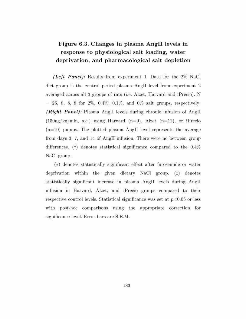
183
Figure 6.3. Changes in plasma AngII levels in response to physiological salt loading, water
deprivation, and pharmacological salt depletion
(Left Panel): Results from experiment 1. Data for the 2% NaCl
diet group is the control period plasma AngII level from experiment 2
averaged across all 3 groups of rats (i.e. Alzet, Harvard and iPrecio). N
= 26, 8, 8, 8 for 2%, 0.4%, 0.1%, and 0% salt groups, respectively.
(Right Panel): Plasma AngII levels during chronic infusion of AngII
(150ng/kg/min, s.c.) using Harvard (n=9), Alzet (n=12), or iPrecio
(n=10) pumps. The plotted plasma AngII level represents the average
from days 3, 7, and 14 of AngII infusion. There were no between group
differences. (†) denotes statistical significance compared to the 0.4%
NaCl group.
(∗) denotes statistically significant effect after furosemide or water
deprivation within the given dietary NaCl group. (‡) denotes
statistically significant increase in plasma AngII levels during AngII
infusion in Harvard, Alzet, and iPrecio groups compared to their
respective control levels. Statistical significance was set at p<0.05 or less
with post-hoc comparisons using the appropriate correction for
significance level. Error bars are S.E.M.

184
Figure 6.3

185
Figure 6.4. Differences in MAP profile of AngII-salt hypertension generated using Alzet, Harvard or
iPrecio pumps
24hr mean arterial pressure (MAP; top panel) and heart rate (HR;
bottom panel) during control and 2 weeks of AngII infusion using
Harvard (white circle; n=12), Alzet (black circle; n=15), or iPrecio
(black triangle; n=10) pumps in rats fed a 2% NaCl diet. MAP on days
1 through 14 of AngII was significantly elevated compared to control
day 3 in all groups. (†), (‡), and (#) denote significant within group
differences compared to values on control day 3 in Alzet, Harvard, and
iPrecio groups, respectively. (∗), (§), and (¶) denote significant
differences between iPrecio and Alzet, iPrecio and Harvard, and Harvard
and Alzet, respectively. Statistical significance was set at p<0.05 or less
in post-hoc comparisons using appropriate correction for significance
level. Error bars are S.E.M.

186
Figure 6.4

187
Figure 6.5. Changes in plasma AngII levels during AngII-salt hypertension generated using Alzet,
Harvard or iPrecio pumps
Plasma AngII levels (top panel) measured on control day 1, and days
3, 7, and 14 of AngII in Harvard (white circle; n=9), Alzet (black circle;
n=12), and iPrecio (black triangle; n=10) groups. 24hr MAP on
corresponding days is also shown (bottom panel). (†), and (‡), denote
significant within group change from control in Alzet, and iPrecio
groups, respectively. (∗), (§), and (¶) denote significant differences
between iPrecio and Alzet, iPrecio and Harvard, and Harvard and Alzet,
respectively. Statistical significance was set at p<0.05 or less in post-hoc
comparisons using appropriate correction for significance level. Error
bars are S.E.M.

188
Figure 6.5