Embed Size (px)
Citation preview

Neuroblastoma
Protocol applies to the examination of specimensfrom patients with neuroblastoma and relatedneuroblastic tumors.
Protocol date: July 2005No AJCC/UICC staging system
Procedures• Cytology (No Accompanying Checklist)• Incisional Biopsy (Needle or Wedge) (No Accompanying Checklist)
• Resection
Authors
Stephen J. Qualman, MD
Department of Laboratory Medicine, Children’s Hospital, Columbus, OhioJay Bowen, MS
Department of Laboratory Medicine, Children’s Hospital, Columbus, Ohio
Patrick L. Fitzgibbons, MD
Department of Pathology, St. Jude Medical Center, Fullerton, CaliforniaSusan L. Cohn, MD
Department of Pediatrics, Northwestern University, Chicago, Illinois
Hiroyuki Shimada, MD, PhDDepartment of Pathology and Laboratory Medicine, Children’s Hospital
Los Angeles, Los Angeles, California
For the Members of the Cancer Committee, College of American Pathologists

Neuroblastoma • Pediatric CAP Approved
2
© 2005. College of American Pathologists. All rights reserved.
The College does not permit reproduction of any substantial portion of these protocolswithout its written authorization. The College hereby authorizes use of these protocols by
physicians and other health care providers in reporting on surgical specimens, in
teaching, and in carrying out medical research for nonprofit purposes. This authorization
does not extend to reproduction or other use of any substantial portion of these protocolsfor commercial purposes without the written consent of the College.
The College of American Pathologists offers these protocols to assist pathologists inproviding clinically useful and relevant information when reporting results of surgical
specimen examinations of surgical specimens. The College regards the reporting
elements in the “Surgical Pathology Cancer Case Summary (Checklist)” portion of theprotocols as essential elements of the pathology report. However, the manner in which
these elements are reported is at the discretion of each specific pathologist, taking into
account clinician preferences, institutional policies, and individual practice.
The College developed these protocols as an educational tool to assist pathologists in
the useful reporting of relevant information. It did not issue the protocols for use in
litigation, reimbursement, or other contexts. Nevertheless, the College recognizes thatthe protocols might be used by hospitals, attorneys, payers, and others. Indeed, effective
July 1, 2004, the Commission on Cancer of the American College of Surgeons
mandated the use of the checklist elements of the protocols as part of its CancerProgram Standards for Approved Cancer Programs. Therefore, it becomes even more
important for pathologists to familiarize themselves with the document. At the same time,
the College cautions that use of the protocols other than for their intended educational
purpose may involve additional considerations that are beyond the scope of thisdocument.

CAP Approved Pediatric • Neuroblastoma
3
Summary
Protocol date: July 2005
This is a new protocol for 2005.
Important NoteFirst priority should always be given to formalin-fixed tissue for morphologic evaluation.
Special studies (eg, ploidy analysis, fluorescence in situ hybridization) are critical to themolecular work-up of neuroblastoma and require at least 100 mg of viable snap-frozen
tissue as the second priority for work-up (Note A).
For more information contact: The Children’s Oncology Group Biopathology Center,
Phone: (614) 722-2890 or (800) 347-2486.

Neuroblastoma • Pediatric CAP Approved
* Data elements with asterisks are not required for accreditation purposes for
the Commission on Cancer. These elements may be clinically important,but are not yet validated or regularly used in patient management.
Alternatively, the necessary data may not be available to the pathologistat the time of pathologic assessment of this specimen.
4
Surgical Pathology Cancer Case Summary (Checklist)
Protocol date: July 2005
Applies to neuroblastoma onlyNo AJCC/UICC staging system
NEUROBLASTOMA: Resection
Patient name:
Surgical pathology number:
Note: Check 1 response unless otherwise indicated.
MACROSCOPIC
Specimen Type___ Subtotal adrenalectomy
___ Total adrenalectomy
___ Other (specify): _________________________
___ Not specified
Tumor Site
Specify: ____________________________ Not specified
Laterality (check all that apply)
___ Right___ Left
___ Midline
___ Not specified
*Specimen Size
*Greatest Dimension: ___ cm*Additional dimensions: ___ x ___ cm
*Specimen Weight
*Specify: ___ g
Tumor Size
Greatest dimension: ___ cm*Additional dimensions: ___ x ___ cm
___ Cannot be determined (see Comment)
Tumor Weight (if separate from total specimen)
Specify: ___ g

CAP Approved Pediatric • Neuroblastoma
* Data elements with asterisks are not required for accreditation purposes for
the Commission on Cancer. These elements may be clinically important,but are not yet validated or regularly used in patient management.Alternatively, the necessary data may not be available to the pathologistat the time of pathologic assessment of this specimen.
5
MICROSCOPIC
Extent of Invasion
Primary Tumor
___ Cannot be assessed___ Encapsulated
___ Capsular extension without other organ involvement
___ Extension into other organs
Regional Lymph Nodes (check all that apply)
___ Cannot be assessed___ No regional lymph node metastasis
___ Right regional lymph node metastasis
Specify: Number examined: ___
Number involved: ______ Left regional lymph node metastasis
Specify: Number examined: ___
Number involved: ___
Distant Metastasis
___ Cannot be assessed___ Distant metastasis
*Specify site(s): ___________________________
Margins
___ Cannot be assessed
___ Margins uninvolved by tumor___ Margin(s) involved by tumor
*Venous/Lymphatic (Large/Small Vessel) Invasion
*___ Absent*___ Present
*___ Indeterminate
International Neuroblastoma Pathology Classification
___ Cannot be determined
Favorable Histopathology
___ Any age; ganglioneuroma (Schwannian stroma-dominant); maturing or mature
___ Any age; ganglioneuroblastoma, intermixed (Schwannian stroma-rich)___ Less than 1.5 years old; neuroblastoma (Schwannian stroma-poor); poorly
differentiated and low or intermediate mitosis-karyorrhexis index (MKI)
___ 1.5 years up to less than 5 years old; neuroblastoma (Schwannian stroma-poor);differentiating and low MKI

Neuroblastoma • Pediatric CAP Approved
* Data elements with asterisks are not required for accreditation purposes for
the Commission on Cancer. These elements may be clinically important,but are not yet validated or regularly used in patient management.
Alternatively, the necessary data may not be available to the pathologistat the time of pathologic assessment of this specimen.
6
Unfavorable Histopathology
___ Any age; ganglioneuroblastoma, nodular (Composite, Schwannian stroma-rich/stroma-dominant and stroma-poor)
___ Any age; neuroblastoma (Schwannian stroma-poor); undifferentiated and any MKI
___ Less than 1.5 years old; neuroblastoma (Schwannian stroma-poor); poorly
differentiated and high MKI, or differentiating and high MKI___ 1.5 years up to less than 5 years old; neuroblastoma (Schwannian stroma-poor);
poorly differentiated and any MKI, or differentiating and intermediate or high MKI
___ Equal to or greater than 5 years old; neuroblastoma (Schwannian stroma-poor); anysubtype and any MKI
International Neuroblastoma Staging System (INSS)#
___ Stage 1
• localized tumor with complete gross excision, with or without microscopic
residual disease
• representative ipsilateral lymph nodes negative for tumor microscopically(nodes attached to and removed with the primary tumor may be positive)
___ Stage 2A
• localized tumor with incomplete gross excision; representative ipsilateralnonadherent lymph nodes negative for tumor microscopically
___ Stage 2B
• localized tumor with or without complete gross excision with ipsilateralnonadherent lymph nodes positive for tumor; enlarged contralateral lymph
nodes must be negative microscopically
___ Stage 3
• unresectable unilateral tumor infiltrating across the midline##, with or withoutregional lymph node involvement
• localized unilateral tumor with contralateral regional lymph node involvement
• midline tumor with bilateral extension by infiltration (unresectable) or by lymphnode involvement
___ Stage 4
• any primary tumor with dissemination to distant lymph nodes, bone, bone
marrow, liver, skin, and/or other organs (except as defined for stage 4S###)___ Stage 4S
• localized primary tumor (as defined for stage 1, 2A, or 2B), with dissemination
limited to skin, liver, and/or bone marrow### (limited to infants less than 1 yearof age)
# Multifocal primary tumors (eg, bilateral adrenal primary tumors) should be staged according to
the greatest extent of disease, as defined above, and followed by a subscript “M” (eg, 3M).
## The midline is defined as the vertebral column. Tumors originating on 1 side and crossing the
midline must infiltrate to or beyond the opposite side of the vertebral column.
### Marrow involvement in stage 4S should be minimal, ie, less than 10% of total nucleated cells
identified as malignant on bone marrow biopsy or marrow aspirate. More extensive marrowinvolvement would be considered stage 4. The MIBG scan (if performed) should be negative inthe marrow.
CAP Approved Pediatric • Neuroblastoma
* Data elements with asterisks are not required for accreditation purposes for
the Commission on Cancer. These elements may be clinically important,but are not yet validated or regularly used in patient management.Alternatively, the necessary data may not be available to the pathologistat the time of pathologic assessment of this specimen.
7
*Additional Pathologic Findings (check all that apply)
*___ None identified*___ Tumor necrosis
*___ Tumor calcification
*___ Other (specify): ______________________
*Comment(s)

Neuroblastoma • Pediatric For Information Only
8
Background Documentation
Protocol date: July 2005
I. Cytologic MaterialA. Clinical Information
1. Patient identificationa. Name
b. Identification number
c. Age (birth date)
d. Sex2. Responsible physician(s)
3. Date of procedure
4. Other clinical informationa. Relevant history (eg, previous diagnoses, treatment, family history) (Note B)
b. Relevant findings (eg, imaging studies, including meta-iodobenzylguanidine
[MIBG] scan; urinary catecholamines) (Notes C and D)c. Clinical diagnosis
d. Procedure (eg, fine-needle aspiration [FNA])
e. Anatomic sites(s) of specimen (eg, right/left adrenal gland, related sites)
B. Macroscopic Examination1. Specimen
a. Unfixed/fixed (specify fixative)
b. Number of slides receivedc. Quantity and appearance of fluid specimen
d. Other materials received (eg, touch preparation from tissue)
e. Results of intraprocedural consultation
2. Material submitted for microscopic examination (eg, smear, cytocentrifuge, touchor filter preparation, cell block)
3. Special studies (specify) (eg, immunohistochemistry, molecular analysis,
cytogenetic analysis) (Notes A and E)C. Microscopic Evaluation
1. Adequacy of specimen (if unsatisfactory for evaluation, specify reason)
2. Tumor, if presenta. Histologic category and subtype, if possible (Note F)
b. Other features (eg, nuclear changes consistent with neuroblastic or
ganglionic differentiation)
3. Other pathologic findings, if present (eg, necrosis, calcification)4. Results/status of special studies (specify)
5. Comments
a. Correlation with intraprocedural consultation, as appropriateb. Correlation with other specimens, as appropriate
c. Correlation with clinical information, as appropriate

For Information Only Pediatric • Neuroblastoma
9
II. Incisional Biopsy(Any Surgical Approach Less Than Complete Adrenalectomy; orOther Primary Tumor Excision, Including Needle or Wedge Biopsy)
A. Clinical Information1. Patient identification
a. Name
b. Identification number
c. Age (birth date)d. Sex
2. Responsible physician(s)
3. Date of procedure4. Other clinical information
a. Relevant history (eg, previous diagnoses, treatment, family history)
b. Relevant findings (eg, imaging studies, including MIBG scan; urinary
catecholamines)c. Clinical diagnosis
d. Procedure (eg, core needle biopsy, wedge biopsy)
e. Anatomic sites(s) of specimen (eg, right/left adrenal gland, related sites)B. Macroscopic Examination (Note G)
1. Specimen
a. Unfixed/fixed (specify fixative)b. Number of pieces
c. Dimensions
d. Descriptive features (eg, hemorrhage, necrosis)
e. Orientation, if designated by surgeonf. Results of intraoperative consultation
2. Tissue submitted for microscopic examination, as appropriate
a. Entire specimenb. Selected sample
c. Frozen section tissue fragment(s), unless saved for special studies
3. Special studies (specify) (eg, immunohistochemistry, ploidy analysis,flourescence in situ hybridization [FISH], cytogenetic analysis) (Notes A and E)
C. Microscopic Evaluation (Note F)
1. Tumor
a. Histologic category (Note F)b. Histologic subtype (Note F)
c. Other features (eg, calcification, amount of neuropil)
d. Venous/lymphatic vessel invasion, if possible to determinee. Mitosis-karyorrhexis index (MKI) of neuroblastoma category (Notes F and H)
2. Additional pathologic findings, if present (eg, necrosis)
3. Results/status of special studies (specify)
4. Commentsa. Correlation with intraoperative consultation, as appropriate
b. Correlation with other specimens, as appropriate
c. Correlation with clinical information, as appropriated. Determination of prognostic group (favorable histology group versus
unfavorable histology group) according to the International Neuroblastoma
Pathology Classification (Note F)
Neuroblastoma • Pediatric For Information Only
10
III. Resection(Adrenalectomy or Other Primary Tumor Excision)
A. Clinical Information
1. Patient identification
a. Nameb. Identification number
c. Age (birth date)
d. Sex2. Responsible physician(s)/clinic(s)
3. Date of procedure
4. Other clinical information
a. Relevant history (Note B)(1) previous diagnoses
(2) surgery and date(s)
(3) radiation and date(s)(4) chemotherapy and date(s)
(5) others
b. Relevant findings (eg, imaging studies, including MIBG scan; urinary
catecholamines) (Notes C and D)c. Clinical diagnosis
d. Procedure (specify anatomic site[s]) (Note G)
(1) excision(2) anatomical structures removed (eg, associated kidney)
(3) lymph node dissection
e. Operative findings (documentation of areas of concern marked by surgeon)B. Macroscopic Examination (Note G)
1. Specimen
a. Organ/tissues included
b. Unfixed/fixed (specify fixative)c. Size (3 dimensions)
d. Weight
e. Orientation, if indicated by surgeonf. Descriptive features (eg, hemorrhage, necrosis)
g. Results of intraoperative consultation
2. Tumor(s)
a. Anatomical site(s) involved by tumorb. Size (3 dimensions)
c. Descriptive characteristics (eg, firm/soft, color, consistency, hemorrhage,
necrosis, biopsy scars)d. Anatomic extent (structures involved by tumor and depth of invasion)
e. Relation to margins
f. Additional tumors3. Additional pathologic findings, if present
4. Lymph nodes, if submitted
a. Number
b. Location, if designated by surgeon5. Margins
6. Stage (Note I)

For Information Only Pediatric • Neuroblastoma
11
7. Tissues submitted for microscopic examination
a. Tumor (adequate sampling of all areas; 1 section for each centimeter ofmaximal tumor diameter and/or different gross appearances)
b. Nodules
c. Margins of resection
d. All lymph nodese. Other lesions
f. Frozen section tissue fragment(s), unless saved for special studies
g. Other organs/tissues8. Special studies (specify) (eg, immunohistochemistry, ploidy analysis, FISH,
cytogenetic analysis) (Notes A and E)
C. Microscopic Examination (Note F)1. Tumor
a. Histologic category (Note F)
b. Histologic subtype (Note F)
c. Other descriptive features (eg, calcification, amount of neuropil)d. Venous/lymphatic vessel invasion, if possible to determine
e. Mitosis-karyorrhexis index (MKI) of neuroblastoma category (Notes F and H)
f. Evaluation of post-therapy tumors for differentiation, necrosis, and fibrosisg. Closest distance to margin
2. Lymph nodes
a. Number (location, if possible)b. Number involved by tumor
3. Additional pathologic findings, if present
4. Results/status of special studies (specify) (Notes A and E)
5. Other organs/tissues6. Comments
a. Correlation with intraoperative consultation, as appropriate
b. Correlation with other specimens, as appropriatec. Correlation with clinical information, as appropriate
d. Determination of prognostic group (favorable histology group versus
unfavorable histology group) according to the International Neuroblastoma
Pathology Classification (Note F)
Explanatory Notes
A. Molecular and Cytogenetic Testing1
MYCN gene amplification (greater than 10 copies by Southern blot or fluorescence in
situ hybridization [FISH]) in neuroblastoma tumor links to a poor prognosis of the patient.
The MYCN gene is located in the short arm of chromosome 2. When amplified, it formsdouble minutes (DMs) and homogeneously staining regions (HSRs), and produces
excess amount of N-myc protein. The myc-max protein complex in the tumor cell
nucleus has been shown to inhibit cellular differentiation and promote cellularproliferation and apoptosis/karyorrhexis.2 Hence, amplification is usually seen in
undifferentiated and poorly differentiated neuroblastomas (Schwannian stroma-poor)
and correlates with a higher mitosis-karyorrhexis index (MKI)3,4 (Note H). MYCN status
of the tumor can be determined by the FISH method within a relatively short period oftime after the surgery/biopsy. A double-staining procedure is recommended for
comparing the number of chromosome 2 and MYCN signals in the same tumor nuclei for
determining MYCN status. Increased MYCN signals associated with a like increase in
Neuroblastoma • Pediatric For Information Only
12
number of chromosome 2 signals does not represent MYCN amplification. MYCN
signals must be seen in excess of chromosome 2 signals by an average of 10 signalsper nucleus to be considered true MYCN amplification.
MYCN amplification is also correlated with advanced stage tumors having chromosome
1p deletions, especially del 1p36.3. The deletion of 14q has also been shown to beunfavorable, as has the 11q deletion and 17q chromosomal gain.
Determination of DNA index by flow cytometry also is very important. A DNA index neardiploid/tetraploid is unfavorable, while hyperdiploid (near-triploid) tumors have a good
prognosis. However, prognostic effects of DNA index are reported to be limited to those
patients diagnosed at less than 1 year of age.5
Higher expression of TrkA (high-affinity nerve growth factor receptor) favors a good
prognosis.1 MYCN-amplified tumors usually have a lower expression of TrkA.
While MYCN FISH studies can be performed on touch preparations, and ploidy analysis
can be performed on frozen tissue also available for touch preparations, cytogenetics
requires fresh tissue. A minimum of 100 mg and preferably 1 g of fresh tumor is requiredfor these purposes (Note G).
B. Clinical PresentationThe clinical presentation of neuroblastoma may provide valuable information in
assessing biologic risk. The abdomen is the most common primary site of
neuroblastoma, with more than 76% of tumors arising either in the adrenals or, less
commonly, in the paramidline sympathetic chains.1 In older children, an abdominal massusually represents an adrenal primary tumor, whereas in infants, it often represents
hepatomegaly secondary to metastatic disease.
The posterior mediastinum is the second most common primary site, and respiratory
symptoms predominate. Cervical neuroblastoma presents as a mass with or without
Horner syndrome.6 All neuroblastomas regardless of biologic risk can extend along
radicular nerves, through spinal foramina, and into the epidural space, forming adumbbell-shaped mass.
Because the spinal cord extends to the level of the T12 to L1 vertebrae, tumors abovethis level are more likely to cause cord compression and paralysis, bladder and bowel
dysfunction, or numbness. Similarly, neuroblastomas primary in the pelvis may present
with constipation or urinary symptoms, including dysuria, infection, flank pain, or urinaryretention.7
The opsoclonus-myoclonus syndrome is a prime example of a paraneoplastic
manifestation of neuroblastoma. Patients with this syndrome usually have an excellentprognosis. This is thought to be secondary to cross-reaction of antineuroblastoma
antibodies with the Purkinje cells of the cerebellum. As many as 70% of such patients
have permanent neurologic deficits despite curative tumor resection.8
C. Imaging Studies
Ultrasound scans are the most common initial screening study to confirm a palpableabdominal or pelvic mass.9 The most useful imaging study is computerized axial
tomography (CT scan) done with simultaneous oral and intravenous contrast.10 This
For Information Only Pediatric • Neuroblastoma
13
gives excellent information about the primary tumor, including location, vascular
encasement, and the status of regional lymph nodes. Hepatic and even gross bonymetastases can be visualized, as can pulmonary metastases (the latter is an extremely
rare site for dissemination).1 Magnetic resonance images (MRI) can give valuable
information about vascular and hepatic involvement and help to determine tumor
resectability but are difficult to perform in active young children.
A diphosphate bone scan and a meta-iodobenzylguanidine (MIBG) scan are requisite to
assess the bone and bone marrow for distant disease.11 A positive bone scan or bonesurvey indicates cortical bone involvement and is a negative prognostic factor; these
patients are at high risk. Approximately 85% of neuroblastomas will take up MIBG.1
D. Endocrine Studies
Urinary catecholamine secretion is increased in neuroblastoma and is useful as a
confirmatory diagnostic marker. Serial determinations are used to assess therapeutic
response and identify recurrence. Vanillylmandelic acid (VMA) and homovanillic acid(HVA) are the 2 catecholamine metabolites commonly measured12 via high-performance
liquid chromatography. In 1 study,13 the sensitivity and specificity of HVA for detection of
neuroblastoma were 72% and 98%; corresponding figures for VMA were 80% sensitivityand 97% specificity. On rare occasions, increased urinary catecholamine secretion may
not be seen with an undifferentiated neuroblastoma.
Rarely, the first diagnostic sign of neuroblastoma may be hypersecretion of vasoactive
intestinal peptide with associated watery diarrhea.14
E. Special StudiesSerology
Serum determinations are useful to help predict prognostic risk. These include serum
lactic dehydrogenase (LDH), neuron-specific enolase (NSE), and ferritin.15 Ferritin levelsare the most important diagnostic marker of the 3, with an elevation above normal
(before transfusion) associated with a worse prognosis. Reference ranges are
dependent on the individual laboratory, but an upper normal limit of 142 ng/mL
frequently is reported.16 Serial LDH levels correlate with disease activity, andpretreatment values of more than 1000 U/L are associated with a worse prognosis.17
Serum levels of NSE more than 30 ng/mL also are associated with a worse prognosis.18
ImmunohistochemistrySchwann cells: S-100 protein-positive.
The following are positive in a variable proportion of cases:
• Neuron-specific enolase
• Chromogranin A• Synaptophysin
• Tyrosine hydroxylase
• Protein gene product 9.5• GD2 (disialoganglioside, a ganglioside on human neuroblastoma cell membrane)
• NB84

Neuroblastoma • Pediatric For Information Only
14
The following are usually negative:
• Actin• Desmin
• Low-molecular-weight cytokeratin
• CD45 (leukocyte common antigen)
• Vimentin
Differential Diagnosis
A cell surface glycoprotein, p30/32 (product of the MIC2 gene detected by CD99antibodies), common in peripheral primitive neuroectodermal tumor (pPNET)/Ewing
sarcoma, usually is negative in neuroblastoma. In contrast, tyrosine hydroxylase
commonly is positive for neuroblastoma and negative for pPNET/Ewing sarcoma.
Undifferentiated cells (in poorly differentiated subtype) may, on rare occasions, express
vimentin and have rhabdoid morphology.
Electron Microscopy
Ultrastructural studies are still of value in the diagnosis of relatively undifferentiated
neuroblastoma, where the diagnosis is not readily evident by light microscopic study orurinary catecholamine study, especially given the variable specificity of immunostaining.
Diagnostic criteria include dense core granules of neurosecretory type and cell
processes (primitive neurites) containing typically arranged microtubules.
F. Morphologic Categories
It is recommended that the International Neuroblastoma Classification19,20 described
below be used when describing tumor samples.
There are 4 categories in this group of tumors:
Neuroblastoma (Schwannian stroma-poor)
Ganglioneuroblastoma, intermixed (Schwannian stroma-rich)
Ganglioneuroma (Schwannian stroma-dominant)
Ganglioneuroblastoma, nodular (composite, Schwannian stroma-rich/stroma-dominantand stroma-poor)
Within each category, 1 or more subtypes are recognized.
Microscopically, tumors in the neuroblastoma category are composed of neuroblastic
cells that form groups or nests separated by delicate, often incomplete stromal septawithout or with limited Schwannian proliferation; whereas tumors in the
ganglioneuroblastoma, intermixed category and the ganglioneuroma category are
characterized by a presence of ganglioneuromatous tissue, where mature and/or
maturing ganglion cells are individually scattered in a background of highly developedSchwannian stroma. Tumors in the ganglioneuroblastoma, nodular category are
composed of multiple clones: one shows an appearance of either ganglioneuroblastoma,
intermixed or ganglioneuroma, and the other(s) show that (those) of neuroblastoma.

For Information Only Pediatric • Neuroblastoma
15
Neuroblastoma (Schwannian Stroma-poor) Category
Three subtypes are distinguished.
Undifferentiated Subtype
Neuropil absent; no tumor cell differentiation; diagnosis relies heavily on ancillary
techniques, such as immunohistochemistry, electron microscopy, and/ormolecular/cytogenetics.
Poorly-differentiated SubtypeNeuropil background evident; 5% or fewer tumor cells show a feature of differentiating
neuroblasts with a synchronous differentiation of nucleus (enlarged, vesicular with a
single prominent nucleolus) and cytoplasm (conspicuous, eosinophilic or amphophilic,and 2 times larger in diameter than nucleus).
Differentiating Subtype
Greater than 5% of tumor cells show an appearance of differentiating neuroblasts (maybe accompanied by mature ganglion-like cells), and neuropil is usually abundant; some
tumors can show substantial Schwannian stromal formation, frequently at their
periphery, and a transition zone between neuroblastomatous and ganglioneuromatousregion can develop, although this zone does not have well-defined borders and
comprises less than 50% of the tumor.
Ganglioneuroblastoma, Intermixed (Schwannian Stroma-rich) Category#
Ganglioneuromatous (stroma-rich) component of tumor exceeds 50%; intermixed or
randomly distributed pattern of microscopic neuroblastic nests present, consisting of
cells in various stages of differentiation (neuroblasts, differentiating neuroblasts,maturing ganglion cells); abundant neuropil; macroscopic hemorrhagic nodules are
absent.
Ganglioneuroma (Schwannian Stroma-dominant) Category
Two subtypes are included; neuroblastic cells (differentiating neuroblasts, maturing and
mature ganglion cells) in the tumor tissue do not form microscopic nests but are
individually distributed in the Schwannian stroma.
Maturing Subtype
Predominately ganglioneuromatous stroma; minor, scattered groups of differentiatingneuroblasts or maturing ganglion cells along with completely mature ganglion cells.
Mature SubtypeMature Schwannian stroma and ganglion cells; neuritic fascicular processes,
accompanied by Schwann cells and perineurial cells; absence of neuroblastomatous
component in complete maturation; satellite cells accompany mature ganglion cells.
Ganglioneuroblastoma, Nodular (Composite Schwannian Stroma-rich/ Stroma-
dominant and Stroma-poor) Category#
Ganglioneuroblastoma, intermixed (stroma-rich) or ganglioneuroma (stroma-dominant)with macroscopic neuroblastic nodules (stroma-poor and usually hemorrhagic); border
between nodule and stroma-rich or stroma-dominant region is often abrupt
microscopically, but may instead be more gradual; the neuroblastoma component maybe found in a metastatic tumor where the primary is ganglioneuroblastoma, intermixed or
ganglioneuroma.

Neuroblastoma • Pediatric For Information Only
16
Neuroblastic Tumor, Unclassifiable
Neuroblastic cells evident; sample insufficient for categorization into 1 of the 4 basic
types. A small biopsy taken from a large tumor can result in this designation.
Neuroblastoma (Schwannian Stroma-poor), Not Otherwise Specified (NOS)
Tumor diagnosis of neuroblastoma (Schwannian stroma-poor); subtyping not possible
due to poor quality of sample or section.
Ganglioneuroblastoma, NOS
Tumor diagnosis of ganglioneuroblastoma (Schwannian stroma-rich); subtyping not
possible due to a limited amount of tissue for evaluation or extensive calcification oftumor.
# Ganglioneuroblastomas are highly variable in both number of neuroblasts and theirextent of differentiation. Variability is seen between tumors, between microscopic fields
in the same tumor, and occasionally between the primary and metastatic tumor.
Ganglioneuroblastoma diagnostic criteria include (a) mature Schwannian stromalcomponent with individually scattered mature and/or maturing ganglion cells and
(b) a neuroblastic component.
The presence of calcification, in any amount in the tumor tissue, tends to indicate animproved prognosis of the patient.21
Differential DiagnosisPrimitive rhabdomyosarcoma
Peripheral primitive neuroectodermal tumor (pPNET)/Ewing sarcoma
Blastematous Wilms tumorBlastic hematopoietic neoplasm
Malignant rhabdoid tumor
Desmoplastic small-round cell tumor
G. Sampling
In the complete macroscopic evaluation of the specimen, sections should be obtained
from central and peripheral areas of the tumor according to common guidelines (at least1 tumor section per centimeter in the longest dimension and sections from the inked
surgical margins).19
The genetic and morphologic heterogeneity of neuroblastoma requires extensivebiologic and histologic study of the tumor. If practical, the entire tumor should be
examined microscopically to facilitate the detection of any neuroblastic nodules that may
have been overlooked on gross examination. In addition to the tissue taken for histologicexamination as described, the International Neuroblastoma Pathology Committee
recommends sampling a neuroblastic surgical specimen for biologic studies as follows.19
For Information Only Pediatric • Neuroblastoma
17
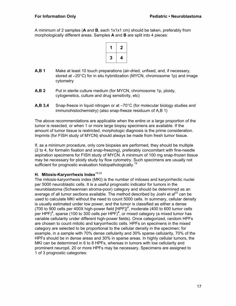
A minimum of 2 samples (A and B, each 1x1x1 cm) should be taken, preferably from
morphologically different areas. Samples A and B are split into 4 pieces:
1 2
3 4
A,B 1 Make at least 10 touch preparations (air-dried, unfixed, and, if necessary,
stored at –20°C) for in situ hybridization (MYCN, chromosome 1p) and image
cytometry
A,B 2 Put in sterile culture medium (for MYCN, chromosome 1p, ploidy,cytogenetics, culture and drug sensitivity, etc)
A,B 3,4 Snap-freeze in liquid nitrogen or at –70°C (for molecular biology studies and
immunohistochemistry) (also snap-freeze residuum of A,B 1)
The above recommendations are applicable when the entire or a large proportion of thetumor is resected, or when 1 or more large biopsy specimens are available. If the
amount of tumor tissue is restricted, morphologic diagnosis is the prime consideration.
Imprints (for FISH study of MYCN) should always be made from fresh tumor tissue.
If, as a minimum procedure, only core biopsies are performed, they should be multiple
(2 to 4, for formalin fixation and snap-freezing), preferably concomitant with fine-needle
aspiration specimens for FISH study of MYCN. A minimum of 100 mg snap-frozen tissuemay be necessary for ploidy study by flow cytometry. Such specimens are usually not
sufficient for prognostic evaluation histopathologically.19
H. Mitosis-Karyorrhexis Index19,22
The mitosis-karyorrhexis index (MKI) is the number of mitoses and karyorrhectic nuclei
per 5000 neuroblastic cells. It is a useful prognostic indicator for tumors in theneuroblastoma (Schwannian stroma-poor) category and should be determined as an
average of all tumor sections available. The method described by Joshi et al22 can be
used to calculate MKI without the need to count 5000 cells. In summary, cellular density
is usually estimated under low power, and the tumor is classified as either a dense(700 to 900 cells per 400X high-power field [HPF])#, moderate (400 to 600 tumor cells
per HPF)#, sparse (100 to 300 cells per HPF)#, or mixed category (a mixed tumor has
variable cellularity under different high-power fields). Once categorized, random HPFsare chosen to count mitotic and karyorrhectic cells. HPFs on specimens in the mixed
category are selected to be proportional to the cellular density in the specimen; for
example, in a sample with 70% dense cellularity and 30% sparse cellularity, 70% of the
HPFs should be in dense areas and 30% in sparse areas. In highly cellular tumors, theMKI can be determined in 6 to 8 HPFs, whereas in tumors with low cellularity and
prominent neuropil, 20 or more HPFs may be necessary. Specimens are assigned to
1 of 3 prognostic categories:

Neuroblastoma • Pediatric For Information Only
18
(1) Low MKI Less than 100 mitotic and karyorrhectic cells/5000 tumor cells,
or less than 2% of tumor consisting of mitotic and karyorrhecticcells
(2) Intermediate MKI 100 to 200 mitotic and karyorrhectic cells/5000 tumor cells, or
2% to 4% of tumor consisting of mitotic and karyorrhectic cells
(3) High MKI Greater than 200 mitotic and karyorrhectic cells/5000 tumor
cells, or greater than 4% of tumor consisting of mitotic andkaryorrhectic cells
# Numbers of neuroblastic cells in each HPF (denominator for MKI determination) canvary based on the type of your microscope (some practice required for assessing the
number of neuroblastic cells in your HPF). Numbers listed above in the parentheses are
for the microscope with a regular ocular. With a super-wide-field type of ocular, you may
be able to have 1200 to 1500 cells per HPF in a dense category.
I. StagingThe International Neuroblastoma Staging System (INSS) is accepted as universally
applicable and should always be recorded for new patients.1 The core of clinical staging
is the size of the primary tumor, locoregional lymph node status, and the presence of
distant metastases.
International Neuroblastoma Staging System (INSS)
Stage 1 Localized tumor with complete gross excision, with or without microscopicresidual disease; representative ipsilateral lymph nodes negative for
tumor microscopically (nodes attached to and removed with the primary
tumor may be positive).Stage 2A Localized tumor with incomplete gross excision; representative ipsilateral
nonadherent lymph nodes negative for tumor microscopically.
Stage 2B Localized tumor with or without complete gross excision, with ipsilateral
nonadherent lymph nodes positive for tumor. Enlarged contralaterallymph nodes must be negative microscopically.
Stage 3 Unresectable unilateral tumor infiltrating across the midline, with or
without regional lymph node involvement; or localized unilateral tumorwith contralateral regional lymph node involvement; or midline tumor with
bilateral extension by infiltration (unresectable) or by lymph node
involvement. The midline is defined as the vertebral column. Tumors
originating on 1 side and crossing the midline must infiltrate to or beyondthe opposite side of the vertebral column.
Stage 4 Any primary tumor with dissemination to distant lymph nodes, bone, bone
marrow, liver, skin, and/or other organs (except as defined for stage 4S).Stage 4S Localized primary tumor (as defined for stage 1, 2A, or 2B), with
dissemination limited to skin, liver, and/or bone marrow (limited to infants
less than 1 year of age). Marrow involvement should be minimal (ie, lessthan 10% of total nucleated cells identified as malignant by bone biopsy
or by bone marrow aspirate). More extensive bone marrow involvement
would be considered to be stage 4 disease. The results of the MIBG scan
(if performed) should be negative for disease in the bone marrow.
For Information Only Pediatric • Neuroblastoma
19
J. Prognostic Groups
Risk group assessment can be defined by clinical and biological variables. A simplifiedapproach is described using either pathologic variables combined with age (Table 1)20,23
or a compendium of biologic and clinical risk factors (Table 2).1 Also included is a risk-
grouping scheme for clinical trials of the Children’s Oncology Group Neuroblastoma
Studies (Table 3) based on the combination of clinical stage, age at diagnosis, MYCNstatus, histopathology classification, and DNA index (only for infants) (Table 3).
The International Neuroblastoma Pathology Classification20 uses age, neuroblasticmaturation, and MKI as prognostic indicators. Unfavorable indicators include
undifferentiated neuroblastoma (especially in older patients) and high MKI.

Neuroblastoma • Pediatric For Information Only
20
Table 1. International Neuroblastoma Pathology Prognostic Classification20
Age Favorable Histology Group Unfavorable Histology Group
Ganglioneuroma(Schwannian stroma-dominant)
• maturing
• mature
Ganglioneuroblastoma,intermixed (Schwannian stroma-
rich)
Ganglioneuroblastoma, nodular(Composite, Schwannian stroma-
rich/stroma-dominant and stroma-poor)#
Any
Neuroblastoma(Schwannian stroma-poor)
• undifferentiated and any MKI
Less
than 1.5years
Neuroblastoma(Schwannian stroma-poor)
• poorly differentiated and low
or intermediate MKI
Neuroblastoma(Schwannian stroma-poor)
• poorly differentiated and high MKI
• differentiating and high MKI
1.5 years
up to lessthan 5
years
Neuroblastoma(Schwannian stroma-poor)
• differentiating and low MKI
Neuroblastoma(Schwannian stroma-poor)
• poorly differentiated and any MKI• differentiating and intermediate or
high MKI
Equal to
or greaterthan 5
years
Neuroblastoma
(Schwannian stroma-poor)• any subtype and any MKI
# All tumors in the category of ganglioneuroblastoma, nodular are classified into an
unfavorable histology group according to the original Shimada classification24 and the
International Neuroblastoma Pathology Classification.20 However, recent analysisdistinguished 2 prognostic subsets, favorable and unfavorable, by applying the same
age-linked histopathology evaluation (see Table 1) to the nodular (neuroblastoma)
components of the tumors in this category.25 The International NeuroblastomaPathology Committee has approved the presence of these 2 subsets and is currently
preparing a new version of the Classification, with modification accordingly.

For Information Only Pediatric • Neuroblastoma
21
Table 2. Biologic and Clinical Risk Factors and Groups in Neuroblastoma1
Parameter Low Risk Intermediate Risk High Risk
MYCN status Normal Normal Amplified (greater than
10 copies)
Ploidy Hyperdiploid Near-diploid Near-diploid
Near-triploid Near-tetraploid Near-tetraploid
17q gain Rare Common Common
11q, 14q loss of
heterozygosity (LOH)
Rare Common Rare
1p LOH Rare Uncommon Common
TRK A expression High Low or absent Low or absent
TRK B expression Truncated Low or absent Low or absent
TRK C expression High Low or absent Low or absent
Age Usually less than
1 year
Usually greater
than 1 year
Usually 1 to 5 years
Stage 1, 2, 4S Usually 3 or 4 Usually 3 or 4
3-year survival rate Greater than
90%
30% to 50% Less than 20%

Neuroblastoma • Pediatric For Information Only
22
Table 3. Risk Grouping Scheme for the Children’s Oncology Group
Neuroblastoma Study
INSS
Stage Age
MYCN
Status
Shimada
Histology
DNA
Ploidy
Risk
Group
1 0-21y Any Any Any Low
2A/2B Less than 365dBetween 1y-21y
Between 1y-21yBetween 1y-21y
AnyNonamplified
AmplifiedAmplified
AnyAny
FavorableUnfavorable
AnyN/A
N/AN/A
LowLow
LowHigh
3 Less than 365dLess than 365dBetween 1y-21y
Between 1y-21y
Between 1y-21y
NonamplifiedAmplifiedNonamplified
Nonamplified
Amplified
AnyAnyFavorable
Unfavorable
Any
AnyAnyN/A
N/A
N/A
IntermediateHighIntermediate
High
High
4 Less than 365dLess than 365d
Between 1y-21y
NonamplifiedAmplified
Any
AnyAny
Any
AnyAny
Any
IntermediateHigh
High
4S Less than 365d
Less than 365dLess than 365d
Less than 365d
Nonamplified
NonamplifiedNonamplified
Amplified
Favorable
AnyUnfavorable
Any
DI>1#
DI=1#
Any
Any
Low
IntermediateIntermediate
High
# DNA ploidy: DNA index (DI) greater than 1 (aneuploid) or equal to 1 (diploid);
hypodiploid tumors (with DI less than 1) will be treated as a tumor with DI greater than 1.

For Information Only Pediatric • Neuroblastoma
23
References
1. LaQuaglia MP. Surgical management of neuroblastoma. Semin Pediatr Surg.2001;10:132-139.
2. Wenzel A, Cziepluch C, Hamann U, Schurmann J, Schwab M. The N-Myc
oncoprotein is associated in vivo with the phosphoprotein Max (p20/22) in human
neuroblastoma cells. EMBO J. 1991;10:3703-3712.3. Shimada H, Stram D, Chatten J, et al. Identification of subsets of neuroblastomas
combined with histopathologic and N-myc analysis. J Natl Cancer Inst.
1995;87:1470-1476.4. Goto S, Umehara S, Gerbing RB, et al. Histopathology and MYCN status in
peripheral neuroblastic tumors: a report from the Children’s Cancer Group. Cancer.
2001;92:2699-2708.5. Hook AZ, Hayes FA, Shuster JJ, et al. Clinical relevance of tumor cell ploidy and N-
myc gene amplification in childhood neuroblastoma: a Pediatric Oncology Group
study. J Clin Oncol. 1991;9:581-591.
6. Abramson SJ, Berdow WE, Ruzal-Shapiro C, et al. Cervical neuroblastoma ineleven infants – a tumor with favorable prognosis: clinical and radiologic (US, ST,
MRI) findings. Pediatr Radiol. 1993;23:253-257.
7. Haase GM, O’Leary MC, Stram DO, et al. Pelvic neuroblastoma – implications for anew favorable subgroup: a Children’s Cancer Group experience. Ann Surg Oncol.
1995;2:516-523.
8. Russo C, Cohn SL, Petruzzi MJ, et al. Long-term neurologic outcome in childrenwith opsoclonus-myoclonus associated with neuroblastoma: a report from the
Pediatric Oncology Group. Med Pediatr Oncol. 1997;28:284-288.
9. Hartman GE, Shochat SJ. Abdominal mass lesions in the newborn: diagnosis and
treatment. Clin Perinatol. 1989;16:123-125.10. Hugosson C, Nyman R, Jorulf H, et al. Imaging of abdominal neuroblastoma in
children. Acta Radiol. 1999;40:534-542.
11. Jacobs A, Delree M, Desprechins B, et al. Consolidating the role of *I-MIBGscintigraphy in childhood neuroblastoma: five years of clinical experience. Pediatr
Radiol. 1990;20:157-159.
12. Laug WE, Siegel SE, Shaw KN, Landing B, Baptista J, Gutenstein M. Initial urinary
catecholamine metabolite concentrations and prognosis in neuroblastoma.Pediatrics. 1978;62:77-83.
13. Horsmans Y, Desager JP, Harveugt C. Sensitivity and specificity of the
determination of urinary catecholamines and their acid metabolites in the diagnosisof neuroblastoma in children. Bull Cancer. 1990;77:985-989.
14. Scheibel E, Rechnitzer C, Fahrenkrug J, et al. Vasoactive intestinal peptide (VIP) in
children with neural crest tumors. Acta Paediatr Scand. 1982;71:721-725.15. Hann HWL. Serum markers and prognosis in neuroblastoma: Ferritin, LDH, and
NSE. In: Brodeur GM, Sawada T, Tsuchida Y, Voute PA, eds. Neuroblastoma.
Amsterdam, The Netherlands: Elsevier; 2000:371-381.
16. Hann HW, Evans AW, Siegel SE, et al. Prognostic importance of serum ferritin inpatients with stages III and IV neuroblastoma: the Children’s Cancer Group
experience. Cancer Res. 1985;43:2843-2848.
17. Berthold F, Trechow R, Utsch S, et al. Prognostic factors in metastaticneuroblastoma: a multivariate analysis of 182 cases. Am J Pediatr Hematol Oncol.
1992;14:207-215.
18. Zeltzer PM, Maroupos PJ, Sateur H, et al. Prognostic significance of serumneuropeptide specific enolase in local and widespread neuroblastoma. Prog Clin
Biol Res. 1985;175:319-329.

Neuroblastoma • Pediatric For Information Only
24
19. Shimada H, Ambros IM, Dehner LP, Hata J, Joshi VV, Roald B. Terminology and
morphologic criteria of neuroblastic tumors: recommendations by the InternationalNeuroblastoma Pathology Committee. Cancer. 1999;86:349-363.
20. Shimada, H, Ambros IM, Dehner LP, et al. The International Neuroblastoma
Pathology Classification (the Shimada system). Cancer. 1999;86:364-372.
21. Joshi VV, Cantor AB, Altschuler A, et al. Age-linked prognostic categorization basedon a new histologic grading system of neuroblastomas: a clinical pathologic study of
211 cases from the Pediatric Oncology Group. Cancer. 1992;69:2197-2211.
22. Joshi VV, Chatten J, Sather HN, Shimada H. Evaluation of the Shimadaclassification in advanced neuroblastoma with a special reference to the mitosis-
karyorrhexis index: a report from the Children’s Cancer Study Group. Mod Pathol.
1991;4:139-147.23. Shimada H, Roald B. Histology: tumors of the neuroblastoma group. In: Brodeur
GM, Sawada T, Tsuchida Y, Voute PA, eds. Neuroblastoma. Amsterdam, The
Netherlands: Elsevier; 2000:341-354.
24. Shimada H, Chatten J, Newton WA Jr, et al. Histopathologic prognostic factors inneuroblastic tumors: definition of subtypes of ganglioneuroblastoma and an age-
linked classification of neuroblastoma. J Natl Cancer Inst. 1984;73:405-416.
25. Umehara S, Nakagawa A, Matthay KK, et al. Histopathology defines prognosticsubsets of ganglioneuroblastoma, nodular: a report from the Children’s Cancer
Group. Cancer. 2000;89:1150-1161.
Bibliography
O'Dorisio MS, Qualman SJ. Neuroblastoma. In: Mazaferri EM, Samaan NA, eds.
Endocrine Tumors. Cambridge, Mass: Blackwell Scientific Publications; 1993:448-454.
Brodeur GM, Sawada T, Tsuchida Y, Voute PA, eds. Neuroblastoma. Amsterdam, The
Netherlands: Elsevier; 2000.

![Detection and therapy of neuroblastoma minimal residual ......Neuroblastoma (NB) is an early childhood cancer that accounts for approximately 15% of pediatric cancer deaths [1]. It](https://img.pdfslide.net/doc/110x75/610184478a80e637194c6683/detection-and-therapy-of-neuroblastoma-minimal-residual-neuroblastoma-nb.jpg)