Embed Size (px)
Citation preview
Neurology Case Presentation
• Vahid Behravan M.D.
• Neurology PGY-4
• August 2012
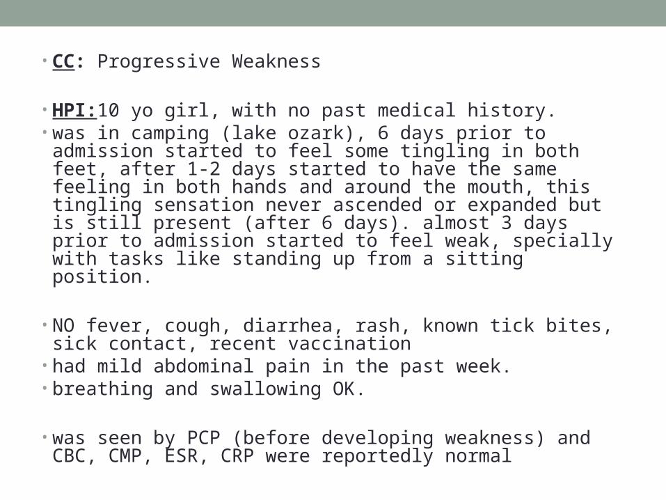
• CC: Progressive Weakness
• HPI:10 yo girl, with no past medical history.• was in camping (lake ozark), 6 days prior to admission started
to feel some tingling in both feet, after 1-2 days started to have the same feeling in both hands and around the mouth, this tingling sensation never ascended or expanded but is still present (after 6 days). almost 3 days prior to admission started to feel weak, specially with tasks like standing up from a sitting position.
• NO fever, cough, diarrhea, rash, known tick bites, sick contact, recent vaccination
• had mild abdominal pain in the past week.• breathing and swallowing OK.
• was seen by PCP (before developing weakness) and CBC, CMP, ESR, CRP were reportedly normal
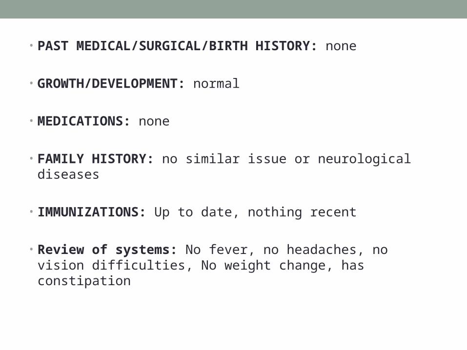
• PAST MEDICAL/SURGICAL/BIRTH HISTORY: none
• GROWTH/DEVELOPMENT: normal
• MEDICATIONS: none
• FAMILY HISTORY: no similar issue or neurological diseases
• IMMUNIZATIONS: Up to date, nothing recent
• Review of systems: No fever, no headaches, no vision difficulties, No weight change, has constipation

Physical Exam:•V/S: T: 36.8, HR: 112, R: 21, BP: 134/88, SpO2: 98 % , NIF in ED -30•well developed, well nourished 10 years old right handed girl, communicating well, cooperating with exam, not in distress•Head atraumatic, normocephalic. Eyes without swelling, redness or discharge. Ears without drainage. Nares patent. Mouth with mucous membranes moist, pink, without lesions.•Neck supple.•Lungs clear on auscultation, with good air movement bilaterally.•Heart normal S1S2, without murmur. •Abdomen soft, no tenderness, not distended. •per ER physician, minimal rectal tone•Musc/Skeletal: AROM and PROM without limitations in all major joints. No contractures noted. very slight tenderness on spine palpation thoracic area.•Skin: no rash
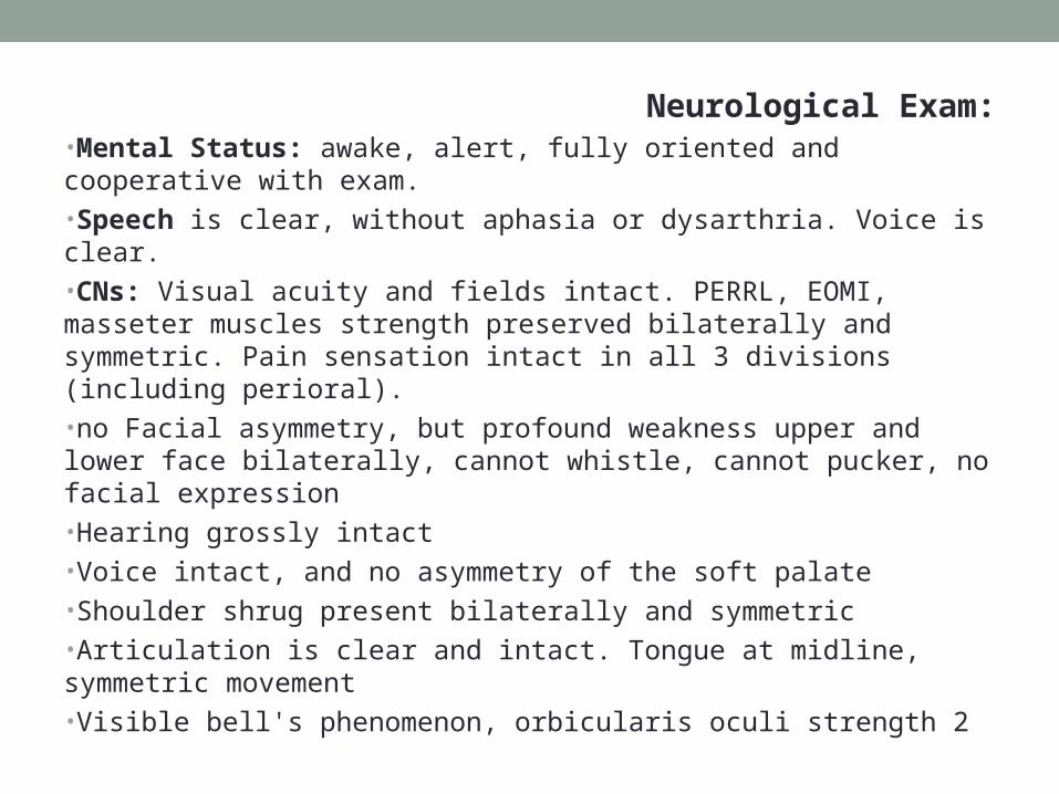
Neurological Exam:•Mental Status: awake, alert, fully oriented and cooperative with exam.•Speech is clear, without aphasia or dysarthria. Voice is clear.•CNs: Visual acuity and fields intact. PERRL, EOMI, masseter muscles strength preserved bilaterally and symmetric. Pain sensation intact in all 3 divisions (including perioral). •no Facial asymmetry, but profound weakness upper and lower face bilaterally, cannot whistle, cannot pucker, no facial expression•Hearing grossly intact•Voice intact, and no asymmetry of the soft palate •Shoulder shrug present bilaterally and symmetric •Articulation is clear and intact. Tongue at midline, symmetric movement•Visible bell's phenomenon, orbicularis oculi strength 2
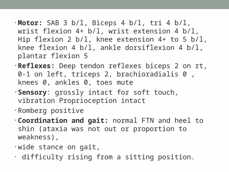
• Motor: SAB 3 b/l, Biceps 4 b/l, tri 4 b/l, wrist flexion 4+ b/l, wrist extension 4 b/l, Hip flexion 2 b/l, knee extension 4+ to 5 b/l, knee flexion 4 b/l, ankle dorsiflexion 4 b/l, plantar flexion 5
• Reflexes: Deep tendon reflexes biceps 2 on rt, 0-1 on left, triceps 2, brachioradialis 0 , knees 0, ankles 0, toes mute
• Sensory: grossly intact for soft touch, vibration Proprioception intact
• Romberg positive• Coordination and gait: normal FTN and heel to shin
(ataxia was not out or proportion to weakness), • wide stance on gait, • difficulty rising from a sitting position.
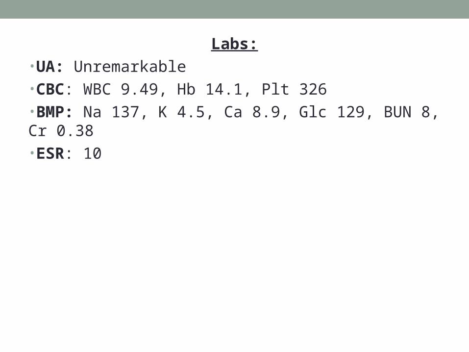
Labs:•UA: Unremarkable•CBC: WBC 9.49, Hb 14.1, Plt 326•BMP: Na 137, K 4.5, Ca 8.9, Glc 129, BUN 8, Cr 0.38•ESR: 10
• Where?
• What?
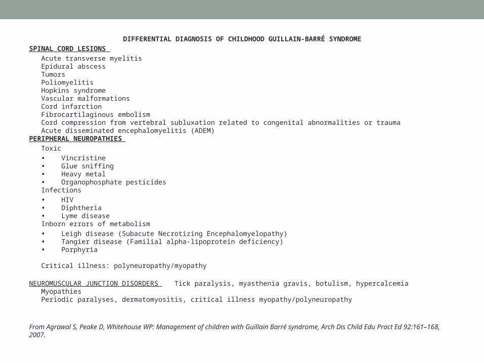
DIFFERENTIAL DIAGNOSIS OF CHILDHOOD GUILLAIN-BARRÉ SYNDROME
SPINAL CORD LESIONS
Acute transverse myelitis Epidural abscess Tumors Poliomyelitis Hopkins syndrome Vascular malformations Cord infarction Fibrocartilaginous embolism Cord compression from vertebral subluxation related to congenital abnormalities or trauma Acute disseminated encephalomyelitis (ADEM)PERIPHERAL NEUROPATHIES
Toxic
• Vincristine • Glue sniffing • Heavy metal • Organophosphate pesticides Infections
• HIV • Diphtheria • Lyme disease Inborn errors of metabolism
• Leigh disease (Subacute Necrotizing Encephalomyelopathy) • Tangier disease (Familial alpha-lipoprotein deficiency) • Porphyria
Critical illness: polyneuropathy/myopathy
NEUROMUSCULAR JUNCTION DISORDERS Tick paralysis, myasthenia gravis, botulism, hypercalcemia Myopathies Periodic paralyses, dermatomyositis, critical illness myopathy/polyneuropathy
From Agrawal S, Peake D, Whitehouse WP: Management of children with Guillain Barré syndrome, Arch Dis Child Edu Pract Ed 92:161–168, 2007.
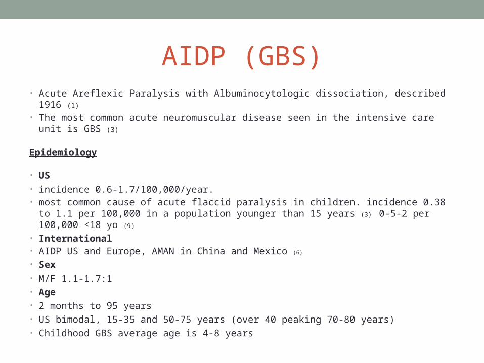
AIDP (GBS)• Acute Areflexic Paralysis with Albuminocytologic dissociation, described 1916 (1)
• The most common acute neuromuscular disease seen in the intensive care unit is GBS (3)
Epidemiology
• US• incidence 0.6-1.7/100,000/year.• most common cause of acute flaccid paralysis in children. incidence 0.38 to 1.1 per 100,000 in a
population younger than 15 years (3) 0-5-2 per 100,000 <18 yo (9)
• International• AIDP US and Europe, AMAN in China and Mexico (6)
• Sex• M/F 1.1-1.7:1• Age• 2 months to 95 years• US bimodal, 15-35 and 50-75 years (over 40 peaking 70-80 years)• Childhood GBS average age is 4-8 years
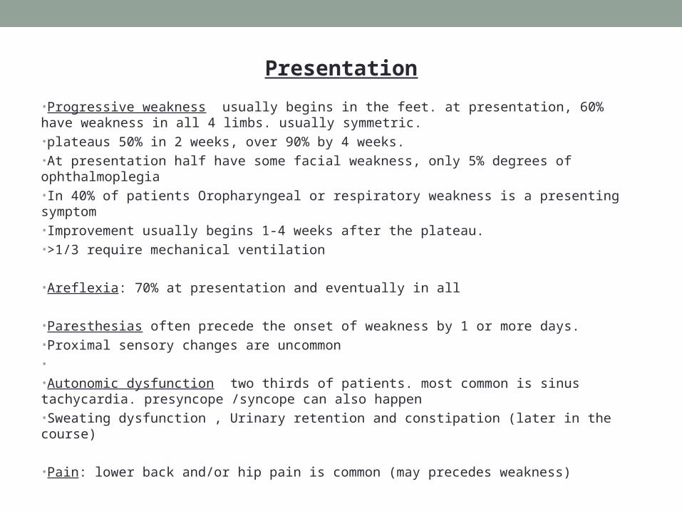
Presentation
•Progressive weakness usually begins in the feet. at presentation, 60% have weakness in all 4 limbs. usually symmetric.•plateaus 50% in 2 weeks, over 90% by 4 weeks. •At presentation half have some facial weakness, only 5% degrees of ophthalmoplegia •In 40% of patients Oropharyngeal or respiratory weakness is a presenting symptom •Improvement usually begins 1-4 weeks after the plateau. •>1/3 require mechanical ventilation
•Areflexia: 70% at presentation and eventually in all
•Paresthesias often precede the onset of weakness by 1 or more days. •Proximal sensory changes are uncommon• •Autonomic dysfunction two thirds of patients. most common is sinus tachycardia. presyncope /syncope can also happen •Sweating dysfunction , Urinary retention and constipation (later in the course)
•Pain: lower back and/or hip pain is common (may precedes weakness)
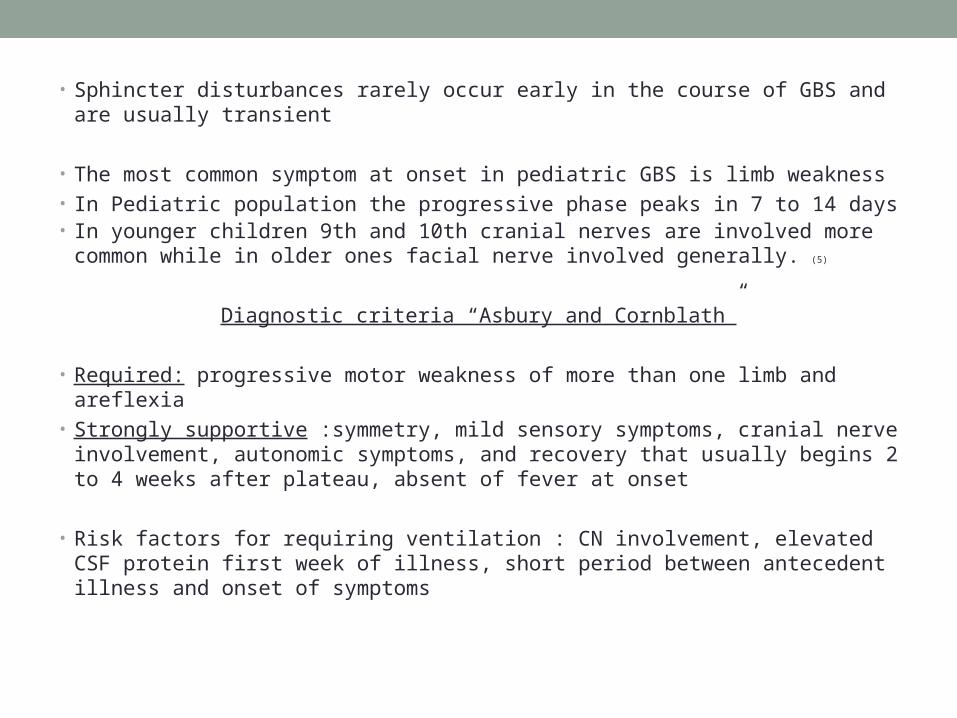
• Sphincter disturbances rarely occur early in the course of GBS and are usually transient
• The most common symptom at onset in pediatric GBS is limb weakness• In Pediatric population the progressive phase peaks in 7 to 14 days• In younger children 9th and 10th cranial nerves are involved more common
while in older ones facial nerve involved generally. (5)
Diagnostic criteria “Asbury and Cornblath”
• Required: progressive motor weakness of more than one limb and areflexia • Strongly supportive :symmetry, mild sensory symptoms, cranial nerve
involvement, autonomic symptoms, and recovery that usually begins 2 to 4 weeks after plateau, absent of fever at onset
• Risk factors for requiring ventilation : CN involvement, elevated CSF protein first week of illness, short period between antecedent illness and onset of symptoms
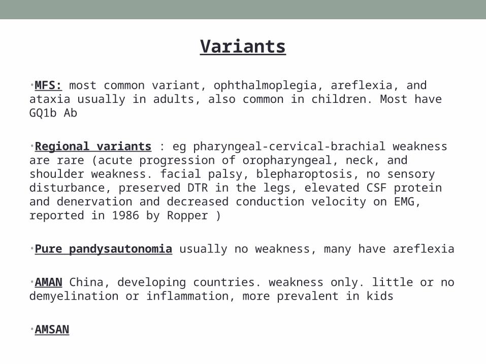
Variants
•MFS: most common variant, ophthalmoplegia, areflexia, and ataxia usually in adults, also common in children. Most have GQ1b Ab
•Regional variants : eg pharyngeal-cervical-brachial weakness are rare (acute progression of oropharyngeal, neck, and shoulder weakness. facial palsy, blepharoptosis, no sensory disturbance, preserved DTR in the legs, elevated CSF protein and denervation and decreased conduction velocity on EMG, reported in 1986 by Ropper )
•Pure pandysautonomia usually no weakness, many have areflexia
•AMAN China, developing countries. weakness only. little or no demyelination or inflammation, more prevalent in kids
•AMSAN
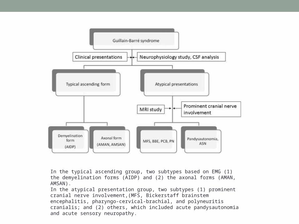
In the typical ascending group, two subtypes based on EMG (1) the demyelination forms (AIDP) and (2) the axonal forms (AMAN, AMSAN). In the atypical presentation group, two subtypes (1) prominent cranial nerve involvement,(MFS, Bickerstaff brainstem encephalitis, pharyngo-cervical-brachial, and polyneuritis cranialis; and (2) others, which included acute pandysautonomia and acute sensory neuropathy.
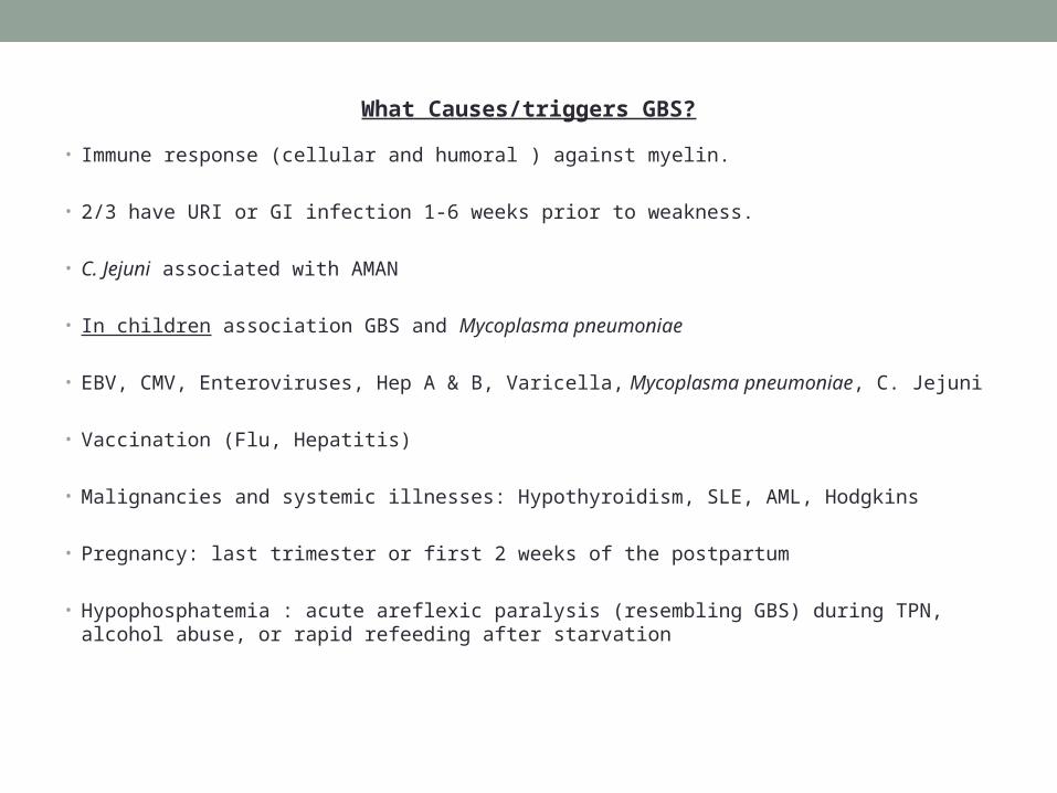
What Causes/triggers GBS?
• Immune response (cellular and humoral ) against myelin.
• 2/3 have URI or GI infection 1-6 weeks prior to weakness.
• C. Jejuni associated with AMAN
• In children association GBS and Mycoplasma pneumoniae
• EBV, CMV, Enteroviruses, Hep A & B, Varicella, Mycoplasma pneumoniae, C. Jejuni
• Vaccination (Flu, Hepatitis)
• Malignancies and systemic illnesses: Hypothyroidism, SLE, AML, Hodgkins
• Pregnancy: last trimester or first 2 weeks of the postpartum
• Hypophosphatemia : acute areflexic paralysis (resembling GBS) during TPN, alcohol abuse, or rapid refeeding after starvation
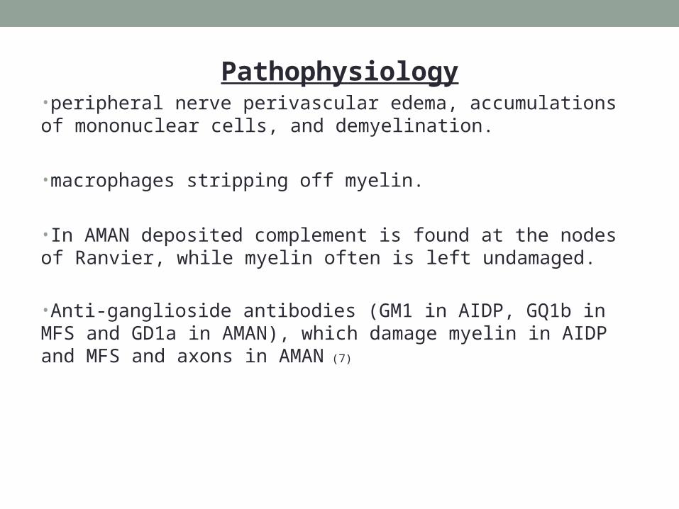
Pathophysiology•peripheral nerve perivascular edema, accumulations of mononuclear cells, and demyelination.
•macrophages stripping off myelin.
•In AMAN deposited complement is found at the nodes of Ranvier, while myelin often is left undamaged.
•Anti-ganglioside antibodies (GM1 in AIDP, GQ1b in MFS and GD1a in AMAN), which damage myelin in AIDP and MFS and axons in AMAN (7)
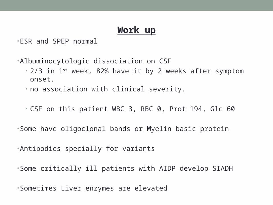
Work up•ESR and SPEP normal
•Albuminocytologic dissociation on CSF• 2/3 in 1st week, 82% have it by 2 weeks after symptom onset. • no association with clinical severity.
• CSF on this patient WBC 3, RBC 0, Prot 194, Glc 60
•Some have oligoclonal bands or Myelin basic protein
•Antibodies specially for variants
•Some critically ill patients with AIDP develop SIADH
•Sometimes Liver enzymes are elevated
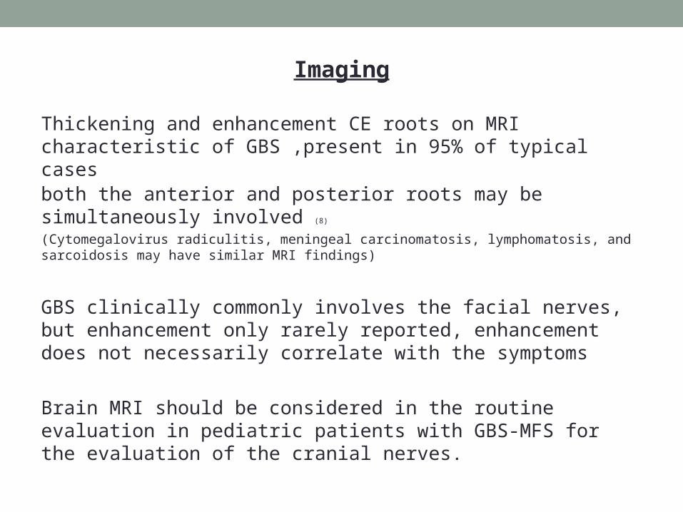
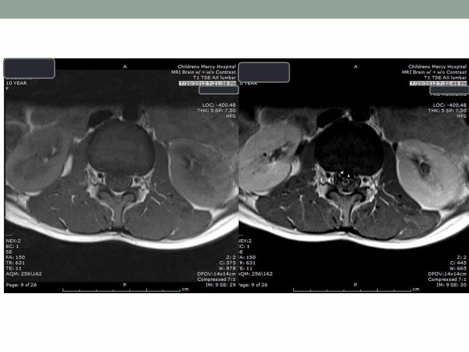
Imaging
Thickening and enhancement CE roots on MRI characteristic of GBS ,present in 95% of typical casesboth the anterior and posterior roots may be simultaneously involved (8)
(Cytomegalovirus radiculitis, meningeal carcinomatosis, lymphomatosis, and sarcoidosis may have similar MRI findings)
GBS clinically commonly involves the facial nerves, but enhancement only rarely reported, enhancement does not necessarily correlate with the symptoms
Brain MRI should be considered in the routine evaluation in pediatric patients with GBS-MFS for the evaluation of the cranial nerves.
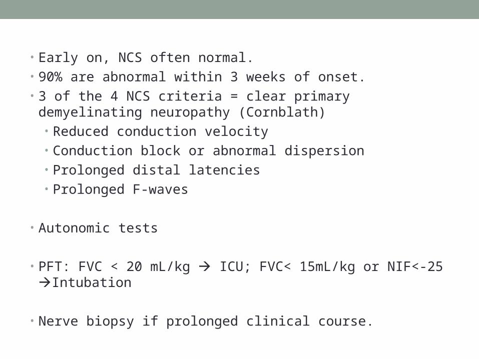
• Early on, NCS often normal. • 90% are abnormal within 3 weeks of onset.• 3 of the 4 NCS criteria = clear primary demyelinating neuropathy
(Cornblath)• Reduced conduction velocity• Conduction block or abnormal dispersion• Prolonged distal latencies• Prolonged F-waves
• Autonomic tests
• PFT: FVC < 20 mL/kg ICU; FVC< 15mL/kg or NIF<-25 Intubation
• Nerve biopsy if prolonged clinical course.
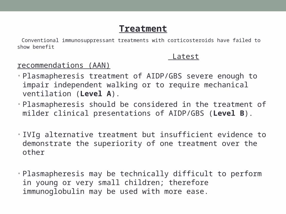
Treatment Conventional immunosuppressant treatments with corticosteroids have failed to show benefit
Latest recommendations (AAN)• Plasmapheresis treatment of AIDP/GBS severe enough to impair
independent walking or to require mechanical ventilation (Level A).• Plasmapheresis should be considered in the treatment of milder
clinical presentations of AIDP/GBS (Level B).
• IVIg alternative treatment but insufficient evidence to demonstrate the superiority of one treatment over the other
• Plasmapheresis may be technically difficult to perform in young or very small children; therefore immunoglobulin may be used with more ease.
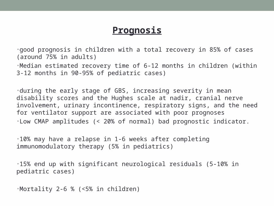
Prognosis
•good prognosis in children with a total recovery in 85% of cases (around 75% in adults)•Median estimated recovery time of 6-12 months in children (within 3-12 months in 90-95% of pediatric cases)
•during the early stage of GBS, increasing severity in mean disability scores and the Hughes scale at nadir, cranial nerve involvement, urinary incontinence, respiratory signs, and the need for ventilator support are associated with poor prognoses•Low CMAP amplitudes (< 20% of normal) bad prognostic indicator.
•10% may have a relapse in 1-6 weeks after completing immunomodulatory therapy (5% in pediatrics)
•15% end up with significant neurological residuals (5-10% in pediatric cases)
•Mortality 2-6 % (<5% in children)
References:• 1) Guillain G, Barré JA, Strohl A. Sur un syndrome de radiculonévrite avec hyperalbuminose du liquide céphalo-rachidien sans réaction
cellulaire: remarques sur les caractères cliniques et graphiques des réflexes tendineux. Bulletins et mémoires de la Société des Médecins des Hôpitaux de Paris 1916;40:1462-70.
• 2) HAYMAKER WE, KERNOHAN JW. The Landry-Guillain-Barré syndrome; a clinicopathologic report of 50 fatal cases and a critique of the literature. Medicine (Baltimore). Feb 1949;28(1):59-141
• 3) Guillain-Barré syndrome in children. Incecik F, Ozlem Hergüner M, Altunbasak S. Neurol Sci. 2011 Jun;32(3):381-5. Epub 2010 Oct 16.• 4) Clinical variants of guillain-barré syndrome in children, Lin JJ, Hsia SH, Wang HS, Lyu RK, Chou ML, Hung PC, Hsieh MY, Lin KL, Pediatr
Neurol. 2012 Aug;47(2):91-6.• 5) Sakakihara Y, Kamoshita S (1991) Age-associated changes in the symptomatology of Guillain–Barré syndrome in children. Dev Med
Child Neurol 33:611–616• 6) Jacobs BC, van Doorn PA, Schmitz PI, Tio-Gillen AP, Herbrink P, Visser LH, et al. Campylobacter jejuni infections and anti-
GM1 antibodies in Guillain-Barré syndrome. Ann Neurol. Aug 1996;40(2):181-7.• 7) Ho TW, Mishu B, Li CY, Gao CY, Cornblath DR, Griffin JW, et al. Guillain-Barré syndrome in northern China. Relationship to
Campylobacter jejuni infection and anti-glycolipid antibodies. Brain. Jun 1995;118 ( Pt 3):597-605• 8) Magnetic resonance imaging of childhood Guillain-Barre syndrome. - Yikilmaz A - Childs Nerv Syst - 01-AUG-2010; 26(8): 1103-8• 9) MANAGEMENT OF CHILDREN WITH GUILLAIN-BARRE´ SYNDROME S Agrawal, D Peake, W P Whitehouse Arch Dis Child Educ Pract
Ed 2007;92:ep161–ep168