Embed Size (px)
Citation preview
Neuromuscular Electrical Stimulation of the Muscles Surrounding the Shoulder
LUCINDA L. BAKER and KAREN PARKER
Neuromuscular electrical stimulation (NMES) can be used to augment range-of-motion, strengthening, and facilitation treatment programs of the muscles surrounding the shoulder. The purposes of this article are 1) to describe the uses of NMES around the shoulder joint as developed through our clinical use and 2) to detail the effects of an NMES program on chronic shoulder subluxation as determined by a clinical study. Because of the complexities of this multiarticular joint, NMES is most useful in the initial phase of the ROM, and stimulated contractions are compromised, relatively, as the humerus moves above the 90-degree horizontal plane. The use of NMES to provide scapular stabilization often entails unwanted alteration of the pressures on the spinal column, occasionally making the treatment program unusable. Electrical stimulation to prevent or correct shoulder subluxation, especially in the neurologically involved patient, provides the therapist with a powerful new treatment technique. In a group of stroke patients, shoulder subluxation was reduced significantly (p < .05) at the completion of a six-week NMES program. Some of the problems, and possible solutions, unique to the development of electrical stimulation programs for the shoulder muscles are discussed. Key Words: Electric stimulation, Physical therapy, Shoulder dislocation, Shoulder
joint.
Neuromuscular electrical stimulation (NMES), in recent years, has been well established as an effective adjunct to range-of-motion, strengthening, facilitation, and spasticity management programs used by the physical therapist.1
The efficacy of NMES in such programs for the shoulder muscles is compromised somewhat, however, because of the large number of muscles surrounding the shoulder joint and the complex synergistic relationships among muscles. The shoulder complex is one of the most intricate joint complexes of the body, creating articular surfaces between three bones and allowing movement in three planes. Although Peat discusses the specific anatomic and ki-nesiologic characteristics of this joint complex (see the article by M. C. Peat in this issue), practical application can be demonstrated in the development of
treatment programs that include NMES for the shoulder. The purposes of this article are 1) to describe several electrical stimulation programs that we have found useful in our clinical practice and 2) to detail the effects of one of these programs as determined through a clinical study.
GENERAL SHOULDER STIMULATION
Guidelines Electrical stimulation can be used to
assist in gaining the initial degrees of motion for patients with frozen shoulder or shoulder-hand syndrome. Because of the limitations in stimulating the muscles around the shoulder and difficulties in restoring the normal synchronization of these muscles, treatment programs including NMES generally are restricted to the initial phase of the ROM. Electrical stimulation programs are less effective than other types of treatment programs when the range increases above the 90-degree horizontal plane. Because of the repetitive nature of cyclical stimulation, however, many patients who have shoulder pain prefer electrically stimulated ROM to other methods of increasing ROM. The repetitive nature
of NMES, in combination with its analgesic effects similar to those of transcutaneous electrical nerve stimulation, provides the therapist with a tool to achieve ROM increases in many patients who are less tolerant of traditional ROM programs. A cyclical stimulation treatment program provides regular and consistent motion in a set range, allowing the patient to relax during the treatment session. The relaxed patient, thus, may achieve increases in ROM by avoiding the "splinting" of muscles that occurs so often during traditional ROM procedures.
In the development of shoulder stimulation programs, electrodes must be cut and shaped individually, depending on the size of the patient. Custom electrode sizes will ensure specific activation of the desired muscles without overlap to other muscle groups. Other factors to consider in developing NMES programs include avoiding stimulation over bony prominences and avoiding placing electrodes too closely together. Both monopolar electrode configurations, with the active negative electrode smaller than the positive electrode, and bipolar electrode configurations, with electrodes of equal size both placed in a single targeted muscle area, are appropriate in
Lucinda Baker is Assistant Professor, Department of Physical Therapy, University of Southern California, 12933 Erickson, Downey, CA 90242 (USA), and Consulting Neurophysiologist for the Rancho Los Amigos Rehabilitation Engineering Center, Downey, CA 90242.
Karen Parker is Physical Therapy Supervisor I, Rancho Los Amigos Medical Center, Downey, CA 90242.
This research was supported in part by National Institute of Handicapped Research Grants G008200045 and G008300077.
1930 PHYSICAL THERAPY
shoulder stimulation programs. In addition, stimulation with symmetrical triphasic waveforms, usually combined with bipolar electrode placements, often is perceived by the patient as being more comfortable2 than stimulation with a compensated monophasic waveform. The increased comfort probably is attributable to the relatively large size of the muscles being activated around the shoulder. A symmetrical biphasic waveform has been shown in other large muscle groups to reduce the amount of amplitude needed to achieve a Fair plus stimulated muscle contraction.2 This reduced current intensity decreases the amount of superficial sensory activation, often making the patient much more comfortable and, thus, more willing to comply with the treatment program.
Shoulder Flexion-Abduction
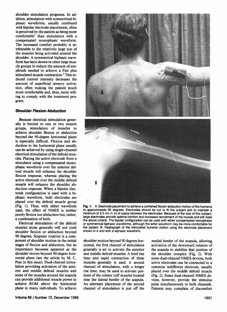
Because electrical stimulation generally is limited to one or two muscle groups, stimulation of muscles to achieve shoulder flexion or abduction beyond the 90-degree horizontal plane is especially difficult. Flexion and abduction to the horizontal plane usually can be achieved by using single-channel electrical stimulation of the deltoid muscles. Placing the active electrode from a stimulator using a compensated mono-phasic waveform over the anterior deltoid muscle will enhance the shoulder flexion response, whereas placing the active electrode over the middle deltoid muscle will enhance the shoulder abduction response. When a bipolar electrode configuration is used with a biphasic waveform, both electrodes are placed over the deltoid muscle group (Fig. 1). Thus, with either waveform used, the effect of NMES is neither purely flexion nor abduction but, rather, a combination of both.
Electrical stimulation of the deltoid muscles alone generally will not yield shoulder flexion or abduction beyond 90 degrees. Scapular rotation is a component of shoulder motion in the initial stages of flexion and abduction, but its importance becomes apparent as the shoulder moves beyond 90-degree horizontal plane (see the article by M. C. Peat in this issue). Dual-channel stimulation providing activation of the anterior and middle deltoid muscles and some of the muscles around the scapula can provide additional muscle power to achieve ROM above the horizontal plane in many individuals. To achieve
Fig. 1. A. Electrode placement to achieve a combined flexion-abduction motion of the humerus to approximately 90 degrees. Electrodes should be cut to fit the subject and to maintain a minimum of 2.5 cm (1 in) of space between the electrodes. Because of the size of this subject, large electrodes provide optimal comfort and increased recruitment of the muscle and still meet the above criteria. The bipolar configuration can be used with either compensated monophasic or symmetrical biphasic waveforms, although the latter waveform may be more comfortable for the patient. B. Radiograph of the stimulated humeral motion using the electrode placement shown in A and with a biphasic waveform.
shoulder motion beyond 90 degrees horizontal, the first channel of stimulation generally is set to activate the anterior and middle deltoid muscles. A brief rise time and rapid contraction of these muscles generally is used. A second channel of stimulation, with a longer rise time, may be used to activate portions of the rotator cuff muscles located near the lateral border of the scapula. An alternate placement of the second channel of stimulation is just off the
medial border of the scapula, allowing activation of the downward rotators of the scapula to stabilize that portion of the shoulder complex (Fig. 2). With some dual-channel NMES devices, both active electrodes can be connected to a common indifferent electrode, usually placed over the middle deltoid muscle (Fig. 2). Some dual-channel NMES devices, however, provide the stimulus pulse simultaneously to both channels. Patients may complain of discomfort
Volume 66 / Number 12, December 1986 1931

Fig. 2. A. Dual-channel, three-electrode placement to achieve a combined flexion-abduction motion of the humerus above the 90-degree horizontal plane. The active electrodes are placed over the subject's anterior deltoid muscle and just off the medial border of the scapula to provide some scapular stabilization. Both active electrodes are connected to the common indifferent electrode positioned over the middle deltoid muscle. For this dual-channel electrode placement, the compensated monophasic waveform is preferred most often. B. Radiograph of the stimulated humeral motion using the electrode placement shown in A.
under the common indifferent electrode because the current of both channels is reaching that electrode at the same time, effectively doubling the current density. The three-electrode, dual-channel configuration sometimes is preferred because of the small surface area available for the placement of electrodes.
The dual-channel stimulated contraction rarely establishes normal synchronization between the scapula and the glenohumeral joint, but it does provide a means of achieving ROM above the horizontal plane. Because the stimu
lated motion does not approximate the normal functioning of the complex glenohumeral joint, stimulation within this range should be used with caution. Pathological conditions of the glenohumeral joint must be considered, because the potential for impingement syndromes exists. Because of the abnormal relationship produced between the stimulated scapular and humeral movements, dual-channel NMES probably would not be the treatment of choice for a muscle facilitation or reeducation program. When motion to the terminal
range is the goal, however, two-channel stimulation may assist in achieving that goal.
Isolated Flexion or Abduction
Electrical stimulation can be used to activate and isolate flexion or abduction of the glenohumeral joint by using monopolar electrode placements and a compensated monophasic waveform. The monopolar electrode configuration includes an active electrode over the anterior deltoid muscle for isolated shoulder flexion or over the middle deltoid muscle for isolated shoulder abduction. The active electrode then is connected to a larger indifferent electrode, usually placed just medial to the border of the scapula. Because of the difference in the size of the electrodes and the use of a compensated monophasic stimulation waveform, the majority of the neuromuscular activation occurs under the negative (ie, active) electrode. Shoulder flexion or abduction to 90 degrees horizontal can be achieved with these monopolar electrode configurations. Isolated flexion or abduction beyond 90 degrees horizontal is more difficult to achieve, even if a second channel of stimulation is instituted
Stimulation of pure flexion or pure abduction may be desirable for the patient who is recovering from traumatic subluxation of the humerus or from a rotator cuff tear.3,4 In our experience, most patients with other types of shoulder involvement benefit from the combined flexion-abduction program and do not require the isolated flexion or isolated abduction treatment program. Because of the combined monophasic waveform and monopolar electrode configuration, isolated flexion or abduction stimulation generally requires higher current levels to achieve the desired quality of muscle contraction. This increased current demand may cause the patient secondary sensory intolerance to the stimulation. A careful training period of three to five days is desirable for all shoulder stimulation programs, but it may be critical to the success of NMES programs isolating specific muscle around the shoulder.
Shoulder Extension
Electrical stimulation of the posterior deltoid muscle generally is restricted to orthopedic patients requiring discrete posterior deltoid muscle strengthening4,5
or to specific applications in the patient with spinal cord injury. Surgical transfer
1932 PHYSICAL THERAPY
of the posterior deltoid muscle to the triceps tendon is a procedure that often is performed to provide the patient with C6-level quadriplegia with active elbow extension.6 After the patient has received the physician's approval to perform active exercises, electrical stimulation is used to facilitate voluntary control of the posterior deltoid muscle with subsequent elbow extension. This facilitation program for elbow extension is brief and often superseded by a voluntary exercise program using electromyographic feedback. When the surgical transfer is determined to be well established, usually after one to three months of healing time, electrical stimulation to increase the strength and power output of the posterior deltoid muscle can be used to augment traditional muscle strengthening programs.
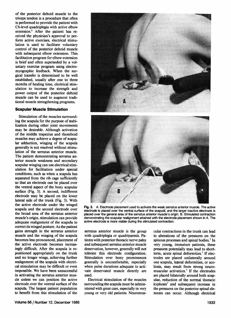
Scapular Muscle Stimulation Stimulation of the muscles surround
ing the scapula for the purpose of stabilization during other joint movements may be desirable. Although activation of the middle trapezius and rhomboid muscles may achieve a degree of scapular adduction, winging of the scapula generally is not resolved without stimulation of the serratus anterior muscle. The patient demonstrating serratus anterior muscle weakness and secondary scapular winging can use electrical stimulation for facilitation under special conditions, such as when a scapula has separated from the rib cage sufficiently so that an electrode can be placed over the ventral aspect of the bony scapular surface (Fig. 3). A second, indifferent electrode may be placed on the lower lateral side of the trunk (Fig. 3). With the active electrode under the winged scapula and the second electrode over the broad area of the serratus anterior muscle's origin, stimulation can provide adequate realignment of the scapula to correct its winged posture. As the patient gains strength in the serratus anterior muscle and the winging of the scapula becomes less pronounced, placement of the active electrode becomes increasingly difficult. After the scapula is repositioned appropriately on the trunk and no longer wings, achieving further realignment of the scapula with electrical stimulation may be difficult or even impossible. We have been unsuccessful in activating the serratus anterior muscle unless we can position the active electrode over the ventral surface of the scapula. The largest patient population to benefit from this stimulation of the
Fig. 3. A. Electrode placement used to activate the weak serratus anterior muscle. The active electrode is placed over the ventral surface of the scapula, and the larger inactive electrode is placed over the general area of the serratus anterior muscle's origin. B. Stimulated contraction demonstrating the scapular realignment attained with the electrode placement shown in A. The active electrode is more visible during the stimulated contraction.
serratus anterior muscle is the group with quadriplegia or quadriparesis. Patients with posterior thoracic nerve palsy and subsequent serratus anterior muscle denervation, however, generally will not tolerate this electrode configuration. Stimulation over bony prominences generally is uncomfortable, especially when pulse durations adequate to activate denervated muscle directly are used.
Electrical stimulation of the muscles surrounding the scapula must be administered with great care, especially in very young or very old patients. Neuromus
cular contractions in the trunk can lead to alterations of the pressures on the spinous processes and spinal bodies.7 In very young, immature patients, these pressures potentially may lead to short-term, acute spinal deformities.7 If electrodes are placed unilaterally around one scapula, lateral deformities, or scoliosis, may result from strong neuromuscular activation.7 If the electrodes are placed bilaterally around both scapulae, reduction of the normal thoracic kyphosis7 and subsequent increase in the pressures on the posterior spinal elements can occur. Although electrical
Volume 66 / Number 12, December 1986 1933
stimulation can be used to manage certain types of idiopathic spinal deformities,8,9 under chronic stimulation conditions NMES potentially may contribute to spinal deformities if used inappropriately.
In the adult, these same types of pressures occur on bony and soft tissues. The spinal column in an adult largely is immobile, however, and pressures on the spinal column may result in pain and discomfort associated with soft tissue distraction, potential pinching of nerve roots, and possibly even the presence of stress fractures of the spinous processes or bodies.7 Special care must be taken with older adults, especially postmenopausal women, who frequently demonstrate a high degree of osteopetrosis, often in the spinal processes and bodies.10-12 Although these complications occur less commonly in the young adult, the use of Fair plus stimulated muscle contractions around the spinal column and the potential complications of such a treatment program should be considered during the development of NMES-assisted scapular facilitation programs.
Shoulder Subluxation Inferior subluxation of the glenohu-
meral joint is a common complication of both neurologic13-18 and musculoskeletal injuries.3-5 Although subluxation may occur after acute trauma, the incidence of chronic subluxation is low in the orthopedic patient population.3-5
In the patient with neurologic involvement, especially the patient with stroke or head injury, chronic subluxation of the shoulder is a frequent problem. Lack of normal muscle activity after an acute insult to the central nervous system is a major contributing factor to the incidence of subluxation. Reduced muscle activity can lead to stretching of the shoulder capsule, which is critical to the maintenance of glenohumeral joint stability.19,20 If the shoulder capsule has been stretched, subluxation often will persist, regardless of whether the patient develops voluntary control or even spasticity in the muscles around the shoulder. Subluxation in patients with stroke has been associated with increased pain, with increased incidence of inappropriate autonomic responses in the upper extremity,21 and occasionally with traction damage to the brachial plexus and other peripheral nerves.22 In the patient with neurologic impairment, therefore, joint-capsule stretching should be avoided. Although the early use of sling
support of the glenohumeral joint has been thought to reduce this problem, the persistently high incidence of chronic subluxation18 indicates that the use of slings may be inadequate in controlling the effects of gravity on the shoulder joint. When subluxation has begun already, use of the sling may retard further joint separation, but generally it is incapable of reducing the existing subluxation. Electrical stimulation for the reduction of shoulder subluxation not only may prevent further joint separation, but also can provide normal glenohumeral alignment for the patient when assuming the upright position. Thus, maintaining normal joint alignment and counteracting the stretching of the shoulder capsule that is caused by gravity are advantages of such stimulation. To assess the effectiveness of an electrical stimulation program in the management of shoulder subluxation, we conducted a study in which we compared the effectiveness of conventional slings to that of NMES.
Study Method Sixty-three patients admitted to the
Stroke Service at Rancho Los Amigos Medical Center were evaluated prospectively for this study. To qualify for the study, all subjects were required to demonstrate a minimum of 5 mm of shoulder subluxation in their involved upper extremity, as compared with radiographs of their uninvolved extremity. This bilateral comparison was made to eliminate the variability of joint laxity from one individual to the next. All radiographs were taken with both upper extremities dependent at the patient's side23,24 and depicted both of the patient's shoulders.
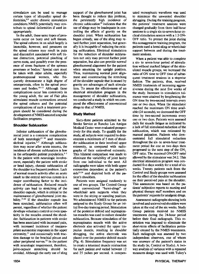
Patients were assigned randomly to one of two groups. The Control Group used conventional "hemi-slings" or wheelchair arm supports when they were in the sitting or standing position. We administered NMES to the patients assigned to the Study Group for an initial five-day training period. Stimulation of the posterior deltoid and supraspina-tus muscles was used to reduce shoulder subluxation. Because stimulation of the supraspinatus muscle with the active electrode also activated the upper trapezius muscle, resulting in shoulder shrugging, the active electrode was placed over the posterior deltoid muscle (Fig. 4). Stimulation frequency was set to create a tetanized muscle contraction for each patient and varied between 12 and 25 pulses per second. A compen
sated monophasic waveform was used to minimize the unwanted shoulder shrugging. During the training program, the patients' treatment sessions were changed gradually from three half-hour sessions to a single six-to-seven-hour cyclical stimulation session with a 1:3 ON-OFF ratio. To protect the joint during this nonaggressive training program, the patients used a hemi-sling or wheelchair support between and during the treatment sessions.
When a patient was able to complete a six- to seven-hour period of stimulation without marked fatigue of the stimulated muscle groups, we increased the ratio of ON time to OFF time of subsequent treatment sessions in a stepwise fashion. The patients, thus, received NMES treatments of increasing aggressiveness during the next five weeks of the study. Increases in stimulation tolerance were attained by lengthening the ON time by two-second intervals every one or two days. When the stimulator reached the maximum ON time available (24 seconds), we decreased the OFF time by two-second increments every one or two days. Patients were assessed daily for muscle fatigue as indicated by a lack of full stimulated reduction of the subluxation, which was estimated by manual palpation. Patients who demonstrated full stimulated reduction throughout the six- to seven-hour treatment period for one or two days then progressed to the next step of the ON; OFF ratio. The maximal ON-OFF ratió allowed by the stimulator was 24:2. This electrical stimulation program was conducted five days weekly for a total of six weeks. Twenty patients each from the Control and Study groups were assessed for the effect of the shoulder subluxation on their perceived pain at the shoulder. This assessment was based on the patients' subjective reports to nursing and physical therapy staff members and on the patients' requests for analgesic drugs.
Assessment radiographs showing both involved and uninvolved shoulders were taken at the end of the six weeks. Study Group patients received no NMES treatments during the 24-hour period before their final radiograph. This restriction was imposed to eliminate the short-term effects of facilitation potentially created by the NMES treatments. Each radiograph was assessed by two independent observers, one of whom was unaware of the patient's status in the study (ie, Control or Study). A two-way analysis of variance with a repeated measures design was used with Tukey's
1934 PHYSICAL THERAPY
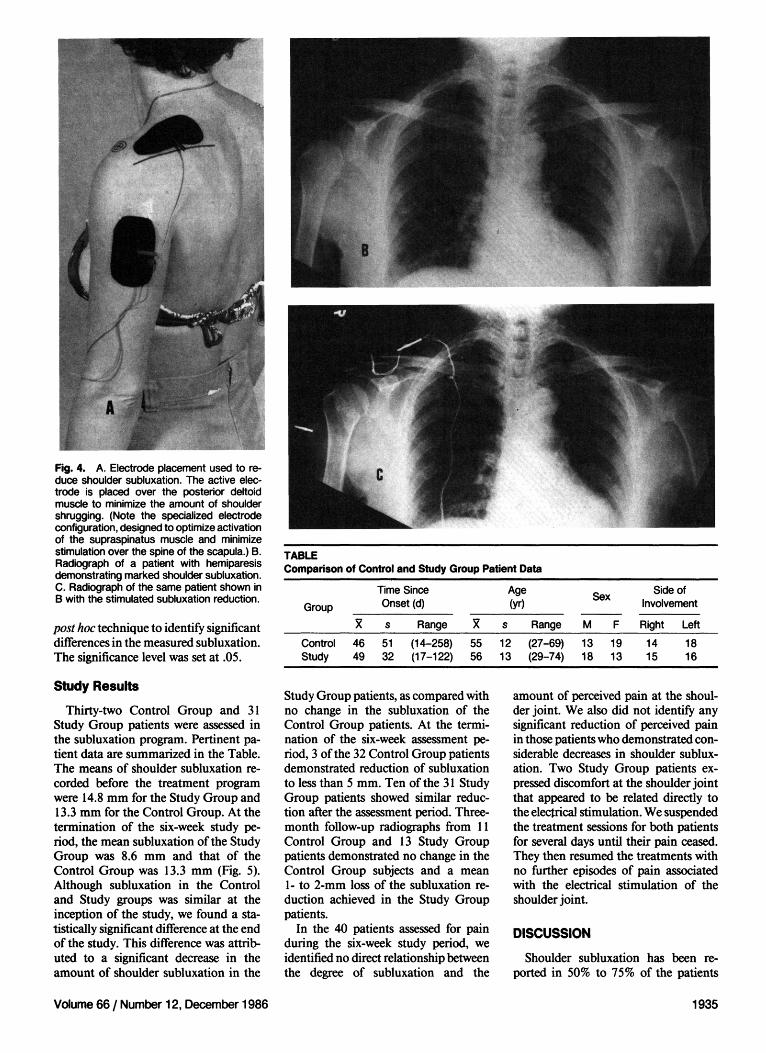
Fig. 4. A. Electrode placement used to reduce shoulder subluxation. The active electrode is placed over the posterior deltoid muscle to minimize the amount of shoulder shrugging. (Note the specialized electrode configuration, designed to optimize activation of the supraspinatus muscle and minimize stimulation over the spine of the scapula.) B. Radiograph of a patient with hemiparesis demonstrating marked shoulder subluxation. C. Radiograph of the same patient shown in B with the stimulated subluxation reduction.
post hoc technique to identify significant differences in the measured subluxation. The significance level was set at .05.
Study Results Thirty-two Control Group and 31
Study Group patients were assessed in the subluxation program. Pertinent patient data are summarized in the Table. The means of shoulder subluxation recorded before the treatment program were 14.8 mm for the Study Group and 13.3 mm for the Control Group. At the termination of the six-week study period, the mean subluxation of the Study Group was 8.6 mm and that of the Control Group was 13.3 mm (Fig. 5). Although subluxation in the Control and Study groups was similar at the inception of the study, we found a statistically significant difference at the end of the study. This difference was attributed to a significant decrease in the amount of shoulder subluxation in the
TABLE Comparison of Control and Study Group Patient Data
Group
Control Study
46 49
Time Since Onset (d)
s 51 32
Range (14-258) (17-122)
55 56
Age (yr)
s 12 13
Range (27-69) (29-74)
Sex
M 13 18
F 19 13
Side of Involvement
Right 14 15
Left 18 16
Study Group patients, as compared with no change in the subluxation of the Control Group patients. At the termination of the six-week assessment period, 3 of the 32 Control Group patients demonstrated reduction of subluxation to less than 5 mm. Ten of the 31 Study Group patients showed similar reduction after the assessment period. Three-month follow-up radiographs from 11 Control Group and 13 Study Group patients demonstrated no change in the Control Group subjects and a mean 1- to 2-mm loss of the subluxation reduction achieved in the Study Group patients.
In the 40 patients assessed for pain during the six-week study period, we identified no direct relationship between the degree of subluxation and the
amount of perceived pain at the shoulder joint. We also did not identify any significant reduction of perceived pain in those patients who demonstrated considerable decreases in shoulder subluxation. Two Study Group patients expressed discomfort at the shoulder joint that appeared to be related directly to the electrical stimulation. We suspended the treatment sessions for both patients for several days until their pain ceased. They then resumed the treatments with no further episodes of pain associated with the electrical stimulation of the shoulder joint.
DISCUSSION Shoulder subluxation has been re
ported in 50% to 75% of the patients
Volume 66 / Number 12, December 1986 1935
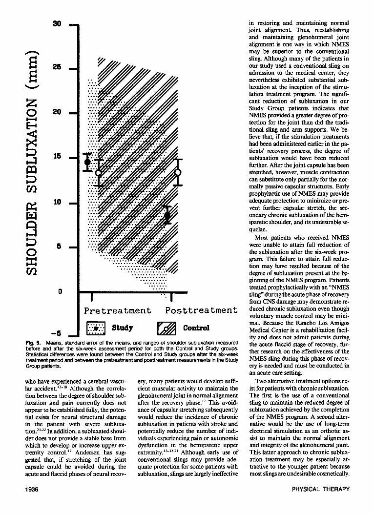
Fig. 5. Means, standard error of the means, and ranges of shoulder subluxation measured before and after the six-week assessment period for both the Control and Study groups. Statistical differences were found between the Control and Study groups after the six-week treatment period and between the pretreatment and posttreatment measurements in the Study Group patients.
who have experienced a cerebral vascular accident.13-18 Although the correlation between the degree of shoulder subluxation and pain currently does not appear to be established fully, the potential exists for neural structural damage in the patient with severe subluxation.21,22 In addition, a subluxated shoulder does not provide a stable base from which to develop or increase upper extremity control.17 Andersen has suggested that, if stretching of the joint capsule could be avoided during the acute and flaccid phases of neural recov
ery, many patients would develop sufficient muscular activity to maintain the glenohumeral joint in normal alignment after the recovery phase.17 This avoidance of capsular stretching subsequently would reduce the incidence of chronic subluxation in patients with stroke and potentially reduce the number of individuals experiencing pain or autonomic dysfunction in the hemiparetic upper extremity.13-18,21 Although early use of conventional slings may provide adequate protection for some patients with subluxation, slings are largely ineffective
in restoring and maintaining normal joint alignment. Thus, reestablishing and maintaining glenohumeral joint alignment is one way in which NMES may be superior to the conventional sling. Although many of the patients in our study used a conventional sling on admission to the medical center, they nevertheless exhibited substantial subluxation at the inception of the stimulation treatment program. The significant reduction of subluxation in our Study Group patients indicates that NMES provided a greater degree of protection for the joint than did the traditional sling and arm supports. We believe that, if the stimulation treatments had been administered earlier in the patients' recovery process, the degree of subluxation would have been reduced further. After the joint capsule has been stretched, however, muscle contraction can substitute only partially for the normally passive capsular structures. Early prophylactic use of NMES may provide adequate protection to minimize or prevent further capsular stretch, the secondary chronic subluxation of the hemiparetic shoulder, and its undesirable sequelae.
Most patients who received NMES were unable to attain full reduction of the subluxation after the six-week program. This failure to attain full reduction may have resulted because of the degree of subluxation present at the beginning of the NMES program. Patients treated prophylactically with an "NMES sling" during the acute phase of recovery from CNS damage may demonstrate reduced chronic subluxation even though voluntary muscle control may be minimal. Because the Rancho Los Amigos Medical Center is a rehabilitation facility and does not admit patients during the acute flaccid stage of recovery, further research on the effectiveness of the NMES sling during this phase of recovery is needed and must be conducted in an acute care setting.
Two alternative treatment options exist for patients with chronic subluxation. The first is the use of a conventional sling to maintain the reduced degree of subluxation achieved by the completion of the NMES program. A second alternative would be the use of long-term electrical stimulation as an orthotic assist to maintain the normal alignment and integrity of the glenohumeral joint. This latter approach to chronic subluxation treatment may be especially attractive to the younger patient because most slings are undesirable cosmetically.
1936 PHYSICAL THERAPY
An additional advantage of a long-term NMES program is the relatively normal alignment of the shoulder joint that is maintained. This normal alignment of the shoulder complex creates a stable platform from which the patient may be able to develop more usable distal motor control. The disadvantages of the chronic use of NMES include the problems associated with long-term use of surface electrical stimulation. Proper electrode placement is difficult for most patients with hemiparesis to achieve and maintain by themselves. In addition, skin irritation from electrodes, gels, and tape frequently occurs with the long-term use of surface electrical stimulation. Although a variety of electrodes are available, many patients with hemiparesis find long-term care of the skin over the supraspinatus and posterior deltoid muscles difficult to maintain. We have found that chronic use of NMES for shoulder subluxation management has been most successful for patients in supervised care settings where supportive individuals can assist the patient in maintaining appropriate electrode placement and skin care. We do not use long-term NMES programs for subluxation management routinely, largely because of these technical difficulties.
The chronic use of NMES for the reduction of shoulder subluxation may become more feasible when implanted or semi-implanted electrode systems become available. Epimysial electrodes may be inserted with minor local surgical procedures and, when connected to a stimulation system, may provide reliable, reproducible electrical stimulation results. With the development of fully implanted electrode systems, stimulation may become a preferred treatment for the patient demonstrating chronic shoulder subluxation because of CNS dysfunction. The stimulation treatment program is not so much an alternative to the traditional sling but a treatment technique that can provide the patient with more normal musculoskeletal alignment that can result in improved function.
CONCLUSION
Neuromuscular electrical stimulation of the muscles surrounding the shoulder joint is an effective method of early mobilization, especially for the patient experiencing shoulder pain. Because of the complexities of the shoulder joint articulations, however, NMES is not recommended for gaining terminal degrees of the ROM. The use of NMES for the reduction of shoulder subluxation, especially in the patient with neurologic involvement, can provide not only an alternative to cumbersome sling-type assistive devices but also a normal musculoskeletal base from which the patient can use both proximal and distal control. The primary advantage of NMES may be its prophylactic use, before stretching the shoulder capsule, to maintain joint integrity throughout the flaccid period of neurologic recovery. For the patient whose chronic subluxation is not reduced fully during a six- to eight-week therapeutic stimulation program, long-term application of NMES may be an effective alternative to the use of the traditional sling support. Until implanted electrode systems become available, however, long-term use of surface electrical stimulation can be managed by only a few patients with hemiparesis and their families.
Acknowledgments. We thank C. Criswell, J. Weiss, D. Menenger, J. Jackson, and C. Zablotny for their assistance in the collection of the shoulder subluxation data. We also thank E. R. Gardner for her assistance in content editing and J. Scofield and P. Robb for their assistance in manuscript preparation.
REFERENCES
1. Benton LA, Baker LL, Bowman BR, et al: Functional Electrical Stimulation: A Practical Clinical Guide, ed 2. Downey, CA, Professional Staff Association of Rancho Los Amigos Medical Center, 1981
2. Bowman BR, Baker LL: Effects of waveform parameters on comfort during transcutaneous neuromuscular electrical stimulation. Ann Biomed Eng 13:59-74,1985
3. Simonet WT, Cofield RH: Prognosis in anterior shoulder dislocation. Am J Sports Med 12:19-24,1984
4. Zarins B, Rowe CR: Current concepts in the diagnosis and treatment of shoulder instability in athletes. Med Sci Sports Exerc 16:444-448, 1984
5. Hawkins RJ, Koppert G, Johnston G: Recurrent posterior instability (subluxation) of the shoulder. J Bone Joint Surg [Am] 66:169-174, 1984
6. Raczka R, Braun R, Waters RL: Posterior deltoid-to-triceps transfer in quadriplegia. Clin Or-thop 187:163-167,1984
7. Axelgaard J, Nordwall A, Brown JC: Correction of spinal curvatures by transcutaneous electrical muscle stimulation. Spine 8:463-481,1983
8. Axelgaard J, Brown JC: Lateral electrical surface stimulation for the treatment of progressive idiopathic scoliosis. Spine 8:242-260, 1983
9. Eckerson LF, Axelgaard J: Lateral electrical surface stimulation as an alternative to bracing in the treatment of idiopathic scoliosis: Treatment protocol and patient acceptance. Phys Ther 64:483-490,1984
10. Conn SH, Vaswani S, Zanzi I, et al: Effect of aging on bone mass in adult women. Am J Physiol 230:143-148,1976
11. Kaplan FS: Osteoporosis. Clin Symp 35:1-32, 1983
12. Ruegsegger P, Dambacher MA, Reugsegger E, et al: Bone loss in premenopausal and postmenopausal women. J Bone Joint Surg [Am] 66:1015-1023, 1984
13. Najenson T, Pikielny SS: Malalignment of the glenohumeral joint following hemiplegia: A review of 500 cases. Annals of Physical Medicine 8:96-99, 1965
14. Taketomi Y: Observations on subluxation of the shoulder joint in hemiplegia. Phys Ther 55:39-40, 1975
15. Jensen EM: Hemiplegic shoulder. Scand J Re-habil Med [Suppl] 7:113-119, 1980
16. Griffin J, Reddin G: Shoulder pain in patients with hemiplegia: A literature review. Phys Ther 61:1041-1045,1981
17. Andersen LT: Shoulder pain in hemiplegia. Am J Occup Ther 39:11-19, 1985
18. Van Ouwenalter C, Laplace PM, Chantraine A: Painful shoulder in hemiplegia. Arch Phys Med Rehabil 67:23-26, 1986
19. Basmajian JV, Bazant FJ: Factors preventing downward dislocation of the adducted shoulder joint: An electromyographic and morphological study. J Bone Joint Surg [Am] 41:1182-1186,1959
20. Basmajian JV, Deluca CJ: Muscles Alive: Their Functions Revealed by Electromyography, ed 5. Baltimore, MD, Williams & Wilkins, 1985, pp 239-276
21. Teppermann PS, Greyson ND, Hilbert L, et al: Reflex sympathetic dystrophy in hemiplegia. Arch Phys Med Rehabil 65:442-447, 1984
22. Chino N: Electrophysiological investigation on shoulder subluxation in hemiplegics. Scand J Rehabil Med 13:17-21, 1981
23. Shai G, Ring H, Costeff H, et al: Glenohumeral malalignment in the hemiplegic shoulder: An early radiologic sign. Scand J Rehabil Med 16:133-136, 1984
24. Lev-Toaff AS, Karasick D, Rao VM: Drooping shoulder: Nontraumatic causes of glenohumeral subluxation. Skeletal Radiol 12:34-36, 1984
Volume 66 / Number 12, December 1986 1937