Embed Size (px)
Citation preview
1
British Journal of Educational Psychology (2004), 74, 1–14
2004 The British Psychological Society
Annual review
Neuroscience and education
Usha Goswami*Faculty of Education, University of Cambridge, UK
Neuroscience is a relatively new discipline encompassing neurology, psychology andbiology. It has made great strides in the last 100 years, during which many aspects ofthe physiology, biochemistry, pharmacology and structure of the vertebrate brain havebeen understood. Understanding of some of the basic perceptual, cognitive,attentional, emotional and mnemonic functions is also making progress, particularlysince the advent of the cognitive neurosciences, which focus specifically onunderstanding higher level processes of cognition via imaging technology. Neuroima-ging has enabled scientists to study the human brain at work in vivo, deepening ourunderstanding of the very complex processes underpinning speech and language,thinking and reasoning, reading and mathematics. It seems timely, therefore, toconsider how we might implement our increased understanding of brain developmentand brain function to explore educational questions.
The study of learning unites education and neuroscience. Neuroscience as broadly
defined investigates the processes by which the brain learns and remembers, from the
molecular and cellular levels right through to brain systems (e.g., the system of neural
areas and pathways underpinning our ability to speak and comprehend language). This
focus on learning and memory can be at a variety of levels. Understanding cell signalling
and synaptic mechanisms (one brain cell connects to another via a synapse) isimportant for understanding learning, but so is examination of the functions of specific
brain structures such as the hippocampus by natural lesion studies or by invasive
methods. Brain cells (or neurons) transmit information via electrical signals, which pass
from cell to cell via the synapses, triggering the release of neurotransmitters (chemical
messengers). There are around 100 billion neurons in the brain, each with massive
connections to other neurons. Understanding the ways in which neurotransmitters
work is a major goal of neuroscience. Patterns of neural activity are thought to
correspond to particular mental states or mental representations. Learning broadlycomprises changes in connectivity, either via changes in potentiation at the synapse or
www.bps.org.uk
* Correspondence should be addressed to Usha Goswami, Faculty of Education, University of Cambridge, Shaftesbury Road,Cambridge CB2 2BX, UK (e-mail: [email protected]).
via the strengthening or pruning of connections. Successful teaching thus directly
affects brain function, by changing connectivity.
Clearly, educators do not study learning at the level of the cell. Successful learning isalso dependent on the curriculum and the teacher, the context provided by the
classroom and the family, and the context of the school and the wider community. All
of these factors of course interact with the characteristics of individual brains. For
example, children with high levels of the MAOA gene (monoamine oxidise A) who
experience maltreatment and adverse family environments seem to be protected from
developing antisocial behaviours (Caspi et al., 2002), possibly via moderating effects on
their neural response to stress. Diet also affects the brain. A child whose diet is poor will
not be able to respond to excellent teaching in the same way as a child whose brain iswell-nourished. It is already possible to study the effects of various medications on
cognitive function. Methylphenidate (Ritalin), a medication frequently prescribed for
children with ADHD (Attention Deficit Hyperactivity Disorder), has been shown to
improve stimulus recognition in medicated children (in terms of attention to auditory
and visual stimuli as revealed by neuroimaging; see Seifert et al., 2003). Neuroimaging
techniques also offer the potential to study the effects of different diets, food additives
and potential toxins on educational performance.
Teaching
It is notable, however, that neuroscience does not as yet study teaching. Successful
teaching is the natural counterpart of successful learning, and is described as a ‘natural
cognition’ by Strauss (2003). Forms of teaching are found throughout the animal
kingdom, usually related to ways of getting food. However, the performance of
intentional acts to increase the knowledge of others (teaching with a ‘theory of mind’)
does seem to be unique to humans, and is perhaps essential to what it means to be a
human being (Strauss, Ziv, & Stein, 2002). The identification and analysis of successfulpedagogy is central to research in education, but is currently a foreign field to cognitive
neuroscience. There are occasional studies of the neural changes accompanying certain
types of highly focused educational programmes (such as remedial programmes for
teaching literacy to dyslexic children, see below), but wider questions involving the
invisible mental processes and inferences made by successful teachers have not begun
to be asked. Strauss suggests that questions such as whether there are specialized neural
circuits for different aspects of teaching may soon be tractable to neuroimaging
methods, and this is a thought-provoking idea. Teaching is a very specialized kind ofsocial interaction, and some of its aspects (reading the minds of others, inferring their
motivational and emotional states) are after all already investigated in cognitive
neuroscience.
Used creatively, therefore, cognitive neuroscience methods have the potential to
deliver important information relevant to the design and delivery of educational
curricula as well as the quality of teaching itself. Cognitive neuroscience may also offer
methods for the early identification of special needs, and enable assessment of the
delivery of education for special needs. At the same time, however, it is worth notingthat ‘neuromyths’ abound. Some popular beliefs about what brain science can actually
deliver to education are quite unrealistic. Although current brain science technologies
offer exciting opportunities to educationists, they complement rather than replace
traditional methods of educational enquiry.
2 Usha Goswami
A quick primer on brain development
Many critical aspects of brain development are complete prior to birth (see Johnson,
1997, for an overview). The development of the neural tube begins during the first
weeks of gestation, and ‘proliferative zones’ within the tube give birth to the cells that
compose the brain. These cells migrate to the different regions where they will be
employed in the mature brain prior to birth. By 7 months gestation almost all of the
neurons that will comprise the mature brain have been formed. Brain development
following birth consists almost exclusively of the growth of axons, synapses and
dendrites (fibre connections): this process is called synaptogenesis. For visual andauditory cortex, there is dramatic early synaptogenesis, with maximum density of
around 150% of adult levels between 4 and 12 months followed by pruning. Synaptic
density in the visual cortex returns to adult levels between 2 and 4 years. For other
areas such as prefrontal cortex (thought to underpin planning and reasoning), density
increases more slowly and peaks after the first year. Reduction to adult levels of density
is not seen until some time between 10 and 20 years. Brain metabolism (glucose uptake,
an approximate index of synaptic functioning) is also above adult levels in the early
years, with a peak of about 150% somewhere around 4–5 years.By the age of around 10 years, brain metabolism reduces to adult levels for most
cortical regions. The general pattern of brain development is clear. There are bursts of
synaptogenesis, peaks of density, and then synapse rearrangement and stabilisation
with myelinisation, occurring at different times and rates for different brain regions (i.e.,
different sensitive periods for the development of different types of knowledge). Brain
volume quadruples between birth and adulthood, because of the proliferation of
connections, not because of the production of new neurons. Nevertheless, the brain is
highly plastic, and significant new connections frequently form in adulthood inresponse to new learning or to environmental insults (such as a stroke). Similarly,
sensitive periods are not all-or-none. If visual input is lacking during early development,
for example, the critical period is extended (Fagiolini & Hensch, 2000). Nevertheless,
visual functions that develop late (e.g., depth perception) suffer more from early
deprivation than functions that are relatively mature at birth (such as colour perception,
Maurer, Lewis, & Brent 1989). Thus more complex abilities may have a lower likelihood
of recovery than elementary skills. One reason may be that axons have already stabilised
on target cells for which they are not normally able to compete, thereby causingirreversible reorganisation.
It is important to realise that there are large individual differences between brains.
Even in genetically identical twins, there is striking variation in the size of different
brain structures, and in the number of neurons that different brains use to carry out
identical functions. This individual variation is coupled with significant localisation of
function. A basic map of major brain subdivisions is shown in Figure 1. Although adult
brains all show this basic structure, it is thought that early in development a number of
possible developmental paths and end states are possible. The fact that developmentconverges on the same basic brain structure across cultures and gene pools is probably
to do with the constraints on development present in the environment. Most children
are exposed to very similar constraints despite slightly different rearing environments.
Large differences in environment, such as being reared in darkness or without contact
with other humans, are thankfully absent or rare. When large environmental differences
occur, they have notable effects on cognitive function. For example, neuroimaging
studies show that blind adults are faster at processing auditory information than sighted
3Neuroscience and education
controls, and that congenitally deaf adults are faster at processing visual information in
the peripheral field than hearing controls (e.g., Neville & Bavelier, 2000; Neville,
Schmidt, & Kutas, 1983; Roder, Rosler, & Neville, 1999).
Nevertheless, neurons themselves are interchangeable in the immature system, and
so dramatic differences in environment can lead to different developmental outcomes.
For example, the area underpinning spoken language in hearing people (used for
auditory analysis) is recruited for sign language in deaf people (visual/spatial analysis)(Neville et al., 1998). Visual brain areas are recruited for Braille reading (tactile analysis)
in blind people (see Roder & Neville, 2003). It has even been reported that a blind adult
who suffered a stroke specific to the visual areas of her brain consequently lost her
proficient Braille reading ability, despite the fact that her somatosensory perception
abilities were unaffected (Jackson, 2000). It has also been suggested that all modalities
are initially mutually linked, as during early infancy auditory stimulation also evokes
large responses in visual areas of the brain, and somatosensory responses are enhanced
by white noise (e.g., Neville, 1995). If this is the case, a kind of ‘synaesthesia’ couldenable infants to extract schemas that are independent of particular modalities,
schemas such as number, intensity and time (see Roder & Neville, 2003). If this mutual
linkage extends into early childhood, it may explain why younger children respond so
well to teaching via multi-sensory methods.
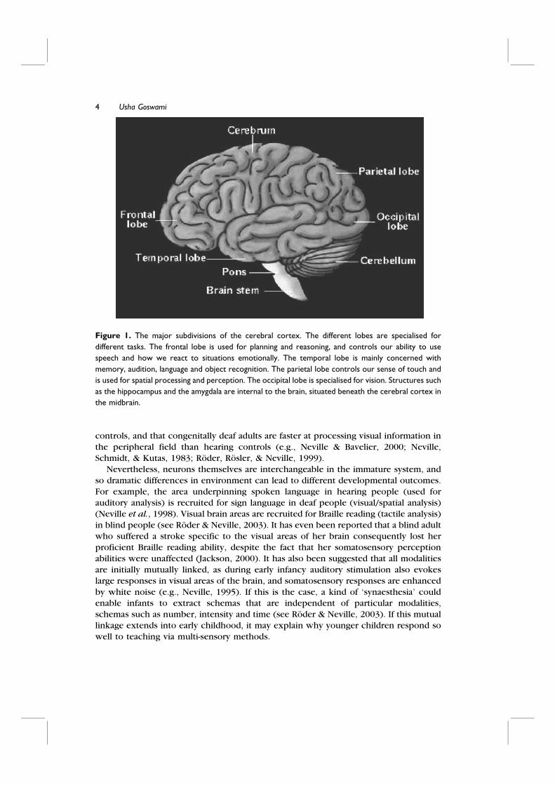
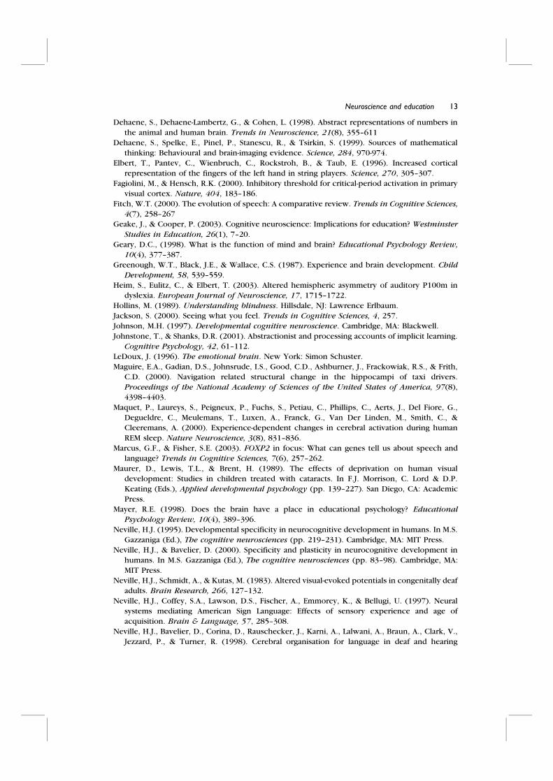
Figure 1. The major subdivisions of the cerebral cortex. The different lobes are specialised for
different tasks. The frontal lobe is used for planning and reasoning, and controls our ability to use
speech and how we react to situations emotionally. The temporal lobe is mainly concerned with
memory, audition, language and object recognition. The parietal lobe controls our sense of touch and
is used for spatial processing and perception. The occipital lobe is specialised for vision. Structures such
as the hippocampus and the amygdala are internal to the brain, situated beneath the cerebral cortex in
the midbrain.
4 Usha Goswami
Neuroimaging tools for developmental cognitive neuroscience
Neuroimaging studies are based on the assumption that any cognitive task makes
specific demands on the brain which will be met by changes in neural activity. These
changes in activity affect local blood flow which can be measured either directly (PET)
or indirectly (fMRI). Dynamic interactions among mental processes can be measured by
ERPs.
PET (positron emission tomography) relies on the injection of radioactive tracers,
and is not suitable for use with children. Brain areas with higher levels of blood flow
have larger amounts of the tracer, allowing pictures of the distribution of radiation to becreated and thereby enabling the localisation of different neural functions. fMRI
(functional magnetic resonance imaging) also enables the localisation of brain activity.
This technique requires inserting the participant into a large magnet (like a big tube),
and works by measuring the magnetic resonance signal generated by the protons of
water molecules in neural cells. When blood flow to particular brain areas increases,
the distribution of water in the brain tissue also changes. This enables measurement of a
BOLD (blood oxygenation level dependent) response which measures changes in the
oxygenation state of haemoglobin associated with neural activity. The change in BOLDresponse is the outcome measure in most fMRI studies. It is very noisy inside the
magnet and participants are given headphones to shield their ears and a panic button
(the magnet is claustrophobic). Because of these factors, it has been challenging to
adapt fMRI for use with children (who also move a lot, impeding scanning accuracy).
However, with the advent of specially adapted coils and less claustrophobic head
scanners, such studies are growing in number.
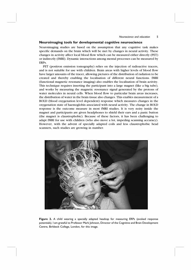
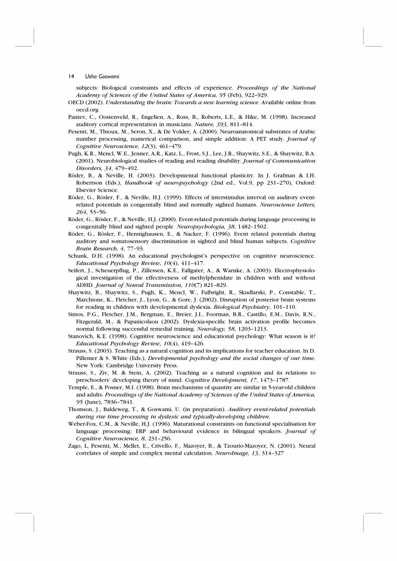
Figure 2. A child wearing a specially adapted headcap for measuring ERPs (evoked response
potentials). I am grateful to Professor Mark Johnson, Director of the Cognitive and Brain Development
Centre, Birkbeck College, London, for this image.
5Neuroscience and education
A different and widely used neuroimaging technique that can be applied to children
is that of the event related potential (ERP). ERPs enable the timing rather than
localisation of neural events to be studied. Sensitive electrodes are placed on the skin ofthe scalp and then recordings of brain activity are taken. Recording of the spontaneous
natural rhythms of the brain is called EEG (electroencephalography). ERP refers to
systematic deflections in electrical activity that may occur to precede, accompany or
follow experimenter-determined events. ERP rhythms are thus time-locked to specific
events designed to study cognitive function. The usual technique is for the child to
watch a video while wearing a headcap (like a swimming cap) that holds the electrodes
(see Figure 2). For visual ERP studies, the video is delivering the stimuli, for auditory
ERP studies, the linguistic stimuli form a background noise and the child sits engrossedin a silent cartoon. The most usual outcome measures are (i) the latency of the
potentials, (ii) the amplitude (magnitude) of the various positive and negative changes
in neural response, and (iii) the distribution of the activity. The different potentials
(characterised in countless ERP studies) are called N100, P200, N400 and so on,
denoting Negative peak at 100 ms, Positive peak at 200 ms and so on. The amplitude
and duration of single ERP components such as the P200 increase until age 3 to 4 years
(in parallel with synaptic density), and then decrease until puberty. ERP latencies
decrease within the first years of life (in parallel with myelinisation) and reach adultlevels in late childhood. ERP studies have provided extensive evidence on the time
course of neural processing and are sensitive to millisecond differences. The sequence
of observed potentials and their amplitude and duration are used to understand the
underlying cognitive processes.
Selected studies from cognitive neuroscience with interestingimplications for education
How valuable is cognitive neuroscience to educational psychologists? Current opinions
vary (Bruer, 1997; Byrnes & Fox, 1998; Geary, 1998; Geake & Cooper, 2003; Mayer,
1998; Schunk, 1998; Stanovich, 1998), but in general the consensus is moving away
from early views that neuroscience is irrelevant because it only confirms what we
already knew. The eventual answer will probably be that it is very valuable indeed. The
tools of cognitive neuroscience offer various possibilities to education, including the
early diagnosis of special educational needs, the monitoring and comparison of theeffects of different kinds of educational input on learning, and an increased
understanding of individual differences in learning and the best ways to suit input to
learner. I will now describe briefly some recent neuroscience studies in certain areas of
cognitive development, and give a flavour of how their methods could contribute to
more specifically educational questions.
LanguageDespite sharing 98.5% of our genome with chimpanzees, we humans can talk and
chimps cannot. Interestingly, genes expressed in the developing brain may hold part of
the answer. For example, a gene called FOXP2 differs in mouse and man by 3 amino
acid differences, two of which occurred after separation from the common human-
chimp ancestor about 200,000 years ago (Marcus & Fisher, 2003). This gene is
implicated in a severe developmental disorder of speech and language that affects the
6 Usha Goswami
control of face and mouth movements, impeding speech. Neurally, accurate vocal
imitation appears to be critical for the development of speech (Fitch, 2000). Hence
when linguistic input is degraded or absent for various reasons (e.g., being hearingimpaired, being orally impaired), speech and language are affected. Studies of normal
adults show that grammatical processing relies more on frontal regions of the left
hemisphere, whereas semantic processing and vocabulary learning activate posterior
lateral regions of both hemispheres. For reasons that are not yet well understood, the
brain systems important for syntactic and grammatical processing are more vulnerable
to altered language input than the brain systems responsible for semantic and lexical
functions. ERP studies show that when English is acquired late due to auditory
deprivation or late immigration to an English-speaking country, syntactic abilities do notdevelop at the same rate or to the same extent (Neville et al., 1997). Late learners do not
rely on left hemisphere systems for grammatical processing, but use both hemispheres
(Weber-Fox & Neville, 1996). ERP studies also show that congenitally blind people
show bilateral representation of language functions (Roder et al., 2000). Blind people
also process speech more efficiently (Hollins, 1989), for example they speed up
cassette tapes, finding them too slow, and still comprehend the speech even though the
recording quality suffers.
ReadingNeuroimaging studies of both children and adults suggest that the major systems for
reading alphabetic scripts are lateralised to the left hemisphere. These studies typically
measure brain responses to single word reading using fMRI or ERPs. Reviews of such
studies conclude that alphabetic/orthographic processing seems mainly associated with
occipital, temporal and parietal areas (e.g., Pugh et al., 2001). The occipital-temporal
areas are most active when processing visual features, letter shapes and orthography.The inferior occipital-temporal area shows electrophysiological dissociations between
words and nonwords at around 180 ms, suggesting that these representations are not
purely visual but are linguistically structured. Activation in temporo-occipital areas
increases with reading skill (e.g., Shaywitz et al., 2002), and is decreased in children
with developmental dyslexia.
Phonological awareness (the ability to recognize and manipulate component sounds
in words) predicts reading acquisition across languages, and phonological processing
appears to be focused on the temporo-parietal junction. This may be the main sitesupporting letter-to-sound recoding and is also implicated in spelling disorders.
Dyslexic children, who typically have phonological deficits, show reduced activation in
the temporo-parietal junction during tasks such as deciding whether different letters
rhyme (e.g., P, T = yes, P, K = no). Targeted reading remediation increases activation in
this area (e.g., Simos et al., 2002). Finally, recordings of event-related magnetic fields
(MEG) in dyslexic children suggest that there is atypical organisation of the right
hemisphere (Heim, Eulitz, & Elbert, 2003). This is consistent with suggestions that
compensation strategies adopted by the dyslexic brain require greater right hemisphereinvolvement in reading.
Although to date neuroimaging studies have largely confirmed what was already
known about reading and its development from behavioural studies, neuroscience
techniques also offer a way of distinguishing between different cognitive theories (e.g.,
whether dyslexia has a visual basis or a linguistic basis in children). Neuroimaging
techniques also offer a potential means for distinguishing between deviance and delay
7Neuroscience and education
when studying developmental disorders. For example, our preliminary studies of basic
auditory processing in dyslexic children using ERPs suggest that the phonological
system of the dyslexic child is immature rather than deviant (Thomson, Baldeweg, &Goswami, in preparation). Dyslexic children show remarkable similarity in N1 response
to younger reading level controls, while showing much larger N1 amplitudes than age-
matched controls. Finally, PET studies have shown that the functional organization of
the brain differs in literate and illiterate adults (Castro-Caldas et al., 1998). Portuguese
women in their sixties who had never learned to read because of lack of access to
education were compared with literate Portuguese women from the same villages in
word and nonword repetition tasks. It was found that totally different brain areas were
activated during nonword repetition for the illiterate versus literate participants.Learning to read and write in childhood thus changes the functional organization of the
adult brain.
MathematicsFor mathematics, cognitive neuroscience is beginning to go beyond existing cognitive
models. It has been argued that there is more than one neural system for the
representation of numbers. A phylogenetically old ‘number sense’ system, found inanimals and infants as well as older participants, seems to underpin knowledge about
numbers and their relations (Dehaene, Dehaene–Lambertz, & Cohen, 1998). This
system, located bilaterally in the intraparietal areas, is activated when participants
perform tasks such as number comparison, whether the comparisons involve Arabic
numerals, sets of dots or number words. Because mode of presentation does not affect
the location of the parietal ERP components, this system is thought to organize
knowledge about number quantities. Developmental ERP studies have shown that
young children use exactly the same parietal areas to perform number comparison tasks(Temple & Posner, 1998). A different type of numerical knowledge is thought to be
stored verbally, in the language system (Dehaene et al., 1999). This neural system also
stores knowledge about poetry and overlearned verbal sequences, such as the months
of the year. Mathematically, it underpins counting and rote-acquired knowledge such as
the multiplication tables. This linguistic system seems to store ‘number facts’ rather
than compute calculations. Many simple arithmetical problems (e.g., 3 + 4, 3 � 4) are so
overlearned that they may be stored as declarative knowledge. More complex
calculation seems to involve visuospatial regions (Zago et al., 2001), possibly attestingto the importance of visual mental imagery in multi-digit operations (an internalized and
sophisticated form of a number line, see Pesenti, Thioux, Seron, & De Volder, 2000).
Finally, a distinct parietal-premotor area is activated during finger counting and also
calculation.
This last observation may suggest that the neural areas activated during finger-
counting (a developmental strategy for the acquisition of calculation skills) eventually
come to partially underpin numerical manipulation skills in adults. If this were the case,
then perhaps finger counting has important consequences for the developing brain,and should be encouraged in school. In any event, neuroimaging techniques offer ways
of exploring such questions. They can also be used to discover the basis of dyscalculia
in children. For example, dyslexic children often seem to have associated mathematical
difficulties. If dyslexia has a phonological basis, then it seems likely that the
mathematical system affected in these children should be the verbal system
underpinning counting and calculation. Dyslexic children with mathematical
8 Usha Goswami
difficulties may show neural anomalies in the activation of this system, but not in the
activation of the parietal and premotor number systems. Children with dyscalculia who
do not have reading difficulties may show different patterns of impairment. Knowledgeof the neural basis of their difficulties could then inform individual remedial curricula.
Direct effects of experienceAlthough it is frequently assumed that specific experiences have an effect on children,
neuroimaging offers ways of investigating this assumption directly. The obvious
prediction is that specific experiences will have specific effects, increasing neural
representations in areas directly relevant to the skills involved. One area of specificexperience that is frequent in childhood is musical experience. fMRI studies have
shown that skilled pianists (adults) have enlarged cortical representations in auditory
cortex, specific to piano tones. Enlargement was correlated with the age at which
musicians began to practise, but did not differ between musicians with absolute versus
relative pitch (Pantev et al., 1998). Similarly, MEG studies show that skilled violinists
have enlarged neural representations for their left fingers, those most important for
playing the violin (Elbert et al., 1996). Clearly, different sensory systems are affected by
musical expertise depending on the nature of the musical instrument concerned. ERPstudies have also shown use-dependent functional reorganization in readers of Braille.
Skilled Braille readers are more sensitive to tactile information than controls, and this
extends across all fingers, not just the index finger (Roder, Rosler, Hennighausen, &
Nacker, 1996). The neural representations of muscles engaged in Braille reading are also
enlarged. Finally, it is interesting to note that London taxi drivers who possess ‘The
Knowledge’ show enlarged hippocampus formations (Maguire et al., 2000). The
hippocampus is a small brain area thought to be involved in spatial representation and
navigation. In London taxi drivers, the posterior hippocampi were significantly largerthan those of controls who did not drive taxis. Furthermore, hippocampal volume was
correlated with the amount of time spent as a taxi driver. Again, localised plasticity is
found in the adult brain in response to specific environmental inputs.
Plasticity in children, of course, is likely to be even greater. Our growing
understanding of plasticity offers a way of studying the impact of specialized remedial
programmes on brain function. For example, on the basis of the cerebellar theory of
dyslexia, remedial programmes are available that are designed to improve motor
function. It is claimed that these programmes will also improve reading. Whether this isin fact the case can be measured directly via neuroimaging. If the effects of such
remedial programmes are specific, then neuroimaging should reveal changes in motor
representations but not in phonological and orthographic processing. If the effects
generalize to literacy (for example, via improved automaticity), then changes in
occipital, temporal and parietal areas should also be observed.
Sleep and cognitionThe idea that sleep might serve a cognitive function dates from at least the time of
Freud, with his analysis of dreams. Recent neuroimaging studies suggest indeed that
Rapid Eye Movement (REM) sleep is not only associated with self-reports of dreaming
but is important for learning and memory. Maquet and colleagues (Maquet et al., 2000)
used PET to study regional brain activity during REM sleep following training on a serial
reaction time task. During task learning, volunteer students were trained to press one of
9Neuroscience and education

6 marked keys on a computer in response to associated visual signals on the computer
screen. Training lasted for 4 hours, from 4 p.m. until 8 p.m. The participants were then
scanned during sleep. Controls were either scanned when awake while receiving thetraining, or were scanned when asleep following no training. It was found that the brain
areas most active in the trained awake group when performing the task were also most
active during REM sleep in the trained participants. They were not active during sleep
in the untrained participants. Hence certain regions of the brain (in occipital and
premotor cortex) were actually reactivated during sleep. It seems that REM sleep either
allows the consolidation of memories or the forgetting of unnecessary material (or both
together). When tested again on the computer task on the following day, significant
improvement in performance was found to have occurred. Although the cellularmechanisms underlying this are not understood, it seems likely that memory
consolidation relies on augmented synaptic transmission and eventually on increased
synaptic density – the same mechanisms that structure the developing brain. Again, this
suggests substantial plasticity even in adulthood, supporting educational emphases on
life-long learning.
Emotion and cognitionIt is increasingly recognized that efficient learning does not take place when the learner
is experiencing fear or stress. Stress can both help and harm the body. Stress responses
can provide the extra strength and attention needed to cope with a sudden emergency,
but inappropriate stress has a significant effect on both physiological and cognitive
functioning. The main emotional system within the brain is the limbic system, a set of
structures incorporating the amygdala and hippocampus. The ‘emotional brain’
(LeDoux, 1996) has strong connections with frontal cortex (the major site for
reasoning and problem solving). When a learner is stressed or fearful, connections withfrontal cortex become impaired, with a negative impact on learning. Stress and fear also
affect social judgments, and responses to reward and risk. One important function of
the emotional brain is assessing the value of information being received. When the
amygdala is strongly activated, it interrupts action and thought, and triggers rapid bodily
responses critical for survival. It is suggested by LeDoux that classroom fear or stress
might reduce children’s ability to pay attention to the learning task because of this
automatic interruption mechanism. To date, however, neuroimaging studies of the
developmental effects of stress on cognitive function are sparse or non-existent. In theeducational arena, studying the role of stress (and emotional affect generally) in
classroom learning seems an area ripe for development. Simple ERP measures of
attentional processes, such as those used by Seifert et al. (2003) to study children with
ADHD receiving Ritalin, could easily be adapted for such purposes.
Neuromyths
The engaging term ‘neuromyths’, coined by the OECD report on understanding the
brain (OECD, 2002), suggests the ease and rapidity with which scientific findings can
be translated into misinformation regarding what neuroscience can offer education.
The three myths given most attention in the OECD report are (1) the lay belief in
hemispheric differences (‘left brain’ versus ‘right brain’ learning etc.), (2) the notion
that the brain is only plastic for certain kinds of information during certain ‘critical
10 Usha Goswami
periods’, and that therefore education in these areas must occur during the critical
periods, and (3) the idea that the most effective educational interventions need to be
timed with periods of synaptogenesis.Regarding neuromyth (1), the left brain/right brain claims probably have their basis
in the fact that there is some hemispheric specialization in terms of the localisation of
different skills. For example, many aspects of language processing are left-lateralised
(although not, as we have seen, in blind people or in those who emigrate in later
childhood to a new linguistic community). Some aspects of face recognition, in
contrast, are lateralised to the right hemisphere. Nevertheless, there are massive cross-
hemisphere connections in the normal brain, and both hemispheres work together in
every cognitive task so far explored with neuroimaging, including language and facerecognition tasks.
Regarding neuromyth (2), optimal periods for certain types of learning clearly exist
in development, but they are sensitive periods rather than critical ones. The term
‘critical period’ implies that the opportunity to learn is lost forever if the biological
window is missed. In fact, there seem to be almost no cognitive capacities that can be
‘lost’ at an early age. As discussed earlier, some aspects of complex processing suffer
more than others from deprivation of early environmental input (e.g., depth perception
in vision, grammar learning in language), but nevertheless learning is still possible. It isprobably better for the final performance levels achieved to educate children in, for
example, other languages during the sensitive period for language acquisition.
Nevertheless, the existence of a sensitive period does not mean that adults are unable
to acquire competent foreign language skills later in life.
Neuromyth (3) concerning synaptogenesis may have arisen from influential work on
learning in rats. This research showed that rodent brains form more connections in
enriched and stimulating environments (e.g., Greenough, Black, & Wallace, 1987). As
discussed earlier, any kind of specific environmental stimulation causes the brain toform new connections (recall the enlarged cortical representations of professional
musicians and the enlarged hippocampi of London taxi drivers). These demonstrations
do not mean that greater synaptic density predicts a greater capacity to learn, however.
Other neuromyths can also be identified. One is the idea that a person can either
have a ‘male brain’ or a ‘female brain’. The terms ‘male brain’ and ‘female brain’ were
coined to refer to differences in cognitive style rather than biological differences
(Baron-Cohen, 2003). Baron-Cohen argued that men were better ‘systemizers’ (good at
understanding mechanical systems) and women were better ‘empathisers’ (good atcommunication and understanding others). He did not argue that male and female
brains were radically different, but used the terms male and female brain as a
psychological shorthand for (overlapping) cognitive profiles.
Another neuromyth is the idea that ‘implicit’ learning could open new avenues
educationally. Much human learning is ‘implicit’, in the sense that learning takes place
in the brain despite lack of attention to/conscious awareness of what is being learned
(e.g., Berns, Cohen, & Mintun, 1997, but see Johnstone & Shanks, 2001). Almost all
studies of implicit learning use perceptual tasks as their behavioural measures (e.g., theparticipant gets better at responding appropriately to ‘random’ letter strings in a
computer task when the ‘random’ strings are actually generated according to an
underlying ‘grammar’ or rule system which can be learned). There are no studies
showing implicit learning of the cognitive skills underpinning educational achieve-
ment. These skills most likely require effortful learning and direct teaching.
11Neuroscience and education
Conclusions
Clearly, the potential for neuroscience to make contributions to educational research is
great. Nevertheless, bridges need to be built between neuroscience and basic research
in education. Bruer (1997) suggested that cognitive psychologists are admirably placed
to erect these bridges, although he also cautioned that while neuroscience has learned a
lot about neurons and synapses, it has not learned nearly enough to guide educational
practice in any meaningful way. This view is perhaps too pessimistic. Cognitive
developmental neuroscience has established a number of neural ‘markers’ that can be
used to assess development, for example of the language system. These markers may beuseful for investigating educational questions. Taking ERP signatures of language
processing as a case in point, different parameters are robustly associated with semantic
processing (e.g., N400), phonetic processing (e.g., mis-match negativity or MMN), and
syntactic processing (e.g., P600). These parameters need to be investigated long-
itudinally in children. Certain patterns may turn out to be indicative of certain
developmental disorders. For example, children at risk for dyslexia may show immature
or atypical MMNs to phonetic distinctions (Csepe, 2003). Children with SLI (specific
language impairment) may have generally immature auditory systems, systemsresembling those of children 3–4 years younger than them (Bishop & McArthur, in
preparation). Characteristic ERPs may also change in response to targeted educational
programmes. For example, the MMN to phonetic distinctions may become sharper (as
indexed by faster latencies) in response to literacy tuition in phonics (see Csepe, 2003).
If this were to be established across languages, education would have a neural tool for
comparing the efficiency of different approaches to the teaching of initial reading. For
example, one could measure whether the MMN to phonetic distinctions sharpened in
response to literacy tuition based on whole language methods. This is only one exampleof the creative application of currently available neuroscience techniques to important
issues in education. Educational and cognitive psychologists need to take the initiative,
and think ‘outside the box’ about how current neuroscience techniques can help to
answer outstanding educational questions.
References
Baron-Cohen, S. (2003). The essential difference: Men, women and the extreme male brain.
London: Penguin/Allen Lane.
Berns, G.S., Cohen, J.D., & Mintun, M.A. (1997). Brain regions responsive to novelty in the
absence of awareness. Science, 276, 1272–1275.
Bishop, D.V.M., & McArthur, G. (in preparation). Using event-related potentials to study
auditory processing in children with language and literacy impairments.
Bruer, J. T. (1997). Education and the brain : A bridge too far. Educational Researcher, 26(8), 4–
16.
Byrnes, J.P., & Fox, N.A. (1998). The education relevance of research in cognitive neuroscience.
Educational Psychology Review, 10(3), 297–342.
Caspi, A., McClay, J., Moffitt, T. E., Mill, J., Martin, J., Craig, I. W., Taylor, A., & Poulton, R. (2002).
Role of genotype in the cycle of violence in maltreated children. Science, 297, 851–854.
Castro-Caldas, A., Petersson, K.M., Reis, A., Stone-Elander S., & Ingvar, M. (1998). The illiterate
brain. Learning to read and write during childhood influences the functional organization of
the adult brain. Brain, 121, 1053–1063.
Csepe, V. (2003). Auditory event-related potentials in studying developmental dyslexia. In V.
Csepe (Ed.), Dyslexia : Different brain, different behaviour (pp. 81–112). New York : Kluwer.
12 Usha Goswami
Dehaene, S., Dehaene-Lambertz, G., & Cohen, L. (1998). Abstract representations of numbers in
the animal and human brain. Trends in Neuroscience, 21(8), 355–611
Dehaene, S., Spelke, E., Pinel, P., Stanescu, R., & Tsirkin, S. (1999). Sources of mathematical
thinking: Behavioural and brain-imaging evidence. Science, 284, 970-974.
Elbert, T., Pantev, C., Wienbruch, C., Rockstroh, B., & Taub, E. (1996). Increased cortical
representation of the fingers of the left hand in string players. Science, 270, 305–307.
Fagiolini, M., & Hensch, R.K. (2000). Inhibitory threshold for critical-period activation in primary
visual cortex. Nature, 404, 183–186.
Fitch, W.T. (2000). The evolution of speech: A comparative review. Trends in Cognitive Sciences,
4(7), 258–267
Geake, J., & Cooper, P. (2003). Cognitive neuroscience: Implications for education? Westminster
Studies in Education, 26(1), 7–20.
Geary, D.C., (1998). What is the function of mind and brain? Educational Psychology Review,
10(4), 377–387.
Greenough, W.T., Black, J.E., & Wallace, C.S. (1987). Experience and brain development. Child
Development, 58, 539–559.
Heim, S., Eulitz, C., & Elbert, T. (2003). Altered hemispheric asymmetry of auditory P100m in
dyslexia. European Journal of Neuroscience, 17, 1715–1722.
Hollins, M. (1989). Understanding blindness. Hillsdale, NJ: Lawrence Erlbaum.
Jackson, S. (2000). Seeing what you feel. Trends in Cognitive Sciences, 4, 257.
Johnson, M.H. (1997). Developmental cognitive neuroscience. Cambridge, MA: Blackwell.
Johnstone, T., & Shanks, D.R. (2001). Abstractionist and processing accounts of implicit learning.
Cognitive Psychology, 42, 61–112.
LeDoux, J. (1996). The emotional brain. New York: Simon Schuster.
Maguire, E.A., Gadian, D.S., Johnsrude, I.S., Good, C.D., Ashburner, J., Frackowiak, R.S., & Frith,
C.D. (2000). Navigation related structural change in the hippocampi of taxi drivers.
Proceedings of the National Academy of Sciences of the United States of America, 97(8),
4398–4403.
Maquet, P., Laureys, S., Peigneux, P., Fuchs, S., Petiau, C., Phillips, C., Aerts, J., Del Fiore, G.,
Degueldre, C., Meulemans, T., Luxen, A., Franck, G., Van Der Linden, M., Smith, C., &
Cleeremans, A. (2000). Experience-dependent changes in cerebral activation during human
REM sleep. Nature Neuroscience, 3(8), 831–836.
Marcus, G.F., & Fisher, S.E. (2003). FOXP2 in focus: What can genes tell us about speech and
language? Trends in Cognitive Sciences, 7(6), 257–262.
Maurer, D., Lewis, T.L., & Brent, H. (1989). The effects of deprivation on human visual
development: Studies in children treated with cataracts. In F.J. Morrison, C. Lord & D.P.
Keating (Eds.), Applied developmental psychology (pp. 139–227). San Diego, CA: Academic
Press.
Mayer, R.E. (1998). Does the brain have a place in educational psychology? Educational
Psychology Review, 10(4), 389–396.
Neville, H.J. (1995). Developmental specificity in neurocognitive development in humans. In M.S.
Gazzaniga (Ed.), The cognitive neurosciences (pp. 219–231). Cambridge, MA: MIT Press.
Neville, H.J., & Bavelier, D. (2000). Specificity and plasticity in neurocognitive development in
humans. In M.S. Gazzaniga (Ed.), The cognitive neurosciences (pp. 83–98). Cambridge, MA:
MIT Press.
Neville, H.J., Schmidt, A., & Kutas, M. (1983). Altered visual-evoked potentials in congenitally deaf
adults. Brain Research, 266, 127–132.
Neville, H.J., Coffey, S.A., Lawson, D.S., Fischer, A., Emmorey, K., & Bellugi, U. (1997). Neural
systems mediating American Sign Language: Effects of sensory experience and age of
acquisition. Brain & Language, 57, 285–308.
Neville, H.J., Bavelier, D., Corina, D., Rauschecker, J., Karni, A., Lalwani, A., Braun, A., Clark, V.,
Jezzard, P., & Turner, R. (1998). Cerebral organisation for language in deaf and hearing
13Neuroscience and education
subjects: Biological constraints and effects of experience. Proceedings of the National
Academy of Sciences of the United States of America, 95 (Feb), 922–929.
OECD (2002). Understanding the brain: Towards a new learning science. Available online from
oecd.org
Pantev, C., Oostenveld, R., Engelien, A., Ross, B., Roberts, L.E., & Hike, M. (1998). Increased
auditory cortical representation in musicians. Nature, 393, 811–814.
Pesenti, M., Thioux, M., Seron, X., & De Volder, A. (2000). Neuroanatomical substrates of Arabic
number processing, numerical comparison, and simple addition: A PET study. Journal of
Cognitive Neuroscience, 12(3), 461–479.
Pugh, K.R., Mencl, W.E., Jenner, A.R., Katz, L., Frost, S.J., Lee, J.R., Shaywitz, S.E., & Shaywitz, B.A.
(2001). Neurobiological studies of reading and reading disability. Journal of Communication
Disorders, 34, 479–492.
Roder, B., & Neville, H. (2003). Developmental functional plasticity. In J. Grafman & I.H.
Robertson (Eds.), Handbook of neuropsychology (2nd ed., Vol.9, pp 231–270), Oxford:
Elsevier Science.
Roder, G., Rosler, F., & Neville, H.J. (1999). Effects of interstimulus interval on auditory event-
related potentials in congenitally blind and normally sighted humans. Neuroscience Letters,
264, 53–56.
Roder, G., Rosler, F., & Neville, H.J. (2000). Event-related potentials during language processing in
congenitally blind and sighted people. Neuropsychologia, 38, 1482–1502.
Roder, G., Rosler, F., Hennighausen, E., & Nacker, F. (1996). Event related potentials during
auditory and somatosensory discrimination in sighted and blind human subjects. Cognitive
Brain Research, 4, 77–93.
Schunk, D.H. (1998). An educational psychologist’s perspective on cognitive neuroscience.
Educational Psychology Review, 10(4), 411–417.
Seifert, J., Scheuerpflug, P., Zillessen, K.E., Fallgater, A., & Warnke, A. (2003). Electrophysiolo-
gical investigation of the effectiveness of methylphenidate in children with and without
ADHD. Journal of Neural Transmission, 110(7) 821–829.
Shaywitz, B., Shaywitz, S., Pugh, K., Mencl, W., Fulbright, R., Skudlarski, P., Constable, T.,
Marchione, K., Fletcher, J., Lyon, G., & Gore, J. (2002). Disruption of posterior brain systems
for reading in children with developmental dyslexia. Biological Psychiatry, 101–110.
Simos, P.G., Fletcher, J.M., Bergman, E., Breier, J.I., Foorman, B.R., Castillo, E.M., Davis, R.N.,
Fitzgerald, M., & Papanicolaou (2002). Dyslexia-specific brain activation profile becomes
normal following successful remedial training. Neurology, 58, 1203–1213.
Stanovich, K.E. (1998). Cognitive neuroscience and educational psychology: What season is it?
Educational Psychology Review, 10(4), 419–426.
Strauss, S. (2003). Teaching as a natural cognition and its implications for teacher education. In D.
Pillemer & S. White (Eds.), Developmental psychology and the social changes of our time.
New York: Cambridge University Press.
Strauss, S., Ziv, M. & Stein, A. (2002). Teaching as a natural cognition and its relations to
preschoolers’ developing theory of mind. Cognitive Development, 17, 1473–1787.
Temple, E., & Posner, M.I. (1998). Brain mechanisms of quantity are similar in 5-year-old children
and adults. Proceedings of the National Academy of Sciences of the United States of America,
95 (June), 7836–7841.
Thomson, J., Baldeweg, T., & Goswami, U. (in preparation). Auditory event-related potentials
during rise time processing in dyslexic and typically-developing children.
Weber-Fox, C.M., & Neville, H.J. (1996). Maturational constraints on functional specialisation for
language processing: ERP and behavioural evidence in bilingual speakers. Journal of
Cognitive Neuroscience, 8, 231–256.
Zago, L, Pesenti, M., Mellet, E., Crivello, F., Mazoyer, B., & Tzourio-Mazoyer, N. (2001). Neural
correlates of simple and complex mental calculation. NeuroImage, 13, 314–327
14 Usha Goswami