Embed Size (px)
Citation preview
1095ISSN 2041-679210.4155/CLI.11.93 © 2011 Future Science Ltd
Clin. Invest. (2011) 1(8), 1095–1106
Friedreich ataxia (FRDA) is an autosomal recessive, neurodegenerative disease. It affects primarily the nervous system and the heart. Progressive gait and limb ataxia, dysarthria, loss of vibration and proprioceptive sense are characteristic neurological symptoms in FRDA. In approximately 96% of patients FRDA is caused by a triplet guanine-adenine-adenine expansion within the first intron of the FXN gene on chromosome 9q13. Increased numbers of guanine-adenine-adenine repeats are suggested to interfere with FXN transcription via heterochromatin-mediated silencing and result in frataxin deficiency in FRDA. Genetic and biological studies support the role of frataxin as a multifunctional protein in iron-dependent mitochondrial pathways. Multicenter, randomized-controlled Phase III trials in FRDA failed to prove disease modifying properties of candidate substances until to date. Phase II studies attributed idebenone, a synthetic short chain quinine analogue of co-enzyme Q10, some clinical benefit. Recent Phase III trials, however, testing idebenone have been negative or are still ongoing. Candidate substances currently tested in small randomized controlled or open-label trials are deferiprone, a mitochondrial iron chelator that forms chemically inert molecules by binding to iron, and conventional recombinant human or carbamylated erythropoietins. Both classes of candidate substances are currently under investigation to assess their efficacy and/or safety profile in Phase II trials. Pioglitazone is a peroxisome proliferator activated receptor g molecule currently tested in a 2-year randomized, double-blind, placebo-controlled safety and efficacy study. Preclinical candidate substances in FRDA are histone deacetylase inhibitors. Promising findings in animal models will have to be replicated in human cellular models such as reprogrammed induced pluripotent stem cells from FRDA patients. A still unmet issue in FRDA is to establish well shaped clinical study designs in small study cohorts within a reasonable time frame. Therefore, large natural history studies as well as the introduction of validated bio- and surrogate markers are essential issues for future clinical trials.
Keywords: biomarker • deferiprone • erythropoietin • frataxin • Friedreich ataxia • idebenone
Friedreich ataxia (FRDA) is an autosomal recessive, neurodegenerative disease. It is caused by a guanine-adenine-adenine (GAA) trinucleotid expansion in intron 1 of the FXN gene, leading to decreased expression of the mitochondrial protein frataxin. FRDA primarily affects the nervous system and the heart. Its first descrip-tion derives from Nicolas Friedreich in the second half of the 19th Century. FRDA is the most common of the recessive ataxias. It seems to be restricted to individuals from Europe, the Middle East, North Africa and India. Its prevalence appears to be closely related to the frequency of large, normal alleles for FXN as these are virtually absent in people from East Asia and in American Indians [1]. In Europe, heterozygous
Therapeutic Perspective
New advances in the treatment of Friedreich ataxia: promises and pitfalls
Wolfgang Nachbauer1 & Sylvia Boesch†1
1Department of Neurology, Medical University Innsbruck, A-6020 Innsbruck, Anichstrasse 35, Austria †Author for correspondence:Tel.: +43 512 5040 Fax: +43 512 5042 6286 E-mail: [email protected]
www.future-science.com future science group1096
Therapeutic Perspective Nachbauer & Boesch
mutation carriers seem to decrease in frequency with a south to north-east gradient [2–4]. Overall prevalence is approximately one in 30,000 to one in 50,000 in most populations, carrier frequency approximately one in 85 in Caucasians [3,5,6]. The high prevalence in Western Europe may be explained by a population bottleneck in one of the so called ‘ice-age refugees’ in which small populations survived. A two-step model merges the duplication of an ancestral (GAA)9 allele and a second mutational event that was restricted to Indo-European and North African populations with an enlargement of the (GAA)18 allele into an unstable range of more than 34 GAA units and explains the lack of FRDA in sub-Saharan Africans [1–3,7–11]. In accordance with this hypothesis, all large normal and expanded allele carriers share a common haplotype.
Clinical presentationThe age of onset in FRDA is typically around puberty, but early and late onset variants do exist. GAA repeat length of the shorter allele correlates with age of onset, accounting for approximately 50% of phenotype varia-tion [12]. Progressive gait and limb ataxia, dysarthria, as well as loss of vibration and proprioceptive sense are char-acteristic neurological symptoms in FRDA. Loss of deep tendon reflexes, extended plantar response and abnormal eye movements accomplish the spectrum of neurologi-cal features often seen in FRDA. Perception of light touch, pain and temperature may initially be normal and decrease with disease progression. Pyramidal involve-ment and progressive weakness becomes severe mainly in late stage disease. Ataxia and balance impairment limit mobility and create the necessity of a wheelchair approximately 15 years after disease onset [13].
Non-neurological features in FRDA include hyper-trophic cardiomyopathy and diabetes mellitus. Heart disease can be severe and can cause premature death, par-ticularly in early-onset cases. Electrocardiography shows widespread T-wave inversion in virtually all patients. Conduction disturbances occur in approximately 10%. Supraventricular ectopic beats and atrial fibrillation are occasionally detectable and increase the risk of throm-boembolic complications and may lead to heart failure. Echocardiography detects left ventricular hypertrophy in 50–65% of FRDA patients, showing increasing inci-dence in patients with larger GAA alleles [12,14]. Severe cardiomyopathy with progressive deterioration of left ventricular ejection fraction and chronic heart failure, however, is rare. The prevalence of foot deformities and scoliosis in FRDA is high [15,16]. Pes cavus can be seen in > 90% of patients, whereas range-of-motion limitations in other joints are uncommon in early FRDA. Secondary to immobility and spasticity a range of joint abnormali-ties may though occur. Scoliosis is seen in approximately
60% of FRDA patients, associated with severe, progres-sive hyperkyphosis [17,18]. Progressive curves are seen before the age of 10 years, whereas nonprogressive curves tend to present during or after puberty. Patients should be carefully screened and surgical treatment should be considered. Diabetes mellitus or impaired fastening glu-cose may be part of the clinical spectrum of FRDA. Oral glucose testing is therefore recommended annually [13].
NeuropathologyCharacteristic neuropathological features in FRDA are atrophy of dorsal root ganglia and thinning of dorsal roots. Especially large myelinated fibers of the dorsal col-umn tend to be sparse in FRDA. A reduction of spinal cord diameters is therefore evident and seems to be most pronounced in the thoracic region [19,20]. In addition, neu-ropathology in FRDA reveals subsequent atrophy of the dentate nucleus, as well as spinocerebellar and corticospi-nal tracts. Histopathological studies indicate for neuronal iron dysmetabolism and inappropriate myelination in FRDA [20]. Atrophy of the cerebellar hemispheres and the vermis cerebelli, however, remains mild and might only be seen in later disease stages. Peripheral neuropathy in FRDA may primarily be related to hypomyelination by deficient interaction between axons and Schwann cells. Axonal degeneration of peripheral nerves is present in FRDA but remains slow in disease progression [21].
Diagnostic criteria, clinical assessment & rating scales Diagnostic criteria for FRDA have been suggested by Anita Harding in the 1980s [22]. They stipulate a slowly progressive, recessive ataxia that is not explained by other primary causes. Ataxia should start before the age of 25 years and is characterized by absent tendon reflexes of the lower limbs as well as dysarthria that occur within 5 years of disease onset [22]. These criteria provide for a sensitivity of 63% and a positive predictive value of 96% [23]. Lower GAA repeat numbers go along with a later onset and slower course of disease, as well as retained tendon reflexes [12].
For the assessment of ataxia in FRDA patients and the monitoring of disease progression three appropri-ate scales - the ‘International Cooperative Ataxia Rating Scale (ICARS)’ [24], the ‘Friedreich Ataxia Rating Scale’ (FARS) [25], as well as the recently validated ‘Scale for the Assessment and Rating of Ataxia (SARA)’ [26] may be used. SARA is composed out of eight items yield-ing in a total numeric score from 0 (no ataxia) to 40 (severe ataxia). Initially SARA was invented for rating of autosomal dominant ataxia. Validity and reliability of the scale in FRDA have been shown recently [27]. Mean time to administer SARA in patients is 14.2 ± 7.5 min (range: 5–40 min). Single SARA items comprise gait
New advances in the treatment of Friedreich ataxia Therapeutic Perspective
future science group Clin. Invest. (2011) 1(8) 1097
(score 0–8), stance (score 0–6), sitting (score 0–4), speech disturbance (score 0–6), finger chase (score 0–4), nose-finger test (score 0–4), fast alternating hand movements (score 0–4) and heel-shin slide (score 0–4). Limb kinetic functions are rated independently for both sides. The mean score is introduced in the total score. SARA scores significantly correlate with ICARS and FARS total scores [27]. FARS is a disease specific scale for rating ataxia severity in FRDA and thus also con-siders symptoms especially occurring in FRDA. FARS consists of five separate sub-scores (bulbar system, upper extremities, lower extremities, peripheral system and upright stability) and results in a score from 0 to 125 with higher scores representing more severe ataxia. Performance measures that have been modified for the use in FRDA have been introduced into the FARS. These measures include a 9-hole peg test for fine motor coordination, a timed 25-foot walk for ambulation, a speech test using the phrase ‘PATA’, and a low-contrast letter acuity vision test. Additionally ‘activities of daily living’ (ADL) and a ‘functional disability scale’ can be implemented into the FARS, resulting in a maximum score of 167 points [28,29]. Progression of FRDA has been measured with FARS, showing more valid data in a 2-year period than after 1 year [30]. ICARS is a widely used semi-quantitative ataxia rating scale designed to represent the classic features of ataxia. ICARS is a hun-dred percent scale with higher percentages indicating more severe ataxia. Postural and stance disturbances (32%) and limb ataxia (52%) engage the largest parts of this scale. The compartmentalization makes it pos-sible to determine sub-scores [24]. ICARS has shown high inter-rater reliability in genetically confirmed ataxias [31]. Usefulness of ICARS for interventional trials was ques-tioned recently concerning practicability and sub-scale structure [32]. Additionally several overlapping ICARS rating items have shown to lead to inconsistent rating [33].
Genetics & pathophysiologyGenetic testing of FRDA is available on a routine basis. In approximately 96% of patients FRDA is caused by a homozygous triplet GAA expansion within the first intron of the FXN gene on chromosome 9q13 [34]. Repeat expansions within the FXN gene can range from 66 repeats (normal is <40 repeats) up to more than 1000 GAA repeats. The majority of expanded alleles contain between 600 and 1200 GAA repeats. There is an inverse correlation between the age at onset, sever-ity of disease and associated systemic symptoms with the size of the smaller GAA repeat expansion probably reflecting residual frataxin expression from the respec-tive allele [5,6,35–37]. Approximately 4% of individuals with FRDA are compound heterozygote for a GAA expansion in one FXN allele and a point mutation in
the other allele resulting in distinct phenotype sever-ity [6,32,35,38]. Penetrance is complete in homozygotes and in compound heterozygotes [34]. Tissue mosaics as often found in mitochondrial disorders may also contribute to an individual clinical phenotype [39,40].
Unlike other triplet repeat diseases such as poly-glutamine expansion and RNA toxicity diseases, GAA expansions in FXN are intronic and do not alter the frataxin protein sequence. GAA repeat expansion leads to triplex DNA formation which may interfere with the transcription of the FXN gene [41–44] and hetero-chromatin-mediated silencing [45], resulting in limited production of frataxin. Frataxin is highly conserved across species with homologs in bacteria, yeast, plants, and animals. In humans, frataxin mRNA is translated into a precursor protein containing 210 amino acids (frataxin1–210) [46,47]. Post-translational processing gen-erates at least two frataxin isoforms (frataxin42–210 and frataxin81–210). Recent reports suggest both isoforms relevant to FRDA pathophysiology [48].
Despite intensive research, the exact physiological functions of frataxin remain the subject of debate. Genetic and biological studies support a pivotal role of frataxin as a multifunctional protein in different iron-dependent mitochondrial pathways [34,49]. Frataxin is suggested to act as a mitochondrial iron chaperone [50] or as an iron sensor regulating the iron-sulfur (Fe-S) cluster biogenesis [51]. In vitro studies suggest that iron binding may trigger the oligomerization of frataxin and lead to radical scavenging of toxic iron in a bioavailable form in consequence [52]. Both monomeric and oligo-meric forms of frataxin were shown to interact with vari-ous potential iron acceptors. In vitro frataxin was shown to interact with ferrochelatase and to provide the iron that is needed in the last step of heme synthesis [53,54]. Frataxin may also interact with mitochondrial aconi-tase, a Fe-S-containing protein, which protects against the disassembly of the Fe-S cluster by facilitating iron transfer to aconitase [55]. Moreover, both monomeric and oligomeric forms of frataxin were proposed to be the iron donor protein for de novo Fe-S cluster biosynthe-sis [51,56–61]. Fe-S clusters are critical prosthetic groups present in proteins involved in essential cellular pro-cesses ranging from nuclear genome stability to protein translation in mitochondrial metabolism [62]. De novo Fe-S cluster assembly, a mitochondrial process in eukaryontes, relies on the assembly of a Fe-S cluster on a scaffold protein (ISCU) from inorganic iron and sul-fur, followed by the transfer of the scaffold bound Fe-S cluster to the target apoproteins. Both the synthesis and the final transfer to apoproteins require the help of addi-tional proteins [63]. The exact function of these proteins is currently unknown. Still, in vitro iron loaded human frataxin has been shown to deliver iron to ISCU [50].
www.future-science.com future science group1098
Therapeutic Perspective Nachbauer & Boesch
The iron-donor function of frataxin has recently been challenged in vitro since it behaves as an iron-dependent inhibitor of Fe-S cluster assembly through specific inter-action with scaffold proteins in kinetic studies of Fe-S cluster biosynthesis [51]. Impaired mitochondrial iron handling in FRDA and mitochondrial iron accumula-tion causes in consequence an impairment of respiratory chain function and contributes to increased oxidative stress and cellular damage [64–66]. Loss of iron sulfur proteins including the respiratory chain complexes I, II, III and aconitase result in reduced ATP generation as confirmed in FRDA ataxia patients by MRI [67]. However, the level of importance of an oxidative stress component in FRDA has been discussed controversially. Still, deficient mitochondrial ATP production in tissue that depends on oxidative phosphorylation such as the nervous system is likely to be responsible for cellular dysfunction and cell death. Moreover, if oxidative stress via dysfunctional lipid peroxidation, impaired scaveng-ing of superoxide radicals and reduced ATP formation may play a role in disease progression of FRDA remains to be elucidated.
Biomarker & surrogate markers ■ Frataxin
Assays to quantitatively measure the amount of frataxin protein have been established recently [68,69]. Frataxin levels are usually quantified per µg total protein content to allow standardized measures. Absolute frataxin levels, however, vary considerably in recently published studies, which may be addressed to distinct cell lysis protocols and frataxin protein detection methods. Compared with healthy controls FRDA patients show a mean residual frataxin expression of 20–35% [68–71]. The amount of frataxin expression is inversely correlated to the number of GAA repeats. Late onset FRDA patients therefore display higher frataxin levels ranging from 40–90% of control values [70]. Until to date frataxin measurements have mainly been performed out of isolated periph-eral blood mononuclear cells and cultured cell lines of FRDA patients. Recent reports also suggest buccal cells, whole blood and skeletal muscle [71,72] as appropri-ate specimens for frataxin detection. Still, considering frataxin’s important role as a biomarker in clinical trials the range of frataxin levels in normal controls, FRDA carriers and FRDA patients in different tissues should be studied in depth.
■ Markers of oxidative stressOxidative stress is caused by the presence of any of a number of reactive oxygen species, which the cell is unable to counterbalance. This may result in damage to one or more biomolecules including DNA, RNA, proteins and lipids. Detection of oxidative stress may be
based on DNA/RNA damage, lipid peroxidation, pro-tein oxidation or the detection of reactive oxygen species in general. In principle, a sole assay addressing oxidative stress may be rather nonspecific for the detection of neurodegenerative cell loss in FRDA patients. Moreover, reactive oxygen species markers in patients may be afflicted by multiple confounders during clinical trials.
■ MRI techniquesMRI has been used as a biomarker in a first clinical FRDA trial by measuring iron content in the den-date nucleus [73]. In addition to special iron detection methods, new MRI techniques such as voxel-based morphometry, fiber tracking or MR-spectroscopy are prone to become valid in vivo surrogate markers in future clinical studies. Several nontherapeutic studies using voxel-based morphometry have revealed a cor-relation of patients’ clinical scores and disease duration with brain white matter atrophy [74–76]. Detection of neurochemical patterns in specific brain areas using MR-spectroscopy was suggested to differentiate distinct ataxias [77]. MRI scans with higher field force (3–5 tesla) will provide for better spatial differentiation. The valid-ity of MRI-techniques as a biomarker in therapeutic trials, however, warrants further investigation.
Cardiac MRI and spectroscopy are adequate meth-ods for detection of interventricular septum thickness, left ventricular mass and estimation of mitochondrial function [78,79]. Their relevance as a biomarker, how-ever, is disputable as cardiomyopathy is not evident in all FRDA patients and ataxia severity lacks correlation with cardiac hypertrophy. Application of a cardiac MRI in clinical trials will therefore be limited to therapeutic compounds addressing cardiac involvement in FRDA.
■ Clinical rating scalesPrecision in ataxia rating is limited because of the semi-quantitative approach in all ataxia rating scales. Moreover, both inter- and intra-rater variability as well as ceiling effects in case of patients with more advanced disease stages contribute to a lack of accuracy in clinical rating. New tools and improvement of rating scales will be necessary for small but significant clinical benefit in future trials [80]. The introduction of quantitative clinical measurements (based on existing clinical ataxia symptoms) using electronic-based portable devices would therefore be helpful. Additionally, functional scores and speech assessment have to be developed to improve clinical outcome measures.
Clinical studiesMost therapeutic approaches in FRDA focus on anti-oxidant treatment to protect mitochondria from oxida-tive stress and iron accumulation. Alternatively, therapy
New advances in the treatment of Friedreich ataxia Therapeutic Perspective
future science group Clin. Invest. (2011) 1(8) 1099
strategies also target enhancing the transcription or pro-tein stabilization of frataxin. Nevertheless, until to date randomized-controlled trials in FRDA are sparse. The following paragraphs provide an overview of candidate substances tested for the use in FRDA, ongoing trials and future perspectives. PubMed was searched for arti-cles published before March 2011 including the search terms ‘Friedreich ataxia’ in combination with ‘therapy’ and ‘treatment’. Studies conducted before availability of genetic testing [34] were given low priority. Ongoing clinical trials were identified via the database of the US NIH [201].
■ Idebenone The basic pathophysiological approach of impaired mitochondrial function due to oxidative stress has led to broad application of antioxidant agents for the treat-ment of FRDA. The majority of clinical therapeutic studies were carried out using idebenone, a synthetic short chain quinine analogue of co-enzyme Q10. It acts as an electron carrier in the mitochondrial respiratory chain and therefore allocates the production of ATP. Besides, idebenone inhibits lipid-peroxidation, serves as a free radical scavenger and may therefore addition-ally prevent mitochondria from oxidative damage [81]. After absorption idebenone is rapidly metabolized and conjugated into several metabolites showing dose pro-portional pharmacokinetics in healthy human subjects up to 2250 mg/day [82,83]. However, cerebrospinal fluid ana lysis suggest idebenone to be less distributed to the brain than to other tissues [84].
Several clinical trials have been conducted with ide-benone in FRDA patients. A majority, however, are lack-ing controlled or blinded study designs. A single-center randomized placebo-controlled trial (Phase II) assessed idebenone in 47 children using different dosages of ide-benone (5, 15 and 45 mg/kg). Primary end point was a change in 8-hydroxy-2’-deoxyguanosine, a urinary marker of oxidative stress, after 6 months of treatment. Secondary outcome measures included ataxia rating (ICARS and FARS) and measures of ADL. Whereas the primary outcome was failed, a subgroup ana lysis of ambulatory patients showed significant improvement in ICARS. Moreover, higher dosages of idebenone were suggested to be superior compared with the lower dosage and placebo [28]. Two subsequent multicenter, randomized-controlled Phase III trials (MICONOS and IONIA) were carried out to assess efficacy of ide-benone treatment for neurological and cardiac symp-toms in FRDA. IONIA included seventy ambulatory children (age 8–18 years) with genetic proven FRDA. Participants were randomized into three treatment arms (placebo, low-dose and high-dose idebenone). Primary outcome was change in ICARS score after 6 months.
Secondary outcomes included changes in FARS rating, performance measures and ADL. IONIA failed to show significant improvement compared with placebo in each of the two end points [85]. Moreover, idebenone did not decrease left ventricular hypertrophy or alter cardiac function [86]. Upcoming results of the MICONOS trial (more than 200 ambulatory as well as wheelchair bound FRDA patients for a study duration of 1 year) will enlighten the issue if larger studies of longer duration are prone to assess the therapeutic potential of idebenone in FRDA [87]. The primary outcome of the MICONOS study is absolute change in ICARS from baseline to year one. Secondary outcome measures include reduc-tion of left ventricular mass index detected by MRI and echocardiography as well as the improvement in peak workload assessed by a modified exercise test.
Numerous earlier studies using idebenone 5 mg/kg/day showed significant impact on cardiac hypertrophy determined by echocardiography [88–91], although only one study was conducted as a randomized-controlled trial [91]. Still, these results could not be replicated in other studies [92,93]. Open label studies investigating clinical effects of idebenone treatment in FRDA patients showed decrease in ICARS Score [92] and stabilization of motor symptoms [94].
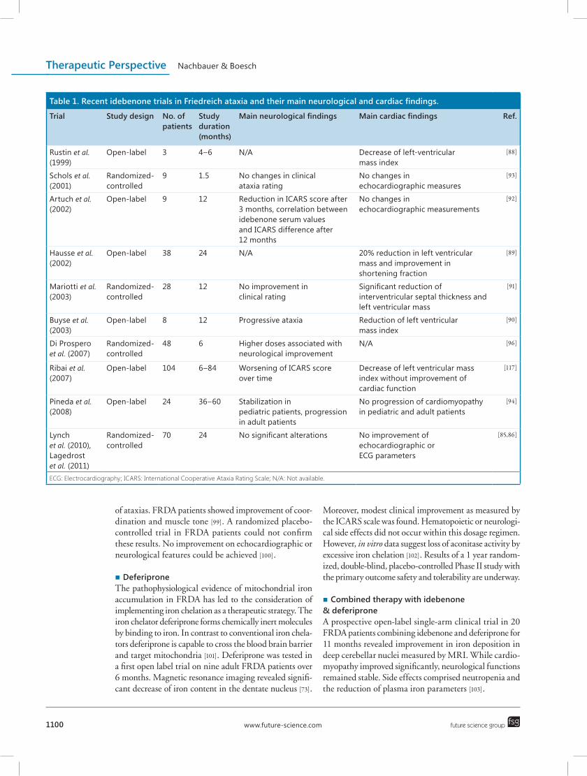
Until to date no multicenter, randomized-controlled trial showing idebenone to have clinical benefit on neurological symptoms in FRDA has been reported. Positive effects of idebenone on cardiac hypertrophy are still lacking clinical significance [95]. By all means idebenone therapy has been shown to be safe and well tolerated as solely low side effects of high dosages have been reported in FRDA patients of all ages [28,87,96]. Benefit of idebenone treatment in FRDA to stabilize disease progression may additionally depend on disease stage and age at initiation of idebenone treatment [92,94], which has been insufficiently considered in clinical tri-als so far. Table 1 gives an overview of recent idebenone trials in FRDA.
■ Other antioxidantsApart from idebenone co-enzyme Q10 and vitamin E have been suggested for antioxidant treatment of FRDA. Co-enzyme Q10 plays a role in mitochondrial ATP pro-duction. Vitamin E is a naturally occurring lipid soluble antioxidant. The combination of co-enzyme Q10 and vitamin E revealed improvement of energy metabolism in cardiac- and skeletal muscle assessed with MRI in an open label trial on ten FRDA patients. Moreover changes in ICARS score suggested stabilization of disease pro-gression as compared with a cross sectional group of FRDA patients [97,98]. Evidence for l-carnitin is based on one double-blind crossover placebo-controlled trial, though conducted in a heterogeneous study population
www.future-science.com future science group1100
Therapeutic Perspective Nachbauer & Boesch
of ataxias. FRDA patients showed improvement of coor-dination and muscle tone [99]. A randomized placebo-controlled trial in FRDA patients could not confirm these results. No improvement on echocardiographic or neurological features could be achieved [100].
■ DeferiproneThe pathophysiological evidence of mitochondrial iron accumulation in FRDA has led to the consideration of implementing iron chelation as a therapeutic strategy. The iron chelator deferiprone forms chemically inert molecules by binding to iron. In contrast to conventional iron chela-tors deferiprone is capable to cross the blood brain barrier and target mitochondria [101]. Deferiprone was tested in a first open label trial on nine adult FRDA patients over 6 months. Magnetic resonance imaging revealed signifi-cant decrease of iron content in the dentate nucleus [73].
Moreover, modest clinical improvement as measured by the ICARS scale was found. Hematopoietic or neurologi-cal side effects did not occur within this dosage regimen. However, in vitro data suggest loss of aconitase activity by excessive iron chelation [102]. Results of a 1 year random-ized, double-blind, placebo-controlled Phase II study with the primary outcome safety and tolerability are underway.
■ Combined therapy with idebenone & deferiproneA prospective open-label single-arm clinical trial in 20 FRDA patients combining idebenone and deferiprone for 11 months revealed improvement in iron deposition in deep cerebellar nuclei measured by MRI. While cardio-myopathy improved significantly, neurological functions remained stable. Side effects comprised neutropenia and the reduction of plasma iron parameters [103].
Table 1. Recent idebenone trials in Friedreich ataxia and their main neurological and cardiac findings.
Trial Study design No. of patients
Study duration (months)
Main neurological findings Main cardiac findings Ref.
Rustin et al. (1999)
Open-label 3 4–6 N/A Decrease of left-ventricular mass index
[88]
Schols et al. (2001)
Randomized-controlled
9 1.5 No changes in clinical ataxia rating
No changes in echocardiographic measures
[93]
Artuch et al. (2002)
Open-label 9 12 Reduction in ICARS score after 3 months, correlation between idebenone serum values and ICARS difference after 12 months
No changes in echocardiographic measurements
[92]
Hausse et al. (2002)
Open-label 38 24 N/A 20% reduction in left ventricular mass and improvement in shortening fraction
[89]
Mariotti et al. (2003)
Randomized-controlled
28 12 No improvement in clinical rating
Significant reduction of interventricular septal thickness and left ventricular mass
[91]
Buyse et al. (2003)
Open-label 8 12 Progressive ataxia Reduction of left ventricular mass index
[90]
Di Prospero et al. (2007)
Randomized-controlled
48 6 Higher doses associated with neurological improvement
N/A [96]
Ribai et al. (2007)
Open-label 104 6–84 Worsening of ICARS score over time
Decrease of left ventricular mass index without improvement of cardiac function
[117]
Pineda et al. (2008)
Open-label 24 36–60 Stabilization in pediatric patients, progression in adult patients
No progression of cardiomyopathy in pediatric and adult patients
[94]
Lynch et al. (2010), Lagedrost et al. (2011)
Randomized-controlled
70 24 No significant alterations No improvement of echocardiographic or ECG parameters
[85,86]
ECG: Electrocardiography; ICARS: International Cooperative Ataxia Rating Scale; N/A: Not available.
New advances in the treatment of Friedreich ataxia Therapeutic Perspective
future science group Clin. Invest. (2011) 1(8) 1101
■ Pioglitazone Pioglitazone is a peroxisome proliferator activated recep-tor g molecule. It induces the expression of enzymes involved in mitochondrial metabolism including super-oxide dismutase. Pioglitazone crosses the blood brain barrier and is suggested to improve antioxidant defense mechanisms. A 2-year randomized, double-blind, placebo-controlled safety and efficacy study in FRDA with the primary out-come measure of stabilization in ICARS is currently ongoing.
■ ErythropoietinsDue to their neuroprotective capacities erythropoi-etins have received considerable attention within the last years [104,105]. In vitro erythropoietin incubation of isolated lymphocytes and fibroblasts of FRDA patients has led to upregulation of the protein frataxin without affecting mRNA expression [106,107]. Open label studies in FRDA patients administering subcutaneously recom-binant human erythropoietin (rhuEPO) have been con-ducted in different dosages and regimens. Continuous rhuEPO application (three-times weekly) showed frataxin upregulation of 27% (range: 15–63%) after 2 months in ten patients. Moreover, reduction of oxidative stress parameters as measured by peroxides and 8-hydroxy-2’-deoxyguanosine were shown [108]. A further open label extension study on eight out of these ten patients revealed clinical improvement as measured by SARA and FARS rating after a study period of 6 months [109]. Intermittent high dose application of rhuEPO in monthly intervals resulted in a cumulative long lasting increase in frataxin levels without clinical improvement in two open label studies on FRDA patients [110,111]. Due to the well known hematopoietic stimulation potential of erythropoietin close meshed monitoring of blood cell count is essen-tial. Elevated hemoglobin and hematocrit levels require phlebotomies, especially in continuous dosing regimens. Though carbamylated erythropoietin could provide the benefit of increase in frataxin levels [112] without affecting red blood cell count or iron metabolism. Currently a first clinical Phase II trial using carbamylated erythropoietin in FRDA is underway.
■ Histone deacetylase inhibitorsHistone deacetylase (HDAC) inhibitors are promis-ing candidate substances for the future treatment as they may directly reverse the primary cause deficient frataxin expression in FRDA. HDAC inhibitors may act on DNA transcription by reversing heterochromatin-mediated silencing of the FXN gene with subsequent increase of frataxin mRNA expression and protein in lymphoid cell lines of FRDA patients [45]. HDAC inhibitors have also been shown to increase frataxin expression in brain and heart in a KIKI mouse model
for FRDA [113]. Moreover, FRDA disease phenotype could partially be reversed in a GAA repeat expansion YG8R mouse model after 5 months of HDAC inhibi-tor treatment [114]. In murine models HDAC inhibitors were well tolerated [113–115], their safety and efficacy pro-file in humans, however, requires further investigation. Preclinical trials are currently underway.
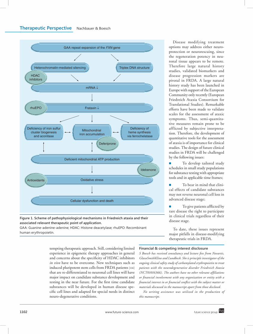
Future perspectivePromising advances for the treatment of FRDA rely on several pathophysiological considerations (Figure 1). The prevention of oxidative stress through an improvement of mitochondrial respiratory chain function and the alloca-tion of ATP production underlies the use of idebenone, a synthetic short chain quinine analogue of co-enzyme Q10. Unfortunately, after some hope during Phase II clinical trials, results of recent Phase III clinical trials using idebenone to achieve clinical benefit were somehow disappointing [85]. Also deferiprone, an intracellular iron chelator entered into a clinical Phase II studies. While findings in MRI surrogates appeared to be promising, secondary clinical outcome measures did not detect consistent benefit [73]. A second Phase II study is cur-rently addressing safety and tolerability and will provide for further data. Recent advances in the understanding of mitochondrial iron metabolism point to a complex interaction between mitochondrial iron content and frataxin function. In the light of these findings the role of iron chelators as a long-term therapeutic approach in FRDA will have to be reconsidered. Pioglitazone is sug-gested to improve antioxidant defense mechanisms and is currently tested in a 2-year randomized, double-blind, placebo-controlled safety and efficacy study in FRDA. Erythropoietins are candidate substances in FRDA therapy. Multimodal properties of erythropoietins may explain laboratory findings, namely an upregulation of cellular frataxin content. They may also explain clinical improvement through symptomatic effects on muscle strength and endurance, similar to enhanced motor performance in athletes with illicit use. Erythropoietins are also prone to influence frataxin function per se considering their multiple functions on intracellular and mitochondrial iron handling. Thus, conventional erythropoietins have considerable short-comings in their safety profile because of their hematopoietic side effects. Therefore nonhemotopoietic derivatives of erythropoi-etin such as carbamylated erythropoietin may have a role in future FRDA therapy [112]. A clinical Phase II study with carbamylated erythropoietin in FRDA is cur-rently under way. Only recently, preclinical studies using HDAC inhibitors in cell cultures and in mouse models provided for promising data in FRDA [45,113]. Reversing heterochromatin-mediated silencing of the FTX gene with subsequent increase of frataxin is an elegant and
www.future-science.com future science group1102
Therapeutic Perspective Nachbauer & Boesch
tempting therapeutic approach. Still, considering limited experience in epigenetic therapy approaches in general and concerns about the specificity of HDAC inhibitors in vivo have to be overcome. New techniques such as induced pluripotent stem cells from FRDA patients [116] that are re-differentiated to neuronal cell lines will have major impact on candidate substance development and testing in the near future. For the first time candidate substances will be developed in human disease spe-cific cell lines and adapted for special needs in distinct neuro-degenerative conditions.
Disease modifying treatment options may address either neuro-protection or neurorescuing, since the regeneration potency in neu-ronal tissue appears to be remote. Therefore large natural history studies, validated biomarkers and disease progression markers are pivotal in FRDA. A large natural history study has been launched in Europe with support of the European Community only recently (European Friedreich Ataxia Consortium for Translational Studies). Remarkable efforts have been made to validate scales for the assessment of ataxic symptoms. Thus, semi-quantita-tive measures remain prone to be aff licted by subjective interpreta-tion. Therefore, the development of quantitative tools for the assessment of ataxia is of importance for clinical studies. The design of future clinical studies in FRDA will be challenged by the following issues:
■ To develop tailored study schedules in small study populations for substance testing with appropriate tools and in applicable time frames;
■ To bear in mind that clini-cal effects of candidate substances may not reverse neuronal cell loss in advanced disease stage;
■ To give patients afflicted by rare disease the right to participate in clinical trials regardless of their disease stage.
To date, these issues represent major pitfalls in disease-modifying therapeutic trials in FRDA.
Financial & competing interest disclosure S Boesch has received consultancy and lecture fees from Novartis, GlaxoSmithKline and Lundbeck. She is principle investigator of the ongoing clinical safety study of carbamylated erythropoietin to treat patients with the neurodegenerative disorder Friedreich Ataxia (NCT01016366). The authors have no other relevant affiliations or financial involvement with any organization or entity with a financial interest in or finan cial conflict with the subject matter or materials discussed in the manuscript apart from those disclosed.
No writing assistance was utilized in the production of this manuscript.
GAA repeat expansion of the FXN gene
Heterochromatin-mediated silencing Triplex DNA structure
mRNA ↓
Frataxin ↓
Mitochondrial iron accumulation
Deficiency of iron sulfurcluster biogenesis
and aconitase
Deficiency of heme synthesis
via ferrochelatase
Deferiprone
rhuEPO
Deficient mitochondrial ATP production
Oxidative stressAntioxidants
Cellular dysfunction and death
Idebenone
HDAC inhibitors
Figure 1. Scheme of pathophysiological mechanisms in Friedreich ataxia and their associated relevant therapeutic point of application.GAA: Guanine-adenine-adenine; HDAC: Histone deacetylase; rhuEPO: Recombinant human erythropoietin.
New advances in the treatment of Friedreich ataxia Therapeutic Perspective
future science group Clin. Invest. (2011) 1(8) 1103
BibliographyPapers of special note have been highlighted as:n of interestnn of considerable interest
1 Labuda M, Labuda D, Miranda C et al. Unique origin and specific ethnic distribution of the Friedreich ataxia GAA expansion. Neurology 54(12), 2322–2324 (2000).
2 Cossee M, Schmitt M, Campuzano V et al. Evolution of the Friedreich’s ataxia trinucleotide repeat expansion: founder effect and premutations. Proc. Natl Acad. Sci. USA 94(14), 7452–7457 (1997).
3 Epplen C, Epplen JT, Frank G, Miterski B, Santos EJ, Schols L. Differential stability of the (GAA)n tract in the Friedreich ataxia (STM7) gene. Hum. Genet. 99(6), 834–836 (1997).
4 Juvonen V, Kulmala SM, Ignatius J, Penttinen M, Savontaus ML. Dissecting the epidemiology of a trinucleotide repeat disease – example of FRDA in Finland. Hum. Genet. 110(1), 36–40 (2002).
5 Pandolfo M. Friedreich’s ataxia: clinical aspects and pathogenesis. Semin. Neurol. 19(3), 311–321 (1999).
6 Pandolfo M. Molecular basis of Friedreich ataxia. Mov. Disord. 16(5), 815–821 (2001).
7 Semino O, Passarino G, Oefner PJ et al. The genetic legacy of Paleolithic homo sapiens sapiens in extant Europeans: a Y chromosome perspective. Science 290(5494), 1155–1159 (2000).
8 Colombo R, Carobene A. Age of the intronic GAA triplet repeat expansion mutation in Friedreich ataxia. Hum. Genet. 106(4), 455–458 (2000).
9 Montermini L, Andermann E, Labuda M et al. The Friedreich ataxia GAA triplet repeat: premutation and normal alleles. Hum. Mol. Genet. 6(8), 1261–1266 (1997).
10 Richards M, Macaulay V, Hickey E et al. Tracing European founder lineages in the near eastern mtDNA pool. Am. J. Hum. Genet. 67(5), 1251–1276 (2000).
11 Tambets K, Rootsi S, Kivisild T et al. The western and eastern roots of the Saami – the story of genetic ‘outliers’ told by mitochondrial DNA and Y chromosomes. Am. J. Hum. Genet. 74(4), 661–682 (2004).
12 Filla A, De Michele G, Cavalcanti F et al. The relationship between trinucleotide (GAA) repeat length and clinical features in Friedreich ataxia. Am. J. Hum. Genet. 59(3), 554–560 (1996).
13 Bidichandani SI, Delatycki MB. In: Friedreich Ataxia (1993). Pagon RA, Bird TD, Dolan CR, Stephens K (Eds). Source GeneReviews. University of Washington, Seattle, WA, USA (updated 25 Jun 2009).
14 Durr A, Cossee M, Agid Y et al. Clinical and genetic abnormalities in patients with Friedreich’s ataxia. N. Engl. J. Med. 335(16), 1169–1175 (1996).
15 Allard P, Duhaime M, Raso JV, Thiry PS, Drouin G, Geoffroy G. Pathomechanics and management of scoliosis in Friedreich ataxia patients: preliminary report. Can. J. Neurol. Sci. 7(4), 383–388 (1980).
16 Cady RB, Bobechko WP. Incidence, natural history, and treatment of scoliosis in Friedreich’s ataxia. J. Pediatr. Orthop. 4(6), 673–676 (1984).
17 Labelle H, Tohme S, Duhaime M, Allard P. Natural history of scoliosis in Friedreich’s ataxia. J. Bone Joint Surg. Am. 68(4), 564–572 (1986).
18 Milbrandt TA, Kunes JR, Karol LA. Friedreich’s ataxia and scoliosis: the experience at two institutions. J Pediatr Orthop 28(2), 234–238 (2008).
19 Koeppen AH. Friedreich’s ataxia: pathology, pathogenesis, and molecular genetics. J. Neurol. Sci. 303(1–2), 1–12 (2011).
20 Koeppen AH, Morral JA, Davis AN et al. The dorsal root ganglion in Friedreich’s ataxia. Acta Neuropathol. 118(6), 763–776 (2009).
21 Morral JA, Davis AN, Qian J, Gelman BB, Koeppen AH. Pathology and pathogenesis of sensory neuropathy in Friedreich’s ataxia. Acta Neuropathol. 120(1), 97–108 (2010).
22 Harding AE. Friedreich’s ataxia: a clinical and genetic study of 90 families with an ana lysis of early diagnostic criteria and intrafamilial clustering of clinical features. Brain 104(3), 589–620 (1981).
23 Filla A, De Michele G, Coppola G et al. Accuracy of clinical diagnostic criteria for Friedreich’s ataxia. Mov. Disord. 15(6), 1255–1258 (2000).
24 Trouillas P, Takayanagi T, Hallett M et al. International cooperative ataxia rating scale for pharmacological assessment of the cerebellar syndrome. The Ataxia Neuropharmacology Committee of the World Federation of Neurology. J. Neurol. Sci. 145(2), 205–211 (1997).
25 Subramony SH, May W, Lynch D et al. Measuring Friedreich ataxia: interrater reliability of a neurologic rating scale. Neurology 64(7), 1261–1262 (2005).
26 Schmitz-Hubsch T, Du Montcel ST, Baliko L et al. Scale for the assessment and rating of ataxia: development of a new clinical scale. Neurology 66(11), 1717–1720 (2006).
Executive summary
■ Therapeutic approaches in Friedreich ataxia (FRDA) focus on antioxidant treatment, as well as on enhancing frataxin transcription or protein stabilization.
■ Until to date no multicenter randomized controlled trial showed clinical benefit of idebenone on neurological symptoms in FRDA. A recent Phase III trial in pediatric FRDA patients failed to show changes in neurological rating scales, as well as cardiac symptoms.
■ Erythropoietins and deferiprone have been suggested as appropriate candidate substances by open label studies and in vitro findings. Ongoing Phase II trials will provide for additional information on safety and efficacy on these compounds in the near future.
■ Reversing heterochromatin-mediated silencing of the frataxin gene as shown in vitro by histone deacetylase inhibitors is a new and elegant therapeutic approach.
■ New techniques such as induced pluripotent stem cells of FRDA patients will have major impact on candidate substance development and testing.
■ Clinical trials will have to use appropriate tools and consider applicable study time frames for efficacy of candidate substances. The right of patients in rare disease to participate in clinical studies even in advanced disease stages has to be taken into account for future study designs.
www.future-science.com future science group1104
Therapeutic Perspective Nachbauer & Boesch
27 Burk K, Malzig U, Wolf S et al. Comparison of three clinical rating scales in Friedreich ataxia (FRDA). Mov. Disord. 24(12), 1779–1784 (2009).
28 Di Prospero NA, Baker A, Jeffries N, Fischbeck KH. Neurological effects of high-dose idebenone in patients with Friedreich’s ataxia: a randomised, placebo-controlled trial. Lancet Neurol. 6(10), 878–886 (2007).
29 Lynch DR, Farmer JM, Tsou AY et al. Measuring Friedreich ataxia: complementary features of examination and performance measures. Neurology 66(11), 1711–1716 (2006).
30 Friedman LS, Farmer JM, Perlman S et al. Measuring the rate of progression in Friedreich ataxia: implications for clinical trial design. Mov. Disord. 25(4), 426–432 (2010).
31 Storey E, Tuck K, Hester R, Hughes A, Churchyard A. Inter-rater reliability of the international cooperative ataxia rating scale (ICARS). Mov. Disord. 19(2), 190–192 (2004).
32 Cossee M, Durr A, Schmitt M et al. Friedreich’s ataxia: point mutations and clinical presentation of compound heterozygotes. Ann. Neurol. 45(2), 200–206 (1999).
33 Schmitz-Hubsch T, Tezenas DU, Montcel S, Baliko L et al. Reliability and validity of the international cooperative ataxia rating scale: a study in 156 spinocerebellar ataxia patients. Mov. Disord. 21(5), 699–704 (2006).
34 Campuzano V, Montermini L, Molto MD et al. Friedreich’s ataxia: autosomal recessive disease caused by an intronic GAA triplet repeat expansion. Science 271(5254), 1423–1427 (1996).
35 Puccio H, Koenig M. Recent advances in the molecular pathogenesis of Friedreich ataxia. Hum. Mol. Genet. 9(6), 887–892 (2000).
36 Schols L, Amoiridis G, Przuntek H, Frank G, Epplen JT, Epplen C. Friedreich’s ataxia. Revision of the phenotype according to molecular genetics. Brain 120(Pt 12), 2131–2140 (1997).
37 Mateo I, Llorca J, Volpini V, Corral J, Berciano J, Combarros O. GAA expansion size and age at onset of Friedreich’s ataxia. Neurology 61(2), 274–275 (2003).
38 Pandolfo M. Molecular pathogenesis of Friedreich ataxia. Arch. Neurol. 56(10), 1201–1208 (1999).
39 Montermini L, Kish SJ, Jiralerspong S, Lamarche JB, Pandolfo M. Somatic mosaicism for Friedreich’s ataxia GAA triplet repeat expansions in the central nervous system. Neurology 49(2), 606–610 (1997).
40 Hellenbroich Y, Schwinger E, Zuhlke C. Limited somatic mosaicism for Friedreich’s ataxia GAA triplet repeat expansions identified by small pool PCR in blood leukocytes. Acta Neurol. Scand. 103(3), 188–192 (2001).
41 Grabczyk E, Usdin K. The GAA*TTC triplet repeat expanded in Friedreich’s ataxia impedes transcription elongation by T7 RNA polymerase in a length and supercoil dependent manner. Nucleic Acids Res. 28(14), 2815–2822 (2000).
42 Bidichandani SI, Ashizawa T, Patel PI. The GAA triplet-repeat expansion in Friedreich ataxia interferes with transcription and may be associated with an unusual DNA structure. Am. J. Hum. Genet. 62(1), 111–121 (1998).
43 Patel PI, Isaya G. Friedreich ataxia: from GAA triplet-repeat expansion to frataxin deficiency. Am. J. Hum. Genet. 69(1), 15–24 (2001).
44 Sakamoto N, Chastain PD, Parniewski P et al. Sticky DNA: self-association properties of long GAA.TTC repeats in R.R.Y triplex structures from Friedreich’s ataxia. Mol. Cell 3(4), 465–475 (1999).
45 Herman D, Jenssen K, Burnett R, Soragni E, Perlman SL, Gottesfeld JM. Histone deacetylase inhibitors reverse gene silencing in Friedreich’s ataxia. Nat. Chem. Biol. 2(10), 551–558 (2006).
n Histone deacetylase inhibitors reverse heterochromatin-mediated silencing of the FTX gene with subsequent increase of frataxin mRNA expression and protein in lymphoid cell lines of Friedreich ataxia (FRDA) patients.
46 Campuzano V, Montermini L, Lutz Y et al. Frataxin is reduced in Friedreich ataxia patients and is associated with mitochondrial membranes. Hum. Mol. Genet. 6(11), 1771–1780 (1997).
47 Koutnikova H, Campuzano V, Foury F, Dolle P, Cazzalini O, Koenig M. Studies of human, mouse and yeast homologues indicate a mitochondrial function for frataxin. Nat. Genet. 16(4), 345–351 (1997).
48 Gakh O, Bedekovics T, Duncan SF, Smith DYT, Berkholz DS, Isaya G. Normal and Friedreich ataxia cells express different isoforms of frataxin with complementary roles in iron-sulfur cluster assembly. J. Biol. Chem. 285(49), 38486–38501 (2010).
49 Schmucker S, Puccio H. Understanding the molecular mechanisms of Friedreich’s ataxia to develop therapeutic approaches. Hum. Mol. Genet. 19(R1), R103–R110 (2010).
50 Yoon T, Cowan JA. Iron-sulfur cluster biosynthesis. Characterization of frataxin as an iron donor for assembly of [2Fe-2S] clusters in ISU-type proteins. J. Am. Chem. Soc. 125(20), 6078–6084 (2003).
51 Adinolfi S, Iannuzzi C, Prischi F et al. Bacterial frataxin CyaY is the gatekeeper of iron-sulfur cluster formation catalyzed by IscS. Nat. Struct. Mol. Biol. 16(4), 390–396 (2009).
52 O’neill HA, Gakh O, Park S et al. Assembly of human frataxin is a mechanism for detoxifying redox-active iron. Biochemistry 44(2), 537–545 (2005).
53 Yoon T, Cowan JA. Frataxin-mediated iron delivery to ferrochelatase in the final step of heme biosynthesis. J. Biol. Chem. 279(25), 25943–25946 (2004).
54 He Y, Alam SL, Proteasa SV et al. Yeast frataxin solution structure, iron binding, and ferrochelatase interaction. Biochemistry 43(51), 16254–16262 (2004).
55 Bulteau AL, O’neill HA, Kennedy MC, Ikeda-Saito M, Isaya G, Szweda LI. Frataxin acts as an iron chaperone protein to modulate mitochondrial aconitase activity. Science 305(5681), 242–245 (2004).
56 Gerber J, Muhlenhoff U, Lill R. An interaction between frataxin and Isu1/Nfs1 that is crucial for Fe/S cluster synthesis on Isu1. EMBO Rep. 4(9), 906–911 (2003).
57 Layer G, Ollagnier-De Choudens S, Sanakis Y, Fontecave M. Iron-sulfur cluster biosynthesis: characterization of Escherichia coli CYaY as an iron donor for the assembly of [2Fe-2S] clusters in the scaffold IscU. J. Biol. Chem. 281(24), 16256–16263 (2006).
58 Leidgens S, De Smet S, Foury F. Frataxin interacts with Isu1 through a conserved tryptophan in its b-sheet. Hum. Mol. Genet. 19(2), 276–286 (2010).
59 Shan Y, Napoli E, Cortopassi G. Mitochondrial frataxin interacts with ISD11 of the NFS1/ISCU complex and multiple mitochondrial chaperones. Hum. Mol. Genet. 16(8), 929–941 (2007).
60 Wang T, Craig EA. Binding of yeast frataxin to the scaffold for Fe-S cluster biogenesis, Isu. J. Biol. Chem. 283(18), 12674–12679 (2008).
61 Li H, Gakh O, Smith DYT, Isaya G. Oligomeric yeast frataxin drives assembly of core machinery for mitochondrial iron-sulfur cluster synthesis. J. Biol. Chem. 284(33), 21971–21980 (2009).
62 Lill R. Function and biogenesis of iron-sulphur proteins. Nature 460(7257), 831–838 (2009).
New advances in the treatment of Friedreich ataxia Therapeutic Perspective
future science group Clin. Invest. (2011) 1(8) 1105
63 Raulfs EC, O’carroll IP, Dos Santos PC, Unciuleac MC, Dean DR. In vivo iron-sulfur cluster formation. Proc. Natl Acad. Sci. USA 105(25), 8591–8596 (2008).
64 Bradley JL, Blake JC, Chamberlain S, Thomas PK, Cooper JM, Schapira AH. Clinical, biochemical and molecular genetic correlations in Friedreich’s ataxia. Hum. Mol. Genet. 9(2), 275–282 (2000).
65 Cavadini P, O’neill HA, Benada O, Isaya G. Assembly and iron-binding properties of human frataxin, the protein deficient in Friedreich ataxia. Hum. Mol. Genet. 11(3), 217–227 (2002).
66 Lodi R, Cooper JM, Bradley JL et al. Deficit of in vivo mitochondrial ATP production in patients with Friedreich ataxia. Proc. Natl Acad. Sci. USA 96(20), 11492–11495 (1999).
67 Lodi R, Taylor DJ, Schapira AH. Mitochondrial dysfunction in friedreich’s ataxia. Biol. Signals Recept. 10(3–4), 263–270 (2001).
68 Willis JH, Isaya G, Gakh O, Capaldi RA, Marusich MF. Lateral-flow immunoassay for the frataxin protein in Friedreich’s ataxia patients and carriers. Mol. Genet. Metab. 94(4), 491–497 (2008).
69 Steinkellner H, Scheiber-Mojdehkar B, Goldenberg H, Sturm B. A high throughput electrochemiluminescence assay for the quantification of frataxin protein levels. Anal. Chim. Acta 659(1–2), 129–132 (2010).
70 Sacca F, Puorro G, Antenora A et al. A combined nucleic acid and protein ana lysis in Friedreich ataxia: implications for diagnosis, pathogenesis and clinical trial design. PLoS One 6(3), e17627 (2011).
71 Deutsch EC, Santani AB, Perlman SL et al. A rapid, noninvasive immunoassay for frataxin: utility in assessment of Friedreich ataxia. Mol. Genet. Metab. 101(2–3), 238–245 (2010).
72 Nachbauer W, Wanschitz J, Steinkellner H et al. Correlation of frataxin content in blood and skeletal muscle endorses frataxin as a biomarker in Friedreich ataxia. Mov. Disord. DOI: 10.1002/mds.23789 (2011) (Epub ahead of print).
73 Boddaert N, Le Quan Sang KH, Rotig A et al. Selective iron chelation in Friedreich ataxia: biologic and clinical implications. Blood 110(1), 401–408 (2007).
n Deferiprone demonstrated decrease of iron content in the dentate nuclei in a first FRDA open label trial on nine adult patients over 6 months.
74 Pagani E, Ginestroni A, Della Nave R et al. Assessment of brain white matter fiber bundle atrophy in patients with Friedreich ataxia. Radiology 255(3), 882–889 (2010).
75 Franca MC Jr, D’abreu A, Yasuda Cl et al. A combined voxel-based morphometry and 1H-MRS study in patients with Friedreich’s ataxia. J. Neurol. 256(7), 1114–1120 (2009).
76 Della Nave R, Ginestroni A, Giannelli M et al. Brain structural damage in Friedreich’s ataxia. J. Neurol. Neurosurg. Psychiatry 79(1), 82–85 (2008).
77 Iltis I, Hutter D, Bushara Ko et al. (1)H MR spectroscopy in Friedreich’s ataxia and ataxia with oculomotor apraxia type 2. Brain Res. 1358, 200–210 (2010).
78 Meyer C, Schmid G, Gorlitz S et al. Cardiomyopathy in Friedreich’s ataxia-assessment by cardiac MRI. Mov. Disord. 22(11), 1615–1622 (2007).
79 Wolf C, Boesch S, Metzler B, Weirich-Schwaiger H, Trieb T, Schocke MF. Phosphorus-31 two-dimensional chemical shift imaging in the myocardium of patients with late onset of Friedreich ataxia. Mol. Imaging Biol. 10(1), 24–29 (2008).
80 Delatycki MB. Evaluating the progression of Friedreich ataxia and its treatment. J. Neurol. 256(Suppl. 1), 36–41 (2009).
81 Meier T, Buyse G. Idebenone: an emerging therapy for Friedreich ataxia. J. Neurol. 256(Suppl. 1), 25–30 (2009).
82 Becker C, Bray-French K, Drewe J. Pharmacokinetic evaluation of idebenone. Expert Opin. Drug Metab. Toxicol. 6(11), 1437–1444 (2010).
83 Bodmer M, Vankan P, Dreier M, Kutz KW, Drewe J. Pharmacokinetics and metabolism of idebenone in healthy male subjects. Eur. J. Clin. Pharmacol. 65(5), 493–501 (2009).
84 Artuch R, Aracil A, Mas A, Monros E, Vilaseca MA, Pineda M. Cerebrospinal fluid concentrations of idebenone in Friedreich ataxia patients. Neuropediatrics 35(2), 95–98 (2004).
85 Lynch DR, Perlman SL, Meier T. A Phase 3, double-blind, placebo-controlled trial of idebenone in friedreich ataxia. Arch. Neurol. 67(8), 941–947 (2010).
nn A Phase III trial on idebenone failed the primary end point change in International Cooperative Ataxia Rating Scale score after 6 months of treatment as compared with placebo, as well as secondary outcome measures (Friedreich ataxia rating scale rating, performance measures and activities of daily living).
86 Lagedrost SJ, Sutton MS, Cohen MS et al. Idebenone in Friedreich ataxia cardiomyopathy-results from a 6-month Phase III study (IONIA). Am. Heart J. 161(3), 639–645 e631 (2011).
nn Idebenone did not alter cardiac function as compared with placebo in a Phase III trial in pediatric patients.
87 Schulz JB, Di Prospero NA, Fischbeck K. Clinical experience with high-dose idebenone in Friedreich ataxia. J. Neurol. 256(Suppl. 1), 42–45 (2009).
88 Rustin P, Von Kleist-Retzow JC, Chantrel-Groussard K, Sidi D, Munnich A, Rotig A. Effect of idebenone on cardiomyopathy in Friedreich’s ataxia: a preliminary study. Lancet 354(9177), 477–479 (1999).
89 Hausse AO, Aggoun Y, Bonnet D et al. Idebenone and reduced cardiac hypertrophy in Friedreich’s ataxia. Heart 87(4), 346–349 (2002).
90 Buyse G, Mertens L, Di Salvo G et al. Idebenone treatment in Friedreich’s ataxia: neurological, cardiac, and biochemical monitoring. Neurology 60(10), 1679–1681 (2003).
91 Mariotti C, Solari A, Torta D, Marano L, Fiorentini C, Di Donato S. Idebenone treatment in Friedreich patients: one-year-long randomized placebo-controlled trial. Neurology 60(10), 1676–1679 (2003).
92 Artuch R, Aracil A, Mas A et al. Friedreich’s ataxia: idebenone treatment in early stage patients. Neuropediatrics 33(4), 190–193 (2002).
93 Schols L, Vorgerd M, Schillings M, Skipka G, Zange J. Idebenone in patients with Friedreich ataxia. Neurosci. Lett. 306(3), 169–172 (2001).
94 Pineda M, Arpa J, Montero R et al. Idebenone treatment in paediatric and adult patients with Friedreich ataxia: long-term follow-up. Eur. J. Paediatr. Neurol. 12(6), 470–475 (2008).
95 Kearney M, Orrell RW, Fahey M, Pandolfo M. Antioxidants and other pharmacological treatments for Friedreich ataxia. Cochrane Database Syst. Rev. 4, CD007791 (2009).
96 Di Prospero NA, Sumner CJ, Penzak SR, Ravina B, Fischbeck KH, Taylor JP. Safety, tolerability, and pharmacokinetics of high-dose idebenone in patients with Friedreich ataxia. Arch. Neurol. 64(6), 803–808 (2007).
97 Lodi R, Hart PE, Rajagopalan B et al. Antioxidant treatment improves in vivo cardiac and skeletal muscle bioenergetics in patients with Friedreich’s ataxia. Ann. Neurol. 49(5), 590–596 (2001).
www.future-science.com future science group1106
Therapeutic Perspective Nachbauer & Boesch
98 Hart PE, Lodi R, Rajagopalan B et al. Antioxidant treatment of patients with Friedreich ataxia: four-year follow-up. Arch. Neurol. 62(4), 621–626 (2005).
99 Sorbi S, Forleo P, Fani C, Piacentini S. Double-blind, crossover, placebo-controlled clinical trial with l-acetylcarnitine in patients with degenerative cerebellar ataxia. Clin. Neuropharmacol. 23(2), 114–118 (2000).
100 Schols L, Zange J, Abele M et al. l-carnitine and creatine in Friedreich’s ataxia. A randomized, placebo-controlled crossover trial. J. Neural. Transm. 112(6), 789–796 (2005).
101 Glickstein H, El RB, Shvartsman M, Cabantchik ZI. Intracellular labile iron pools as direct targets of iron chelators: a fluorescence study of chelator action in living cells. Blood 106(9), 3242–3250 (2005).
102 Goncalves S, Paupe V, Dassa EP, Rustin P. Deferiprone targets aconitase: implication for Friedreich’s ataxia treatment. BMC Neurol. 8, 20 (2008).
103 Velasco-Sanchez D, Aracil A, Montero R et al. Combined therapy with idebenone and deferiprone in patients with Friedreich’s ataxia. Cerebellum 10(1), 1–8 (2011).
104 Grasso G, Sfacteria A, Meli F et al. The role of erythropoietin in neuroprotection: therapeutic perspectives. Drug News Perspect. 20(5), 315–320 (2007).
105 Siren AL, Fasshauer T, Bartels C, Ehrenreich H. Therapeutic potential of erythropoietin and its structural or functional variants in the nervous system. Neurotherapeutics 6(1), 108–127 (2009).
106 Sturm B, Stupphann D, Kaun C et al. Recombinant human erythropoietin: effects on frataxin expression in vitro. Eur. J. Clin. Invest. 35(11), 711–717 (2005).
107 Acquaviva F, Castaldo I, Filla A et al. Recombinant human erythropoietin increases frataxin protein expression without increasing mRNA expression. Cerebellum 7(3), 360–365 (2008).
108 Boesch S, Sturm B, Hering S, Goldenberg H, Poewe W, Scheiber-Mojdehkar B. Friedreich’s ataxia: clinical pilot trial with recombinant human erythropoietin. Ann. Neurol. 62(5), 521–524 (2007).
109 Boesch S, Sturm B, Hering S et al. Neurological effects of recombinant human erythropoietin in Friedreich’s ataxia: a clinical pilot trial. Mov. Disord. 23(13), 1940–1944 (2008).
n Continuous subcutaneous application of recombinant human erythropoietin showed frataxin upregulation and clinical improvement in ten FRDA patients after 6 months.
110 Sacca F, Piro R, De Michele G et al. Epoetin a increases frataxin production in Friedreich’s ataxia without affecting hematocrit. Mov. Disord. 26(4), 739–742 (2010).
n Intermittend high-dose epoetin a administration revealed cumulative increase of frataxin levels without change in clinical rating scales in an open label trial.
111 Nachbauer W, Hering S, Seifert M et al. Effects of erythropoietin on frataxin levels and mitochondrial function in Friedreich ataxia – a dose-response trial. Cerebellum DOI: 10.1007/s12311-011-0287-9 (2011) (Epub ahead of print).
112 Sturm B, Helminger M, Steinkellner H, Heidari MM, Goldenberg H, Scheiber-Mojdehkar B. Carbamylated erythropoietin increases frataxin independent from the erythropoietin receptor. Eur. J. Clin. Invest. 40(6), 561–565 (2010).
113 Rai M, Soragni E, Jenssen K et al. HDAC inhibitors correct frataxin deficiency in a Friedreich ataxia mouse model. PLoS One 3(4), e1958 (2008).
114 Sandi C, Pinto RM, Al-Mahdawi S et al. Prolonged treatment with pimelic o-aminobenzamide HDAC inhibitors ameliorates the disease phenotype of a Friedreich ataxia mouse model. Neurobiol. Dis. 42(3), 496–505 (2011).
115 Thomas EA, Coppola G, Desplats PA et al. The HDAC inhibitor 4b ameliorates the disease phenotype and transcriptional abnormalities in Huntington’s disease transgenic mice. Proc. Natl Acad. Sci. USA 105(40), 15564–15569 (2008).
116 Liu J, Verma PJ, Evans-Galea MV et al. Generation of induced pluripotent stem cell lines from Friedreich ataxia patients. Stem Cell Rev. DOI: 10.1007/s12015-010-9210-x (2010) (Epub ahead of print).
117 Ribai P, Pousset F, Tanguy Ml et al. Neurological, cardiological, and oculomotor progression in 104 patients with Friedreich ataxia during long-term follow-up. Arch. Neurol. 64(4), 558–564 (2007).
■ Website201 ClinicalTraisl.gov
www.clinical.trials.gov (Accessed 1 March 2011)


![Antioxidants and other pharmacological treatments for ... · [Intervention Review] Antioxidants and other pharmacological treatments for Friedreich ataxia Mary Kearney1, Richard W](https://img.pdfslide.net/doc/110x75/5b03caf57f8b9a2d518caa5e/antioxidants-and-other-pharmacological-treatments-for-intervention-review-antioxidants.jpg)


![Vestibular impact of Friedreich ataxia in early onset patients · Friedreich ataxia (FRDA) [1] is the most frequent form of inherited ataxias with an estimated carrier frequency of](https://img.pdfslide.net/doc/110x75/6036c07736391664a86aee72/vestibular-impact-of-friedreich-ataxia-in-early-onset-patients-friedreich-ataxia.jpg)