Embed Size (px)
Citation preview
New Indian Journal of Pediatrics NIJP: ISSN no. 2277-9507
Editorial Team Contents Page No
Editor- in- Chief: Dr Satish Tiwari
Managing Editors:
Dr Amar Varma,
Dr Jayant Vagha,
Associate Editors: Dr Utpal kanta Singh,
Dr Sandhya Khadse,
Dr Prasanth Saboth
Ethical Issues: Dr Akash Bang
Legal Issues: Dr Balraj Yadav,
Dr Vishesh Kumar
Executive Members: Dr Sukanto Chattarjee,
Dr Arun Thakur,
Dr Jyanindranath Behera,
Dr Satish Agrawal,
Dr Ranbir Laishram
Dr Nimain Mohanty
International
Members:
Dr Pushpa Chaturvedi
(UAE)
Editorial: Clinical Trials: Are we forgetting the ethics? Dr Satish Tiwari, Dr. C M Chhajer, Dr R K Agarwal
Research Study: Role of knowledge and perception of parents and
involvement of fathers in immunization uptake in an
urban community: Dr Shantibala Konjengbam, Dr B
Akoijam, Dr J Laishram, Dr H. Sanayaima Devi, Dr P.
Romola Devi, L. Ranbir Singh
Prevalence and risk factors of soil -transmitted helminth
infections among the under-five children in an urban
slum of Manipur: Dr Romola Pukhrambam, Dr RK
Ranjan, Dr Susmita Chaudhuri, Dr Salona Mukhia, Dr Th.
Achouba, Dr A Golmei
Study of prevalence of various hemoglobinopathies in
sindhi community of Amravati: Dr AT Deshmukh, Dr RR
Soni, Dr AT Harwani, Dr AP Bajaj, Dr AS Thakare
Case Reports:
1. Anhidrotic Ectodermal Dysplasia: Dr R.K. Rupabati
Devi, Dr. Amita M, Dr Ksh. Chourjit Singh, Dr.L.Ranbir
Singh, Dr Sunilbala K
2. Iatrogenic pneumocephalus secondary to ventricular
tap.: Dr Sachin Damke, Dr Amit Nagpal, Dr G S P Thakur,
Dr Jayant Vagha
1. 3. Congenital left hemiparesis due to schizencephaly: Dr
V Chauhan, Dr. K.Y. Vilhekar, Dr. M Srivastava, Dr. R
Chaudhary, Dr. M Singh
4. TRAP sequence: A Case Report: Dr. S. K. Patil, Dr.
B. G. Boricha, Dr. N. C. Mohanty, Dr. M. S. Choudhary
Media Watch / Around the World: Dr Anil Lohar, Dr
Satish Agrawal
Guidelines for authors
Membership forms
50
56
63
70
75
79
81
84
87
95
97
Volume 2 Number 2 Apr- Jun: 2013

Published by:
Pediatrics Association of India
Cuttack, Odisha
Editorial office:
Dr Satish Tiwari
Editor-in-chief,
Professor of Pediatrics
Dr PDM Medical College Amravati
Address for correspondence:
Yashodanagar no 2 Amravati, 444606
Maharashtra, India
Ph: 0721-2541252, 9422857204
E-mail: [email protected], [email protected]
Disclaimer:
Views expressed in the articles or letters are of individual writers. Publisher or editor may
not necessarily agree with the same.
All efforts have been taken not to include any unethical or unlawful advertisements in this
journal. Publisher or editor may not necessarily agree with the advertisements or
exaggerated claims in any particular advertisements.
Important information: All possible care has been taken to ensure accuracy of the
material, but, the editor, printer or publisher shall not be responsible for any inadvertent
error(s) or consequences arising out of it.
All rights reserved:
Any part of these publications can’t be reproduced or transmitted in any form, by any
means or otherwise without the permission of the editor, printer or publisher
NIJP: ISSN no. 2277-9507

NIJP Volume 2 Apr-Jun 2013Page 50
Editorial:
Clinical Trials: Are we forgetting the ethics? Dr. Satish Tiwari*, Dr. Chhajer CM,** Dr. R K Agarwal***
*Prof. of Pediatrics, Dr PDM Medical College Amravati, ** Asstt Prof of Pediatrics Tripura Medical College & Dr
BRAM teaching hospital Agartala, *** President IAP 2008, e-mail: [email protected]
In this era of legal or judicial activism,
human rights are gaining more and more
importance. Health related decisions and
rights are being discussed not only at
individual level but also at community level.
In recent decades bio-medical research has
changed care of individuals. No medical
research is complete unless experimentation
on human subjects, including children, is
undertaken. Experimentation on human
beings is subject to ethical standards that
promote respect for all and protect their
health and rights. Children as research
subjects are more vulnerable than adults.
Hence, research on children should only be
conducted, if the importance of the objective
outweighs the inherent risks and burden to
the subject. Now, it is widely expected that
any new therapeutic, diagnostic or
preventive product, that is likely to be used
in patient, should undergo adequate safety
and efficacy investigations. (1) Well
designed and well executed clinical research
is necessary to improve health care. The
scenario has worsened in last few years. In
order that every research on human subjects
is planned with a view to cause maximum
benefits to the mankind while causing
minimum damage to the research subjects, it
is essential that an appropriately constituted
Ethics Committee approves all the research
proposals.
Definition:
Clinical trials means a systematic study of a
new drug in human subject to generate data
for discovering or verifying the clinical
claims or pharmacological and adverse
effects with an aim to determine the safety
and efficacy of the drug in question. The
stages of clinical trials includes; Phase I
(Non blind open trial), Phase II (Single blind
or double blind), Phase III (Large scale
multi-centered) and Phase IV (Post
surveillance). (2)
Pre-requisites for clinical trials:
The pre-requisites for the clinical trials
include the following:
a) Identifying or selecting a new drug
b) Details of the mode of study to be
conducted
c) Agreement from the sponsors
d) An informed consent from the human
volunteers
e) Approval of the Ethical committee
What is the role?
The clinical or drug trials are important to
assess the efficacy, safety, cost-effectiveness
etc before accepting any drug for the
therapeutic, prophylactic or diagnostic
purposes. Drug trials are an essential step for
pharmaceutical companies in order to win
regulatory approval to bring new drugs to
market.
NIJP Volume 2 Apr-Jun 2013Page 51
What are the controversies?
As revealed in a Fairfax Media
investigation, clinical drug trials are at the
centre of a growing controversy in India, as
evidence emerges before courts and, in
government inquiries, of patients being put
onto drug trials without their knowledge or
consent, of patients dying and their families
being left without compensation, and of
doctors being paid generous commissions to
enlist as many subjects as they can. As we
are becoming more and more information
oriented or dependent we are trying to have
better access to various indications, adverse
effects, affordability of drugs. This
ultimately results in many confusions,
conflicts and controversies. So far, drug
companies have been getting away with
meagre and arbitrary payments sometimes
as less as Rs. 50,000 in case of a life lost
during a trial including biological and
medical devices.
The role of stakeholders:
The role of various stakeholders involved
from discovery of drug through its
manufacturing, marketing, ethical
promotion, profit making and of course
various deleterious effects has always been
questioned time and again. 12 doctors were
accused of conducting secret trials on
children and patients with learning
disabilities. They paid fines of less than
$100 each. Faced with mounting criticism,
the Indian Council of Medical Research in
2011 sought proposals from doctors and
health activists on new draft guidelines for
compensation for people used in drug trials.
The misrepresentation of research by drug
industry for their benefit is global.
According to a study on the quality of
pharmaceutical advertisements in medical
journals from 26 countries concluded that
the quality of advertising is not what is
desirable. (3)
Conflict of interests:
The conflict of interest includes the interest
of the researcher, company or person
promoting the research or the drug itself,
manufacturers, distributors, prescribers and
of course the end users. The interest may
again be related to profit making (which
includes purchase and sale), claim of the
discovery of new molecule, academic
interest etc. They have compiled and
submitted a report on more than 200 cases in
which patients were subjected to trials to
check the efficacy of various new treatments
without their permission. Indians are being
used by companies to make money selling
expensive medicines in the West". "[They
are] using illiterate and poor Indians who
will never be able to afford these kinds of
medicines. One of the World‘s largest
Drugs & Vaccine manufacturing Multi
National Company (GlaxoSmithKline) was
ordered to pay $3 billion in the largest health
care fraud settlement in US history for its
use of kickbacks, misbranding and other
misconduct to market drugs. The agreement
is the largest healthcare fraud settlement
which is ―unprecedented in both size and
scope,‖ in history, spanning nearly every
state, according to the Justice Department.
It‘s also the largest payment ever made by a
drug company. The British company
illegally marketed the drug, even sponsoring
dinners and spa programs in the drug‘s
name. Glaxo also used sham advisory
boards and speakers at lavish resorts to

NIJP Volume 2 Apr-Jun 2013Page 52
promote drug. Customers were urged to use
higher-than-approved dosages and also
making false claims about the safety and
usefulness of such drugs. Glaxo Smith Kline
was accused of withholding important safety
information about the drug and illegally
promoting two other drugs for unapproved
uses. "The goal was a culture of greed where
patient safety took a back seat to profit and
where drugs were promoted for disorders
though there was no medical evidence that
these would help. (4)
Role of Judiciary:
The judiciary has to be very alert, vigilant,
prompt, sincere and strict in implementing
the various laws and the provisions as far as
the market of spurious drugs are concerned.
The judiciary will also have to check,
monitor the violations of various safety
regulations and exaggerated claims
especially for a newer drug.
Supreme Court said that unregulated clinical
trials of new drugs were causing "havoc" in
the country as it ordered the health ministry
to monitor any new applications for tests.
The comments were made during a hearing
on a petition detailing deaths and health
problems caused by clinical trials carried out
on Indians, often without their knowledge or
consent. According to the court, the Indians
are being used like "guinea pigs", and
ordered the health secretary to monitor all
new applications for trials from
pharmaceutical companies. The bench said,
"There has to be some sense of
responsibility (on the part of the
government).
You have to protect the health of the citizens
of our country. It is your obligation. Death
must be arrested and illegal trials must be
stayed‖. (5)
Role of Government:
The role of policy makers and government is
very important if we want to control the
menace of drug trials, ethical use of the
essential/ proper drugs and prevention of the
misuse of spurious drugs. The role starts
from drafting of a proper legislation to the
proper implementation of the existing laws.
According to data provided by DGCI,
Serious Adverse Events of deaths in clinical
trials reported during 2008-11 were 2031.
During last four years, 2008-12 the total
number of clinical trials registered was
2376, according to Health Ministry data.
Most of these trials were spread across
number of centers and involved number of
patients. After over 2,242 deaths during
clinical drug trials in last five years,
government plans to regulate the $500
million sector by bringing changes in drug
laws to make lapses by pharma Multi
National Corporations (MNCs) a punishable
offence and enhance compensation among
other steps. Recently, the health ministry
issued a gazette notification making
amendments to the Drugs and Cosmetics
Rules, 1945. The prescribed changes make it
compulsory for all such panels to be
registered before giving approvals to clinical
trials.
Role of academic organizations:
The academic organizations or academicians
can provide the evidence based, scientific,
unbiased recommendation for the use of any
drugs / medicine. There should not be any
conflict of interest between the researchers
or academicians as far as the ethical and

NIJP Volume 2 Apr-Jun 2013Page 53
scientific studies are concerned. The results
shouldn‘t be manipulated for personal
interests, profits or gains. It was observed
that a group of doctors and a voluntary
organization - claim several patients seeking
medical help in the central state of Madhya
Pradesh were used in drug trials. Doctors are
being told what to say - word for word - by
the drug manufacturers in their assessment
of the drugs they are supposed to be
trialling, a parliamentary committee has
found. There must be a „code of conduct‟
on which the academia- industry
relationship must subsist. (6)
The Role of Medical councils:
As per Medical Council of India regulations
7.22, Clinical drug trials or other research
involving patients or volunteers as per the
guidelines of ICMR can be undertaken,
provided ethical considerations are borne in
mind. Violation of existing ICMR guidelines
in this regard shall constitute misconduct.
Consent taken from the patient for trial of
drug or therapy which is not as per the
guidelines shall also be construed as
misconduct. According to, ―Indian Medical
Council (Professional Conduct, Etiquette
and Ethics) (Amendment) Regulations,
2009; in dealing with pharmaceutical and
allied healthcare industry a medical
practitioner shall always ensure that there
shall never be any compromise either with
his / her own professional autonomy and / or
with the autonomy and freedom of the
medical institution.
Ethical Issues:
There are many ethical issues which needs
discussion when we think of any drug or
clinical trials. There are so many legal and
ethical issues involved with clinical trials
and the government has to take initiative in
this context. Low costs, weak laws and
inadequate enforcement and penalties have
made India an attractive destination for the
tests, activists say. It has been alleged that
illegal and unethical clinical trials were
being done on poor persons including
juveniles, tribals and dalits who were used
as guinea pigs for testing of drugs and
vaccines produced by multinational
corporations. Ethics committees are set up
by various healthcare institutions to ensure
the protection of the rights, safety and well-
being of human subjects involved in clinical
trials. Ethics committees are required to
have at least seven members —
pharmacologists, legal expert, clinicians,
scientists, etc. The idea is to ensure that
there are proper ethics committees in place
to monitor ongoing trials. Till now, there
were no guidelines or rules for such ethics
committees operating in the country. "There
are ethical violations at every level". "There
is a lack of accountability, a lack of
monitoring and regulation."
Medical errors and medicines:
It is a well accepted fact that any drug
which has action will also have some
adverse reaction. It is the balance between
the adverse reactions and safety margins
which increases the pharmacological value
of a drug. Today healthcare delivery has
become a very complex and tightly inter-
locked system. A lapse at any one step is
bound to have repercussions on the next
steps. Adverse Drug Event is injury
resulting due to the administration of drug.
It also includes any unintended and
undesirable effect of a drug. Medication

NIJP Volume 2 Apr-Jun 2013Page 54
errors that lead to iatrogenic injuries are a
well-known worldwide phenomenon and
are common, costly, and clinically
important. Incidence rates of adverse drug
events amongst adults admitted to the
hospital have ranged from 2 to 7 per 100
admissions. What should be done is
generally known as the five rights - the
right drug, right dose, right route, right time
and right patient. Safety is more than just
the absence of errors. Safety has multiple
dimensions. (7)
What needs to be done?
As a qualified medical graduate our aim is to
see that we don‘t cause any harm to the
patient who has approached us for his
sufferings.
1) There is need to form a Committee of
Experts, consisting of members of civil
society especially, the All India Drug
Action Network, to examine the present
legal provisions concerning clinical trials
both in India and abroad and to make
recommendations for framing guidelines.
2) Strengthen the approval procedures and
monitoring mechanism for clinical trials
and to ensure safety, rights and well-
being of trial subjects, which includes the
amendments notified in the Drugs and
Cosmetics Rules, 1945.
3) The government should further amend the
Drugs and Cosmetics Act to make lapses
by pharma MNCs a punishable offence
under law,
4) Ensure accountability of this hitherto
unregulated sector.
5) Categories for grant of compensation
shall be fixed depending on death, severe
injury and minor injury and minimum
compensation amount should be notified.
6) Transparency is one of the core guiding
principles in the ICMR Ethics Guidelines.
Institutions and investigators need to put
more information into the public domain.
New regulations in medical research mean
that is now more costly and difficult than
ever before to conduct trials into new
medicines. New regulatory approval and
research strategies are urgently needed to
speed the development of new, effective,
and safer treatments for children if we are to
continue to improve the cure rate, reduce
toxicity compared to existing treatments,
and minimize side effects in later life.
Finding subjects for clinical trials can pose
challenges for drug makers. The Food and
Drug Administration is looking into ways to
approve what it calls ―breakthrough‖
therapies. The law defines a ―breakthrough‖
therapy as one that is intended, alone or in
combination with one or more other drugs,
to treat a serious or life-threatening disease
or condition. There‘s always the human
factor that sometimes need to understand
that even if a particular treatment may not
benefit them, it may help others in the
future. It‘s all about finding the right site
with the direct access to the patient being
sought it becomes an art at some point.
Ethics committees, set up to monitor clinical
trials in the country, will now have to be
mandatorily registered with the central drug
regulator. India, as an emerging economy
needs to continue to promote a strong
culture of research and development,
including in the health sector. However,
attention needs to be paid to ensuring that
stringent quality checks are built in, and that

NIJP Volume 2 Apr-Jun 2013Page 55
investigators conduct research in an
impeccable manner. Failure to do so will
dent the credibility of the research
enterprise, affecting not just investigators or
institutions conducting research, but also
those planning to do so.(8)
References:
1) Shah N, Paul Y. Legal safety in Pediatrics
research, In: Baldwa M, Tiwari S, Tiwari
M, Shah N editors; Legal problems in
day-to-day pediatrics practice: 2nd
edition, Hyderabad Paras Publishing
2010; 199-201
2) Sharma HL, Sharma KK. Drug discovery
and clinical evaluation of new drug, In:
Sharma HL, Sharma KK eds, Principles
of pharmacology 2nd edition, Hyderabad
Paras Publishing 2011; 94-105
3) Othman N, Vitry A, Roughead EE.
Quality of pharmaceutical advertisements
in medical journals: a systematic review.
PLoS one. 2009;4: e6350
4) Varma S. Unfair practices: Pharma
companies paid $ 13 billion fine in 4 yrs.
The Times of India, 2012; 16th
July
Mumbai p.9 (Col.1-2)
5) Human guinea pigs in dark on drug trials.
http://www.smh.com.au/world/human-
guinea-pigs-in-dark-on-drug-trials-
20130126-2ddl2.html#ixzz2Lj4XD0Ni
Accessed 12 April 2013
6) Gupta P, Vashishtha V. The game
industry plays. Indian Pediatr. 2012; 49:
699
7) Sardana SM. Errors in medical practice;
In: Tiwari S, Baldwa M, Tiwari M, Kuthe
A editors; Text Book on medico-legal
issues related to various medical
specialties: 1st edition, Jaypee brothers
medical Publishers, New Delhi 2012; 70-
92
8) Bhan A. Clinical trial ethics in India: One
step forward, two steps back. J Pharmacol
Pharmacother. 2012 Apr-Jun; 3(2): 95–97
NIJP Volume 2 Apr-Jun 2013Page 56
Research Study:
Knowledge and perception of parents and involvement of
fathers in immunization uptake in an urban community
Dr Shantibala Konjengbam, Dr B Akoijam, Dr J Laishram, Dr H. Sanayaima Devi, Dr P. Romola
Devi, *Dr L. Ranbir Singh
Department of Community Medicine and *Department of Pediatrics, RIMS, Imphal, India
e-mail: [email protected]
Keywords: Immunization, Vaccination
status, Perception, Knowledge
Abstract
Background: Immunization is an important
public health measure for disease control
and preventing deaths of millions of
children. Involvement of parents is an
important factor for the achievement of high
immunization coverage. However, mothers
and not fathers are the usual focus of
strategies to maximize immunization
coverage in developing countries. Demand
for immunization requires the recipient or
the parent they be aware of the threat of
vaccine preventable diseases, and must
know that a safe and effective vaccine is
available for the diseases.
Objectives: The present study was
conducted to determine the knowledge and
perception of parents and involvement of
fathers in immunization uptake.
Methods: This cross sectional study was
conducted in an urban community in Imphal
among parents of children ≤ 5 years of age
during November and December, 2009. A
structured interview schedule was used for
data collection.
Results: Parents of 150 children were
interviewed. 93 (61.6%) children were fully
vaccinated for age. Parents of fully
immunized children had significantly better
knowledge about routine immunization. The
vaccination status of the children was
significantly associated with the parents‘
positive perception regarding distance to the
immunization centre, travelling cost to
immunization centre, waiting time at
immunization centre and attitude of health
personnel at the immunization centre.
Children were more likely to be fully
vaccinated for age when the fathers
accompanied the children for immunization
and it was found to be statistically
significant. When fathers took the decision
for immunization, children were more likely
to be fully vaccinated for age.
Conclusion: Good knowledge and positive
perception regarding immunization and
involvement of father plays a significant role
in immunization uptake.
Introduction
Immunization is an important cost effective,
large scale public health measure for disease
control and preventing death of millions of
children. The goal of immunization is to
protect all children against the chief diseases
responsible for child mortality and
morbidity. However it is not an easy task to
achieve. Involvement of the parents is an
important factor for the achievement of high
NIJP Volume 2 Apr-Jun 2013Page 57
immunization coverage. Previous studies
have highlighted that ―not aware of the
needs of vaccination‖ was the main reason
for children not being fully immunized.(1,2)
Lack of knowledge about immunization can
exist at many levels. Demand for
immunization requires the recipient or the
parent that they be aware of the threat of
vaccine preventable diseases, and must
know that a safe and effective vaccine is
available for the diseases. The importance of
parents‘ level of education in influencing the
health and immunization status of their
children is well recognized.(3)
However,
mothers and not fathers are the usual focus
of strategies to maximize immunization
coverage in developing countries. Fathers do
make important contributions to many
aspects of children‘s wellbeing and health.
Involvement of father is associated with
positive cognitive, developmental and socio
behavioral child outcomes such as improved
weight gain in preterm infants, improved
breastfeeding rates, higher receptive
language skills and higher academic
achievement.(4) Where both parents
reported that father participated in the
decision making process for immunizations,
the child was more likely to have completed
the immunization schedule by 12 months.(5)
So both parents need to be involved in every
aspect in the upbringing of their children.
Few studies have been carried out on this
region in this aspect.
Therefore this study was undertaken to
assess the knowledge and perception of the
parents regarding routine immunization and
to determine the role of fathers in
immunization uptake in an urban area.
Materials and Methods
A cross sectional study was conducted from
November to December, 2009 in an urban
community in Imphal, Manipur. The study
population was parents of children less than
or equal to five years of age who were
residing in that community for at least one
year. Those parents who refused to
participate and those who could not be
contacted even after three visits were
excluded from the study. A structured
interview schedule was used as tool for data
collection. The schedule consisted of socio
demographic characteristics, vaccination
related knowledge, perceptions on routine
immunization, immunization status of child
and involvement of father in immunization.
For vaccination related knowledge, parents
were asked to name the childhood vaccines,
diseases for which the vaccines were given,
common adverse reactions and routine
immunization schedule. A correct response
was given a score of one while an incorrect
response was given zero. Based on the score
obtained the parents were categorized as
having poor, average and good knowledge.
Involvement of father was assessed by the
number of times he accompanied the child
to immunization centre, his involvement in
decision making for immunization and
health care and whether he accompanied the
mother at the time of child‘s delivery or
whether he was around at home in case of
home delivery.
Any child who had received at the time of
survey all the vaccines appropriate for
his/her age is said to be fully immunized for
his/her age for this study.

NIJP Volume 2 Apr-Jun 2013Page 58
If there were more children less than or
equal to 5 years in a family, information
about the vaccination status was collected
only for the youngest child so as to
minimize recall bias and it could reflect the
recent practice.
Separate questionnaires were filled for the
father and mother. Whenever possible both
parents were interviewed. Informed consent
was taken before each interview and
confidentiality was maintained. The study
was reviewed and approved by the
Institutional Ethics Committee, RIMS,
Imphal. Statistical analysis was done using
descriptive statistics and chi-square test was
employed as test of significance and p-value
of <0.05 was taken as significant.
Results
There were 352 households in the study area
and 160 households had children aged 5
years and below. Parents of 150 children
were interviewed and parents of 9 children
refused to participate in the study. One child
was staying with her grandmother and she
was excluded from the study. A total of 150
mothers and 122 fathers were interviewed.
Both parents of the same child were
interviewed for 122 children. The age of the
mothers ranged between 18 and 45 years,
the mean age being 29(±6) years while the
age of the fathers ranged between 20 and 52
years, the mean being 34(±6) years.
Majority of the children were males
87(57.6%) and majority were above 2 years
of years 91(60.3%). More than two-fifth of
the children belonged to 2nd
birth order.
Majority of the parents were educated and
only 10 (6.6%) fathers and 40 (26.5%)
mothers were illiterate. Majority of the
mothers 125 (82.8%) were housewives and
only 3 (2%) fathers were unemployed.
Regarding family income nearly half of the
families had income less than Rs 5000 per
month.
Almost all the respondents 141 (94.7%)
went to a government setting and only 8
(5.3%) went to a private clinic for
immunization of their children.
When enquired into who accompanied the
sick child for health care, 106 (70.7%) of
mother and 90 (73.8%) fathers responded
that both parents went together for health
care when their child was sick. Most of the
parents took their children to doctor‘s
residence for consultation (126, 84% -
mothers‘ response and 98, 80.3% - fathers‘
response).
Ninety-three (61.6%) children were fully
vaccinated for age. Female children
42(65.6%) and children of 2nd
birth order
43(65.2%) were better vaccinated for age
but the difference was not statistically
significant.
Children of educated parents were more
likely to be vaccinated for age. However,
only mothers‘ education was observed to be
significantly associated with vaccination
status (χ2 = 9.41; P=0.024). Children of
higher income group were more likely to be
vaccinated for age and the finding was
statistically significant (χ2= 25.39; P=0.000).
However, there was no significant
association between occupation of parents
and vaccination status.
Parents of fully immunized children had
significantly better knowledge about routine
immunization. As vaccination knowledge of
the mother increases, the proportion of fully
NIJP Volume 2 Apr-Jun 2013Page 59
immunized children also increases (Chi-
square for linear trend=25.58,
P=0.000).Similar finding was also observed
in case of father too.
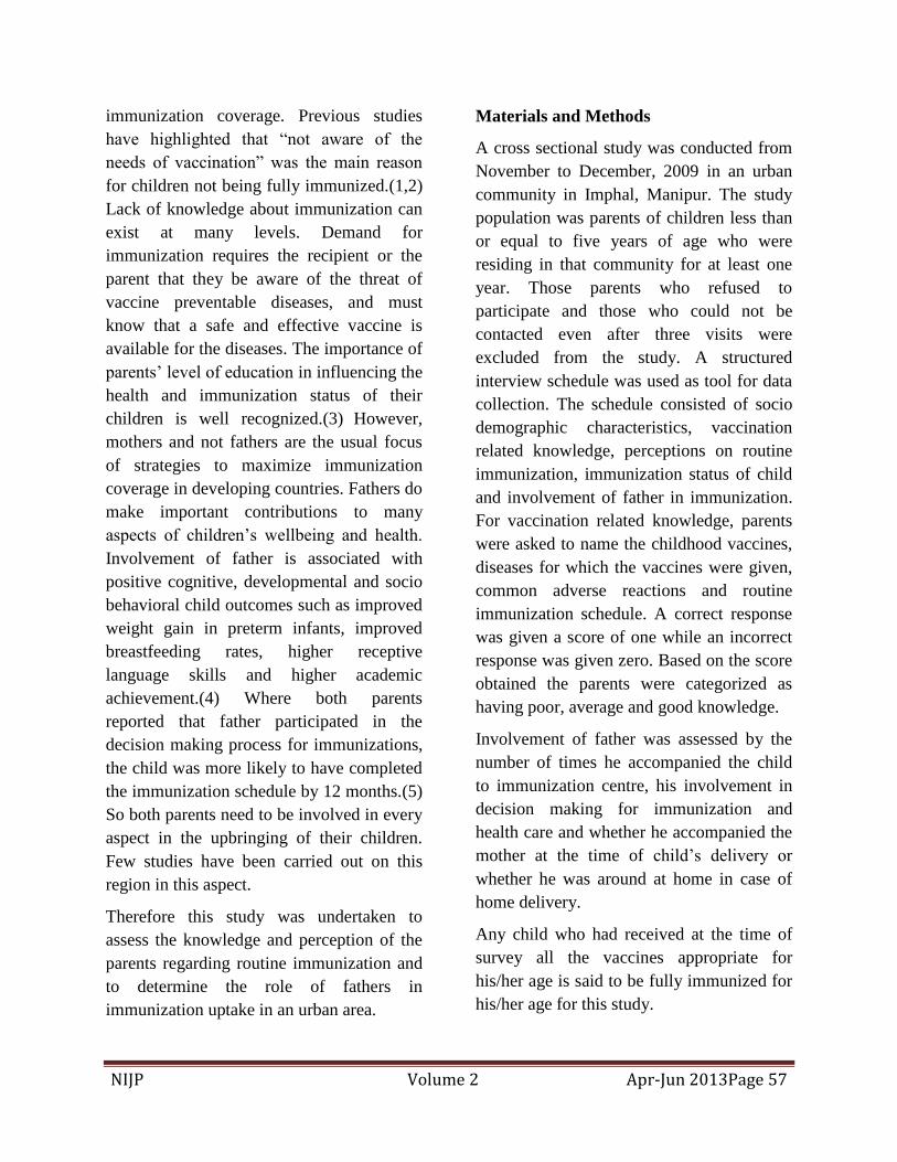
Table 1 showed the perception of parents on
routine immunization and its association
with vaccination status of the child. The
vaccination status of the children was
significantly associated with the parents‘
perception that distance to the immunization
centre was convenient, travelling cost was
affordable, waiting time was not long and
health personnel at the centre were helpful.
*P<0.05
Table 1: Parents’ perception and vaccination status
Perception Mothers‟ response (N=150) Fathers‟ response(N=122)
Vaccination status
P-
value
Vaccination status
P-
value
Routine
immunization is safe
and beneficial
Vaccinated
for age
N (%)
Not
vaccinated
for ageN (%)
Vaccinated
for age
N (%)
Not
vaccinated
for ageN (%)
Yes 84(63.6) 48(36.4)
0.109
70(60.9) 45(39.1)
0.290 Not sure 3(30.0) 7(70.0) 1(100) 0(0)
No 5(62.5) 3(37.5) 2(33.3) 4(66.7)
Distance to immunization centre is convenient
Yes 48(70.6) 20(29.4)
0.035*
41(71.9) 16(28.1)
0.013* Not sure 38(57.6) 28(42.4) 30(52.6) 27(47.4)
No 6(37.5) 10(62.5) 2(25.0) 6(75.0)
Travelling cost to immunization centre is affordable
Yes 45(68.2) 21(31.8)
0.043*
43(69.4) 19(30.6)
0.002* Not sure 42(60.9) 27(39.1) 29(58.0) 21(42.0)
No 5(33.3) 10(66.7) 1(10.0) 9(90.0)
Waiting time at immunization centre is not long
Yes 49(76.6) 15(23.4)
0.002*
36(75.0) 12(25.0)
0.018* Not sure 35(53.0) 31(47.0) 32(51.6) 30(48.4)
No 8(40.0) 12(60.0) 5(41.7) 7(58.3)
Health personnel at immunization centre are helpful
Yes 53(80.3) 13(19.7)
0.000*
43(78.2) 12(21.8)
0.001* Not sure 37(49.1) 40(51.9) 29(46.0) 34(54.0)
No 2(28.6) 5(71.4) 1(25.0) 3(75.0)
They have sufficient information regarding immunization
Yes 45(73.8) 16(26.2)
0.024*
28(68.3) 13(31.7)
0.210 Not sure 33(55.9) 26(44.1) 35(59.3) 24(40.7)
No 14(46.7) 16(53.3) 10(45.5) 12(54.5)
Their child is susceptible to vaccine preventable diseases
Yes 84(63.6) 48(36.4)
0.109
84(63.6) 48(36.4)
0.870 Not sure 3(30.0) 7(70.0) 3(30.0) 7(70.0)
No 5(62.5) 3(37.5) 5(62.5) 3(37.5)

NIJP Volume 2 Apr-Jun 2013Page 60
However, though vaccination status was
significantly associated with the mothers‘
perception that they have sufficient
knowledge regarding immunization, it was
not so with fathers‘ perception. Parents‘
perception that routine immunization was
safe and beneficial and that their children
were susceptible to vaccine preventable
diseases had no significant association with
vaccination status.
Table 2 showed the involvement of fathers
and its association with vaccination status. It
was observed from both mothers‘ and
father‘s response that when the father took
the decision for immunization, the child was
more likely to be fully vaccinated for age.
The findings were however not found to be
statistically significant.
Regarding decision taking in healthcare,
when both father and mother were involved,
children were more likely to be fully
vaccinated for age. However, the finding
was statistically significant only for
mother‘s response. Children were also
significantly more likely to be fully
vaccinated for age when the father
accompanied the wife at the times of child‘s
delivery.
*P<0.05, # not included in analysis
Table 2: Fathers’ involvement and vaccination status
Perception Mothers‟ response (N=150) Fathers‟ response(N=122)
Vaccination status
P-value
Vaccination status
P-
value
Fathers‟
involvement in
child care
Vaccinated
for age
N (%)
Not
vaccinated
for age
N (%)
Vaccinated
for age
N (%)
Not
vaccinated
for age
N (%)
Decision taking for immunization
Father decides 15(68.2) 7(31.8)
0.902
13(68.4) 6(31.6)
0.705 Mother decides 24(61.5) 15(38.5) 16(53.3) 14(46.7)
Both decides 49(59.8) 33(40.2) 41(61.2) 26(38.8)
Others decides 4(57.1) 3(42.9) 3(50.0) 3(50.0)
Decision taking for health care
Father decides 48(70.6) 20(29.4)
0.045*
0(0) 3(100)
0.070 Mother decides 38(57.6) 28(42.4) 14(50.0) 14(50.0)
Both decides 6(37.5) 10(62.5) 58(64.4) 32(35.6)
Others decides 2(100) 0 1(100) 0(0)
Accompanying wife to hospital for delivery of the child
Yes 77(65.3) 41(34.7)
0.03*
60(63.2) 35(36.8)
0.37 No 2(22.2) 7(77.8) 1(20.0) 4(80.0)
Not
applicable(home
delivery)#
13(56.5) 10(43.5) 12(54.5) 10(4.5)
Father accompanying child to vaccination centre
Yes, all the time 22(64.7) 12(35.3)
0.013*
18(72.0) 7(28.0)
0.008
*
Most of the time 25(78.1) 7(21.9) 15(62.5) 9(37.5)
Sometimes 37(60.7) 24(36.3) 37(63.8) 21(36.2)
Not at all 8(34.8) 15(65.2) 3(20.0) 12(80.0)
NIJP Volume 2 Apr-Jun 2013Page 61
However presence of father at home
deliveries had no significant association
with vaccination status. It was also observed
that when fathers accompanied the children
for immunization, the children were more
likely to be fully vaccinated for age and it
was found to be statistically significant.
Discussion
Participation of the fathers in decision
making regarding immunization of their
children was significantly and positively
associated with timely completion of
immunization. Similar findings were also
observed in a study by Brugha and co-
workers.(1)The responses given by the
fathers that 68.4% of them took the decision
agreed with the mothers‘ response that
68.2% of the fathers took the decision for
immunization. Children were also more
likely to be fully vaccinated for age when
the father accompanied them for
vaccination. Father taking active role at the
time of delivery of the child was also
significantly and positively associated with
vaccination status. It can be expected that
such persons may prove better care taker of
their wives and children. Children of parents
with higher educational status and birth
order of up to three were more vaccinated
for age and this was in agreement with
another study conducted by Odiit and co-
investigators.(6,7) As the parents‘
knowledge about vaccination increased the
proportion of fully vaccinated for age
children increased and the findings were
found to be statistically significant. This
finding was supported by a study conducted
by Teklay and co-investigators that mothers
of fully vaccinated for age had significantly
higher maternal immunization awareness
score than mother of children not fully
vaccinated for age.(8) When the number of
children are more and had seen their
children growing up without serious health
problems parents might become negligent of
immunization.
Positive perception of parents regarding the
benefits and barriers of routine
immunization was significantly associated
with vaccination status. This finding was
also seen in a study conducted by
Vishwanathan and Rohde where mothers of
fully immunized children were twice as
likely to believe in the possibility of disease
prevention, than the mothers of non
immunized children, and had a slight
tendency to fear disability than death for
their child. Mothers of fully immunized
children were also better informed about the
benefits of vaccination.(3) Another study
conducted by Cutts and co-workers reported
that completion of vaccination was
significantly associated with maternal
education, mother considering vaccination
to be affordable, positive experience with
vaccination services like short waiting time,
not being turned away from vaccination and
not having a child with post-vaccination
abscess.(9)
Conclusion
This study provides evidence of a positive
relationship that could be of considerable
help in planning program to increase
immunization uptake. Good knowledge and
positive perception regarding immunization
and involvement of fathers play a significant
role in immunization uptake in the study
area. Campaigns to educate and involve the
fathers could be conducted through the

NIJP Volume 2 Apr-Jun 2013Page 62
media. Programs which educate and at the
same time involve them in decision making
process about their children could make a
significant change in the immunization
coverage.
References
1. Coverage evaluation survey for PPI,
Routine Immunization and Maternal Care
in some districts of West Bengal and
Assam by Indian Public Health
Association. Available from:
http://www.iphaonline.org/pages/downlo
ad/16c.%20report%20ces%20survey.pdf
2. Jain SK, Chawla U, Gupta N, Gupta RS,
Venkatesh S, Lal S. Child survival and
safe motherhood program in Rajasthan.
Indian J Pediatr 2006; 73: 43-47.
3. Vishwanathan H, Rohde JE.
Immunization-The Effect of Maternal
Knowledge and Attitudes on
Immunization Coverage. Indian Journal
of Community Medicine 1990; XV(4):
207-212.
4. Garfield CF, Isacco A. Fathers and well
child visits. Pediatrics Apr 2006; 117(4):
637-645.
5. Brugha RF, Kevany JP, Swan AV. An
investigation of the role of fathers in
immunization uptake. Int J Epidemiol
Aug 1996; 25(4): 840-845.
6. Odiit A, Amuge B. Comparison of
vaccination status of children born in
health units and those born at home. East
African Medical Journal Jan 2003; 80(1):
3-6.
7. Pragti C, Parvathy N, Anita G,
Meenakshi S, Kannan AT. Immunization
in Urbanized villages of Delhi. Indian J
Pediatr 2007; 74(2): 131-134.
8. Teklay K, Michael T. Factor influencing
child immunization coverage in a rural
District of Ethiopia, 2000. Ethiop. J.
Health Dev 2003; 17(2): 105-110.
9. Cutts FT, Rodrigues LC, Colomeso S,
Bennett S. Evaluation of Factors
Influencing Vaccine Uptake in
Mozambique. International Journal of
Epidemiology 1989; 18(2): 427-433.

NIJP Volume 2 Apr-Jun 2013Page 63
Research Study:
Prevalence and risk factors of soil -transmitted helminth
infections among the under-five children in an urban slum
of Manipur
Dr Romola Pukhrambam, Dr RK Ranjan, Dr Susmita Chaudhuri, Dr Salona Mukhia, Dr Th.
Achouba, *Dr A Golmei Departments of Community Medicine and
* Departments of Microbiology, Regional Institute of Medical Sciences,
Imphal, Manipur, India
e-mail: [email protected]
Key Words: soil transmitted helminthes,
under-five children
Abstract
Objectives: To determine the prevalence of
soil- transmitted helminth infections and to
assess the association between helminthes
infection with some important variables.
Materials and Methods: A cross-sectional
study was conducted among the under-five
children of an urban slum (Hatta, Golapatti)
of Manipur during May-July 2012. Data was
collected using purposive sampling with an
interview schedule containing questions on
various socio-demographic variables,
awareness of worm infestation and practices
like hand washing, type of latrine use etc.
Stool samples for qualitative STH analysis
(saline and iodine mount method) were
collected. Data was described using
descriptive statistics and analyzed by Chi
square test. Results: Response rate was
96.8%. Prevalence of STH infection
(Ascariasis) was 21.5%. Majority of the
participants were Muslims (90.7%) and
living in non-pucca house (83%). Awareness
about worm infestation was 45.7%. Tap
water was the main household source of
water (83.4%) but majority drank without
boiling or filtering (54.3%). Disposal of
kitchen garbage in open area was practiced
widely (40.08%) and 29.6% of the children
played in that area. One third (30.4%) of the
children defecated indiscriminately. More
than one third (36.8%) of the respondents
said that food stuffs were accessible to flies
and rodents. Over-crowding (51%),
dampness (38.1%) and water logging
(42.9%) were common. STH Infection was
significantly associated with age (p=0.04),
education of father (p=0.007), type of family
(p=0.04), history of passing or vomiting
worms (p=0.000), washing hands after
playing with pets (p=0.04).
Introduction
Soil-transmitted helminthes, commonly
known as intestinal worms, are the most
common infections worldwide affecting the
most deprived communities.
The causal agent of soil-transmitted
helminthiasis is any of the following worms:
Ascaris lumbricoides, Trichuris trichiura
and the hookworms. Infection is caused by
ingestion of eggs from contaminated soil (A.
lumbricoides and T. trichiura) or by active
penetration of the skin by larvae in the soil
(hookworms). (1)
NIJP Volume 2 Apr-Jun 2013Page 64
Soil-transmitted helminthes produce a wide
range of symptoms including intestinal
manifestations (diarrhea, abdominal pain),
general malaise and weakness that may
affect working and learning capacities and
impair physical growth. Hookworms cause
chronic intestinal blood loss that result in
anemia.(1)
Recent estimates suggest that A.
lumbricoides infects over 1 billion people, T.
trichiura 795million, and hookworms
(Ancylostoma duodenale and Necator
americanus) 740 million.(1) The greatest
numbers of STH infections occur in sub-
Saharan Africa, East Asia, China, India and
South America.(2)
The United Nations estimates that 182
million preschool children-33% of those
living in developing countries- are stunted
when their height-for-age is compared with
the norms for well-nourished children living
in good environments. Underweight and
stunted children are known to be at greater
risk of dying during childhood and may not
achieve their full potential in education and
physical performance. It is now accepted
that parasitic disease is a major contributor
to the etiology of the malnutrition-infection
complex. Much of the growth faltering in
children occurs between the ages of 6
months and 2 years—in other words, at the
time when several helminthic infections
begin to be established and the immune
system is challenged to respond. The more
we learn about child-helminthes
interactions, the greater will be the success
of efforts to reduce the burden of disease
due to helminthes.(3)
This study was conducted among under-five
children in an urban area of Imphal East
district of Manipur to determine the
prevalence of soil-transmitted helminthes
among under-five children and to assess the
association between helminthes infection
among these children with various socio-
demographic variables.
Materials and Methods
This Cross sectional study was conducted
during May to July 2012 in an urban
community of Imphal East district of
Manipur. In this study, this urban area was
purposively selected and sample size of 255
under five children was calculated using
prevalence rate 91% (allowable error 4%
and non-response rate 25%).(4) Data was
collected using a pretested structured
interview schedule which consisted of socio-
demographic variables from the parents or
guardian of under- five children after taking
their informed consent. Those who refused
to participate and who could not be
contacted even after 3 visits were excluded
from the study. Stool samples for qualitative
STH analysis (saline and iodine mount
method) were collected and all the positive
cases were given treatment for helminth
infection.
Statistical analysis: Data so collected was
checked for consistency and completeness
and fitted in data base software. Descriptive
statistics were used to describe the findings.
Analysis was done using Chi-square test and
p-value of <0.05 was taken as significant.
Ethical issue: The study proposal was
approved by the Institutional Ethics
committee, RIMS, Imphal. Informed
consent from the parents/guardian and

NIJP Volume 2 Apr-Jun 2013Page 65
assent from the children was taken.
Confidentiality of the respondents was
maintained.
Results
Response rate was 96.8%. Number of
children by age was almost equally
distributed in the first four years except age
between 4- 5 years which constituted only 7
(2.8%). Males (50.6%) and females (49.4%)
were also almost equally distributed and
majority of the children were of the 1st birth
order (35.6%) and 2nd
birth order (31.8%).
Majority of the respondents were Muslims
(90.7%) and majority were living in non-
pucca houses (83%) and belonged to nuclear
family (73.3%). Majority of the mothers
(44.9%) and fathers (59.9%) was below
class X standard. Majority of the mothers
were housewives (92.7%) and majority of
fathers were self employed (81.0%). Two
fifths (45.7%) of the caretakers (mostly
mothers) were not aware of worm
infestation in their children. Tap water was
the main household source of water (83.4%)
but more than half of them drank without
boiling or filtering (54.3%). More than two
third (70.4%) of the respondents used
sanitary latrines but disposal of kitchen
garbage in open area was widely practiced
(40.08%) and 29.6% of the children played
in that area. One third (30.4%) of the
children defecated indiscriminately and
more than one third (36.8%) of the
respondents said that food stuffs were
accessible to flies and rodents. Over-
crowding (51%), dampness (38.1%) and
water logging (42.9%) were common.
(Table 1)
Characteristics Number Percentage
Age (Years)
1 52 21.1
2 61 24.7
3 62 25.1
4 65 26.3
5 7 2.8
Sex
Male 125 50.6
Female 122 49.4
Religion
Muslim 2
2
4
90.7
Others 2
3
9.3
Type of family
Nuclear 1
8
1
73.3
Joint 6
6
26.7
Type of house
Pucca 4
2
17.0
Not pucca 2
0
5
83.0
Educational status of the mother
Illiterate 9
9
40.1
< Class 10 1
1
1
44.9
Class 10-12 2
9
11.7
>Class 12 8 3.2
Educational status of the father
Illiterate 3
7
15.0
< Class 10 1
4
8
59.9
Class 10-12 3
7
15.0
>Class 12 2
5
10.1
Table 1a: Socio-demographic
characteristics of the respondents
NIJP Volume 2 Apr-Jun 2013Page 66
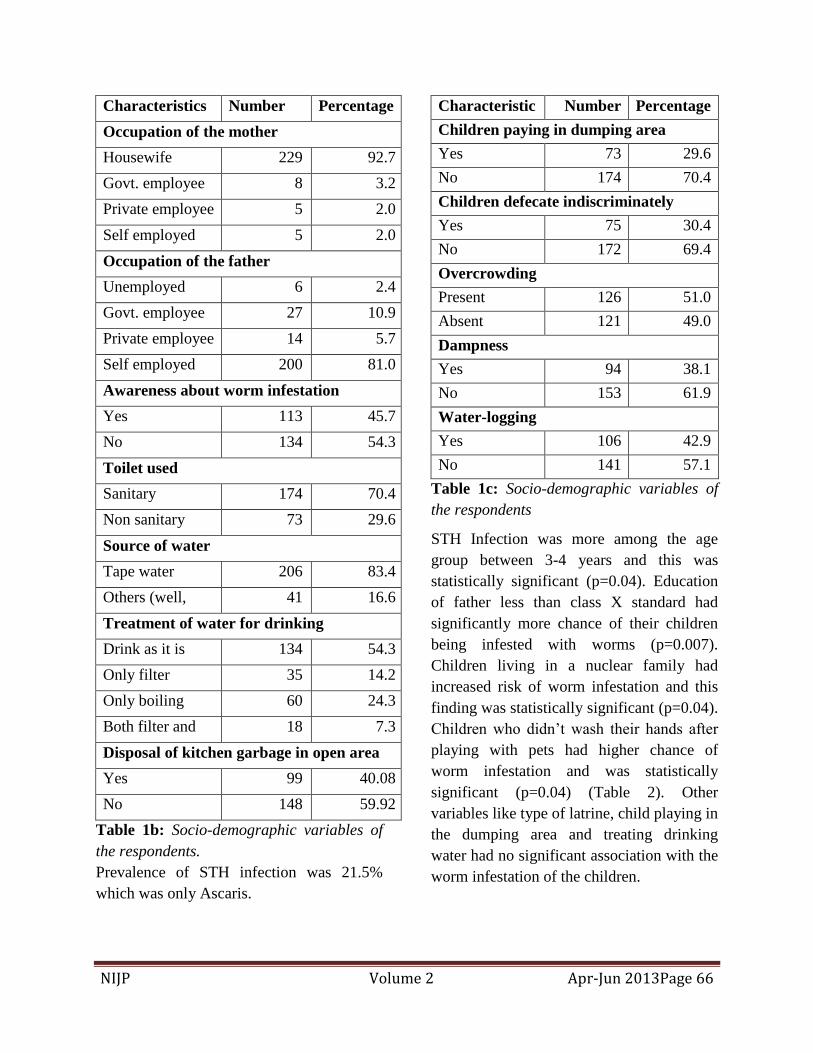
Characteristics Number Percentage
Occupation of the mother
Housewife 229 92.7
Govt. employee 8 3.2
Private employee 5 2.0
Self employed 5 2.0
Occupation of the father
Unemployed 6 2.4
Govt. employee 27 10.9
Private employee 14 5.7
Self employed 200 81.0
Awareness about worm infestation
Yes 113 45.7
No 134 54.3
Toilet used
Sanitary 174 70.4
Non sanitary 73 29.6
Source of water
Tape water 206 83.4
Others (well,
pond etc.)
41 16.6
Treatment of water for drinking
Drink as it is 134 54.3
Only filter 35 14.2
Only boiling 60 24.3
Both filter and
boiling
18 7.3
Disposal of kitchen garbage in open area
Yes 99 40.08
No 148 59.92
Table 1b: Socio-demographic variables of
the respondents.
Prevalence of STH infection was 21.5%
which was only Ascaris.
Characteristic
s
Number Percentage
Children paying in dumping area
Yes 73 29.6
No 174 70.4
Children defecate indiscriminately
Yes 75 30.4
No 172 69.4
Overcrowding
Present 126 51.0
Absent 121 49.0
Dampness
Yes 94 38.1
No 153 61.9
Water-logging
Yes 106 42.9
No 141 57.1
Table 1c: Socio-demographic variables of
the respondents
STH Infection was more among the age
group between 3-4 years and this was
statistically significant (p=0.04). Education
of father less than class X standard had
significantly more chance of their children
being infested with worms (p=0.007).
Children living in a nuclear family had
increased risk of worm infestation and this
finding was statistically significant (p=0.04).
Children who didn‘t wash their hands after
playing with pets had higher chance of
worm infestation and was statistically
significant (p=0.04) (Table 2). Other
variables like type of latrine, child playing in
the dumping area and treating drinking
water had no significant association with the
worm infestation of the children.
NIJP Volume 2 Apr-Jun 2013Page 67
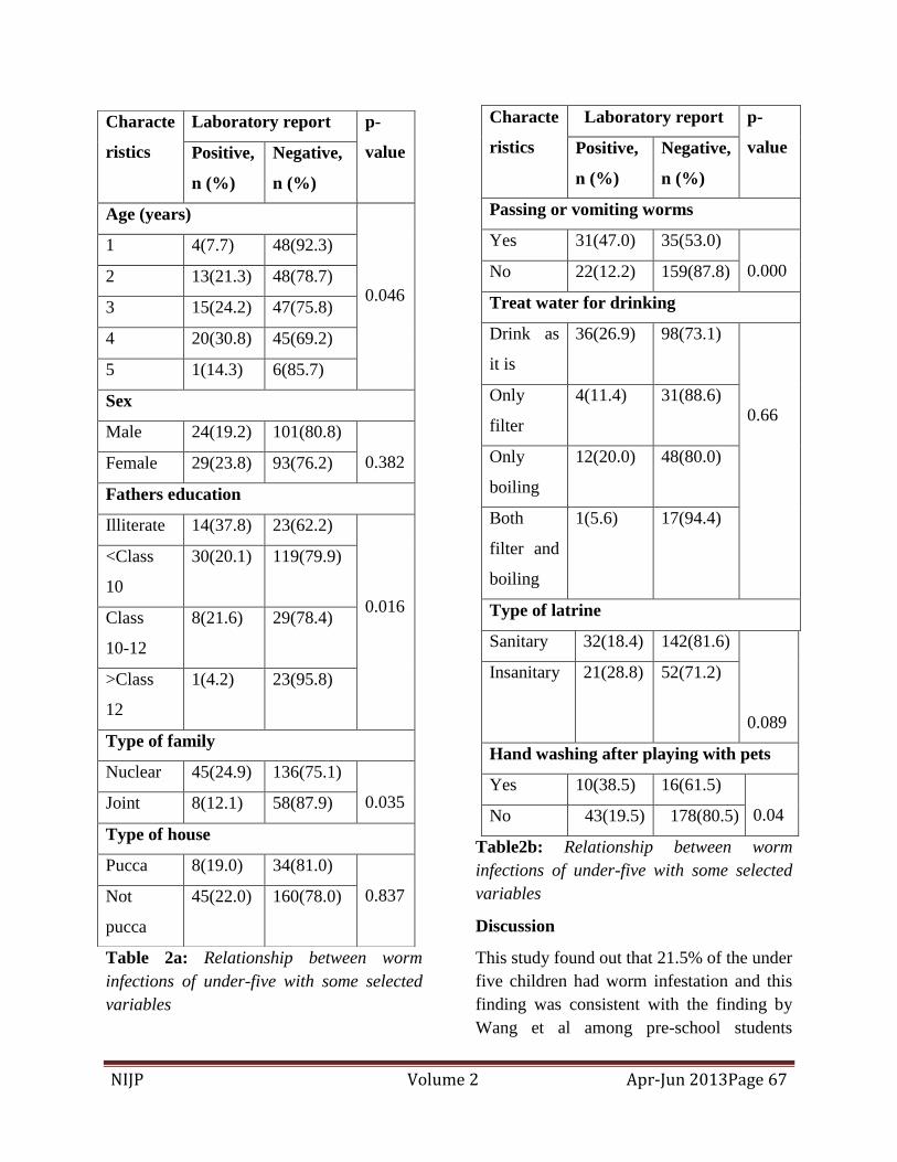
Table 2a: Relationship between worm
infections of under-five with some selected
variables
Table2b: Relationship between worm
infections of under-five with some selected
variables
Discussion
This study found out that 21.5% of the under
five children had worm infestation and this
finding was consistent with the finding by
Wang et al among pre-school students
Characte
ristics
Laboratory report p-
value Positive,
n (%)
Negative,
n (%)
Age (years)
0.046
1 4(7.7) 48(92.3)
2 13(21.3) 48(78.7)
3 15(24.2) 47(75.8)
4 20(30.8) 45(69.2)
5 1(14.3) 6(85.7)
Sex
Male 24(19.2) 101(80.8)
0.382 Female 29(23.8) 93(76.2)
Fathers education
Illiterate 14(37.8) 23(62.2)
0.016
<Class
10
30(20.1) 119(79.9)
Class
10-12
8(21.6) 29(78.4)
>Class
12
1(4.2) 23(95.8)
Type of family
Nuclear 45(24.9) 136(75.1)
0.035 Joint 8(12.1) 58(87.9)
Type of house
Pucca 8(19.0) 34(81.0)
0.837 Not
pucca
45(22.0) 160(78.0)
Characte
ristics
Laboratory report p-
value Positive,
n (%)
Negative,
n (%)
Passing or vomiting worms
Yes 31(47.0) 35(53.0)
0.000 No 22(12.2) 159(87.8)
Treat water for drinking
Drink as
it is
36(26.9) 98(73.1)
0.66 Only
filter
4(11.4) 31(88.6)
Only
boiling
12(20.0) 48(80.0)
Both
filter and
boiling
1(5.6) 17(94.4)
Type of latrine
Sanitary 32(18.4) 142(81.6)
0.089
Insanitary 21(28.8) 52(71.2)
Hand washing after playing with pets
Yes 10(38.5) 16(61.5)
0.04 No 43(19.5) 178(80.5)
NIJP Volume 2 Apr-Jun 2013Page 68
(21.2%).(5) In India, the overall prevalence
rates ranges from 13-66% with varying
prevalence rates for individual parasites.
Similar reports have been seen in several
developing countries like Malaysia, Nepal,
Kuwait, and several parts of Kenya, Guyana
and South Africa.(6,7) All the detected
worms were Ascaris and this finding was
concurrent with many studies that majority
of the worms detected was Ascaris.(8,9) No
hookworm was identified in our study which
is consistent with results obtained in studies
conducted in urban localities. There was no
significant difference between males and
female children in this study and was also
found in a study by Anbumani N et al.(10)
Living in a rented house had more chance of
being infested (23.6% of rented house Vs
15.4% of owned house). This finding was
found to be statistically insignificant. This
contradicts the finding by Mehraj et al that
living in rented house as a proxy measure
for socio-economic status increased the
chance of worm infestation.(10) The reason
might be both the rented and owned were
having the same condition in this urban area.
With the increase in age up to 4 years of the
child there was increased chance of being
infested with worms and this was
statistically significant. This finding was
consistent with the finding by Mehraj et
al.(11) The less chance of worm infestation
between above 4 years and below 5 years
might be due to less number of children in
this group. Education of father less than
class X standard had significantly more
chance of their children being infested with
worms. This was expected as less education
led to poor knowledge regarding worms and
its etiology and thus not preventing their
children from worm infestation. Children
living in a nuclear family had increased risk
of worm infestation and this finding was
statistically significant (p=0.04). This might
be because fewer members would be there
to look after their children avoiding
contamination and thus worm infestation. .
Children who didn‘t wash their hands after
playing with pets had higher chance of
worm infestation and was statistically
significant (p=0.04). Other variables like
type of latrine, child playing in the dumping
area and drinking treated water had no
significant association with the worm
infestation of the children.
Since this study was conducted in an urban
community of Imphal East district it is
representative of that community only and
that could be the limitation of this study.
Still our study could give some important
insight regarding prevalence of worm
infestation and the risk factors associated
with worm infestation among under-five
children, this is the first survey of its kind
among under-five conducted in Manipur and
particularly in RIMS.
Conclusion
Soil transmitted helminthes infection among
under-five children was more than one fifth
(21.5%). Risk factors associated with
infection suggest that de-worming strategies
must be employed and also address
provision of safe water and health education
about hand-washing and thereby indirectly
impacting the achievement of national goal
in particular Millennium Development Goal.

NIJP Volume 2 Apr-Jun 2013Page 69
References
1. WHO. Intestinal worms, soil-transmitted
helminthes 2012; [2 screens]. Available
at: URL:
http://www.who.int/intestinal_worms/en/.
Accessed June 20, 2012.
2. Brooker S, Clements AC, Bundy DA.
Global epidemiology, ecology and
control of soil-transmitted helminth
infections. Adv Parasitol 2006;62:221-61.
3. Crompton DWT, Montresor A, Nesheim
MC, Savioli L. Controlling disease due to
helminth infections. [268 pages].
Available at: URL:
http://www.who.int/wormcontrol/docume
nts/en/Controlling%20Helminths.pdf.
Accessed June 20, 2012.
4. Naish S, McCarthy J, Williams GM.
Prevalence, intensity and risk factors for
soil-transmitted helminth infection in a
South Indian fishing village. Acta Trop
2004 Jul;91(2):177-87.
5. Wang X, Zhang L, Luo R, Wang G, Chen
Y, Medina A, et al. Soil-Transmitted
Helminth Infections and Correlated Risk
Factors in Preschool and School-Aged
Children in Rural Southwest China. PLoS
ONE 2012 Sep; 7(9). Available at: URL:
http://www.plosone.org/article/info%3Ad
oi%2F10.
1371%2Fjournal.pone.0045939.
Accessed July 22, 2012.
6. WHO. Prevention and Control of
intestinal parasitic Infections. Report of a
WHO Expert Committee. World Health
Organ Tec Rep Ser 1987;749. Available
at: URL:
http://whqlibdoc.who.int/trs/WHO_TRS_
749. Accessed on July 20, 2012.
7. Gagandeep kang, Marys Mathew D,
Parasanna Rajan, Jasper D. Daniel,
Minnie M. Mathan, V.I. Mathan, J.P.
Prevalence of intestinal parasites in Rural
Southern India. Tropical Medicine and
International Health 1998;3(1):70-5.
8. Saldira SR, Silveria AS, Philippi ST.
Ascaris–Trichuris association and
malnutrition in Brazilian children.
Pediatric and perinatal
Epidemiology 1999;13:89-98.
9. Carla Scolari, Carla Torti. Prevalence and
distribution of soil transmitted helminth
(STH) infection in urban and indigenous
school children in oritigueira, state of
panama, Brazil; implications for control.
Tropical medicine and International
health 2000;5:302-7.
10. Anbumani N, Mallika M. Prevalence
and Distribution of Soil Transmitted
Helminths (STH) among Asymptomatic
School Going Children in South
Chennai, Tamil Nadu, India. Int. J. Med.
Public health 2011;1(2):57-9.
11. Mehraj V, Hatcher J, Akhtar S, Rafique
G, Beg MA. Prevalence and factors
associated with intestinal parasitic
infection among children in an urban
slum of Karachi. PLoS ONE 2008
Nov;3(11). Available at: URL:
http://www.plosone.org/article/info%3A
doi%2F10.1371%2Fjournal.pone.00036
80. Accessed July 22, 2012.

NIJP Volume 2 Apr-Jun 2013Page 70
Research Study:
Study of Prevalence of Various Hemoglobinopathies in
Sindhi Community of Amravati
Dr AT Deshmukh, Dr RR Soni, *Dr AT Harwani, Dr AP Bajaj, Dr AS Thakare
Department of Pathology, Dr PDMMC, Amravati, Consultant Physician, Amravati
E-mail: [email protected]
Key words: hemoglobinopathy,
Hemoglobin D, β thalassemia, Sindhi
community.
Abstract:
Among the inherited disorders of blood,
hemoglobinopathies constitute a major bulk
of non-communicable genetic diseases in
India. The carrier frequency of
hemoglobinopathy varies from 3 to 17% in
different population groups of India. The
frequency of Hb D among various
communities in India varies from 0.5 to 3%
(3% in khatris while 0.5 % among the other
hindus and 2.4% in Sindhi community in
various studies). The frequency of β
thalassemia is very high among Sindhi
communities in various states in India.
Aims: To determine the prevalence of
Hemoglobinopathies in Sindhi community
in Amravati.
Material & Methods: The study is being
carried out among Sindhi population of pre-
marital age group aiding pre marital
counseling. Selected person in the pre-
marital age group of Sindhi community were
subjected to a battery of investigations.
These investigations include i) CBC ii)
Sickling test iii) Hemoglobin electrophoresis
on cellulose acetate at alkaline & acidic pH.
Result: Till date total 240 cases are studied,
among which 19 cases turned out positive
for hemoglobin D. (i.e. about 7.9%) Out of
these 18 were trait and 1was homozygous
for Hb D. the prevalence of β thalassemia
trait was 21.25% (51 out of 240).
Conclusion: This study concludes that
prevalence of hemoglobin D in the Sindhi
community of Amravati is high as compared
to general population and Sindhi community
of other regions, whereas the prevalence of
β thalassemia is comparable to Sindhi
community of other areas.
Introduction:
The inherited disorders of blood include
hemoglobinopathies as one of the major
public health problems in India. The carrier
frequency of hemoglobinopathy varies from
3 to 17% in different population groups of
India (1) . The cumulative gene frequency of
the three most predominant abnormal
hemoglobins, i.e. sickle cell, hemoglobin D
and hemoglobin E has been estimated to be
5.35% in India (2). Thus, there is a
tremendous amount of burden of
hemoglobinopathies in India.
The hemoglobinopathies are characterized
by the production of structurally defective
hemoglobin due to abnormalities in the
formation of the globin moiety of the
molecule. When biological function is
altered owing to a mutation in the
hemoglobin, the condition is known as a
hemoglobinopathy (3). The globin moiety of
NIJP Volume 2 Apr-Jun 2013Page 71
hemoglobin (Hb) molecule is composed of
several different types of polypeptide chains,
varying in number and arrangement of
amino acids during different stages of
human intra-uterine development(4) and are
designated by the Greek letters, alpha (α),
beta (β),gamma (γ), delta (δ), epsilon (ε),
and zeta (δ). Epsilon, zeta and some alpha
chains are synthesized in early embryonic
life, alpha and gamma chains in fetal life,
and alpha, beta and delta chains predominate
in the postnatal life. HbF (α2γ2), forms
about 80–90% of the total hemoglobin at
birth, whereas in the postnatal (postpartum)
life it is replaced by the human adult
hemoglobin HbA (α2β2) comprising about
97% and HbA2 (a2δ2), the latter constitutes
about one-fortieth (1.5–3.5%) of the total
adult hemoglobin (5).
Hemoglobin D is a beta chain variant of
hemoglobin A. The 121st position amino
acid glutamate is replaced by glutamine in
the beta chain. Different variants are
identified from different places represented
by suffix, eg. Hb D Punjab, Hb D Los
Angeles.(6,7)
Materials and methods:
Patients:
This study was performed on 240 Sindhi
youths (129 male and 111 female) with the
aim of providing pre-marital counseling for
inheritable globin chain disorders. This work
had been approved by Institutional Ethics
Committee of our College. Every individual
included in this study underwent a battery of
investigations including: i) CBC ii) sickling
test iii) Hb electrophoresis at acidic and
alkaline pH iv) HPLC as and when required.
Informed consent was taken from all
participants.
Laboratory Methods:
Blood samples from all participants were
subjected to complete cell count by Coulter
machine, Sickling test, electrophoresis at
alkaline pH and at acidic pH when required
and High performance liquid
chromatography if required for the
confirmation of hemoglobin type. Definitive
tests like electrophoresis and HPLC are
indicated in suspected hemoglobinopathies,
even in presence of normal blood cell count
and smear, to detect variant hemoglobins.(8)
Electrophoresis:
All the samples were processed and applied
to fully automated Genio S electrophoresis
machine. This machine utilizes Mylar
stratified cellulose acetate strips both at
acidic and alkaline pH. This fully automated
machine facilitates separation of HbA, F,
S/D/G/O, C/E/A2, H with quantitation at
alkaline pH and separation of Hb S from
D/G and C from E, C-Harlem a acidic pH.
So when a band is obtained at alkaline pH at
the position of S/D/G then sickling test of
the same sample and also electrophoresis at
acidic pH is done for confirmation of type of
hemoglobin
Results:
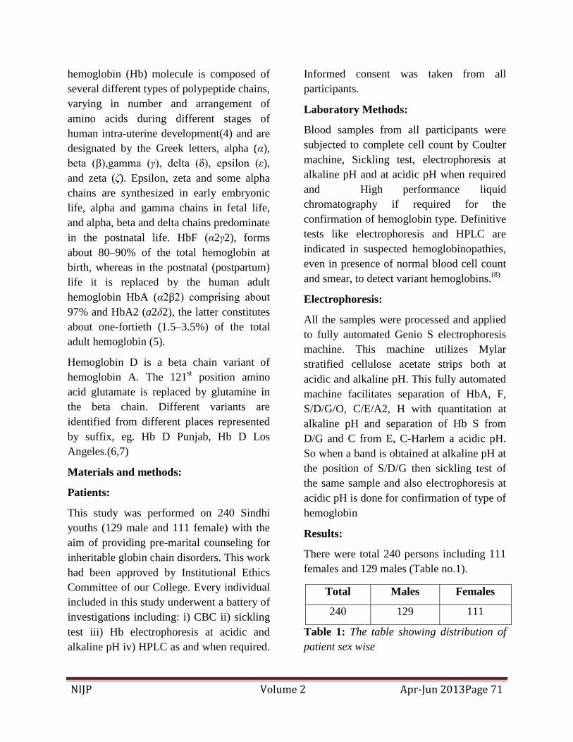
There were total 240 persons including 111
females and 129 males (Table no.1).
Table 1: The table showing distribution of
patient sex wise
Total Males Females
240 129 111
NIJP Volume 2 Apr-Jun 2013Page 72
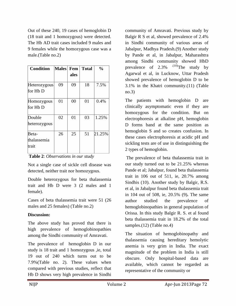
Out of these 240, 19 cases of hemoglobin D
(18 trait and 1 homozygous) were detected.
The Hb AD trait cases included 9 males and
9 females while the homozygous case was a
male.(Table no.2)
Table 2: Observations in our study
Not a single case of sickle cell disease was
detected, neither trait nor homozygous.
Double heterozygous for beta thalassemia
trait and Hb D were 3 (2 males and 1
female).
Cases of beta thalassemia trait were 51 (26
males and 25 females) (Table no.2)
Discussion:
The above study has proved that there is
high prevalence of hemoglobinopathies
among the Sindhi community of Amravati.
The prevalence of hemoglobin D in our
study is 18 trait and 1 homozygous ,ie, total
19 out of 240 which turns out to be
7.9%(Table no. 2). These values when
compared with previous studies, reflect that
Hb D shows very high prevalence in Sindhi
community of Amravati. Previous study by
Balgir R S et al, showed prevalence of 2.4%
in Sindhi community of various areas of
Jabalpur, Madhya Pradesh.(9) Another study
by Pande et al, in Jabalpur, Maharashtra
among Sindhi community showed HbD
prevalence of 2.3% .(10)
The study by
Agarwal et al, in Lucknow, Uttar Pradesh
showed prevalence of hemoglobin D to be
3.1% in the Khatri community.(11) (Table
no.3)
The patients with hemoglobin D are
clinically asymptomatic even if they are
homozygous for the condition. But on
electrophoresis at alkaline pH, hemoglobin
D forms band at the same position as
hemoglobin S and so creates confusion. In
these cases electrophoresis at acidic pH and
sickling tests are of use in distinguishing the
2 types of hemoglobin.
The prevalence of beta thalassemia trait in
our study turned out to be 21.25% whereas
Pande et al; Jabalpur, found beta thalassemia
trait in 106 out of 511, ie, 20.7% among
Sindhis (10). Another study by Balgir, R.S.
et al, in Jabalpur found beta thalassemia trait
in 104 out of 508, ie, 20.5% (9). The same
author studied the prevalence of
hemoglobinopathies in general population of
Orissa. In this study Balgir R. S. et al found
beta thalassemia trait in 18.2% of the total
samples.(12) (Table no.4)
The situation of hemoglobinopathy and
thalassemia causing hereditary hemolytic
anemia is very grim in India. The exact
magnitude of the problem in India is still
obscure. Only hospital-based data are
available, which cannot be regarded as
representative of the community or
Condition Males Fem
ales
Total %
Heterozygous
for Hb D
09 09 18 7.5%
Homozygous
for Hb D
01 00 01 0.4%
Double
heterozygous
02 01 03 1.25%
Beta-
thalassemia
trait
26 25 51 21.25%
NIJP Volume 2 Apr-Jun 2013Page 73
population.(13)
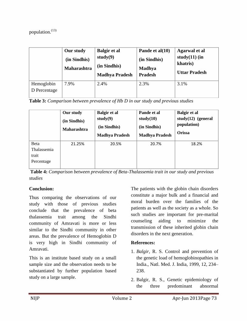
Table 3: Comparison between prevalence of Hb D in our study and previous studies
Table 4: Comparison between prevalence of Beta-Thalassemia trait in our study and previous
studies
Conclusion:
Thus comparing the observations of our
study with those of previous studies
conclude that the prevalence of beta
thalassemia trait among the Sindhi
community of Amravati is more or less
similar to the Sindhi community in other
areas. But the prevalence of Hemoglobin D
is very high in Sindhi community of
Amravati.
This is an institute based study on a small
sample size and the observation needs to be
substantiated by further population based
study on a large sample.
The patients with the globin chain disorders
constitute a major bulk and a financial and
moral burden over the families of the
patients as well as the society as a whole. So
such studies are important for pre-marital
counseling aiding to minimize the
transmission of these inherited globin chain
disorders in the next generation.
References:
1. Balgir, R. S. Control and prevention of
the genetic load of hemoglobinopathies in
India., Natl. Med. J. India, 1999, 12, 234–
238.
2. Balgir, R. S., Genetic epidemiology of
the three predominant abnormal
Our study
(in Sindhis)
Maharashtra
Balgir et al
study(9)
(in Sindhis)
Madhya Pradesh
Pande et al(10)
(in Sindhis)
Madhya
Pradesh
Agarwal et al
study(11) (in
khatris)
Uttar Pradesh
Hemoglobin
D Percentage
7.9% 2.4% 2.3% 3.1%
Our study
(in Sindhis)
Maharashtra
Balgir et al
study(9)
(in Sindhis)
Madhya Pradesh
Pande et al
study(10)
(in Sindhis)
Madhya Pradesh
Balgir et al
study(12) (general
population)
Orissa
Beta
Thalassemia
trait
Percentage
21.25% 20.5% 20.7% 18.2%
NIJP Volume 2 Apr-Jun 2013Page 74
hemoglobins in India. J. Assoc. Phys.
India, 1996, 44, 25–28.
3. Rodwell, V. W. Proteins: Myoglobin and
Hemoglobin. In eds Murray, R.
K.,Granner, D. K., Mayes, P. A. and
Rodwell, V. W.Harper‘s Biochemistry,
Appleto and Lange, Stamford, 2003,26th
edn, pp. 40–48.
4. Serjeant, G. R., Sickle Cell Disease,
Oxford University Press, Oxford, 1985.
5. Firkin, F., Chesterman, C., Penington, D.
and Rush, B., De Gruchy‘s Clinical
Hematology in Medical Practice,
Blackwell Scientific Publications,
Oxford, 1989, 5th edn, pp. 137–172.
6. Baglioni C. Abnormal human
hemoglobins. VIII. Chemical studies on
hemoglobin D. Biochim Biophys Acta
1962; 59:437-49.
7. S Worthington, H Lehmann The first
observation of Hb D Punjab beta zero
thalassemia in an English family with 22
cases of unsuspected beta zero
thalassemia minor among its members, J
Med Genet 1985; 22:377-381
8. B J Bain. The laboratory diagnosis of
hemoglobinopathies-Guideline. British
Journal of Hematology, 101: 783–792.
doi: 10.1046/j.1365-2141.1998.00809.x
9. Balgir RS. Inherited hemolytic disorders
with high occurrence of ß-thalassemia in
Sindhi community of Jabalpur town In
Madhya Pradesh, India. Online J Health
Allied Scs. 2009;8(4):5
10. P.L. Pande, M.P.S.S. Singh and N.K.
Choudhary, Occurrence of Thalassemia
among the Sindhi Community of
Jabalpur, Madhya Pradesh (Central
India), J. Hum. Ecol.,17(2),2005,157-
158
11. Agarwal, S., Gupta, U. R., Kohli, N.
Verma, C. and Agarwal,S. S.,
Prevalence of Hemoglobin D in Uttar
Pradesh, Indian J. Med. Res., 1989, 90,
39–43.
12. Balgir RS. Spectrum of
hemoglobinopathies in the state of
Orissa, India: A ten years cohort study. J
Assoc Phys India 2005; 53:1021-1026.
13. Balgir, R.S. The burden of
hemoglobinopathies in India and the
challenges ahead Current Science, Vol.
79, No. 11, 10 December 2000,1536-
1547
NIJP Volume 2 Apr-Jun 2013Page 75
Case Reports:
Anhidrotic Ectodermal Dysplasia
Dr R.K. Rupabati Devi*, Dr. Amita M**, Dr Ksh. Chourjit Singh*, Dr.L.Ranbir Singh**,
Dr Sunilbala K**
*Department of Pediatrics, Jawaharlal Nehru Institute of Medical Sciences (JNIMS), and **Department of Pediatrics,
Regional Institute of medical Sciences(RIMS), Imphal, Manipur
E-mail:[email protected]
Keywords: Ectodermal dysplasia (ED),
Hypotrichosis, Hypodontia
Abstract:-
Anhidrotic ectodermal dysplasia (AED) is a
rare disorder characterized by primary
defects in the development of two or more
tissues derived from embryonic ectoderm.
The tissues which are affected include teeth,
skin and appendageal structures like hair,
nails and eccrine glands. We report a male
child who had typical features of this
disorder.
Introduction
The ectodermal dysplasias are a group of
disorder characterized by constellation of
defects involving the teeth, skin and
appendageal structures including hair, nails,
eccrine and sebaceous glands (1).More than
192 distinct ED have been described (2).The
incidence in male is estimated 1 in 100,000
births and carriers- incidence is around 17 in
100,000 women (3). Anhidrotic ectodermal
dysplasia, most common of ED, also known
as Christ -Siemens-Touraine syndrome is a
rare X--linked recessive disorder. Rarely
autosomal recessive and autosomal
dominant inheritance have also been seen.
The predicted gene product responsible for
this disorder is 135 amino acid proteins with
no significant homology to previously
known proteins (4). The phenotype is seen
only in affected males, and the major
clinical problem is regulation of body
temperature in the absence of sweating. The
less specific symptoms include
hyperpigmentation around the eyes, mild
facial dysmorphic features, prominent low
set ear, eczema and increased susceptibility
to respiratory infections. The diagnosis of
EDs is mainly clinical. Some studies showed
that horizontally section punch biopsy of
scalp or palms that lack eccrine structures
are diagnostic of hypohidrotic ED (5). The
management of ED is to be directed to
whichever ectodermal structures are
abnormal. The prognosis for most patients
with EDA is good. They have a normal life
expectancy and normal intelligence. We
report here a case of Anhidrotic Ectodermal
Dysplasia.
Case Report:-
A 2 year old male child born of non-
consanguinous marriage presented with
prolonged fever, intolerance to heat and
absence of sweating since birth. There was
history of ear discharge and nasal block off
and on. His motor and mental developments
were normal. Physical examination revealed
sparse scalp hair and eye lashes. There was
absence of eyebrows, thick everted lip,
prominent low set ear and pegged teeth
(Fig.1). Skin was rough and dry. Other
systemic examinations were found to be
within normal limits. Skin biopsy from
palmer ridge showed absence of
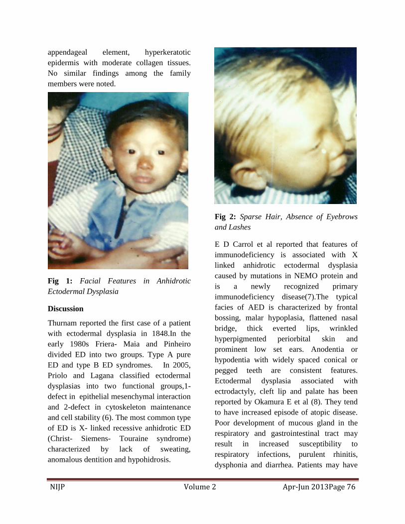
NIJP Volume 2 Apr-Jun 2013Page 76
appendageal element, hyperkeratotic
epidermis with moderate collagen tissues.
No similar findings among the family
members were noted.
Fig 1: Facial Features in Anhidrotic
Ectodermal Dysplasia
Discussion
Thurnam reported the first case of a patient
with ectodermal dysplasia in 1848.In the
early 1980s Friera- Maia and Pinheiro
divided ED into two groups. Type A pure
ED and type B ED syndromes. In 2005,
Priolo and Lagana classified ectodermal
dysplasias into two functional groups,1-
defect in epithelial mesenchymal interaction
and 2-defect in cytoskeleton maintenance
and cell stability (6). The most common type
of ED is X- linked recessive anhidrotic ED
(Christ- Siemens- Touraine syndrome)
characterized by lack of sweating,
anomalous dentition and hypohidrosis.
Fig 2: Sparse Hair, Absence of Eyebrows
and Lashes
E D Carrol et al reported that features of
immunodeficiency is associated with X
linked anhidrotic ectodermal dysplasia
caused by mutations in NEMO protein and
is a newly recognized primary
immunodeficiency disease(7).The typical
facies of AED is characterized by frontal
bossing, malar hypoplasia, flattened nasal
bridge, thick everted lips, wrinkled
hyperpigmented periorbital skin and
prominent low set ears. Anodentia or
hypodentia with widely spaced conical or
pegged teeth are consistent features.
Ectodermal dysplasia associated with
ectrodactyly, cleft lip and palate has been
reported by Okamura E et al (8). They tend
to have increased episode of atopic disease.
Poor development of mucous gland in the
respiratory and gastrointestinal tract may
result in increased susceptibility to
respiratory infections, purulent rhinitis,
dysphonia and diarrhea. Patients may have
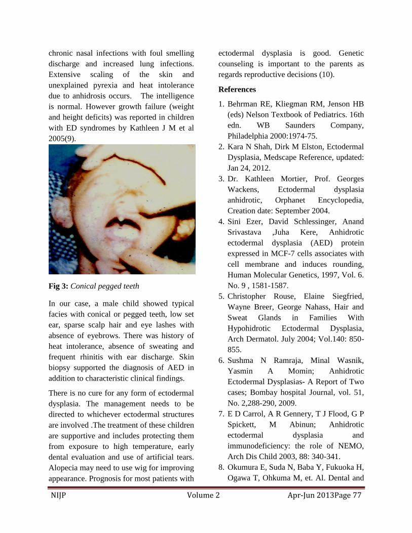
NIJP Volume 2 Apr-Jun 2013Page 77
chronic nasal infections with foul smelling
discharge and increased lung infections.
Extensive scaling of the skin and
unexplained pyrexia and heat intolerance
due to anhidrosis occurs. The intelligence
is normal. However growth failure (weight
and height deficits) was reported in children
with ED syndromes by Kathleen J M et al
2005(9).
Fig 3: Conical pegged teeth
In our case, a male child showed typical
facies with conical or pegged teeth, low set
ear, sparse scalp hair and eye lashes with
absence of eyebrows. There was history of
heat intolerance, absence of sweating and
frequent rhinitis with ear discharge. Skin
biopsy supported the diagnosis of AED in
addition to characteristic clinical findings.
There is no cure for any form of ectodermal
dysplasia. The management needs to be
directed to whichever ectodermal structures
are involved .The treatment of these children
are supportive and includes protecting them
from exposure to high temperature, early
dental evaluation and use of artificial tears.
Alopecia may need to use wig for improving
appearance. Prognosis for most patients with
ectodermal dysplasia is good. Genetic
counseling is important to the parents as
regards reproductive decisions (10).
References
1. Behrman RE, Kliegman RM, Jenson HB
(eds) Nelson Textbook of Pediatrics. 16th
edn. WB Saunders Company,
Philadelphia 2000:1974-75.
2. Kara N Shah, Dirk M Elston, Ectodermal
Dysplasia, Medscape Reference, updated:
Jan 24, 2012.
3. Dr. Kathleen Mortier, Prof. Georges
Wackens, Ectodermal dysplasia
anhidrotic, Orphanet Encyclopedia,
Creation date: September 2004.
4. Sini Ezer, David Schlessinger, Anand
Srivastava ,Juha Kere, Anhidrotic
ectodermal dysplasia (AED) protein
expressed in MCF-7 cells associates with
cell membrane and induces rounding,
Human Molecular Genetics, 1997, Vol. 6.
No. 9 , 1581-1587.
5. Christopher Rouse, Elaine Siegfried,
Wayne Breer, George Nahass, Hair and
Sweat Glands in Families With
Hypohidrotic Ectodermal Dysplasia,
Arch Dermatol. July 2004; Vol.140: 850-
855.
6. Sushma N Ramraja, Minal Wasnik,
Yasmin A Momin; Anhidrotic
Ectodermal Dysplasias- A Report of Two
cases; Bombay hospital Journal, vol. 51,
No. 2,288-290, 2009.
7. E D Carrol, A R Gennery, T J Flood, G P
Spickett, M Abinun; Anhidrotic
ectodermal dysplasia and
immunodeficiency: the role of NEMO,
Arch Dis Child 2003, 88: 340-341.
8. Okumura E, Suda N, Baba Y, Fukuoka H,
Ogawa T, Ohkuma M, et. Al. Dental and
NIJP Volume 2 Apr-Jun 2013Page 78
maxillofacial characteristics of six
Japanese individuals with ectrodactyly-
ectodermal dysplasia- cleting syndrome.
Cleft Palate Craniofac J, 2013 Mar; 50
(2): 192-200.
9. Kathleen J. Motil, Timothy J. Fete, J.
Kennard Fraley, Rebecca J. Schultz,
Thomas M. Foy, Ulrike Ochs et al.
Pediatrics 2005; 116; e229 DOI: 10.
1542/ peds 2004- 2830.
10. Rajwant K Vaswani, Sandipan
Sabde; Approach to a malformed infant,
Dysplasias: Clinical Genetics,2005,Keya
Lehiri ( chief editor),page: 42-49.
NIJP Volume 2 Apr-Jun 2013Page 79
Case Report:
Iatrogenic Pneumocephalus Secondary to Ventricular Tap
Dr Sachin Damke, Dr Amit Nagpal, Dr G S P Thakur, Dr Jayant Vagha
Department of Pediatrics, JNMC, AVBR hospital, Sawangi (M), Wardha, Maharashtra,
Email: [email protected]
Keywords: Pneumocephalus, Ventricular
tap, Hydrocephalus, “Mount Fuji” sign
Abstract
The presence of air in cranial vault is called
pneumocephalus. A child with tubercular
meningitis with hydrocephalus was
subjected to ventricular tap, after which he
developed pneumocephalus. He underwent
ventriculo-peritoneal shunt for
hydrocephalus. This is probably the first
case reported in literature of
pneumocephalus secondary to a ventricular
tap.
Introduction
Pneumocephalus is the presence of air
within the cranial cavity and is most
commonly caused by trauma, neurosurgery
tumor and infection (1). It has also been
described secondary to iatrogenic causes
like insertion of intracranial pressure
monitor, lumbar puncture, bag and mask
resuscitation (2). We report a case of
pneumocephalus secondary to ventricular
tapping procedure.
Case
HJ, 18 months boy was referred from a
private nursing home as a case of suspected
tuberculous meningitis. The parents
complained of irritability, lethargy, low
grade fever and anorexia since past two
months. He also had vomiting since past 5
days. There was no history of convulsions.
The child was investigated at the nursing
home. A ventricular tap was also done there.
He was started on parenteral antibiotics and
anti tubercular treatment with steroids and
was referred to us. On examination, he was
lethargic and malnourished. Sunsetting sign
was present, the deep tendon reflexes were
brisk and plantars were extensors. A CT
scan head (Fig. 1) done by us showed,
communicating hydrocephalus with air in
bilateral frontal horn of lateral ventricles
with few air foci in bilateral frontal lobe
consistent with history of ventricular
tapping. A diagnosis of tuberculous
meningitis with communicating
hydrocephalus with iatrogenic
pneumocephalus was made. We continued
the antibiotics, anti tubercular treatment with
steroids and subjected him to ventriculo-
peritoneal shunt.
Discussion
Ventricular tap/puncture (3) refers to the
removal of cerebrospinal fluid directly from
the intracranial ventricular system by way of
an open fontanel or suture. The main
indication for this procedure is cerebral
herniation due to hydrocephalus that is
unresponsive to hyperventilation and
diuretics in a patient with an open fontanel
or coronal suture. It is also performed for
diagnosing ventriculitis and for
administering intraventricular drugs.
Complications of ventricular tap are

NIJP Volume 2 Apr-Jun 2013Page 80
intracranial haemorrhage, parenchymal
damage and ventriculitis.
Pneumocephalus is the presence of air
within the cranial cavity. Tension
pneumocephalus occur when air enters the
extradural or intradural spaces in sufficient
volume to exert a mass or pressure effect on
the brain, leading to brain herniation (1, 2).
Pneumocephalus is commonly caused by
trauma (75%), tumor, infection, and
abnormal communication between
intracranial cavity and external air spaces
(hydrocephalus, encephalocele, open
myelomeningocele, chronic otitis media)
and iatrogenic causes. Iatrogenic
pneumocephalus have been reported,
following ventriculopleural shunting,
epidural anaesthesia, lumbar puncture,
nasotracheal intubation, bag-mask
ventilation, skull base radiation, and even
frequent Valsalva manoeuvre. Barotraumas
leading to pneumocephalus in a pilot and
scuba divers have been reported.
Fig 1: CT scan head showing
pneumocephalus (arrows).
Pneumocephalus is diagnosed by X ray or
CT scan of brain. CT scan done in tension
pneumocephalus shows the ―Mount Fuji‖
sign (4) due to massive accumulation of air
that separates and compresses both frontal
lobes which mimics the profile of the large
volcano in Japan. Most cases of
pneumocephalus resolve spontaneously, and
conservative management should be
provided in form of bed rest in an upright
position, avoidance of the Valsalva
manoeuvre, analgesia and high
concentration oxygen (1). The treatment for
tension pneumocephalus includes urgent
decompression via burr hole (1, 2). We
could not find a reported case of
pneumocephalus in literature secondary to
ventricular tap.
References
1. Schirmer CM, Heilman CB, Bhardwaj A.
Pneumocephalus: case illustrations and
review. Neurocrit Care. 2010
Aug;13(1):152-8.
2. Vitali AM, Roux AA. Tension
pneumocephalus as a complication of
intracranial pressure monitoring: a case
report. Indian Journal of Neurotrauma.
2007;4(2):115-7.
3. James F Wiley II, Ann-Christine
Duhaime. Ventricular puncture. In:
Christopher King, Fred M. Henretig
editors. Textbook of pediatric emergency
procedures, 2nd
edition, Philadelphia US:
Lippincott, Williams and Wilkins; 2008.
p. 527-9.
4. Michel SJ. The Mount Fuji sign.
Radiology. 2004 Aug;232(2):449-50.
NIJP Volume 2 Apr-Jun 2013Page 81
Case Report:
Congenital Left Hemiparesis Due to Schizencephaly
Dr V Chauhan, Dr. K.Y. Vilhekar, Dr. M Srivastava, Dr. R Chaudhary, Dr. M Singh
Department of Pediatrics, Mahatma Gandhi Institute of Medical Sciences, Sevagram, Wardha, Maharashtra E mail: [email protected]
Keywords: Schizencephaly, hemiparesis
Abstract:
Congenital hemiparesis due to
schizencephaly is a rare condition. It is a
developmental disorder of neuronal
migration, characterized by early focal
destruction of the germinal matrix and
surrounding brain before the cerebral
hemispheres are fully formed at 1-5 months
of gestation. The lesion is most likely related
to multiple etiologies including genetic,
toxic, metabolic, vascular or infectious
agents. This case is reported due to its
worldwide rarity.
Introduction:
Schizencephaly, a developmental disorder of
neuronal migration is characterized by cleft
in cerebral mantle, which communicates
between the subarachnoid spaces laterally
and ventricular system medially. This
disorder was originally described by
Wadsworth and Yakolev. Their original
work describes schizencephaly to result
from failure of normal migration of
primitive normoblasts resulting in cerebral
cleft. The cardinal neuropathological
features are і) Hemispheric cleft іі)
communication of subarachnoid space with
lateral ventricle medially ііі) Infolding of
grey matter along the cleft iv) Multiple
associated intracranial malformations
including polymicrogyria, absent septum
pellucidum, optic nerve hypoplasia. The
presentation and outcome are variable i.e.
hemiparesis, developmental delay,
microcephaly, mental retardation and most
patients have seizures.
Case Report
A 5 year aged female child was admitted in
pediatric ward with complain of weakness
of the left half of the body since early
infancy. The child was delivered full term
normally at hospital with unremarkable
antenatal maternal history. She cried soon
after birth and there were no postnatal
complications. There is no history of
consanguineous marriage. On physical
examination child had normocephaly, no
dysmorphism of face. Muscle tone of the left
upper and lower limbs showed 3/5 power
grade with left plantar reflexes flexor.
Vitals were normal. Anthropometric
measurements of weight and length were
within normal limits. There was no history
of seizures. Scholastic performance of the
child was within normal limits. Investigation
profile revealed normal blood count and
hematocrit. MRI brain Fig: 1-
Schizencephaly: Axial T2-weighted MRI
showed gray matter lined cleft extending
from ependymal surface of right later
ventricle with absent septum pellucidum.
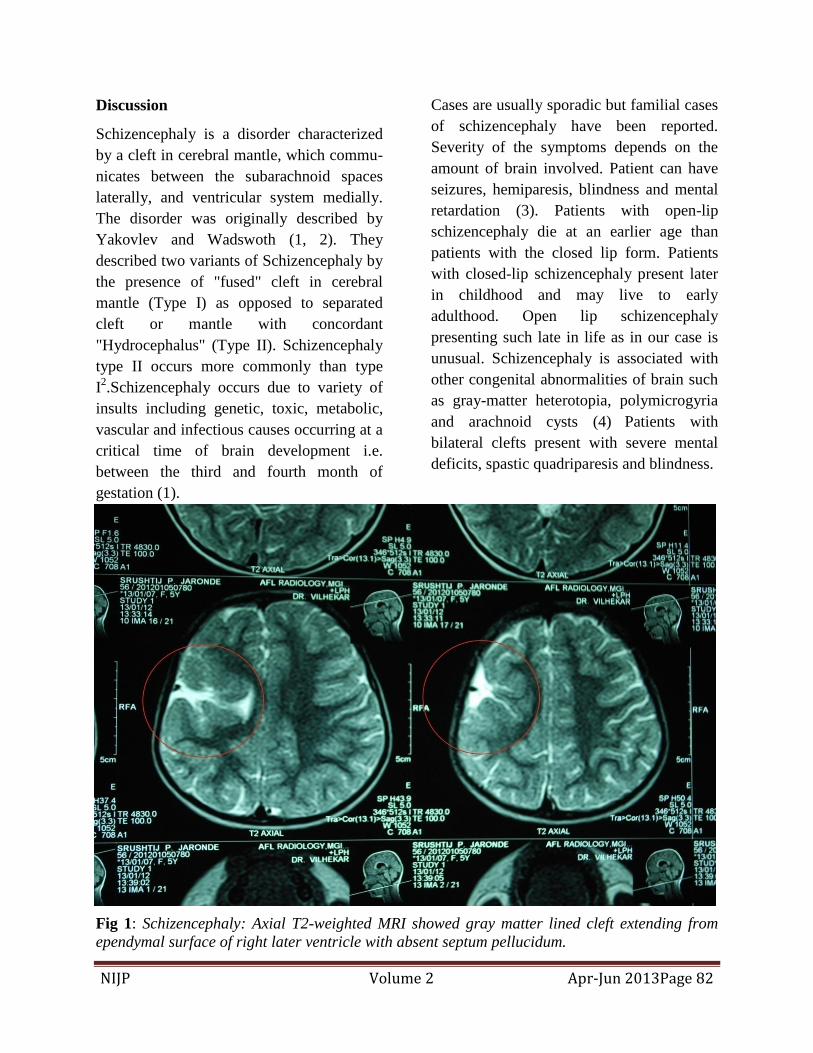
NIJP Volume 2 Apr-Jun 2013Page 82
Discussion
Schizencephaly is a disorder characterized
by a cleft in cerebral mantle, which commu-
nicates between the subarachnoid spaces
laterally, and ventricular system medially.
The disorder was originally described by
Yakovlev and Wadswoth (1, 2). They
described two variants of Schizencephaly by
the presence of "fused" cleft in cerebral
mantle (Type I) as opposed to separated
cleft or mantle with concordant
"Hydrocephalus" (Type II). Schizencephaly
type II occurs more commonly than type
I2.Schizencephaly occurs due to variety of
insults including genetic, toxic, metabolic,
vascular and infectious causes occurring at a
critical time of brain development i.e.
between the third and fourth month of
gestation (1).
Cases are usually sporadic but familial cases
of schizencephaly have been reported.
Severity of the symptoms depends on the
amount of brain involved. Patient can have
seizures, hemiparesis, blindness and mental
retardation (3). Patients with open-lip
schizencephaly die at an earlier age than
patients with the closed lip form. Patients
with closed-lip schizencephaly present later
in childhood and may live to early
adulthood. Open lip schizencephaly
presenting such late in life as in our case is
unusual. Schizencephaly is associated with
other congenital abnormalities of brain such
as gray-matter heterotopia, polymicrogyria
and arachnoid cysts (4)
Patients with
bilateral clefts present with severe mental
deficits, spastic quadriparesis and blindness.
Fig 1: Schizencephaly: Axial T2-weighted MRI showed gray matter lined cleft extending from
ependymal surface of right later ventricle with absent septum pellucidum.

NIJP Volume 2 Apr-Jun 2013Page 83
Language development is also impaired in
patients with bilateral clefts as compared to
patients with unilateral schizencephaly.MRI
provides excellent demonstration of the
anatomic changes in Schizencephaly and is
better than CT- head in finding the clefts. (5)
Differential diagnosis of schizencephaly is
focal cortical dysplasia and Porencephaly.
Focal cortical dysplasia sometime may have
a cleft on the ventricular surface that does
not extend completely to the ventricular
surface.
Conclusion:
Schizencephaly should be considered in the
differential diagnosis of hemiplegia/paresis
in childhood. MRI is the best imaging
modality for demonstrating the lesions.
References:
1. M. Salek, H. Alam, M. Islam, M. Zaman:
Childhood convulsion and hemiparesis
due to schizencephaly –a case report. The
Internet Journal of
Neurosurgery.2009,Volume:6, Number 1
2. S. Garg, N. Gupta: Schizencephaly in
Images, Indian Journal for the Practicing
Doctor, 2008, Volume 1.
3. P. K. Chhetri, S. Raut: Schizencephaly –A
case report, Journal of College of Medical
Sciences-Nepal, 2010, Volume; 6 No.1,
54-56.
4. Gouranga Santra, Shankha Shubhra Sen:
Schizencephaly, Journal, Indian Academy
of Clinical Medical, 2008, July-
September, Volume: 9, No.3, 233-236.
5. A. K. Dubey, R. K. Gupta, P. Sharma, R.
K. Sharma: Case report – Schizencephaly
Type I, Indian Pediatr, 2001: 38: 1049-
1051.
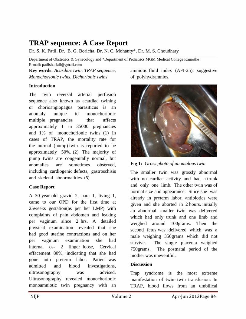
NIJP Volume 2 Apr-Jun 2013Page 84
TRAP sequence: A Case Report Dr. S. K. Patil, Dr. B. G. Boricha, Dr. N. C. Mohanty*, Dr. M. S. Choudhary
Department of Obstetrics & Gynecology and *Department of Pediatrics MGM Medical College Kamothe
E-mail: [email protected]
Key words: Acardiac twin, TRAP sequence,
Monochorionic twins, Dichorionic twins
Introduction
The twin reversal arterial perfusion
sequence also known as acardiac twining
or chorioangiopagus parasiticus is an
anomaly unique to monochorionic
multiple pregnancies that affects
approximately 1 in 35000 pregnancies
and 1% of monochorionic twins. (1) In
cases of TRAP, the mortality rate for
the normal (pump) twin is reported to be
approximately 50%. (2) The majority of
pump twins are congenitally normal, but
anomalies are sometimes observed,
including cardiogenic defects, gastroschisis
and skeletal abnormalities. (3)
Case Report
A 30-year-old gravid 2, para 1, living 1,
came to our OPD for the first time at
25weeks gestation(as per her LMP) with
complaints of pain abdomen and leaking
per vaginum since 2 hrs. A detailed
physical examination revealed that she
had good uterine contractions and on her
per vaginum examination she had
internal os- 2 finger loose, Cervical
effacement 80%, indicating that she had
gone into preterm labor. Patient was
admitted and blood investigations,
ultrasonography was advised.
Ultrasonography revealed monochorionic
monoamniotic twin pregnancy with an
amniotic fluid index (AFI-25), suggestive
of polyhydramnios.
Fig 1: Gross photo of anomalous twin
The smaller twin was grossly abnormal
with no cardiac activity and had a trunk
and only one limb. The other twin was of
normal size and appearance. Since she was
already in preterm labor, antibiotics were
given and she aborted in 2 hours. initially
an abnormal smaller twin was delivered
which had only trunk and one limb and
weighed around 100grams. Then the
second fetus was delivered which was a
male weighing 350grams which did not
survive. The single placenta weighed
750grams. The postnatal period of the
mother was uneventful.
Discussion
Trap syndrome is the most extreme
manifestation of twin- twin transfusion. In
TRAP, blood flows from an umbilical

NIJP Volume 2 Apr-Jun 2013Page 85
artery of the pump twin in the reversed
direction into the umbilical artery of the
perfused twin, via an arterio- arterial
anastomosis. The perfused twin is a true
parasite. Its blood supply is poorly
oxygenated and hypoxemia contributes to
variable degrees of deficient development
of the head heart and upper limb
structures. The lower half of the body is
usually better developed, which may be
explained by the mechanism of
transfusion of the acardiac twin. Blood
enters the acardiac twin via the umbilical
artery and flows through the common
iliac artery and aorta. Whatever little
oxygen is present is extracted in the
lower part of the body, causing partial
development of lower limbs and
abdomen. By the time it reaches the
upper body most of the oxygen has
already been extracted, leading to poor
development of upper body structures. (4)
Fig 2: X-ray of anomalous twin
Two criteria must be fulfilled for the
development of TRAP sequence. The
first is an arterio-arterial anastomosis and
the second is discordant development (5)
or IUFD (6) in one monochorionic twins,
allowing for blood flow reversal. Not
infrequently, chromosomal abnormalities
are identified in the acardiac twin, while
the pump twin has a normal karyotype.
(7,8) Embryos / fetuses with chromosomal
abnormalities have a high rate of
spontaneous IUFD, which in dichorionic
twins would lead to a ―vanishing‖ twin.
However, in monochorionic twins, the
―vanishing‖ can be prevented by the
occurrence of persistence , yet reverse,
flow to the chromosomally abnormal twin
via vascular anastomosis. Therefore,
whenever single IUFD is suspected in
monochorionic twins on first trimester
scan, the differential diagnosis or
development of TRAP should be kept in
mind and follow-up scan arranged. (9)
TRAP sequence is characterized by a
grossly abnormal fetus that grows, may
even show movements, but has no
functional cardiac activity of its own.
Rarely, a rudimentary heart may show
pulsatility. Marked hydrops and cystic
hygroma are frequently present, especially
toward the end of pregnancy. The
diagnosis can be reliably made on
ultrasound scan in first trimester. (10)
Early diagnosis of TRAP sequence
enables both invasive and conservative
treatment to be offered to the mother to
reduce the amount of compromise of the
pump twin.

NIJP Volume 2 Apr-Jun 2013Page 86
References:
1. Healey M: Acardia: predictive risk
factors for the co-twin's
survival.Teratology 1994, 50:205-213.
2. Gembruch U, Viski S, Bagamery K, Berg
C, Germer U: Twin reversed arterial
perfusion sequence in twin-to-twin
transfusion syndrome after the death of
the donor co-twin in the second trimester.
Ultrasound Obstet Gynecol 2001, 17:153-
156.
3. Chen CY, Wu YC, Chen CL, Yang
ML: Acardiac syndrome coexisting with
complex skeletal dysplasia in the co-twin:
3 D sonographic findings. J Clin
Ultrasound 2007, 35:387-389.
4. Sebire NJ, Sepulveda W, Jeanty P,
Nyberg DA, Nicolaides KH. Multiple
gestations. In: Nyberg DA,McGahan JP,
Pretorius DH, Pilu G (eds). Diagnostic
Imaging of Fetal Anomalies.
Philadelphia: Lippincott Williams
&Wilkins; 2003; p. 777-813.
5. Van Allen MI, Smith DW, Shepard TH.
Twin reversed arterial perfusion (TRAP)
sequence: a study of 14 twin pregnancies
with acardius. Semin Perinatol 1983;
7:285-93.
6. Gembruch U, Viski S, Bagamery K, Berg
C, Germer U.: Twin reversed arterial
perfusion sequence in twin-to-twin
transfusion syndrome after the death of
the donor co-twin in the second trimester.
Ultrasound Obstet Gynecol. 2001
Feb;17(2):153-6.
7.Moore TR, Gale S, Benirsehle K. Perinatal
outcome of forty-nine pregnancies
complicated by acardiac twinning. Am J
Obstet Gynecol 1990; 163:907-12.
8. Chaliha, C., Schwarzler, P., Booker, M.
Trisomy 2 in an acardiac twin in a triplet
in-vitro fertilization pregnancy. Hum.
Reprod. 1999; 14, 1378–1380.
9. Petersen BL, Broholm H, Skibsted L
Acardiac twin with preserved brain. Fetal
Diagn Ther 2001;16:231-233.
10.Schwarzler P, Ville Y, Moscosco G.
Diagnosis of twin reversed arterial
perfusion sequence in the first trimester
by trans vaginal color Doppler
ultrasound. Ultrasound Obstet Gynecol
1999;13(2):143-146.

NIJP Volume 2 Apr-Jun 2013Page 87
Media Watch / Around the World Dr. Anil Lohar*, Dr. Satish Agrawal**
* Senior. Resident Dr PDMMC Amravati ** Practicing Pediatrician Amravati. e-mail: [email protected]
Abdominal epilepsy in chronic recurrent
abdominal pain
Chronic recurrent abdominal pain is a
perplexing problem for pediatricians.
Among the various causes of pain,
abdominal epilepsy (AE) is a relatively
uncommon cause and hence, is frequently
ignored. In this study, an attempt will be
made to determine the incidence of AE in
children suffering from chronic recurrent
abdominal pain, with the help of clinical
symptoms and EEG, and to study the
response to empirical anti-epileptic drug
(AED).
This prospective study involved 150
children, in the age group of 6-15 years,
suffering from chronic recurrent abdominal
pain (i.e., pain severe enough to affect
child's activity or three episodes of
abdominal pain within 3 months and
abdominal pain associated with nausea,
vomiting, bloating, headache and diarrhea)
from May 2007 to March 2010.
Children below 6 years and above 15 years
of age and those who were unable to give a
proper history and those having acute
abdominal pain of duration less than 3
months, having visceral disease, were
excluded from the study.
All children were investigated for stool and
urine examination to rule out worm
infestation, acute gastrointestinal infection
and urinary tract infection. Urine
porphobilinogen was performed before
starting AED and to rule out intermittent
porphyria. Complete blood count was
performed to rule out anemia and systemic
infections. Abdomen and pelvis
ultrasonography was done to rule out
organic intra-abdominal cause for chronic
recurrent abdominal pain. EEG was done in
every child to correlate the findings of EEG
with clinical diagnosis of AE.
An EEG was performed on all children with
a Recorders and Medicare systems (RMS)
EEG 24 BRAINVIEW PLUS (24 channel
EEG machine) All EEGs were reported by
experts.
Of the 150 cases, EEG changes were seen in
111 (74%) patients between the ages of 6
and 15 years with chronic recurrent
abdominal pain. Of the 111 patients with an
abnormal EEG suggestive of AE, 75
(67.56%) were girls and 36 (32.43%) were
boys Of the 111 abnormal EEG cases, 39
(35.13%) cases had temporal type of EEG
changes, 36 (32.43%) cases had fronto-
temporal type of EEG changes, 33 (29.72%)
cases had generalized type of EEG changes
and 3 (2.70%) cases had parieto-temporal
type of EEG changes the most common
pattern was sharp wave in 99 (89.18%) cases
and 12 (10.81%) cases had spike and wave
pattern .
Of the 111 (74%) patients with abnormal
EEG, all the patients had symptomatic
improvement after treatment with the AED
oxcarbazepine. Of the 39 (24%) patients
with a normal EEG record, 27 (18%)
patients had asymptomatic improvement
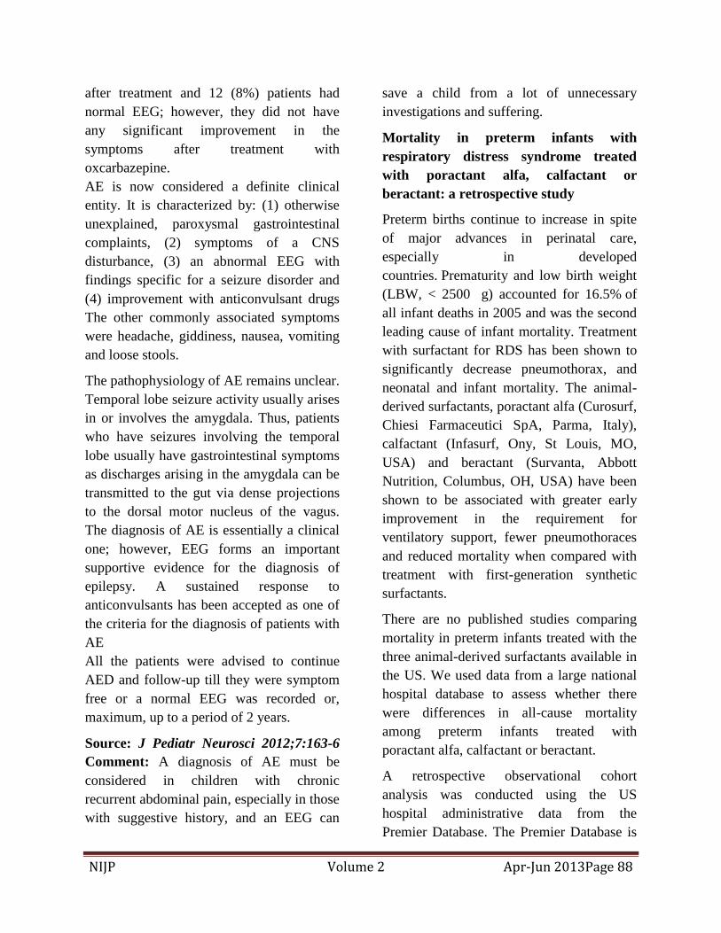
NIJP Volume 2 Apr-Jun 2013Page 88
after treatment and 12 (8%) patients had
normal EEG; however, they did not have
any significant improvement in the
symptoms after treatment with
oxcarbazepine.
AE is now considered a definite clinical
entity. It is characterized by: (1) otherwise
unexplained, paroxysmal gastrointestinal
complaints, (2) symptoms of a CNS
disturbance, (3) an abnormal EEG with
findings specific for a seizure disorder and
(4) improvement with anticonvulsant drugs
The other commonly associated symptoms
were headache, giddiness, nausea, vomiting
and loose stools.
The pathophysiology of AE remains unclear.
Temporal lobe seizure activity usually arises
in or involves the amygdala. Thus, patients
who have seizures involving the temporal
lobe usually have gastrointestinal symptoms
as discharges arising in the amygdala can be
transmitted to the gut via dense projections
to the dorsal motor nucleus of the vagus.
The diagnosis of AE is essentially a clinical
one; however, EEG forms an important
supportive evidence for the diagnosis of
epilepsy. A sustained response to
anticonvulsants has been accepted as one of
the criteria for the diagnosis of patients with
AE
All the patients were advised to continue
AED and follow-up till they were symptom
free or a normal EEG was recorded or,
maximum, up to a period of 2 years.
Source: J Pediatr Neurosci 2012;7:163-6
Comment: A diagnosis of AE must be
considered in children with chronic
recurrent abdominal pain, especially in those
with suggestive history, and an EEG can
save a child from a lot of unnecessary
investigations and suffering.
Mortality in preterm infants with
respiratory distress syndrome treated
with poractant alfa, calfactant or
beractant: a retrospective study
Preterm births continue to increase in spite
of major advances in perinatal care,
especially in developed
countries. Prematurity and low birth weight
(LBW, < 2500 g) accounted for 16.5% of
all infant deaths in 2005 and was the second
leading cause of infant mortality. Treatment
with surfactant for RDS has been shown to
significantly decrease pneumothorax, and
neonatal and infant mortality. The animal-
derived surfactants, poractant alfa (Curosurf,
Chiesi Farmaceutici SpA, Parma, Italy),
calfactant (Infasurf, Ony, St Louis, MO,
USA) and beractant (Survanta, Abbott
Nutrition, Columbus, OH, USA) have been
shown to be associated with greater early
improvement in the requirement for
ventilatory support, fewer pneumothoraces
and reduced mortality when compared with
treatment with first-generation synthetic
surfactants.
There are no published studies comparing
mortality in preterm infants treated with the
three animal-derived surfactants available in
the US. We used data from a large national
hospital database to assess whether there
were differences in all-cause mortality
among preterm infants treated with
poractant alfa, calfactant or beractant.
A retrospective observational cohort
analysis was conducted using the US
hospital administrative data from the
Premier Database. The Premier Database is

NIJP Volume 2 Apr-Jun 2013Page 89
a large US hospital-based database,
containing information on approximately 5.5
million annual hospital discharges The study
was designed to compare all-cause in-
hospital mortality, defined by the discharge
status of ‗expired‘, in preterm infants treated
with poractant alfa, calfactant or beractant.
Infants were included in the study if they
were discharged as an inpatient from a
Premier Database hospital from 1 January
2005, through 31 December 2009. Inclusion
criteria for the study included having
gestational age of 25–32 weeks, BW 500–
1999g, diagnosis of RDS, age 2 calendar
days, when they received the first dose of
surfactant, and having received only one of
the three study surfactants. Patients were
excluded from the study in case of missing
values for any of the variables planned a
priori to be included in the logistic
regression model, if they received more than
one surfactant during their hospitalization,
or if there was evidence of congenital
abnormalities such as trisomy 13 or 18,
anencephaly or dwarfism. Information on
diagnosis of RDS, gestational age at birth,
BW and congenital anomalies was obtained
from International Classification of
Diseases, 9th Revision, Clinical
Modification (ICD-9) codes.
A total of 14173 infants discharged from
236 hospitals were included in the study
population. Overall, the unadjusted all-cause
in-hospital mortality rates were
3.61% (n=184) in the poractant alfa group,
5.95% (n=201) in the calfactant group, and
4.58% (n=261) in the beractant group. The
lowest mortality rate was always observed in
the poractant alfa group, except for the
category 1250–1499 g, where beractant-
treated infants had the lowest mortality.
Mortality was significantly lower for infants
500–749 g, who received poractant alfa
(11.72%) than for those who received
calfactant (20.67%,P<0.001) or beractant
(17.39%, P=0.011). In the 1000–1249 g
BW category, mortality was significantly
higher in the calfactant group (5.46%) than
in the poractant alfa (2.67%, P=0.002) and
beractant (3.54%, P=0.035) groups.
In conclusion, this large retrospective study
of preterm infants with RDS found lower
mortality among infants who received
poractant alfa, compared with infants who
received either calfactant or beractant, even
after adjusting for patient characteristics
such as gestational age and BW, and after
accounting for hospital characteristics and
center effects. These results in real-world
settings are consistent with prior RCTs, but
provide additional significant findings that
most RCTs have not been able to provide
due to their relatively small sample size.
Source: Journal of Perinatology (2013) 33,
119–125.
Comment: More prospective studies are
needed to establish the fact in such
commonly encountered issue like RDS.
Effective dose of Topiramate in pediatric
migraine prophylaxis
Migraine is a common neurological disorder
in childhood and adolescence. It has a
prevalence of 5% in children aged 7-10
years and of 17% in adolescents.
Management of pediatric migraine includes
lifestyle changes (in order to avoid foods,
habits or environmental factors that may
trigger a migraine attack), the use of
abortive medications, and preventive

NIJP Volume 2 Apr-Jun 2013Page 90
measures, which can be either non-
pharmacological or pharmacological.
Topiramate has been shown to be effective
for migraine prevention in adults. Limited
data is available for the efficacy and safety
of topiramate in the pediatric population. It
is proposed that epilepsy and migraine share
some of the same pathophysiological
mechanisms, including abnormal function of
voltage-gated sodium and calcium channels,
reduced gamma-aminobutyric acid (GABA)
-mediated inhibition, and increased
glutamate-mediated excitation.
The present study was planned and
performed to evaluate the efficacy of low-
dose topiramate in pediatric migraine
prophylaxis.
The study was designed as a prospective
study and was performed in 60 children
(between 5 and 15 years of age) treated with
topiramate for prevention of migraine
headaches, at a pediatric neurology clinic in
north of Iran from March 2011 through
September of 2011.
A general evaluation and physical
examination were performed in the first
visit, and patients were asked baseline
laboratory screening tests and a migraine
checklist, including demographic and
clinical details of the patients such as
frequency, duration, and intensity of
headache was filed for each person.
Exclusion criteria were as follows:
1. Prior or current prophylactic treatment for
migraine in the last two months
2. Persistent increasing headache
3. Change of behavior and school
performance
4. Abnormal neurologic and persistent focal
signs (e.g., papilledema)
5. Neuro-imaging studies indicative of a
focal neurological lesion
6. Contraindications for topiramate (e.g.,
glaucoma, renal stone)
7. Patients with known case of epilepsy.
After simple randomized dividing the
patients into two groups, topiramate was
started at a dose of greater or lower than 2
mg/kg/day. There were three stages of this
study such as at the beginning of study
(Base-line), period which was 4-weeks after
receiving drugs (phase I) and at the end of 8-
weeks after receiving drugs (phase II).
Headache severity was scored on a 1-3 point
scale with one presenting no effect on daily
activity, two for partial inhibition of daily
activity and three for loss of daily activities
based on the Visual Analog Scale (VAS).
The response to topiramate treatment was
recorded according to the patients and
parents' reports during follow-up visits, and
was defined as: Complete cessation of
headache attacks (excellent response),
reduction of headache frequency more than
50% of baseline (good response), reduction
of headache frequency less than 50% of
baseline (fair response), No response or
increase of headache attacks (poor
response). The data analysis was performed
by statistical package for social sciences
version 13. We analyzed number of
headaches per month, changes in Body Mass
Index (BMI) and dosage before and after
treatment according to the presence or
absence of adverse effects, using the
Student's t test.

NIJP Volume 2 Apr-Jun 2013Page 91
Sixty patients who met the inclusion criteria
were enrolled in the study. Five of the
patients were excluded due to no follow-up,
patient's choice or drug intolerance.
Ultimately, out of 55 patients who
completed the course of study, 30 (54.5%)
were girls, and 25 (45.5%) were boys. The
mean age was 8 years (range: 6.3-14.5
years). Patients in the two groups were not
significantly different regarding age, gender,
and other characteristics (P > 0.05). Mean
(±SD) numbers of monthly headache
frequency at the beginning of the study with
topiramate in two groups receiving > 2
mg/kg/day and < 2 mg/kg/day were 6.9
(±2.1) and 6.2 (±2.4), and mean (±SD)
duration of each episode of headache at the
baseline were 5.2 (±2.4), and 5.4 (±2.1) h,
respectively. The mean (±SD) headache
intensity at the beginning of the trial was
7.11 (±1.4) and 7.2 (±1.9) and the base line
data show that there were no statistically
significant differences between the two
groups according to base line headache
characteristics (P > 0.05)and data about
comparison of headache parameters at
baseline and after 4 and 8 weeks of
treatment. The most common side effects of
topiramate were paresthesias (five patients),
anorexia (four patients), and drowsiness
(four patients).
In conclusion, the results of this study
demonstrated that low-dose of topiramate
(<2 mg/kg/day) is effective, well-tolerated,
safe, and suggested as an alternative
prophylactic treatment for pediatric
migraine.
Source: J Pediatr Neurosci 2013 Feb
23;7:171-4.
Comment: If more data support the same
conclusion, the drug may prove to be very
useful
Application of the Yamaguchi criteria for
classification of “suspected” systemic
juvenile idiopathic arthritis (sJIA)
Many children with sJIA may have a
delayed onset of arthritis and so fail to fulfill
the ILAR criteria for sJIA. This study was
undertaken to determine whether the
Yamaguchi criteria (for adult onset Still‘s
disease) is useful in classification of children
with systemic juvenile idiopathic arthritis
(sJIA) particularly in ―pre-arthritic‖, pure
systemic, phase of the illness. A secondary
objective was to determine the time delay
between disease onset and onset of arthritis
in our sJIA cohort.
In comparison to the other diseases which
are jointly grouped under the term juvenile
idiopathic arthritis (JIA), systemic juvenile
idiopathic arthritis (sJIA or Still‘s disease as
it was previously known) has many distinct
features One important difference is that, at
least in a subset of sJIA patients, arthritis
may be a non-prominent and/or a delayed
manifestation. Patients have been reported to
have a delay of up to 10 years before the
onset of arthritis .The International League
Against Rheumatism (ILAR) criteria require
the presence of arthritis to classify a patient
as having sJIA Since morbidity and
mortality may develop early in sJIA
patients, a strict adherence to the ILAR
criteria may lead to an unacceptable delay in
diagnosis.
Still‘s disease is also known to occur in
adults, in whom the disease is labeled as
Adult Onset Still‘s Disease (AOSD). Most

NIJP Volume 2 Apr-Jun 2013Page 92
studies comparing AOSD and Still‘s disease
in children have not found significant
differences in the clinical features at
presentation Among the different criteria
available for the classification of AOSD, the
Yamaguchi criteria are the most sensitive
(96%) and still manage to retain a high
degree of specificity (92%) The present
study was undertaken in an attempt to
determine the utility of the Yamaguchi
criteria in the classification of children
presenting with typical systemic features of
sJIA who had still not developed arthritis
(labeled as ―suspected‖ sJIA). We also
wished to determine the time lag between
onset of fever and onset of arthritis among
sJIA patients in our cohort who had fulfilled
the ILAR criteria at presentation to our
hospital.
The study was a retrospective chart review
conducted in the pediatrics department of a
tertiary level teaching hospital. Electronic
medical records were mined using key
words ―arthritis‖, ―JIA‖ and ―JRA‖. All
patients with these keywords mentioned
anywhere in their notes, and had been
diagnosed between Jan 1, 2004 and Jan 1,
2010 were identified. Their detailed case
records were reviewed and those patients
finally diagnosed by the treating
pediatrician, as ―definite‖ sJIA (fulfilling the
ILAR classification criteria) or ―suspected‖
sJIA (not fulfilling the ILAR criteria) were
included. For inclusion in the cohort, the
patients had to have been investigated at
length to rule out infections and
malignancies as causes of fever of unknown
origin (FUO).
The fulfillment of the various variables in
both the Yamaguchi criteria and the ILAR
criteria were identified in each patient at the
time of first presentation to our institute.
Exclusionary components of the criteria set
were also considered. Variables representing
symptoms (characteristic fever, arthralgia,
and sore throat,) were considered positive
irrespective of whether they had it at
presentation to our institute or at any time in
the past during the course of the disease.
Laboratory criteria (leukocyte count, liver
function test, serositis and negative ANA,
RF) and clinical signs (typical rash,
splenomegaly, hepatomegaly, and
lymphadenopathy) were considered positive
only if they had been performed/observed in
our department at the time of presentation
and had been fulfilled. In case of
parent/patient reported arthritis, the child
was considered to have had arthritis only if
in addition to arthralgia the patient had had
either significant swelling which had been
documented by a doctor or any 2 of the
following physical findings: limitation of
range of motion/deformity, pain on motion
leading to limitation of motion or increased
warmth over the joint.
The time duration between onset of fever
and onset of arthritis as reported by the
patients or their parents (based on
information available in the charts or
information obtained via a telephonic
interview) was also recorded. The statistical
significance of any difference was inferred
using the Fisher test for nominal values and
the independent t test for continuous
variables. The coefficient of association
between the criteria was calculated using the
Phi coefficient. Approval for the study was
obtained from the Institutional Ethics
Committee.
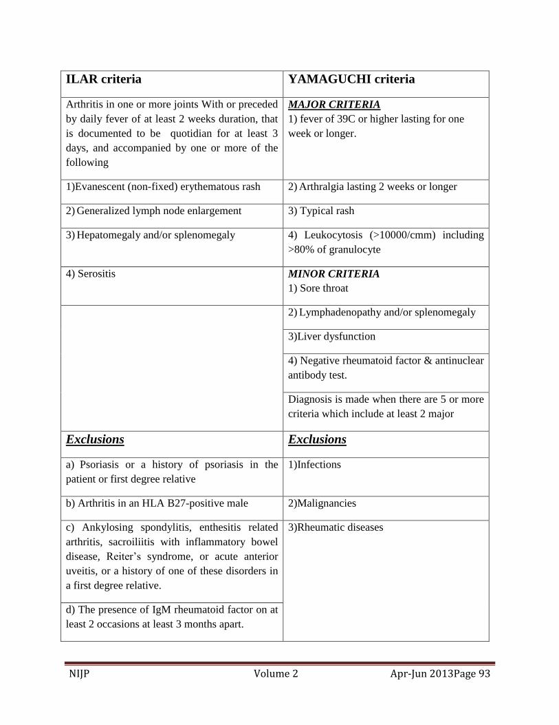
NIJP Volume 2 Apr-Jun 2013Page 93
ILAR criteria YAMAGUCHI criteria
Arthritis in one or more joints With or preceded
by daily fever of at least 2 weeks duration, that
is documented to be quotidian for at least 3
days, and accompanied by one or more of the
following
MAJOR CRITERIA
1) fever of 39C or higher lasting for one
week or longer.
1)Evanescent (non-fixed) erythematous rash 2) Arthralgia lasting 2 weeks or longer
2) Generalized lymph node enlargement 3) Typical rash
3) Hepatomegaly and/or splenomegaly 4) Leukocytosis (>10000/cmm) including
>80% of granulocyte
4) Serositis MINOR CRITERIA
1) Sore throat
2) Lymphadenopathy and/or splenomegaly
3)Liver dysfunction
4) Negative rheumatoid factor & antinuclear
antibody test.
Diagnosis is made when there are 5 or more
criteria which include at least 2 major
Exclusions Exclusions
a) Psoriasis or a history of psoriasis in the
patient or first degree relative
1)Infections
b) Arthritis in an HLA B27-positive male 2)Malignancies
c) Ankylosing spondylitis, enthesitis related
arthritis, sacroiliitis with inflammatory bowel
disease, Reiter‘s syndrome, or acute anterior
uveitis, or a history of one of these disorders in
a first degree relative.
3)Rheumatic diseases
d) The presence of IgM rheumatoid factor on at
least 2 occasions at least 3 months apart.

NIJP Volume 2 Apr-Jun 2013Page 94
Thirty-four patients were diagnosed with
sJIA (definite or suspected) in the last 6
years. Three patients (all girls) were
excluded due to insufficient data in their
medical records. Twenty boys and eleven
girls (n=31) formed the study cohort. All of
the patients were DMARD naïve at
presentation and none had received steroids
for more than 2 weeks. Thirteen patients
failed to fulfill the ILAR criteria, all of them
due to a lack of arthritis. The mean age of
children who fulfilled the ILAR criteria
(11.9 years) was significantly higher than
patients who did not fulfill the ILAR criteria
(6.9 years, p=0.006)
Among the 13 patients who did not fulfill
the ILAR criteria, nearly all children (12
patients) fulfilled the Yamaguchi criteria.
Thus the total number of patients who
fulfilled the Yamaguchi criteria was 23
(74%), compared to only 18 patients (58%)
who fulfilled the ILAR criteria.
There is a subgroup of sJIA patients in
whom arthritis is absent or quite delayed and
in this group, the diagnosis may be very
challenging. Comparison of the ILAR and
Yamaguchi criteria in our cohort of sJIA
patients suggests that some changes to the
ILAR criteria for sJIA are probably
necessary to make it more inclusive.
Source: Pediatric
rheumatology 2012, 10:40
Comment: Possible inclusion of recently
described biomarkers or application of the
Yamaguchi criteria (which do not consider
arthritis as an essential feature), might
enable the criteria to be more sensitive and
clinically useful. These changes may
facilitate early and timely diagnosis of
children with sJIA and thus, allow early
treatment.

NIJP Volume 2 Apr-Jun 2013Page 95
Guidelines for Authors
Manuscript Preparation:
All Manuscripts sent to the NIJP should not
be sent in any other journal or publication
and becomes the sole property of the
publishers. The manuscripts submitted
should be accompanied by a certificate
signed by all the authors that ―The
undersigned author(s) certifies (fy) that the
submitted article is original and is not under
consideration with other publisher and has
not been published anywhere in the past. All
copyright ownership of the manuscript is
transferred to the publisher of NIJP‖. The
manuscript should include the ethical
clearance certificate from appropriate
authorities.
Articles are edited for grammar and style.
The spelling pattern in the journal is of
American style. The statement and opinions
expressed in the manuscripts are essentially
of the author(s) and the editors/ publishers
don‘t owe any responsibility for these.
The product, service or the claim made by
the manufacturer as advertised in the journal
are those of the manufacturer. The
publishers don‘t endorse these claims.
Preparation of Manuscripts:
Title Page: This contains the title of the
manuscripts along with the name of the
author(s) with their affiliations.
The name of the institute where the study
was carried out, the address for
correspondence with e-mail of the author
should be mentioned at the bottom of the
page.
Abstract: The objective, method of the
study, results and conclusion should be sent
in a paragraph as abstract of the manuscript.
It should contain the purpose of the study,
selection of study subjects, specific data
denoting the main findings and the chief
conclusions. The new and important aspects
of the study should be revealed by the
abstract.
Text: Every article must follow the
following format:
Introduction, Material and method, Results,
Discussion and Conclusions. The text should
preferably be in a simple language which is
easy to understand, pertaining to the topic
concerned and should avoid vocabulary
jargon.
Contributors: At the end of the article, the
contribution of all the authors should be
mentioned which includes:
(1) Each author‘s contribution
(2) Conflict of interest
(3) Funding source and its role in the study
The last element of the text is
acknowledgment. Abbreviations if used
should be in accordance with the
international guidelines e.g. cm, ml etc.
Abbreviation use should be kept to the
minimum and if they are used, they should
be preceded by the full form.
References: Only those references which
were consulted in original should be cited.
They should be arranged sequentially. The
references should be visible on the line after
punctuation. The author(s) shall be

NIJP Volume 2 Apr-Jun 2013Page 96
responsible for the accuracy and
completeness of the references. Vancouver
System is followed in the journal:
References should be listed and numbered in
the order in which they appear in the text.
The list of references at the end of the text
includes:
Names of author(s): If more authors are
involved in the study, the first 6 names
should appear ending with et al, the title of
the paper, Name of the journal as appears in
Index Medicus, publication year, volume
number and first and last page numbers.
References from the book includes, name of
the book, editors, place of publication,
publisher, edition, the year of publication
and first and last page numbers.
If multiple authors are involved then the
chapter name, first and last page numbers
and names and initials of the editors should
be give.
From journals:
Mahesh Baldawa, Satish Tiwari: Legal
issues and preventable medical errors, New
Indian Journal of Pediatrics 2012, 1: 8-12.
From Books:
Bhattacharya A, Mishra S.DNR, Euthanasia
and Life support systems; In: Tiwari S,
Baldwa M, Tiwari M, Kuthe A editors; Text
Book on medico-legal issues related to
various medical specialties: 1st edition,
Jaypee brothers medical Publishers, New
Delhi 2012; 179-185
From Newspapers:
Aamir ko ho raha pachhatava. Dainik
Bhaskar (Hindi), 2011; 4th
December.
Nagpur p.5 (Col.3)
From internet:
Harms Roger W. Breast-feeding while
pregnant: Is it safe?
http://www.mayoclinic.com/health/breast-
feeding-while-pregnant/AN01840
Accessed on 16th
Oct 2011.
Those papers which have been accepted but
not published yet should be mentioned as
―in press‖. Those which are in preparation,
personal communication or unpublished
observation should be mentioned as such.

NIJP Volume 2 Apr-Jun 2013Page 97
INDIAN MEDICO-LEGAL & ETHICS ASSOCIATION
Dear Colleagues, Warm regards
Please accept the seasons greeting on behalf of we all. The practice of medicine has
changed drastically in the twenty first century. There have been many positive as well as
negative changes in medical sciences. The good age-old doctor-patient relationship is in
doldrums. The communication skills have almost been forgotten. Commercialization is the
obvious agenda especially with the development of corporate culture in the health sector. The
concept of privatization has added fuel to the fire. The patient, who are willing to pay feel
that the life can also be purchased with money. This has resulted in soaring expectations.
Because of all these doctors are not only affected by medico-legal cases but many other legal
problems arising out of other related issues of staff, instruments & infrastructure. The
Government is coming up with newer and newer laws and restrictions on medical fraternity
and hospitals. We have experienced this on many occasions, which prompted us along with
some other colleagues & friends to form a medico-legal & ethics association.
In last few years, we found various problems, which as a medical consultants /
medico-legal experts we were trying to solve single handedly. It was then, that we realized
the need of a fleet of experts to work in co-ordination. The association has thus being formed
to help you in preventing a disaster in your practice. We hope that we will succeed in
achieving the aims and objects of guiding the medical practitioners in their difficult times.
The various membership benefits include:
1) Personal / individual professional indemnity cover for upto five years (Amount and
terms decided by Executive Board) included in life membership.
2) Hospital insurance at concessional rate (as compared to other insurance / risk
management companies).
3) Free med-legal guidance in hours of crisis.
4) Services of crisis management committee at city / district level.
5) Free expert opinion if there are cases in court of law.
6) Services of legal experts at concessional rates (wherever available).
7) Participation in academic activities related to med-legal issues.
All this can‘t be achieved without the help of dedicated, hard working, sincere
members of the association. Hence, we would like you to become the member of this
association. We hope that with active & enthusiastic members like you, our association
will attain greater heights as we progress further. Please send your constructive
criticism, suggestions, and programs for the future.
Yours truly
Dr. Balraj SinghYadav
(Secretary)
Address for correspondence:
Krishna Medicare Center
9, Friends Colny, Jharsa Rd Gurgaon 122001
Phone: 0124-2339244; 09811108230 E-mail: [email protected]

NIJP Volume 2 Apr-Jun 2013Page 98
INDIAN MEDICO-LEGAL & ETHICS ASSOCIATION
Membership Form
Name of the applicant:………………………………………………………
(Surname) (First name) (Middle name)
Date of Birth:………………………… ……. Sex: ………………….
Address for Correspondence:………………………………………………………………...……..
………………………………………………………………………………………………………
Telephone No:- Resi. ,,,,,,,,,,,,,,,,,,,,,,,,,,,,,,,,,,,,,,,,,,,,,,,,,,,,Hosp:,,,,,,,,,,,,,,,,,,,,,,,,,,,,,,,,,,,,,,,,,,,,,,,,,,,,,,,,,,
Mobile:………………………………Other:…………………………………….
Fax:…………………………..E-mail ……………………………………………………………..
Registration no. ……………………………………. Date of Reg. ………………………………
Name of the Council (MCI/Dental/Homeo/Ayurvedic/ Others: ---------------------
Medical/legal qualification University Year
Experience in legal field (if any) ----------------------------------------
Name, membership no. & signature of proposer:--------------------
Name, membership no & signature of seconder:-------------------
A) Was / Is there any med-legal case against you /your Hospital: Yes / No
If, Yes (Give details) --------------------------------------------------------
(Attach separate sheet)--------------------------------------------------------- B) Do you have a Professional Indemnity Policy: Yes / No
Name of the Company: Amount: C) Do you have Hospital Insurance: Yes / No
Name of the Company: Amount: D) Do you have Risk Management Policy: Yes / No
Name of the company: Amount: E) Is your relative / friend practicing Law: Yes / No
If Yes, Name: Qualification:
Place of Practice: Specialization: F) Any other information you would like to share: (Please attach the details)
I hereby declare that above information is correct. I shall be responsible for any incorrect / fraudulent
declarations.
Place:
Date: (signature of applicant)
Enclosures: Degree & Registration Certificates
Life Membership fee (individual 2500/-, couple 4000/-) by CBS (At Par, Multicity Cheque) or
DD, in the name of Indian medico-legal & ethics association (IMLEA) payable at Amravati.
Please send the Cheque/DD to Dr. Satish Tiwari, Yashodanagar No.2 Amravati 444606.
NIJP Volume 2 Apr-Jun 2013Page 99
MEMBERSHIP FORM
PEDIATRICS ASSOCIATION OF INDIA (PAI)
THE CHILD, B.K. ROAD, RANIHAT CUTTACK-753001, ODISHA
Name of the Applicant: __________________________________________________________
Date of Birth: DD/MM/YYYY Sex: Male / Female
Communication Address:-________________________________________________________
______________________________________________________________________________
______________________________________________________________________________
State:________________________________ Nationality: ______________________________
Telephones: (STD Code) _________________________________________________________
Mobile: ______________________________________________________________________
Email ID: _____________________________________________________________________
Medical / Pediatric Qualification Name of the University Qualifying Year
MBBS Registration No. & Registering Authority (e.g. MCI or State Medical Council):-
______________________________________________________________________________
Short Curriculum Vitae with area of interest (within 10 lines):-
______________________________________________________________________________
______________________________________________________________________________
______________________________________________________________________________
______________________________________________________________________________
______________________________________________________________________________
____________________________________________________________________________ .
Place: Signature of the Applicant
Date:
(In hardcopy)
Recommendation of the Nodal Person:-
The statements above are true to best of my knowledge. His/her membership may be
accepted.
Name of the Nodal Person: _______________________________________________________


![Indian Academy of Pediatrics Guidelines on the Fast and Junk … · 2019-08-23 · Gupta et al. IAP JUNCS GUIDELINES INDIAN PEDIATRICS 5 AUGUST 10, 2019 [E-PUB AHEAD OF PRINT] unhealthy](https://img.pdfslide.net/doc/110x75/5e98d51a8110f06b574bc8b8/indian-academy-of-pediatrics-guidelines-on-the-fast-and-junk-2019-08-23-gupta.jpg)


![16-00717R1 edited by N approved - Indian Pediatrics · INDIAN PEDIATRICS 1 SEPTEMBER 26, 2017 [E-PUB ... [E-PUB AHEAD OF PRINT] Research Paper Reference Centile Curves for Body Fat](https://img.pdfslide.net/doc/110x75/5af0ed387f8b9aa9168e7ad0/16-00717r1-edited-by-n-approved-indian-pediatrics-1-september-26-2017-e-pub.jpg)