Embed Size (px)
Citation preview

W H I T EPAPER
New Insights into Neuroscience ResearchUsing nCounter® Technology for Comprehensive Gene Expression Profiling and Biomarker Identification
GLPCP_PM0024 | Nov 2017
NanoString Technologies, Inc., Seattle, WA 98109
Authors: Katie Buchanan and Joseph M. Beechem
2 | Introduction N O V 3 , 2017
Introduction
Diagnosis and treatment of neurodegenerative disease has long
been impeded by the difficulty in accessing the affected areas
of the brain. Unlike a suspicious mole on the skin or inflamed
tissue around a wound, the brain presents a greater challenge
to examine. Most neurodegenerative disease diagnoses have
relied on post mortem confirmation of disease pathology or
medical imaging during disease progression. Early diagnosis
and treatment have remained elusive but have accelerated using
advanced molecular biology methods including gene expression
and profiling, mRNA analysis, and even investigations of single
cells derived from neural cell lines.
One challenge facing all these advanced techniques is the scarcity
of sample. To accurately analyze brain tissue, blood samples, or
microvolumes of single cell supernatant requires highly sensitive and
robust technology beyond traditional methods. Using NanoString’s
nCounter technology, scientists have been able to profile
specifically curated gene expression panels with minimal hands-
on time to generate accurate, insightful, and repeatable results.
Already researchers are making significant strides in understanding
neurodegenerative disease and mechanisms of neuroinflammation.
The following five publications represent groundbreaking research
in neuropathology, including Alzheimer’s disease, Parkinson’s
disease. and Amyotrophic Lateral sclerosis. In each study,
NanoString technology was the cornerstone for research and results
beyond today’s standards of qPCR and microarray analysis. The
studies are leading to a more comprehensive understanding of
neurodegenerative disease and treatment.
Comprehensive Analysis from FFPEResearchers unlock gene expression data from many different sample types
There is an increasing body of evidence that noncoding,
expanded repeats in RNA can contribute to the pathogenesis of
neurodegenerative and neuromuscular disorders. In the case of
familial amyotrophic lateral sclerosis (ALS) and frontotemporal
dementia (FTD) patients, a newly discovered hexanucleotide
GGGGCC repeat expansion in the noncoding region of the
C9ORF72 gene has been confirmed in over 40% of patients.
While the function of the C9ORF72 protein remains unknown, it
represents a potential candidate for screening and therapeutic
intervention. In this study by Jeffrey D. Rothstein et al., the group
characterized C9ORF72 to determine if expansion of the repeat
contributes to the disease progression1.
The team directly assayed samples of patient-derived human
brain tissue, ALS fibroblasts, iPSCs and iPSNs to determine
the levels of C9ORF72 RNA. NanoString RNA detection allows
highly sensitive, direct screening of tissue without any nucleotide
amplification and confirmed C9ORF72 iPSCs (and ALS patient
tissue) exhibit the GGGGCC repeat expansion and have reduced
C9ORF72 RNA levels. Complicating matters is that there are
three mRNA products transcribed from the C9ORF72 gene:
C9ORF72 Variants 1, 2, and 3 (V1, V2, V3, respectively). The
roles of these three variants is also unknown but V1 and V3
contain ORFs upstream of the expanded GGGGCC repeat. To
detect the levels of each C9ORF72 RNA variant and to compare
each variant between samples, the group generated two 50-
mer NanoString probes for each transcript that targets one of
the C9ORF72 variants. In order for the specific RNA variant
to be detected, each 50-mer probe must bind in tandem to
the transcript. C9ORF72 ALS patient tissue and iPSNs showed
approximately a 50% reduction in expression of C9ORF72 V1 &
V2 (Fig. 1). Patient-derived fibroblasts showed low expression
with no differences between control and diseased tissue.
The group then used microarrays to assess other genes that
have different expression levels in C9ORF72 iPSNs, fibroblasts,
and human motor cortex. Genes were selected that code for
proteins expressed in the CNS and that are predicted to be
secreted, thus allowing for easy detection and monitoring in
patient CSF samples. This led to the successful identification
of sixteen aberrantly expressed target genes in C9ORF72 ALS
patient tissues, seven of which showed similar dysregulation
patterns when compared with iPSNs. These seven genes, whose
expression mirrors the C9ORF72 disease state, can be potential
candidates for disease biomarkers to monitor therapeutic
intervention and are easily collected from patient blood or CSF
samples. The NanoString profiling will continue to facilitate
analysis of gene expression in human patient samples without
the need for amplification steps. This not only reduces error
and variability, but maximizes the amount of data that can be
gathered from these patient samples and biomarkers.
New Insights into Neuroscience Research

3 | Cell Type Profiling N O V 3 , 2017
Further Reading: Using Nanostring’s nCounter Human
Inflammation Gene Expression panel, researchers at the
University of Alberta studied the expression of 184 inflammation-
related genes from FFPE brain samples to compare host innate
immune response and bacterial polyglycan (PGN) presence in
MS and non-MS samples. They found that PGN presence was
inversely correlated with the intensity of LFB (luxol fast blue)
stained sections of MS lesions, and host gene expression analysis
revealed the induction of immune genes NFKB1, RIPK1, and IL-
12A, demonstrate the highest immune response correlation with
PGN in MS brains. They concluded that demyelination, PGN, and
inflammatory gene expression accounts for 86% of observed
variance in MS, and inflammatory demyelination could contribute
to underlying disease mechanisms2.
Cell Type ProfilingDetection of protein in neurons and astrocytes down to a single cell
Amyloid precursor protein (APP) is an integral membrane
protein found in high levels in the synapses of neurons. APP
can be cleaved into soluble amyloid precursor protein-α
(sAPPα); alternatively, APP can be cleaved to generate the
insoluble amyloid β-protein (Aβ). Alzheimer’s disease is
neurodegenerative disease characterized by accumulation
of Aβ, creating extracellular amyloid plaques, presenting as
lesions on the brain. To understand why either sAPPα or Aβ
are generated, most research has utilized heterologous cell
lines that were not derived from neuronal cells. As a result,
the contributions of different cell types to the overall disease
progression has not been well characterized.
There is an increasing body of evidence that specific cell types
may secrete products that contribute to the pathology of AD.
Tracy L. Young-Pearse et al. had already established protocols
to differentiate human induced pluripotent stem cells (iPSC)
to neuronal and glial cells found in the forebrain3. In order to
examine the roles of individual cells in more depth, they adapted
a technique known as microengraving to their studies of AD. In
microengraving, cells are cultured in a dense array of nanowells,
allowing analysis of secreted products from individual cells. In
particular, the group examined the differential processing of APP
from single living neuronal and glial cells derived from iPSCs.
Each nanowell array contains 84,672 wells on a glass microscope
slide; each well is only 50 x 50 x 50 μm. Each nanowell constitutes
an individual experiment and because the cells are live, they
can be revisited over time. Optimizing data collection from this
undertaking requires tools that are sensitive, robust, and can
handle large amounts of data. The team used NanoString’s Single
Cell Multiplexing capabilities to analyze gene expression of single
neurons and glia. The custom-designed 150 gene NanoString
CodeSet amplified gene products from each microwell, targeting
cell fate markers, AD-related genes, and housekeeping genes.
When they analyzed the data with the nCounter Digital Analyzer
and software, they found they were able to detect Aβ and sAPPα
from these individual cells at multiple time points (Fig 2).
FIGURE 1: C9ORF72 ALS-Specific Gene Expression1 Researchers performed targeted gene expression analysis of aberrantly expressed genes on NanoString‘s nCounter platform, and revealed dysregulation of seven genes that were also found to be similarly dysregulated in C9ORF72 iPSNs.
FIGURE 2: Examination of gene expression profiles after detection of sAPPα and Aβ from single iPSC-derived neural cells3 HiPSC-derived neural cells were lysed, cDNA was synthesized and hybridized to a custom NanoString codeset for analysis. A heat map of expression data of select genes are shown for each cell, as well as for pools of 500 cells each.

4 | Gene Signature Generation N O V 3 , 2017
FIGURE 3: Identification of a miRNA microglia signature5 NanoString 600 miRNA nCounter chip was used to create a heatmap and hierarchical clustering of differentially expressed miRNAs in microglia, organ specific macrophages and immune cell populations to identify an novel miRNA gene signature for microglia.
Whereas previously only pooled groups of cells could be
studied, now individual cells can be profiled to understand
how they contribute to disease progression. More importantly,
demonstrating that iPSCs are compatible with microengraving
can reduce the reliance on immortalized cell lines that are less
relevant or possibly even misleading. Taken together, this paper
presents groundbreaking new methodology and a turning point
in the study of AD and other neuronal diseases.
Further Reading: Researchers at UC Irvine published in Neuron
in 2017 a process for producing induced microglia-like cells
(iMGLs) from induced-pluripotent stem cells (iPSCs). In order
to demonstrate that genomic integrity was maintained after
differentiation, the team used NanoString’s nCounter Human
Karyotype CNV panel and found a high correlation (r2 > .92)
between the iMGL and iPSC genomes. This study demonstrates that
iMGLs can be used as a renewable source of patient-derived cells for
determining gene function in neurodegenerative diseases4.
Gene Signature GenerationnCounter platform allows rapid identification of microglia gene signatures
Microglia are a key component of the immune response in the
central nervous system. Studying the role of these cells in the
immune response is hampered because they lack clearly identifiable
features that would distinguish them from infiltrating macrophages.
Howard L. Weiner et al. used extensive gene and microRNA
(miRNA) array screens and were able to identify a unique signature
dependent on TGF-β in both mouse and human microglial cells5.
To this end, the team worked with NanoString to create a custom
mouse codeset containing 354 microglial enriched genes, 40
inflammation-related genes, and six housekeeping genes. This chip,
referred to as MG400, formed the basis of their studies.
The MG400 chip analysis enabled the group to analyze immune
cell populations and create both a heatmap and clusters of the
key microglial molecules as grouped by cell location and function
(Fig. 3). Six of the genes identified showed little to no expression
in immune cells or macrophages from other organs—they were
specific to microglial cells. Additionally, the genes displayed
conserved expression patterns in human microglia.
The team then used a NanoString mouse miRNA panel of 600
miRNAs to assess microglial cells with respect to other immune
cells. This identified eight miRNAs highly expressed in microglial
cells, three of which were unique to microglial over immune cells,
resulting in the identification of a miRNA signature specific to
microglia. Subsequent investigation showed that the signature is
specific to developing microglial cells in vivo.
Surprisingly, this signature is not present in monocytes recruited
during neuroinflammation nor is it expressed in established
microglial cell lines in vitro. The team determined that the
signature is inducible by culturing adult microglia cells in the
presence of TGF-β1, and confirmed this by showing a loss of
microglia in mice deficient for TGF-β1 in the CNS.
While an undertaking of this magnitude is possible using
standard qPCR techniques, it was no doubt greatly enhanced
and expedited by utilizing the NanoString nCounter platform.
This extensive transcriptomic comparison of sorted mouse cells
to identify the microglia gene signature required the efficient
analysis of over 400 gene targets in at least 30 distinct cell
populations, as well as 600 miRNA targets in 24 immune cell
types and multiple CNS cell subsets. Now that this signature has
been identified, the role of microglia in protection and damage
to the CNS can be characterized and eventually modulated to
potentially combat neurodegenerative disease.
5 | Microglia Activation N O V 3 , 2017
Further Reading: A team of researchers led by Sellgren at
Massachusetts General Hospital set out to generate human
microglia-like cells from human somatic cells and demonstrate
psychiatric disease-relevant application. The team isolated total
RNA for a custom panel of probes developed with NanoString,
and a miRNA panel of 800 miRNAs to compare the patient-
derived human microglia-like cells to human fetal microglia cells.
They were able to demonstrate that patient-derived human
microglia-like cells performed similarly to actual microglia cells
and they provide an opportunity for high-throughput drug
screening that is not possible with post-mortem tissue6.
Microglia ActivationResearchers demonstrate a therapeutic approach for neurodegenerative diseases by regulating microglia activation
Early studies on the brain and its cellular structure described it
as a static environment, where neurons did not regenerate and
non-neuronal cells played a minimal role. However, that view is
changing rapidly, with studies revealing neuronal regeneration
and the critical and varying roles played by supporting cells.
One of those supporting cell types, microglia, are particularly
important in maintaining a healthy environment as the brain
changes during development or in response to damage.
Microglia are the phagocytes of the brain. They detect
damaged or dying tissue, engulf the cellular debris, and clear
the area so that normal growth or repair can occur. In addition
to phagocytosis, microglia can enter either a pro- or anti-
inflammatory state. In the anti-inflammatory state, microglia are
thought to aid healing by minimizing inflammation. Conversely,
microglia in the pro-inflammatory state are believed to
contribute to cell damage, even while they continue to remove
potentially damaging cellular debris. To further complicate the
role of microglia, phagocytic cells produce reactive oxygen
species (ROS) to break down ingested debris. However, the ROS
produced can be damaging to nearby cells. It remains unclear
whether these activities are independent or interrelated. A clear
understanding of the molecular mechanisms regulating microglia
activity could provide new therapeutic targets for a range of
neurodegenerative diseases and cancers.
In a recent publication in the Journal of Neuroinflammation, Siddiqui
et al, sought to address the question of how microglial states and
activities interact and whether their state can be altered7.
The authors took advantage of NanoString’s high throughput
nCounter system to analyze mRNA expression levels of six
independent microglia cultures for each of three conditions:
control, pro-inflammatory and anti-inflammatory; examining
each with or without myelin.
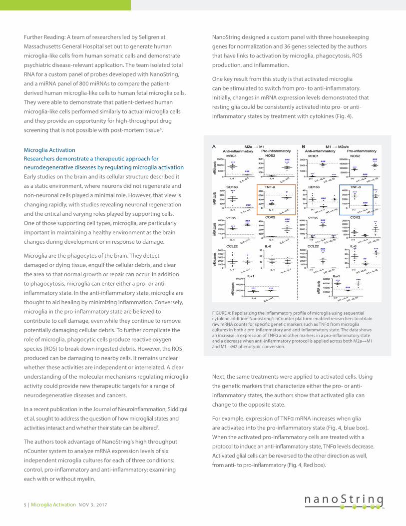
NanoString designed a custom panel with three housekeeping
genes for normalization and 36 genes selected by the authors
that have links to activation by microglia, phagocytosis, ROS
production, and inflammation.
One key result from this study is that activated microglia
can be stimulated to switch from pro- to anti-inflammatory.
Initially, changes in mRNA expression levels demonstrated that
resting glia could be consistently activated into pro- or anti-
inflammatory states by treatment with cytokines (Fig. 4).
Next, the same treatments were applied to activated cells. Using
the genetic markers that characterize either the pro- or anti-
inflammatory states, the authors show that activated glia can
change to the opposite state.
For example, expression of TNFα mRNA increases when glia
are activated into the pro-inflammatory state (Fig. 4, blue box).
When the activated pro-inflammatory cells are treated with a
protocol to induce an anti-inflammatory state, TNFα levels decrease.
Activated glial cells can be reversed to the other direction as well,
from anti- to pro-inflammatory (Fig. 4, Red box).
FIGURE 4: Repolarizing the inflammatory profile of microglia using sequential cytokine addition7 Nanostring’s nCounter platform enabled researchers to obtain raw mRNA counts for specific genetic markers such as TNFα from microglia cultures in both a pro-inflammatory and anti-inflammatory state. The data shows an increase in expression of TNFα and other markers in a pro-inflammatory state and a decrease when anti-inflammatory protocol is applied across both M2a→M1 and M1→M2 phenotypic conversion.

6 | Early Predictors of Parkinson’s Disease N O V 3 , 2017
The extent of changes in expression levels varies by gene,
but the changes are consistent. This suggests that regulating
the activation state of microglia cells could be a therapeutic
approach to supplement current therapies for cancer and
neurodegenerative diseases.
Further Reading: In a 2017 study in Translational Psychiatry,
Muhie’s team showed that PTSD activates and sustains
inflammatory pathways by analyzing differentially expressed
genes on Nanostring’s nCounter Analysis System. Sustained
neuroinflammation appears to drive the developmental and
behavioral manifestations of PTSD. Blood samples show promise
as a suitable and accessible stand in for brain specimens for
clinical translation8.
Early Predictors of Parkinson’s DiseaseAnalysis of SNCA isoforms as potential early stage biomarkers
The α–synuclein (SNCA) gene has traditionally been thought of as
a “neuron-specific” gene because high levels of the α–synuclein
protein accumulate in the brain in Parkinson’s disease. When it was
discovered that high levels of α–synuclein are found in circulating
blood cells, including red blood cells, it raised interest in this gene
as an easily accessible indicator of the disease. Researchers began
investigating how transcripts of α–synuclein in the blood could be
used to track disease progression.
Under the direction of Clemens R. Scherzer, a team of researchers
evaluated the levels of SNCA mRNA in three independent
biomarker cohorts9. Using qPCR, microarray analysis, and
NanoString technology, the group found that SNCA transcripts
were consistently reduced in patients with Parkinson’s disease
in each cohort. This finding seemed counterintuitive—the α–
synuclein protein accumulates in the brain in Parkinson’s disease
and yet there was an overall decrease in SNCA transcripts. It is
notable that there are several isoforms of the SNCA transcript
and some of these isoforms have been thought to have a role
in Parkinson’s disease. Taken together, this led the group to
extend their studies into patients who were earlier in the disease
progression. The Parkinson’s Progressive Markers Initiative
(PPMI) identifies individuals who are clinically at the early stage
of Parkinson’s but not yet at the full disease state. Detailed
multiplexed digital analysis of 340 patient samples using the
NanoString nCounter Analysis System demonstrated that disease-
relevant SNCA transcript isoforms were already reduced in the
blood samples at this early stage in the PPMI cohort.
Specifically, a long 3’ UTR-SNCA mRNA transcript and E4E6-
SNCA mRNA (a transcript skipping exon 5) were reduced by
27% and 19%, respectively, whereas other Parkinson’s related
transcripts (such as DJ-1), were unchanged (Fig. 5). It is
possible that these specific isoforms could be used as sensitive
biomarkers of early stage disease.
Using NanoString technology, the researchers were able
to examine the SNCA isoforms directly from the blood
sample without the need for additional steps such as reverse
transcription or PCR amplification. Additionally, NanoString
data were precise and reliable; template controls generated no
signal and reference RNA counts were highly correlated with R2
> 0.999 within and between different plates. Moreover, when a
subset of samples was randomly resampled to verify the retest
reliability of the technique, the average correlation value R2
was 0.98. Taken together, NanoString nCounter technology
demonstrates a clear advantage over existing methods to
identify early disease markers in Parkinson’s disease.
Further Reading: A team of scientists led by the Cure
Huntington’s Disease Initiative (CHDI) used Nanostring’s
nCounter to demonstrate the benefits of PDE10 inhibition
for Huntington’s disease mouse models. They found that
PDE10 inhibition showed improvements and partial reversal of
deregulated transcripts and the prevention of neurophysiological
deficits related to Huntington’s disease10.
Figure 5: Disease-relevant SNCA isoforms are already reduced near disease onset9 SNCA isoforms were probed on Nanostring’s nCounter platform across 202 cases of Parkinson’s disease (less than two years of disease) and found that disease-relevant isoforms with long the 3’ UTR (A) or skipping exon 5 (B) were reduced by 27% and 19% respectively compared to control. However, levels of PARK7 (DJ-1), mutated in rare autosomal recessive Parkinson’s disease (C), were not significantly different from the control group.

7 | Advanced Neuroscience Research Starts Here N O V 3 , 2017
Advanced Neuroscience Research Starts Here
The pace of neuroscience research is accelerating and
personalized medicine allows new therapeutic approaches that
are highly targeted to the individual disease state. Whereas
previous generations of patients faced a dim prognosis when
diagnosed with a neurodegenerative disease, current technology
allows patients to be diagnosed faster and treated more
effectively. The publications discussed here represent new
potential avenues for early detection, improved diagnosis, and
better clarity of disease pathogenesis.
As more biomarkers are identified and specific cell contributions
are better understood it creates opportunities to transform
knowledge into actions. NanoString’s nCounter platform is
playing a significant role in this progress by enabling discovery
on a cellular and molecular level. NanoString technology is
faster than PCR and more convenient and targeted than NGS.
Researchers can work with NanoString scientists to custom
configure their codesets and primers to expedite data collection
and optimize analytical software to maximize their time
interpreting results.
These advancements have the promise of earlier detections and
therapeutic intervention for those impacted by neurodegenerative
diseases. As new research explores neurodegenerative pathways,
processes, and cellular profiles, there is greater potential to
develop personalized therapies for patients that are more
successful and less toxic than previous treatments.

R E F E R E N C E S
1 Donnelly CJ et al. (2013) RNA toxicity from the ALS/FTD C9ORF72 expansion is mitigated by antisense intervention. Neuron 80(4):1102.
2 Branton WG et al. (2016) Brain microbiota disruption within inflammatory demyelinating lesions in multiple sclerosis. Scientific Reports 6:37344.
3 Liao MC et al. (2016) Single-Cell Detection of Secreted Aβ and sAPPα from Human IPSC-Derived Neurons and Astrocytes. Journal of Neuroscience 36(5):1730-46.
4 Abud EM et al. (2017) iPSC-Derived Human Microglia-like Cells to Study Neurological Diseases. Neuron 94(2):278-293.
5 Butovsky O et al. (2014) Identification of a unique TGF-β-dependent molecular and functional signature in microglia. Nature Neuroscience 17(1):131-43.
6 Sellgren CM et al. (2017) Patient-specific models of microglia-mediated engulfment of synapses and neural progenitors. Molecular Psychiatry 22(2):170-177.
7 Siddiqui TA et al. (2016) Complex molecular and functional outcomes of single versus sequential cytokine stimulation of rat microglia. Journal of Neuroinflammation 13(1):66.
8 Muhie S et al. (2017) Molecular indicators of stress-induced neuroinflammation in a mouse model simulating features of post-traumatic stress disorder. Translational Psychiatry 7(5):e1135.
9 Locascio JJ et al. (2015) Association between α-synuclein blood transcripts and early, neuroimaging-supported Parkinson's disease. Brain 138(9):2659-71.
10 Beaumont V et al. (2016) Phosphodiesterase 10A Inhibition Improves Cortico-Basal Ganglia Function in Huntington’s Disease Models. Neuron 92(6):1220-1237.
FOR RESEARCH USE ONLY. Not for use in diagnostic procedures. © 2017 NanoString Technologies, Inc. All rights reserved. NanoString, NanoString Technologies, the NanoString logo and nCounter are trademarks or registered trademarks of NanoString Technologies, Inc., in the United States and/or other countries. All other trademarks and/or service marks not owned by NanoString that appear in this document are the property of their respective owners.
For more information, please visit nanostring.com
NOV 2017 GLPCP_PM0024
Sales ContactsUnited States [email protected]: [email protected]
NanoString Technologies, Inc. 530 Fairview Avenue NorthSeattle, Washington 98109
Asia Pacific & Japan [email protected] Regions [email protected]
T (888) 358-6266F (206) 378-6288
nanostring.com [email protected]