Embed Size (px)
Citation preview
Alcocer-Gómez et al
1
NLRP3 inflammasome inhibition rescues Hutchinson-Gilford Progeria cellular
phenotype and extend longevity of an animal model
Elísabet Alcocer-Gómez1, Beatriz Castejón-Vega2, Jéssica Nuñez-Vasco3, Débora
Lendines-Cordero3, José M. Navarro-Pando4, Mario D. Cordero4
1 Departamento de Psicología Experimental, Facultad de Psicología, Universidad de
Sevilla, Sevilla, Spain
2 Institute of Molecular, Cell and Systems Biology, University of Glasgow, Glasgow G12
8QQ, UK.
3 Research Laboratory, Oral Medicine Department, University of Sevilla, Sevilla, Spain
4 Cátedra de Reproducción y Genética Humana del Instituto para el Estudio de la Biología
de la Reproducción Humana (INEBIR)-Universidad Europea del Atlántico
(UNEATLANTICO)-Fundación Universitaria Iberoamericana (FUNIBER).
Running Title: The inhibition of NLRP3 delays progeria phenotype
Corresponding Author:
Cátedra de Reproducción y Genética Humana del Instituto para el Estudio de la
Biología de la Reproducción Humana (INEBIR)-Universidad Europea del Atlántico
(UNEATLANTICO)-Fundación Universitaria Iberoamericana (FUNIBER), Email:
.CC-BY-ND 4.0 International licenseperpetuity. It is made available under apreprint (which was not certified by peer review) is the author/funder, who has granted bioRxiv a license to display the preprint in
The copyright holder for thisthis version posted September 10, 2020. ; https://doi.org/10.1101/2020.09.09.288290doi: bioRxiv preprint
Alcocer-Gómez et al
2
Abstract
Inflammation is a hallmark of aging and accelerated aging syndromes. In this context,
inflammation has been associated to the pathophysiology of Hutchinson–Gilford
progeria syndrome (HGPS). In this study, we report that progeroid skin fibroblasts and
animal models present an hyperactivation of the NLRP3-inflammasome complex. High
expression of NLRP3 and caspase 1 was also observed in skin fibroblasts from HGPS
associated to the nuclei morphology. Lymphoblast from HGPS also showed increased
basal levels of NLRP3 and caspase 1 independent to the induction from metabolic
factors. Consistent with these results, Zmpste24−/− showed high expression of Nlrp3 and
caspase 1 in heart, liver and kidney and reduced levels of Nlrc3, however these changes
were not observed in other inflammasomes. We also show that pharmacological
inhibition of NLRP3 using a direct NLRP3 inhibitor, MCC950, improved cellular
phenotype, significantly extends the lifespan of these progeroid animals and reduced
inflammasome-dependent inflammation. These findings suggest the NLRP3-
inflammasome comples as a therapeutic approach for patients with HGPS.
.CC-BY-ND 4.0 International licenseperpetuity. It is made available under apreprint (which was not certified by peer review) is the author/funder, who has granted bioRxiv a license to display the preprint in
The copyright holder for thisthis version posted September 10, 2020. ; https://doi.org/10.1101/2020.09.09.288290doi: bioRxiv preprint
Alcocer-Gómez et al
3
Ageing involves a progressive impairment of physiological homeostasis, situation that is
reflected in the cell tissues, and at organismal level (1). In this context, inflammation is
highly associated to the aging process and age-related diseases and the inflamm-ageing,
a low-grade sterile chronic inflammation, has been described as a progressive event
during biological ageing with accumulation of pro-inflammatory mediators (2). In the last
years, the role of NLRP3-inflammasome has been studied in many age-related diseases.
The NLRP3-inflammasome is one of the most well-studied inflammasomes in humans
and mice (3). It is a multiprotein complex comprising NLRP3 itself as an intracellular
sensor, the adapter protein ASC and pro-caspase-1. The NLRP3 inflammasome is
activated by a range of danger and stress signals (3), some of which rise during aging (4).
The Nlrp3 ablation in mice has been shown to improves lifespan and health by attenuating
multiple age-related degenerative changes such as cardiac aging, insulin sensitivity, bone
loss, and ovarian aging (5-7). According to this, the role of NLRP3-inflammasome has
been assessed in normal aging yet remain unexplored in genetic models of accelerated
aging. Hutchinson-Gilford progeria syndrome (HGPS) is a rare premature aging
condition in which a point mutation in the LMNA gene (c.1824C > T; GGC > GCT;
p.G608G) (8) causes the accumulation at the nuclear envelope of an aberrant precursor
of lamin A, named progerin, which disrupts the nuclear membrane architecture and causes
multiple cellular alterations, including abnormal gene transcription and signal
transduction. The clinical phenotype is characterized by delayed loss of primary teeth,
alopecia, osteoporosis, abnormal skin pigmentation, accelerated cardiovascular disease,
growth impairment, lipodystrophy, dermal and bone abnormalities, and metabolic
alterations (9). Inflammation has been associated to the pathophysiology of progeroid
syndromes. Nuclear factor κB (NF-κB)-mediated secretion of high levels of
proinflammatory cytokines have been shown in two different mouse models (10).
.CC-BY-ND 4.0 International licenseperpetuity. It is made available under apreprint (which was not certified by peer review) is the author/funder, who has granted bioRxiv a license to display the preprint in
The copyright holder for thisthis version posted September 10, 2020. ; https://doi.org/10.1101/2020.09.09.288290doi: bioRxiv preprint
Alcocer-Gómez et al
4
However, the genetic and pharmacological inhibition of NF-κB signaling prevented age-
associated features in these animal models, and extended their longevity (10).
Interestingly, NF-κB is a central mediator of the priming signal of NLRP3-inflammasome
complex (11). In the present work, we report that human skin fibroblasts and lymphocytes
from patients with HGPS and Zmpste24−/− mice, an appropriate murine model of HGPS,
demonstrate a clear activation of the NLRP3-inflammasome complex. In addition, we
show that this alteration is detrimental, given that skin fibroblasts from patients treated
with MCC950, a specific inhibitor of NLRP3, show improved phenotype and
Zmpste24−/− mice treated with MCC950 show a clear amelioration of progeroid features
and inflammation and extended longevity compared with untreated Zmpste24−/− mice.
Material and methods
Reagents.
Trypsin was purchased from Sigma Chemical Co., (St. Louis, Missouri). Anti-actin
monoclonal antibody from Calbiochem-Merck Chemicals Ltd. (Nottingham, UK). Lamin
A/C, NLRP3, NLRC3 and caspase 1 were obtained from Cell Signaling Technology.
MAP-LC3, NLRP4, Nalp1 and Nalp10 were obtained from Santa Cruz Biotechnology.
NLRP3 inhibitors MCC950 and 16673-34-0 were obtained from Sigma-Aldrich (Saint
Louis, USA). A cocktail of protease inhibitors (complete cocktail) was purchased from
Boehringer Mannheim (Indianapolis, IN). Grace's insect medium was purchased from
Gibco. The Immun Star HRP substrate kit was from Bio-Rad Laboratories Inc. (Hercules,
CA).
Fibroblast culture
All fibroblasts from patients with HGPS were obtained from The Progeria Research
Foundation Cell and Tissue Bank (http://www.progeriaresearch.org). The following
.CC-BY-ND 4.0 International licenseperpetuity. It is made available under apreprint (which was not certified by peer review) is the author/funder, who has granted bioRxiv a license to display the preprint in
The copyright holder for thisthis version posted September 10, 2020. ; https://doi.org/10.1101/2020.09.09.288290doi: bioRxiv preprint
Alcocer-Gómez et al
5
fibroblasts were used: HGADFN367 (3-year-old male) and HGADFN155 (1.2-year-old
female). Control fibroblasts were obtained from the Coriell Institute for Medical Research
(Camden, NJ, USA). Fibroblasts were cultured in high glucose DMEM (Dulbecco’s
modified media) (Gibco, Invitrogen, Eugene, OR, USA) supplemented with 15% fetal
bovine serum (FBS) (Gibco, Invitrogen, Eugene, OR, USA), 1% GlutaMAX
(ThermoFisher) and antibiotics (Sigma Chemical Co., St. Louis, MO, USA). Cells were
incubated at 37ºC in a 5% CO2 atmosphere. The medium was changed every two days to
avoid changes in pH.
We also used the following lymphoblasts: HGALBV009 (5.1-year-old male) and
HGALBV021 (father of the proband, 37-year-old female). Lymphoblasts were cultured
in RPMI-1640 (Gibco, Invitrogen, Eugene, OR, USA) supplemented with 15% fetal
bovine serum (FBS) (Gibco, Invitrogen, Eugene, OR, USA), and antibiotics (Sigma
Chemical Co., St. Louis, MO, USA). Cells were incubated at 37ºC in a 5% CO2
atmosphere
Western Blotting
Whole cellular lysate from fibroblasts was prepared by gentle shaking with a buffer
containing 0.9% NaCl, 20 mM Tris-ClH, pH 7.6, 0.1% Triton X-100, 1 mM
phenylmethylsulfonylfluoride and 0.01% leupeptin. The protein content was determined
by the Bradford method. Electrophoresis was carried out in a 10–15% acrylamide
SDS/PAGE and proteins were transferred to Immobilon membranes (Amersham
Pharmacia, Piscataway, NJ). Next, membranes were washed with PBS, blocked over
night at 4°C and incubated with the respective primary antibody solution (1:1000).
Membranes were then probed with their respective secondary antibody (1:2500).
Immunolabeled proteins were detected by chemiluminescence method (Immun Star HRP
.CC-BY-ND 4.0 International licenseperpetuity. It is made available under apreprint (which was not certified by peer review) is the author/funder, who has granted bioRxiv a license to display the preprint in
The copyright holder for thisthis version posted September 10, 2020. ; https://doi.org/10.1101/2020.09.09.288290doi: bioRxiv preprint
Alcocer-Gómez et al
6
substrate kit, Bio-Rad Laboratories Inc., Hercules, CA). Western blot images were
quantified using ImageJ software.
NLRP3 immunofluorescence
NLRP3 distribution in cytosol was assessed by immunofluorescence techniques using
antibodies against NLRP3 and DAPI as a marker of the nuclei.
Proliferation rate
Two hundred thousand fibroblasts were cultured with or without the MCC950 at two
different concentrations (0.6 and 1.2mM) for 24, 48, and 120h. After discharging
supernatant with dead cells, cells from three high-power fields were counted with an
inverted microscope using a 40X objective.
Animals
Animal studies were performed in accordance with European Union guidelines
(2010/63/EU) and the corresponding Spanish regulations for the use of laboratory animals
in chronic experiments (RD 53/2013 on the care of experimental animals). All
experiments were approved by the local institutional animal care committee. For all
experiments, only male mice were used. Mutant mice deficient in Zmpste24
metalloproteinase have been described previously (10). All groups had ad libitum access
to their prescribed diet and water throughout the whole study. Body weight was monitored
weekly. Animal rooms were maintained at 20–22°C with 30–70% relative humidity.
For all experiments with NLRP3 inhibitors, Zmpste24+/+ (wild type) and Zmpste24-/- were
maintained on a regular 12 h light/dark cycle at 20–22°C. Treatments were started at
1month of age after randomization into three groups (wild type vehicle, Zmpste24-/-
vehicle and Zmpste24-/-MCC950). These groups correspond to the following treatment:
.CC-BY-ND 4.0 International licenseperpetuity. It is made available under apreprint (which was not certified by peer review) is the author/funder, who has granted bioRxiv a license to display the preprint in
The copyright holder for thisthis version posted September 10, 2020. ; https://doi.org/10.1101/2020.09.09.288290doi: bioRxiv preprint
Alcocer-Gómez et al
7
i) standard diet with i.p vehicle (saline) treatment (vehicle groups) from Teklad Global
14% Protein Rodent Maintenance Diet, Harlan Laboratories (carbohydrate:protein:fat
ratio of 48:14:4 percent of kcal) and ii) standard diet with MCC950 treatment (MCC950
group). MCC950 was administered 20mg/kg daily by i.p. route. All groups had ad libitum
access to their prescribed diet and water throughout the study. Individuals were monitored
daily and weighed monthly but were otherwise left undisturbed until they died. Survival
was assessed using male mice, and all animals were dead by the time of this report.
Kaplan–Meier survival curves were constructed using known birth and death dates, and
differences between groups were evaluated using the logrank test. A separate group of
male mice were sacrificed at age 4 months to study (western blots).
Statistical Analysis
Data in the figures is shown as mean ± SD. Data between different groups were analysed
statistically by using ANOVA on Ranks with Sigma Plot and Sigma Stat statistical
software (SPSS for Windows, 19, 2010, SPSS Inc. Chicago, IL, USA). For cell-culture
studies, Student’s t test was used for data analyses. A value of p<0.05 was considered
significant
Results
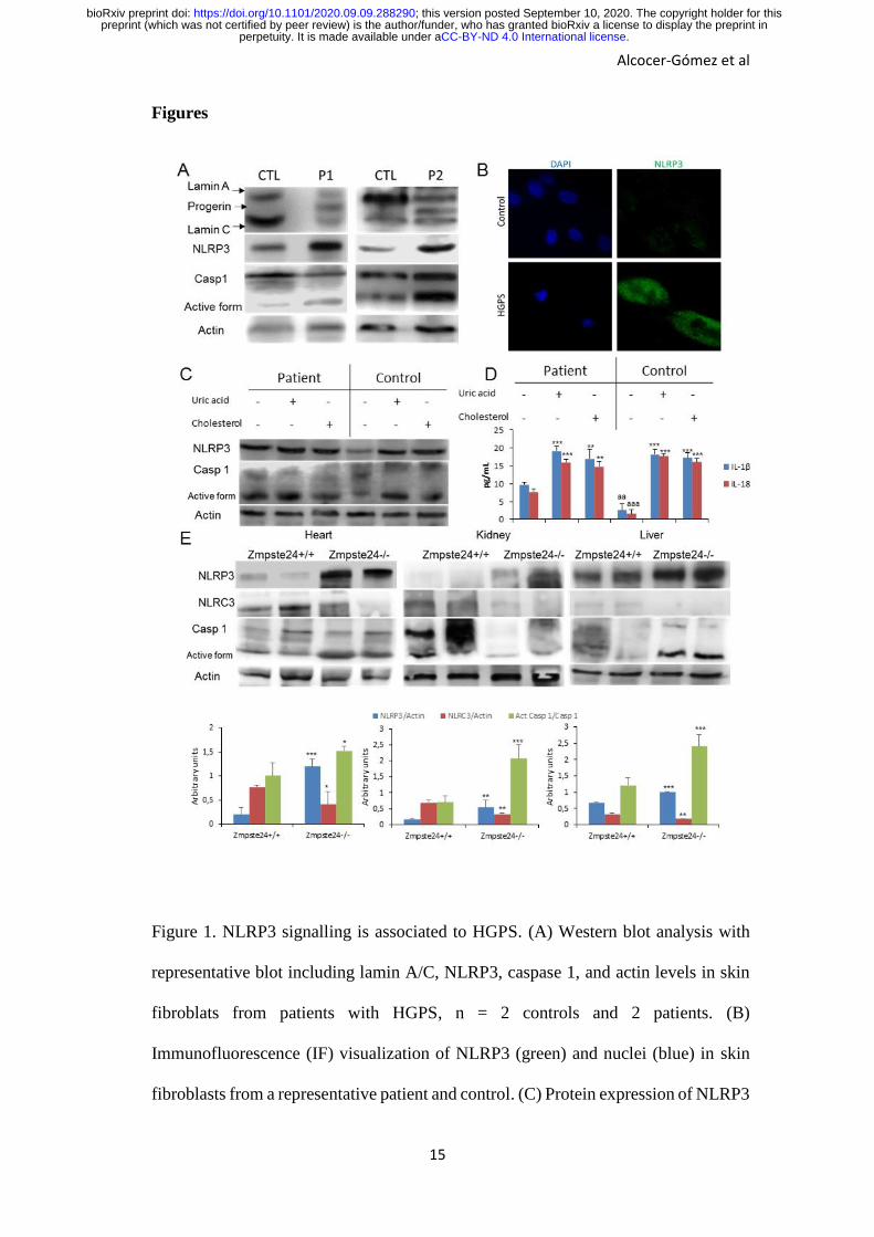
HGPS patients show NLRP3-inflammasome hyperactivation
To evaluate the role of the NLRP3-inflammasome in progeria syndrome, we examined
the NLRP3-inflammasome complex expression in HGPS fibroblasts and age‐ and
passage‐matched controls. As shown in Fig. 1A, HGPS demonstrates a significant
increment in NLRP3 and caspase 1 protein expression. This high expression of NLRP3
was also observed in skin fibroblasts by immunofluorescence, in which, NLRP3 was
localized in cytosol (Fig. 1B).
.CC-BY-ND 4.0 International licenseperpetuity. It is made available under apreprint (which was not certified by peer review) is the author/funder, who has granted bioRxiv a license to display the preprint in
The copyright holder for thisthis version posted September 10, 2020. ; https://doi.org/10.1101/2020.09.09.288290doi: bioRxiv preprint
Alcocer-Gómez et al
8
It is known that some soluble circulating factors can induce cardiac and metabolic damage
and systemic inflammation (12). Furthermore, metabolic alterations have been observed
in animal models HGPS with lipid accumulation (13). To evaluate the grade of response
of NLRP3-inflammasome complex in HGPS after metabolic alterations, we exposed
HGPS lymphoblasts and age‐ and passage‐matched controls to cholesterol crystals and
uric acid, two well known metabolic inductor of the NLRP3 activation and associated to
aging (1, 4). Interestingly, HGPS cells showed increased basal levels of NLRP3 and
caspase 1 protein expression with a moderate increment after cholesterol and uric acid
compared to control lymphoblasts (Fig.1C). These high expression was accompanied to
increased IL-1β and IL-18 release with high basal levels in HGPS, as well (Fig. 1D).
Zmpste24-deficient mice show NLRP3-inflammasome hyperactivation
To try to extend these observations to an in vivo model, we evaluated the inflammasome
complex status in a murine model of progeria. For this purpose, we used a mouse model
with the absence of Zmpste24 metalloproteinase which leads to a progeroid phenotype
similar to the human premature aging syndrome. After to exam the basal inflammasome
levels of diverse tissues from wild-type and Zmpste24-/- mice, we observed an evident
increment of the Nlrp3 and caspase 1 protein expression in heart, kidney and liver tissues
associated to reduced expression of Nlrc3 (Fig. 1E) which was not observed in other
tissues such as muscle and lung (Fig. S1). The specific function of the NLRP3-
inflammasome complex in the pathophysiology of HGPS was reinforced by the
observation that there were no changes in other inflammasomes (Fig. S2).
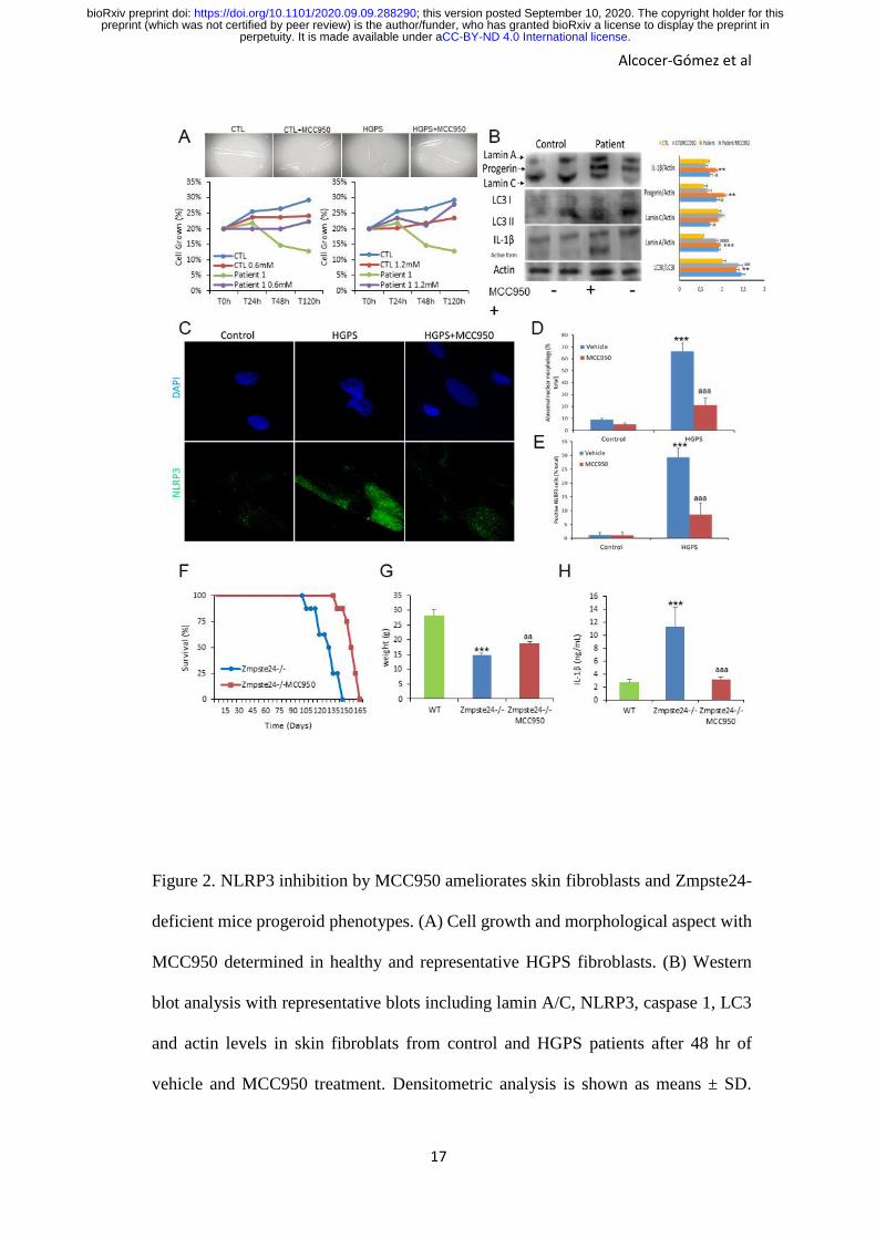
NLRP3-inflammasome inhibition improves progerian phenotype
To examine whether pharmacological inhibition of NLRP3 could be an effective
treatment in HGPS, we next assessed the effect of MCC950 on mutant fibroblasts. Control
and HGPS fibroblasts from a representative patient were treated at two different doses
.CC-BY-ND 4.0 International licenseperpetuity. It is made available under apreprint (which was not certified by peer review) is the author/funder, who has granted bioRxiv a license to display the preprint in
The copyright holder for thisthis version posted September 10, 2020. ; https://doi.org/10.1101/2020.09.09.288290doi: bioRxiv preprint
Alcocer-Gómez et al
9
(0.6 and 1.2mM) of MCC950. The results showed a statistically significant dose-
dependent increment of growth rate in patient fibroblasts with cell morphology
normalization (Fig. 2A). Western blot analyses also showed that the levels of lamin A
and C levels remained relatively constant in both control and HGPS cells treated with
MCC950 (to low doses 0.6mM after 48h); conversely, progerin signals in MCC950-
treated HGPS cells were decreased accompanied by a reduction of IL-1β and an
increment of autophagy protein LC3 (Fig. 2B). Interestingly, the NLRP3 inhibition
reduced the frequency of abnormal nuclear morphology in both control and HGPS
fibroblasts after 48 hours with a correlative inhibition of NLRP3 expression by
immunostaining (Fig. 2C-E).
NLRP3-inflammasome extend longevity in Zmpste24-deficient mice
Finally, in this work we explored the in vivo effect of the pharmacological inhibition of
Nlrp3 in Zmpste24-/- mice. MCC950 was administered by i.p. route at 20mg/kg daily.
Interestingly, Zmpste24-/- mice treated with MCC950 significantly extended the longevity
of Zmpste24-/- mice with an increment in mean lifespan of 19.2% and in maximum
lifespan of 13.9%, improved body weight from 14.7 ± 0.7 g to 18.6 ± 0.7 g (P < 0.001),
and reduced IL-1β (Fig. 2F-H).
Discussion
During the last years, the role of the NLRP3-inflammasome during aging has been subject
to study. Genetic deletion of Nlrp3 in mice has been shown to improve lifespan and health
by attenuating multiple age-related degenerative changes such as cardiac aging, insulin
sensitivity with glycemic control, bone loss, cognitive function and motor performance
(4-6). Further, NLRP3 has been studied in cardiovascular diseases. NLRP3
inflammasome is up-regulated in atherosclerosis, myocardial infarction, ischemic heart
.CC-BY-ND 4.0 International licenseperpetuity. It is made available under apreprint (which was not certified by peer review) is the author/funder, who has granted bioRxiv a license to display the preprint in
The copyright holder for thisthis version posted September 10, 2020. ; https://doi.org/10.1101/2020.09.09.288290doi: bioRxiv preprint
Alcocer-Gómez et al
10
disease, chronic heart failure, or hypertension (5). In this context, cardiovascular diseases
have been shown accelerated in HGPS patients and progerin, the abnormal form of
prelamin-A, has also been shown to induces atherosclerosis and cardiac
electrophysiological alterations (14). Furthermore, exogenously expressed progerin was
showed increases inflammation (15). On the other hand, the AIM2 inflammasome has
been shown to be activated after pharmacological alterations of the nuclear envelope
integrity compatible with laminopathies (16). For this reasons, it is tempting to speculate
that the inflammasomes could contribute to increased aging observed in progeroid
syndromes.
During the last years, an effort has been done to find specific pharmacological inhibitors
of NLRP3. While several of these inhibitors have been shown to have a specific direct
action on NLRP3, others have been shown indirect inhibitory effects (17). MCC950 and
analogues are a specific small-molecule inhibitors of the NLRP3, with remarkable
therapeutic potential in human diseases.
Many strategies to treat HGPS have been studied but anti-inflammatories have been
poorly studied (9). Interestingly, several of these strategies have anti-inflammatory
potential and many of these have been shown to induce indirect inhibition of NLRP3 such
metformin, resveratrol, rapamycin, quercetin or spermidine (4).
In the current study, we provide evidence showing that the NLRP3-inflammasome
complex is an important determinant of HGPS in human cells and mice. We show that
the specific expression of NLRP3 and other components of the complex rapidly increase
in the skin fibroblasts from patients and the specific tissues associated to the human
progeroid phenotype of an animal model such as heart, liver and heart (8,9) where
previously were described inflammatory phenotype (10). Interestingly, we also observed
reduced levels of Nlrc3 which is a negative regulator of inflammatory signaling pathways
.CC-BY-ND 4.0 International licenseperpetuity. It is made available under apreprint (which was not certified by peer review) is the author/funder, who has granted bioRxiv a license to display the preprint in
The copyright holder for thisthis version posted September 10, 2020. ; https://doi.org/10.1101/2020.09.09.288290doi: bioRxiv preprint
Alcocer-Gómez et al
11
and NLRP3 inflammasome (18, 19). Moreover, we show the continuous activation of the
NLRP3 inflammasome in immunological cells like lymphocytes from patients. Finally,
pharmacological inhibition of NLRP3 by MCC950 treatment improved cell survival and
morphology, reduced inflammation and, in HGPS mice resulted in extension of longevity
and the prevention of inflammasome-dependent inflammatory event. Therefore, this
HGPS treatment strategy, focused on the inhibition of NLRP3-inflammasome complex,
could constitute an alternative therapy to slow disease progression in patients with
progeria.
Acknowledgments
This study was supported by a grant from the Andalusian regional government (Grupo
de Investigacion Junta de Andalucia CTS113 and Consejería de Salud de la Junta de
Andalucia: PI-0036-2014).
Author Disclosure Statement
The authors declare that no conflict of interest exists for any of them.
.CC-BY-ND 4.0 International licenseperpetuity. It is made available under apreprint (which was not certified by peer review) is the author/funder, who has granted bioRxiv a license to display the preprint in
The copyright holder for thisthis version posted September 10, 2020. ; https://doi.org/10.1101/2020.09.09.288290doi: bioRxiv preprint
Alcocer-Gómez et al
12
References
1. C. López-Otín, M.A. Blasco, L. Partridge, M. Serrano, G. Kroemer, The
hallmarks of aging. Cell. 153, 1194-217 (2013).
2. C. Franceschi, et al., Inflamm-aging. An evolutionary perspective on
immunosenescence. Ann N Y Acad Sci. 908, 244–254 (2006).
3. I.S. Afonina, Z. Zhong, M. Karin, R. Beyaert, Limiting inflammation-the negative
regulation of NF-κB and the NLRP3 inflammasome. Nat Immunol. 18,861-869
(2017).
4. M.D. Cordero, M.R. Williams, B. Ryffel, AMP-Activated Protein Kinase
Regulation of the NLRP3 Inflammasome during Aging. Trends Endocrinol
Metab. 29, 8-17 (2018).
5. F. Marín-Aguilar, et al, NLRP3 inflammasome suppression improves longevity
and prevents cardiac aging in male mice. Aging Cell. 19:e13050 (2020).
6. Y.H. Youm, et al, Canonical Nlrp3 inflammasome links )systemic low-grade
inflammation to functional decline in aging. Cell Metab. 18,519-32 (2013).
7. J.M. Navarro-Pando, et al., Inhibition of the NLRP3 inflammasome prevents
ovarian aging. bioRxiv 2020.04.26.062646; doi:
https://doi.org/10.1101/2020.04.26.062646
8. K.H. Schreiber, B.K. Kennedy, When lamins go bad: nuclear structure and
disease. Cell. 152, 1365-75 (2013).
9. W.F. Lai, W.T. Wong, Progress and trends in the development of therapies for
Hutchinson-Gilford progeria syndrome. Aging Cell. 19, e13175 (2020).
.CC-BY-ND 4.0 International licenseperpetuity. It is made available under apreprint (which was not certified by peer review) is the author/funder, who has granted bioRxiv a license to display the preprint in
The copyright holder for thisthis version posted September 10, 2020. ; https://doi.org/10.1101/2020.09.09.288290doi: bioRxiv preprint
Alcocer-Gómez et al
13
10. F. G. Osorio, et al., Nuclear lamina defects cause ATM‐dependent NF‐kappaB
activation and link accelerated aging to a systemic inflammatory response.
Genes & Development. 2620, 2311–2324 (2012).
11. E.I. Elliott, F.S. Sutterwala, Initiation and perpetuation of NLRP3
inflammasome activation and assembly. Immunol Rev. 265,35-52 (2015).
12. P. Libby, M. Nahrendorf, F.K. Swirski, Leukocytes Link Local and Systemic
Inflammation in Ischemic Cardiovascular Disease: An Expanded "Cardiovascular
Continuum". J Am Coll Cardiol. 67,1091-103 (2016).
13. G. Mariño, et al., Premature aging in mice activates a systemic metabolic
response involving autophagy induction. Hum Mol Genet. 17, 2196-211 (2008).
14. M.R. Hamczyk, L. del Campo, V. Andrés, Aging in the Cardiovascular System:
Lessons from Hutchinson-Gilford Progeria Syndrome. Annu Rev Physiol. 80,
27-48 (2018).
15. G. Bidault, et al., Progerin Expression Induces Inflammation, Oxidative Stress
and Senescence in Human Coronary Endothelial Cells. Cells. 9, 1201 (2020).
16. A. Di Micco, et al., AIM2 inflammasome is activated by pharmacological
disruption of nuclear envelope integrity. Proc Natl Acad Sci U S A. 113, E4671-
80 (2016).
17. A. Zahid, B. Li, A.J.K. Kombe, T. Jin, J. Tao, Pharmacological Inhibitors of the
NLRP3 Inflammasome. Front Immunol. 10, 2538 (2019).
18. T. Uchimura, et al., The Innate Immune Sensor NLRC3 Acts as a Rheostat that
Fine-Tunes T Cell Responses in Infection and Autoimmunity. Immunity. 49,
1049-1061.e6 (2018).
.CC-BY-ND 4.0 International licenseperpetuity. It is made available under apreprint (which was not certified by peer review) is the author/funder, who has granted bioRxiv a license to display the preprint in
The copyright holder for thisthis version posted September 10, 2020. ; https://doi.org/10.1101/2020.09.09.288290doi: bioRxiv preprint
Alcocer-Gómez et al
14
19. E. Eren, M. Berber, N. Özören, NLRC3 protein inhibits inflammation by
disrupting NALP3 inflammasome assembly via competition with the adaptor
protein ASC for pro-caspase-1 binding. J Biol Chem. 292, 12691-12701 (2017).
.CC-BY-ND 4.0 International licenseperpetuity. It is made available under apreprint (which was not certified by peer review) is the author/funder, who has granted bioRxiv a license to display the preprint in
The copyright holder for thisthis version posted September 10, 2020. ; https://doi.org/10.1101/2020.09.09.288290doi: bioRxiv preprint
Alcocer-Gómez et al
15
Figures
Figure 1. NLRP3 signalling is associated to HGPS. (A) Western blot analysis with
representative blot including lamin A/C, NLRP3, caspase 1, and actin levels in skin
fibroblats from patients with HGPS, n = 2 controls and 2 patients. (B)
Immunofluorescence (IF) visualization of NLRP3 (green) and nuclei (blue) in skin
fibroblasts from a representative patient and control. (C) Protein expression of NLRP3
.CC-BY-ND 4.0 International licenseperpetuity. It is made available under apreprint (which was not certified by peer review) is the author/funder, who has granted bioRxiv a license to display the preprint in
The copyright holder for thisthis version posted September 10, 2020. ; https://doi.org/10.1101/2020.09.09.288290doi: bioRxiv preprint
Alcocer-Gómez et al
16
and caspase 1 in lymphocytes from control and one patient after stimulation with uric
acid and cholesterol crystal. (D) IL-1β and IL-18 medium release from lymphoblasts.
which were assessed after a 24 hr incubation with uric acid and cholesterol. ***P <
0.001, **P < 0.005, *P < 0.05 treatment vs no treatment; aaaP < 0.001; aaP < 0.01
control cells vs patient cells. (E) Western blot analysis with representative blot
including NLRP3, NLRC3, caspase 1, and actin levels in heart, kidney and liver
tissues from wild-type and Zmpste24-/- mice. Densitometric analysis is shown as
means ± SD, n = 5 mice per group. ***P < 0.001, **P < 0.005, *P < 0.05 wild-type
vs Zmpste24-/- mice.
.CC-BY-ND 4.0 International licenseperpetuity. It is made available under apreprint (which was not certified by peer review) is the author/funder, who has granted bioRxiv a license to display the preprint in
The copyright holder for thisthis version posted September 10, 2020. ; https://doi.org/10.1101/2020.09.09.288290doi: bioRxiv preprint
Alcocer-Gómez et al
17
Figure 2. NLRP3 inhibition by MCC950 ameliorates skin fibroblasts and Zmpste24-
deficient mice progeroid phenotypes. (A) Cell growth and morphological aspect with
MCC950 determined in healthy and representative HGPS fibroblasts. (B) Western
blot analysis with representative blots including lamin A/C, NLRP3, caspase 1, LC3
and actin levels in skin fibroblats from control and HGPS patients after 48 hr of
vehicle and MCC950 treatment. Densitometric analysis is shown as means ± SD.
.CC-BY-ND 4.0 International licenseperpetuity. It is made available under apreprint (which was not certified by peer review) is the author/funder, who has granted bioRxiv a license to display the preprint in
The copyright holder for thisthis version posted September 10, 2020. ; https://doi.org/10.1101/2020.09.09.288290doi: bioRxiv preprint
Alcocer-Gómez et al
18
***P < 0.001, **P < 0.005, *P < 0.05 no treatment vs treatment; aaaP < 0.001; aaP <
0.01 control cells vs patient cells. (C and D) Representative fluorescence images of
HGPS and control fibroblasts to evaluate the effect of the MCC950 in the nuclear
morphology and NLRP3 expression. (E) Kaplan-Meier graph showing a significant
increase in the maximum lifespan in WT mice compared with Zmpste24-/- mice. N=7
per group (F) Body weights of the groups over time. (G) Analysis of serum
concentrations of IL-1β measured by ELISA. N=6 per group. Data are shown as
means ± SD. ***P < 0.001, **P < 0.005, *P < 0.05 wild-type vs Zmpste24-/- mice;
aaaP < 0.001; aaP < 0.01 vehicle vs MCC950.
.CC-BY-ND 4.0 International licenseperpetuity. It is made available under apreprint (which was not certified by peer review) is the author/funder, who has granted bioRxiv a license to display the preprint in
The copyright holder for thisthis version posted September 10, 2020. ; https://doi.org/10.1101/2020.09.09.288290doi: bioRxiv preprint

Alcocer-Gómez et al
19
Supplementary Figure 1. NLRP3-inflammasomes expression in lung and muscle from
progeroid animals. Western blot analysis with representative blot including NLRP3,
NLRC3, caspase 1, and actin levels in lung and muscle tissues from wild-type and
Zmpste24-/- mice. Densitometric analysis is shown as means ± SD, n = 5 mice per
group.
.CC-BY-ND 4.0 International licenseperpetuity. It is made available under apreprint (which was not certified by peer review) is the author/funder, who has granted bioRxiv a license to display the preprint in
The copyright holder for thisthis version posted September 10, 2020. ; https://doi.org/10.1101/2020.09.09.288290doi: bioRxiv preprint

Alcocer-Gómez et al
20
Supplementary Figure 2. Other inflammasomes are not involved in heart and liver from
pathophysiology of progeroid animals. Western blot analysis with representative blot
including Nlrc4, Nalp1, Nalp10, and actin levels in heart and liver tissues from wild-
type and Zmpste24-/- mice. Densitometric analysis is shown as means ± SD, n = 5 mice
per group.
.CC-BY-ND 4.0 International licenseperpetuity. It is made available under apreprint (which was not certified by peer review) is the author/funder, who has granted bioRxiv a license to display the preprint in
The copyright holder for thisthis version posted September 10, 2020. ; https://doi.org/10.1101/2020.09.09.288290doi: bioRxiv preprint