Embed Size (px)
Citation preview
Newborn Infant With Inherited Ring and De NovoInterstitial Deletion on HomologousChromosome 22s
Sharon L. Wenger,3* Leslie Y. Boone,1 James H. Cummins,1 Maria A. Del Vecchio,1 Carolyn A. Bay,1,2
Marybeth Hummel,4 and Patricia A. Mowery-Rushton1
1Children’s Hospital of Pittsburgh, Division of Medical Genetics, Pittsburgh, Pennsylvania2Department of Human Genetics, University of Pittsburgh, Graduate School of Public Health,Pittsburgh, Pennsylvania
3Department of Pathology, West Virginia University, Morgantown, West Virginia4Department of Pediatrics, West Virginia University, Morgantown, West Virginia
A 2-day-old infant was evaluated and sus-pected of having 22q11.2 deletion based onmicrocephaly, short and narrow palpebralfissures, a prominent nose with hypoplasticalae nasi, thin fingers, and a right aorticarch. He also had an imperforate anus,which is not in the del 22q11.2 syndrome.Karyotype analysis identified a ring 22,while fluorescence in situ hybridization(FISH) for the DiGeorge syndrome criticalregion identified a 22q deletion on the otherhomologue. The karyotype designation was46,XY,r(22)(p13q13.3).ish del(22)(q11.2q11.2)(D22S75-). Both parents function in themildly mentally retarded range. The fa-ther’s karyotype was normal whereas themother had the ring 22 that was inheritedby her son. This is the first case reported forabnormalities on both 22 homologues. Am.J. Med. Genet. 91:351–354, 2000.© 2000 Wiley-Liss, Inc.
KEY WORDS: anal atresia; fluorescence insitu hybridization; velocar-diofacial syndrome; 22qterdeletion
INTRODUCTION
Constitutional chromosomal abnormalities occurringon homologous chromosomes, with the exception of ac-rocentrics, are extremely rare.Three previous cases in-clude 46,XX,t(4;11)(q31.3;q14)de novo,t(6;11)(q24;q21)mat [Eiben et al., 1988], 46,XX,t(9;9)(9pter→cen→
9pter;9qter→cen::9q13→9qter)de novo [Nakamura etal., 1990] and 46,XY,t(2;3)(p25;p23),t(3;12)(p25;p13),t(4;6)(p16;q21)de novo [Del Porto et al., 1991].
The incidence of del(22)(q11.2), which is associatedwith variable expressivity for DiGeorge syndrome, ve-locardiofacial syndrome, and conotruncal anomaly facesyndrome has been estimated to be 1/4,000 [Devriendtet al., 1998a]. Common characteristics include mentalimpairment, cardiac defects, hypocalcemia, thymic andparathyroid abnormalities, cleft palate or velopharyn-geal insufficiency, renal abnormalities, and minor fa-cial anomalies [Demczuk and Aurias, 1995; Leana-Coxet al., 1996; Ryan et al., 1997].
Ring 22 chromosomes are rare. The breakpoint in-volving loss of short arm material is of no clinical con-sequence, whereas breakpoint on the long arm, whichcan vary in size, can affect the phenotypic expressiondependent on the size of the deletion. Manifestations of22q telomere deletions include developmental delay,normal or accelerated growth, hypotonia, delay or ab-sence of speech, and minor facial anomalies [Doheny etal., 1997; Nesslinger et al., 1994; Schroder et al., 1998],which is similar to the phenotype reported for ring 22[Hunter et al., 1977].
We report on a case with different chromosome ab-normalities involving both 22 homologues.
CLINICAL REPORT
The patient was the product of a 39-week gestationto a 22-year-old G1 white woman and 25-year-old fa-ther. The pregnancy was further complicated by mildpolyhydramnios noted on ultrasonography in the thirdtrimester. The infant was delivered by C-section sec-ondary to variable decelerations of heart rate on a non-stress test. Birth weight was 3.12 kg (25–50th centile),length was 48.9 cm (20–25th centile), and head circum-ference was 32 cm (<5th centile). Apgar scores were 7and 8 at one and five minutes. An imperforate anuswith a perianal fistula were noted. The infant was in-
*Correspondence to: Sharon L. Wenger, Ph.D., Department ofPathology, West Virginia University, P.O. Box 9203, Morgan-town, WV 26506-9203. E-mail: [email protected]
Received 26 July 1999; Accepted 11 January 2000
American Journal of Medical Genetics 91:351–354 (2000)
© 2000 Wiley-Liss, Inc.
tubated due to respiratory distress and transported toa tertiary care hospital for further evaluation andtreatment.
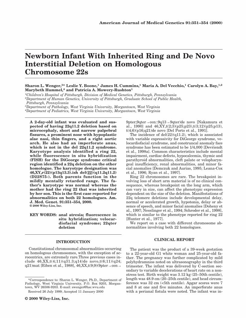
The infant underwent a colostomy on his second dayof life. Cardiology evaluation noted a right aortic arch,vascular ring, patent foreman ovale, and patent ductusarteriosus. Renal and spinal ultrasound findings werenormal. A genetic evaluation at 13 days showed signifi-cant microcephaly (OFC 32.5 cm; <5th centile);“square” face; bitemporal narrowing; a ridge on theskull perpendicular to the sagittal suture; short, nar-row palpebral fissures; anteriorly folded superior heli-ces of the ears; two small but prominent tags on theright lower ear lobe and one on the left ear lobe; aprominent nose, nasal root, and tip with hypoplasticalae nasi; a slightly simple philtrum; and a smallmouth. The patient also had a pilonidal dimple, thinfingers with hyperconvex nails of the fourth and fifthdigits on his left hand, and hypoplastic distal toenails,especially of the great toes bilaterally. Neurologic sta-tus was unremarkable at 2 weeks. The short narrowpalpebral fissures, prominent nasal root and tip, hypo-plastic alae nasi, and right aortic arch are frequentmanifestations of del 22q11.2 (Fig.1a,b).
At 20 months the patient was enrolled in an earlyintervention program. His motor skills are age-appropriate with minimal expressive language (10–14month level). His growth acceleration decreased and is3–4 standard deviations below the mean. His weight isin the 5–10th centile.
The mother has two normal half sibs and a niece onher father’s side with hyperactivity. She has learningdisabilities and graduated from high school attendingspecial education classes. She has a head circumfer-
ence at the 75th centile. The father also has learningdisabilities and a head circumference >95th centile.The father reportedly has a normal daughter by a pre-vious relationship and a 3-year-old nephew who is re-ported to have seizures, developmental delay, and ab-sence of speech.
MATERIALS AND METHODS
Peripheral blood samples from the patient and hisparents were processed for karyotype analysis usingroutine cytogenetic techniques and GTG-banded. FISHstudies were performed using the DiGeorge syndromecritical region probe D22S75 at 22q11.2 and markerprobe STS WI-941 at 22q13 (direct labeled single colorprobes on patient) or D22S39 at 22q13.3 (indirect la-beled single color probes on mother) (Oncor) followingmanufacturer’s instructions.
RESULTS
The child’s karyotype contained a ring 22 in all 21cells analyzed with both the DiGeorge and markerprobes present, whereas FISH for the DiGeorge probewas deleted at 22q11.2 on the other 22 homologue. Thefather’s karyotype was 46,XY.ish 22(q11.2)(D22S75x2).The mother’s karyotype was 46,XX,r(22) (p13q13.3).ish22(q11.2)(D22S75x2) in 50 cells examined; there wasno evidence of mosaicism for a normal cell line. Neitherthe DiGeorge syndrome region nor the marker probewas deleted in the ring chromosome, suggesting thatthe breakpoints are 22p and distal to D22S39. Among50 cells examined, 3 cells lacked the ring, and 1 cell hada double-size ring, which could be attributed to tissue
Fig. 1. Patient at (a) 3 months and (b) 6 months of age
352 Wenger et al.

culture artifact. Therefore, the child’s karyotype was46,XY,r(22)(p13q13.3) mat.ish del(22)(q11.2q11.2)(D22S75-)de novo (Figs. 2 and 3).
DISCUSSION
The occurrence of different abnormalities on homolo-gous chromosomes is rare, and to our knowledge ourpatient is the first case reported with abnormalitiesaffecting both chromosome 22s. The r(22) was inheritedfrom the mother, whereas the deletion 22q11.2 was anew mutation, presumably paternal. Therefore, the pa-tient was hemizygous for 22q11.2 and 22qter.
The incidence of the 22q11.2 deletion is 1/4,000[Devriendt et al., 1998a]. The high incidence is the re-sult of unequal meiotic recombination in this regiondue to sequence homology at the breakpoints [Baumeret al., 1998]. Over 25% of 22q deletions are inheritedfrom a parent [Brondum-Nielsen and Christensen,1996; Ryan et al. 1997]. The phenotype among relativesis highly variable [Lindsay et al., 1995], even amongmonozygotic twins [Goodship et al., 1995; Yamagishi etal., 1998]. Our patient’s abnormalities consistent withthe 22q11.2 deletion included facial anomalies and car-diac defect. Polyhydramnios has been reported in eightpatients with del(22)(q11.2) and conotruncal heart de-fect or uropathy [Devriendt et al., 1998b]. Anal atresiawas reported recently in 22q11.2 deletions with cono-truncal anomaly face syndrome and velocardiofacial/DiGeorge syndrome [Kerstjens-Frederikse et al., 1999;Matsuoka et al., 1998; Yamagishi et al., 1998].
Ring 22 chromosomes are rare, and only four caseshave been reported as being inherited. Two familieshad a normal phenotype in both parent and child.
While no breakpoints were given, missing material ap-peared to be minimal by size of the ring chromosome[Crusi and Engel, 1986; Teyssier and Moreau, 1985]. Inanother family the mentally retarded child had inher-ited the ring from a normal mosaic mother, 46,XX/46,XX,r(22)/45,XX,t(15;22)(p11;q11); no breakpointwas given for the ring 22 [Fryns and Van den Berghe,1979]. A fourth family had a 22q13 breakpoint withlittle chromosomal material lacking by size. Relativeswere reported to be mosaic for a normal cell line, with40–50% of cells containing the ring in a mother withmild mental retardation and minor anomalies, in herunaffected father, and in her unaffected children [Stolland Roth, 1983]. Formation of ring chromosomes in-volves breakpoints on the p and distal q arms, suchthat the phenotype is related to a terminal 22q dele-tion. Reports in the literature have not included mo-lecular studies to define breakpoints in these ring 22cases. However, molecular studies have been con-ducted on 22q terminal deletions. Of those reported,the deletion has included D22S39 (Table I). Our pa-tient shows little effect due to the 22qter deletion. At 20months of age he has language delay but currently doesnot show significant developmental delay. The 22qtelomeric deletion on his ring chromosome is smallerthan cryptic terminal deletions recently identified[Doheny et al., 1997; Precht et al., 1998] since theD22S39 FISH probe was present on the ring chromo-some. The de novo 22q deletion most likely is unrelatedto the ring 22, since these microdeletions are relativelyfrequent. Although the phenotypes for both of theseconditions are variable, the occurrence of hemizygosityfor both of these regions in our patient present as arelatively mild phenotype.
REFERENCES
Baumer A, Dutly F, Balmer D, Riegel M, Tukel T, Krajewska-Walasek M,Schinzel AA. 1998. High level of unequal meiotic crossovers at theorigin of the 22q11.2 and 7q11.23 deletions. Hum Mol Genet 7:887–894.
Brondum-Nielsen K, Christensen K. 1996. Chromosome 22q11 deletionand other chromosome aberrations in cases with cleft palate, congeni-tal heart defects and/or mental disability. A survey based on the Dan-ish facial cleft register. Clin Genet 50:116–120.
TABLE I. Characteristics in Terminal and InterstitialDeletions on Chromosome 22
FindingsRing(22)
del(22)(q13.31)a
del(22)(q11.2q11.2)
Ourpatient
Mental retardation/delay X X X X
Small head X XHeart defect XThymic abnormality XHypocalcemia XCleft palate XRenal abnormality XFacial dysmorphism X X X XHypotonia X XSpeech delay X X X
aRing 22 phenotype: Hunter et al., 1977; Stoll and Roth, 1983; a breakpointis proximal to D22S39 phenotype: Nesslinger et al., 1994; Doheny et al.,1997; Schroder et al., 1998; Precht et al., 1998; interstitial deletion22q11.2: Demczuk and Aurias, 1995; Leana-Cox, 1996; Ryan et al., 1997.
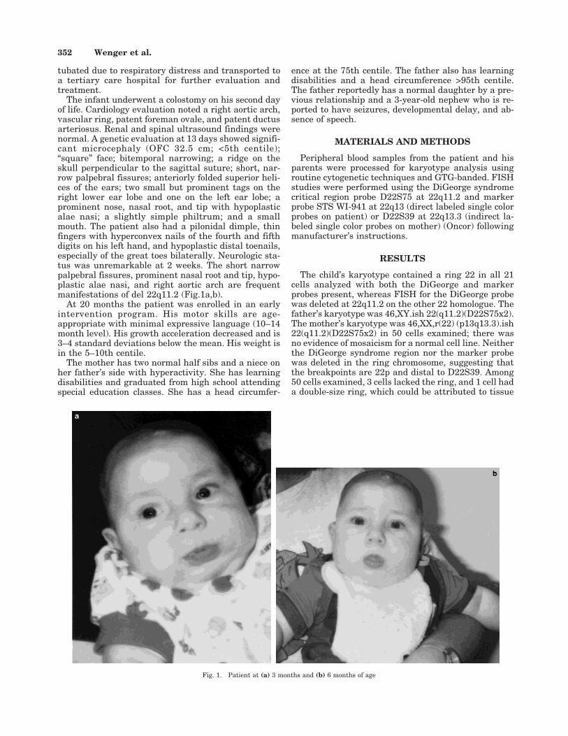
Fig. 2. Partial 22 karyotypes for the mother (left) and patient (right).Ring chromosomes are on right of each pair.
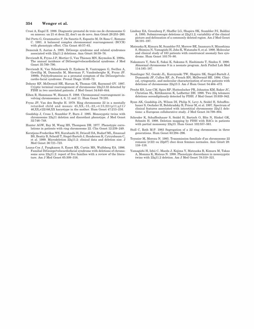
Fig. 3. FISH for DiGeorge deletion in patient; deleted chromosome is onthe left with two signals for the telomere probe, and ring chromosome is onthe right with two signals for the telomere probe and single smaller signalfor the DiGeorge syndrome critical region above the right marker signal.
Ring 22 and 22q11.2 Deletion 353
Crusi A, Engel E. 1986. Diagnostic prenatal de trois cas de chromosome Gen anneau: un 21 et deux 22, don’t un de novo. Ann Genet 29:253–260.
Del Porto G, Grammatico P, De Sanctis S, Esposito M, Di Rosa C, RomanoC. 1991. A balanced complex chromosomal rearrangement (BCCR)with phenotypic effect. Clin Genet 40:57–61.
Demczuk S, Aurias A. 1995. DiGeorge syndrome and related syndromesassociated with 22q11.2 deletions. Ann Genet 38:59–76.
Devriendt K, Fryns J-P, Mortier G, Van Thienen MN, Keymolen K. 1998a.The annual incidence of DiGeorge/velocardiofacial syndrome. J MedGenet 35:789–790.
Devriendt K, Van Schoubroeck D, Eyskens B, Vantrappen G, Swillen A,Gewillig M, Dumoulin M, Moerman P, Vandenberghe K, Fryns JP.1998b. Polyhydramnios as a prenatal symptom of the DiGeorge/velo-cardio-facial syndrome. Prenat Diagn 18:68–72.
Doheny KF, McDermid HE, Harum K, Thomas GH, Raymond GV. 1997.Cryptic terminal rearrangement of chromosome 22q13.32 detected byFISH in two unrelated patients. J Med Genet 34:640–644.
Eiben B, Hammans W, Hansen S. 1988. Chromosomal rearrangement in-volving chromosomes 4, 6, 11 and 11. Hum Genet 79:193.
Fryns JP, Van den Berghe H. 1979. Ring chromosome 22 in a mentallyretarded child and mosaic 45,XX,-15,-22,+t(15;22)(p11;q11)/46,XX,r(22)/46,XX karyotype in the mother. Hum Genet 47:213–216.
Goodship J, Cross I, Scambler P, Burn J. 1995. Monozygotic twins withchromosome 22q11 deletion and discordant phenotype. J Med Genet32:746–748.
Hunter AGW, Ray M, Wang HS, Thompson DR. 1977. Phenotypic corre-lations in patients with ring chromosome 22. Clin Genet 12:239–249.
Kerstjens-Frederikse WS, Kurahashi H, Driscoll DA, Budarf ML, EmanuelBS, Beatty B, Scheidl T, Siegel-Bartelt J, Henderson K, Cytrynbaum C,et al. 1999. Microdeletion 22q11.2: clinical data and deletion size. JMed Genet 36:721–723.
Leana-Cox J, Pangkanon S, Eanet KR, Curtin MS, Wulfsberg EA. 1996.Familial DiGeorge/velocardiofacial syndrome with deletions of chromo-some area 22q11.2: report of five families with a review of the litera-ture. Am J Med Genet 65:309–316.
Lindsay EA, Greenberg F, Shaffer LG, Shapira SK, Scambler PJ, BaldiniA. 1995. Submicroscopic deletions at 22q11.2; variability of the clinicalpicture and delineation of a commonly deleted region. Am J Med Genet56:191–197.
Matsuoka R, Kimura M, Scambler PJ, Morrow BE, Imamura S, MinoshimaS, Shimizu N, Yamagishi H, Joho K, Watanabe S, et al. 1998. Molecularand clinical study of 183 patients with conotruncal anomaly face syn-drome. Hum Genet 103:70–80.
Nakamura Y, Sato E, Sakai K, Sakuma S, Hashimoto T, Sindou S. 1990.Abnormal chromosome 9 in a neonate program. Arch Pathol Lab Med114:185–187.
Nesslinger NJ, Gorski JL, Kurczynski TW, Shapira SK, Siegel-Bartelt J,Dumanski JP, Cullen RF, Jr, French BN, McDermid HE. 1994. Clini-cal, cytogenetic, and molecular characterization of seven patients withdeletions of chromosome 22q13.3. Am J Hum Genet 54:464–472.
Precht KS, Lese CM, Spiro RP, Huttenlocher PR, Johnston KM, Baker JC,Christian SL, Kittikamron K, Ledbetter DH. 1998. Two 22q telomeredeletions serendipitously detected by FISH. J Med Genet 35:939–942.
Ryan AK, Goodship JA, Wilson DI, Philip N, Levy A, Seidel H, Schuffen-hauer S, Oechsler H, Belohradsky B, Prieur M, et al. 1997. Spectrum ofclinical features associated with interstitial chromosome 22q11 dele-tions: a European collaborative study. J Med Genet 34:798–804.
Schroder K, Schuffenhauer S, Seidel H, Bartsch O, Blin N, Hinkel GK,Schmitt H. 1998. Deletion mapping by FISH with BACs in patientswith partial monosomy 22q13. Hum Genet 102:557–561.
Stoll C, Roth M-P. 1983 Segregation of a 22 ring chromosome in threegenerations. Hum Genet 63:294–296.
Teyssier M, Moreau N. 1985. Transmission familiale d’un chromosome 22remanie [r(22) ou 22p0?] chez deux femmes normales. Ann Genet 28:116–118.
Yamagishi H, Ishii C, Maeda J, Kojima Y, Matsuoka R, Kimura M, TakaoA, Momma K, Matsuo N. 1998. Phenotypic discordance in monozygotictwins with 22q11.2 deletion. Am J Med Genet 78:319–321.
354 Wenger et al.