Embed Size (px)
Citation preview

THORACIC ANESTHESIA
A. NEYRINCK

LUNG ISOLATION DEVICES

indications for one -lung ventilation
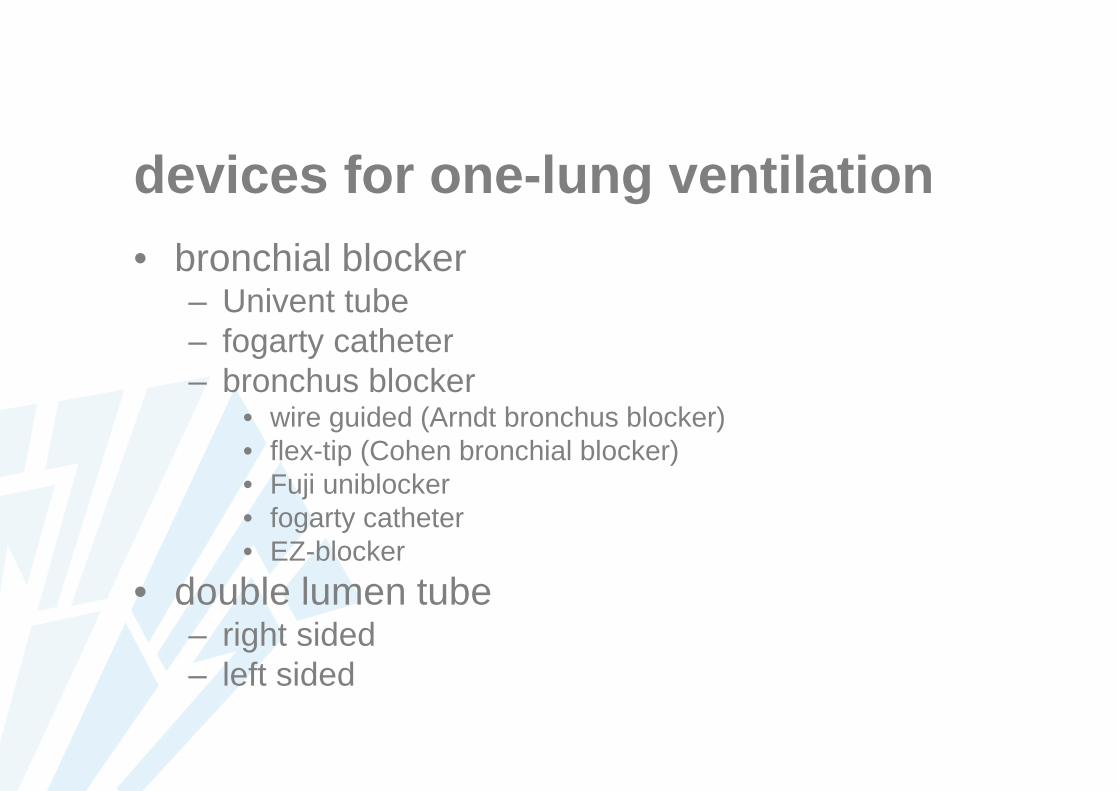
devices for one -lung ventilation• bronchial blocker
– Univent tube– fogarty catheter– bronchus blocker
• wire guided (Arndt bronchus blocker)• flex-tip (Cohen bronchial blocker)• Fuji uniblocker• fogarty catheter• EZ-blocker
• double lumen tube– right sided– left sided
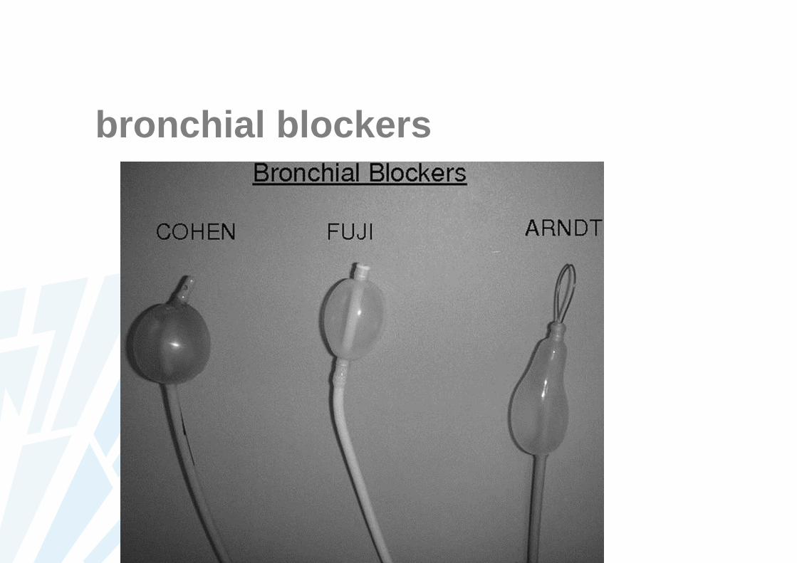
bronchial blockers

bronchial blockers : Arndt

bronchial blocker: arndt

bronchial blocker: EZ -blocker

bronchial blocker: univent tube

double lumen tube: left versus right sided

double lumen tube: left versus right sided
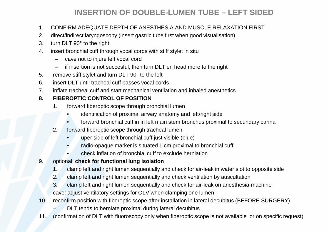
INSERTION OF DOUBLE-LUMEN TUBE – LEFT SIDED
1. CONFIRM ADEQUATE DEPTH OF ANESTHESIA AND MUSCLE RELAXATION FIRST2. direct/indirect laryngoscopy (insert gastric tube first when good visualisation)3. turn DLT 90° to the right4. insert bronchial cuff through vocal cords with stiff stylet in situ
– cave not to injure left vocal cord– if insertion is not succesful, then turn DLT en head more to the right
5. remove stiff stylet and turn DLT 90° to the left6. insert DLT until tracheal cuff passes vocal cords7. inflate tracheal cuff and start mechanical ventilation and inhaled anesthetics8. FIBEROPTIC CONTROL OF POSITION
1. forward fiberoptic scope through bronchial lumen• identification of proximal airway anatomy and left/right side• forward bronchial cuff in in left main stem bronchus proximal to secundary carina
2. forward fiberoptic scope through tracheal lumen• uper side of left bronchial cuff just visible (blue)• radio-opaque marker is situated 1 cm proximal to bronchial cuff• check inflation of bronchial cuff to exclude herniation
9. optional: check for functional lung isolation1. clamp left and right lumen sequentially and check for air-leak in water slot to opposite side2. clamp left and right lumen sequentially and check ventilation by auscultation3. clamp left and right lumen sequentially and check for air-leak on anesthesia-machinecave: adjust ventilatory settings for OLV when clamping one lumen!
10. reconfirm position with fiberoptic scope after installation in lateral decubitus (BEFORE SURGERY)– DLT tends to herniate proximal during lateral decubitus
11. (confirmation of DLT with fluoroscopy only when fiberoptic scope is not available or on specific request)
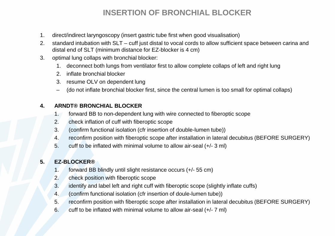
INSERTION OF BRONCHIAL BLOCKER
1. direct/indirect laryngoscopy (insert gastric tube first when good visualisation)2. standard intubation with SLT – cuff just distal to vocal cords to allow sufficient space between carina and
distal end of SLT (minimum distance for EZ-blocker is 4 cm)3. optimal lung collaps with bronchial blocker:
1. deconnect both lungs from ventilator first to allow complete collaps of left and right lung2. inflate bronchial blocker3. resume OLV on dependent lung– (do not inflate bronchial blocker first, since the central lumen is too small for optimal collaps)
4. ARNDT® BRONCHIAL BLOCKER1. forward BB to non-dependent lung with wire connected to fiberoptic scope2. check inflation of cuff with fiberoptic scope3. (confirm functional isolation (cfr insertion of double-lumen tube))4. reconfirm position with fiberoptic scope after installation in lateral decubitus (BEFORE SURGERY)5. cuff to be inflated with minimal volume to allow air-seal (+/- 3 ml)
5. EZ-BLOCKER®1. forward BB blindly until slight resistance occurs (+/- 55 cm)2. check position with fiberoptic scope3. identify and label left and right cuff with fiberoptic scope (slightly inflate cuffs)4. (confirm functional isolation (cfr insertion of doule-lumen tube))5. reconfirm position with fiberoptic scope after installation in lateral decubitus (BEFORE SURGERY)6. cuff to be inflated with minimal volume to allow air-seal (+/- 7 ml)

FIBEROPTIC ANATOMY
http:
Campos JH. Curr Opin Anaesthesiol 2009; 22: 4-10

FIBEROPTIC ANATOMY
Campos JH. Curr Opin Anaesthesiol 2009; 22: 4-10
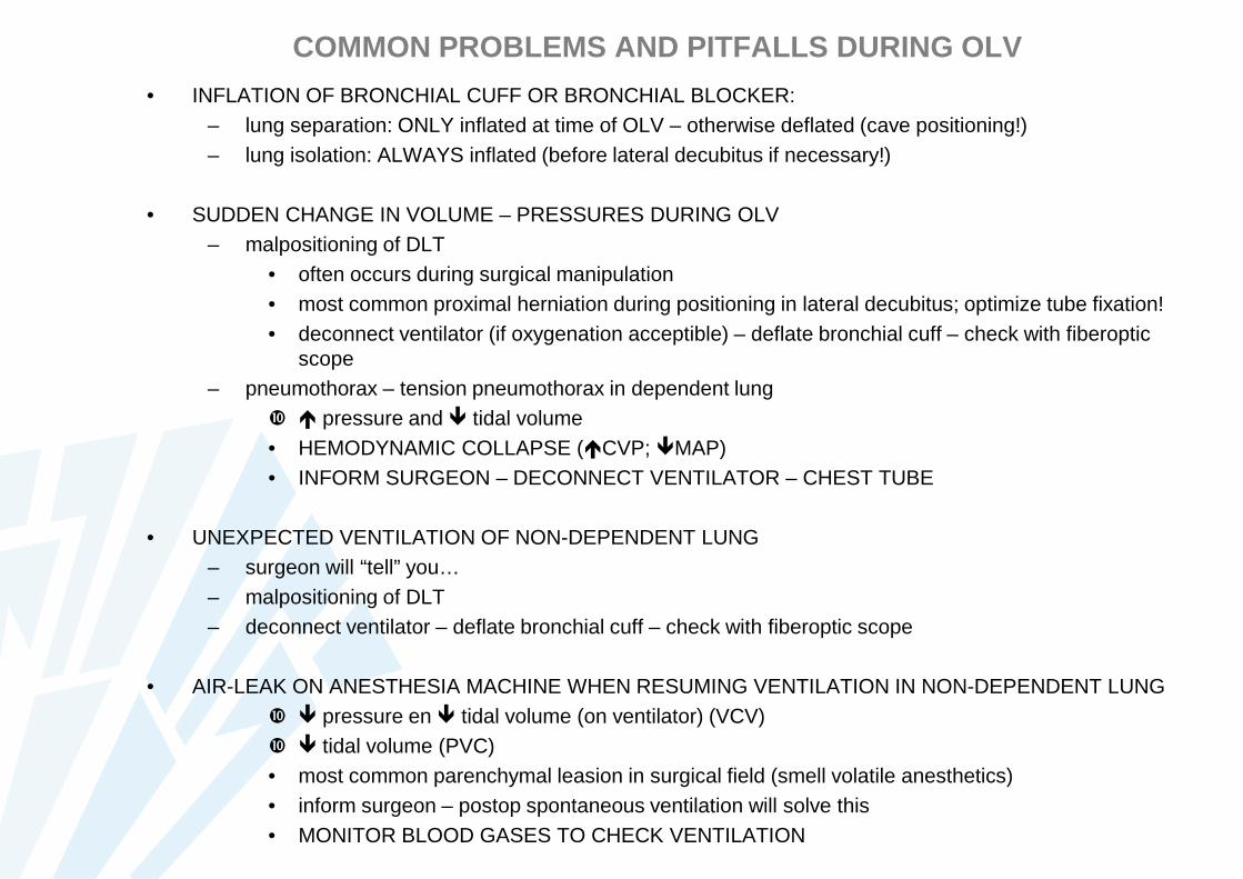
COMMON PROBLEMS AND PITFALLS DURING OLV
• INFLATION OF BRONCHIAL CUFF OR BRONCHIAL BLOCKER:– lung separation: ONLY inflated at time of OLV – otherwise deflated (cave positioning!)– lung isolation: ALWAYS inflated (before lateral decubitus if necessary!)
• SUDDEN CHANGE IN VOLUME – PRESSURES DURING OLV– malpositioning of DLT
• often occurs during surgical manipulation• most common proximal herniation during positioning in lateral decubitus; optimize tube fixation!• deconnect ventilator (if oxygenation acceptible) – deflate bronchial cuff – check with fiberoptic
scope– pneumothorax – tension pneumothorax in dependent lung
� � pressure and � tidal volume• HEMODYNAMIC COLLAPSE (�CVP; �MAP)• INFORM SURGEON – DECONNECT VENTILATOR – CHEST TUBE
• UNEXPECTED VENTILATION OF NON-DEPENDENT LUNG– surgeon will “tell” you…– malpositioning of DLT– deconnect ventilator – deflate bronchial cuff – check with fiberoptic scope
• AIR-LEAK ON ANESTHESIA MACHINE WHEN RESUMING VENTILATION IN NON-DEPENDENT LUNG� � pressure en � tidal volume (on ventilator) (VCV)� � tidal volume (PVC)• most common parenchymal leasion in surgical field (smell volatile anesthetics)• inform surgeon – postop spontaneous ventilation will solve this• MONITOR BLOOD GASES TO CHECK VENTILATION

MALPOSITIONING OF DLT DURING PROCEDURE
• MALPOSITIONING/DISLOCATION OF DLT-LEFT SIDED DURING PROCEDURE– most common problem: PROXIMAL HERNIATION OF ETT– deconnect ventilator (when oxygenation acceptible) – deflate bronchial cuff – check with fiberoptic
scope
during 2-lung ventilation remark
VCV PCV Sat
DLT too deep � PIP �TV � right lung not ventilated
during OLV remark
VCV PCV Sat
DLT too deep � PIP �TV � LUL not ventilated
during 2-lung – cuff inflated remark
VCV PCV Sat
DLT proximal herniation � PIP �TV � tracheal obstruction
during OLV – cuff inflated remark
VCV PCV Sat
DLT proximal herniation � PIP �TV � bilateral lung ventilation

malpositioning


DOUBLE-LUMEN TUBE (DLT) OR BRONCHIAL BLOCKER (BB) FOR ONE-LUNG VENTILATION (OLV)
OLVLUNG ISOLATION LUNG SEPARATIONto avoid contamination with
blood, pus, secretions, lavagefunctional – to optimize
surgical exposure
DLT DLT
BBRIGHT LEFT
•lesion left main stem bronchus•(large thoracic aortic aneurysm)
•bilateral intervention•pneumectomy•sleeve lobectomy•lobectomy•lung transplantation(SLTX – SSLTX)
LEFT
•ETT – SLT in situ•unanticipated OLV required during procedure•(tracheal bronchus)
•segmentecomy•mediastinal surgery•esophagectomy•cardiac surgery•other non-pulmonarysurgery requiring OLV
•IF PATIENT REQUIRES PROLONGED POSTOP INTUBATION: SW ITH TO SINGLE LUMEN TUBE (SLT)AT THE END OF PROCEDURE (EXCEPT IF LUNG ISOLATION I S REQUIRED)•POST SSLTX: SLT 8 FOR FEMALE AND SLT 9 FOR MALE•ALWAYS INDIRECT/DIRECT LARYNGOSCOPY WHEN SWITCHING TO SLT – CONSIDER USE OFTUBE EXCHANGER
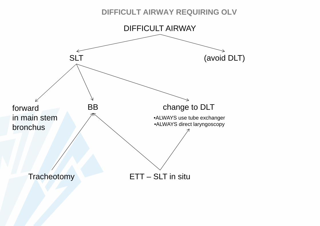
DIFFICULT AIRWAY REQUIRING OLV
DIFFICULT AIRWAY
SLT (avoid DLT)
forwardin main stembronchus
BB change to DLT•ALWAYS use tube exchanger•ALWAYS direct laryngoscopy
ETT – SLT in situTracheotomy
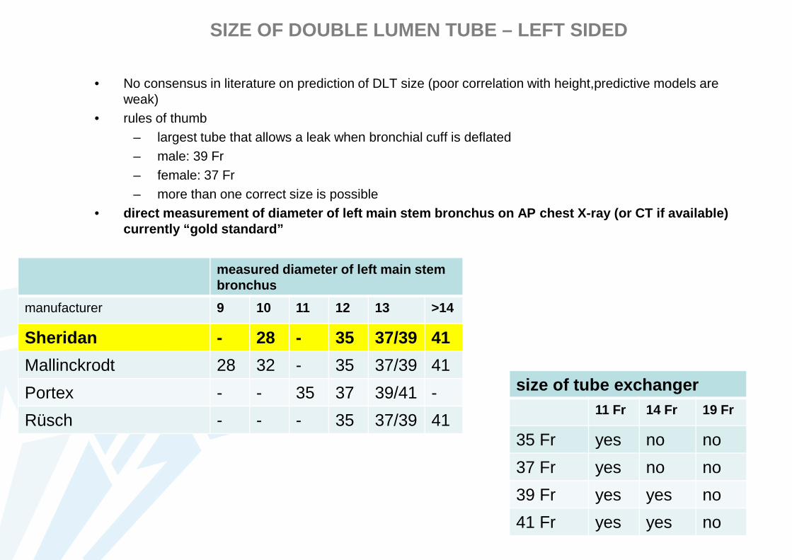
SIZE OF DOUBLE LUMEN TUBE – LEFT SIDED
• No consensus in literature on prediction of DLT size (poor correlation with height,predictive models are weak)
• rules of thumb– largest tube that allows a leak when bronchial cuff is deflated– male: 39 Fr– female: 37 Fr– more than one correct size is possible
• direct measurement of diameter of left main stem bronc hus on AP chest X-ray (or CT if available) currently “gold standard”
measured diameter of left main stem bronchus
manufacturer 9 10 11 12 13 >14
Sheridan - 28 - 35 37/39 41
Mallinckrodt 28 32 - 35 37/39 41
Portex - - 35 37 39/41 -
Rüsch - - - 35 37/39 41
size of tube exchanger11 Fr 14 Fr 19 Fr
35 Fr yes no no
37 Fr yes no no
39 Fr yes yes no
41 Fr yes yes no
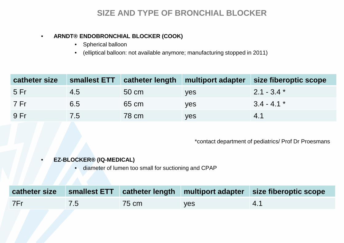
SIZE AND TYPE OF BRONCHIAL BLOCKER
• ARNDT® ENDOBRONCHIAL BLOCKER (COOK)• Spherical balloon• (elliptical balloon: not available anymore; manufacturing stopped in 2011)
• EZ-BLOCKER® (IQ-MEDICAL) • diameter of lumen too small for suctioning and CPAP
catheter size smallest ETT catheter length multiport adapt er size fiberoptic scope
5 Fr 4.5 50 cm yes 2.1 - 3.4 *
7 Fr 6.5 65 cm yes 3.4 - 4.1 *
9 Fr 7.5 78 cm yes 4.1
*contact department of pediatrics/ Prof Dr Proesmans
catheter size smallest ETT catheter length multiport adapt er size fiberoptic scope
7Fr 7.5 75 cm yes 4.1

LATERAL DECUBITUS AND ONE-LUNG VENTILATION
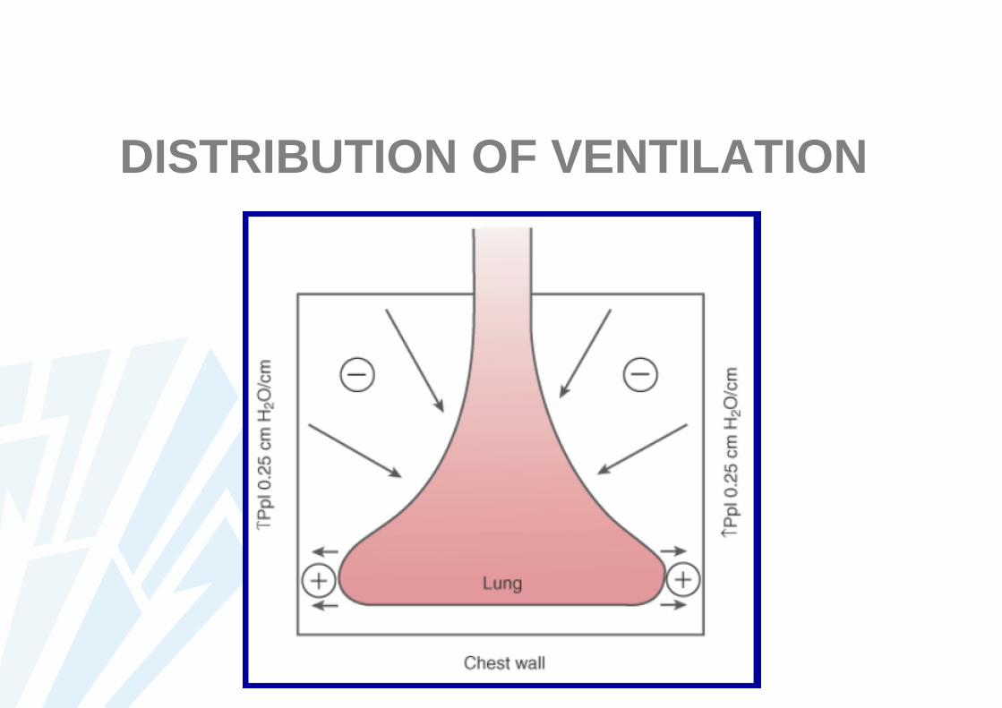
DISTRIBUTION OF VENTILATION

DISTRIBUTION OF VENTILATION
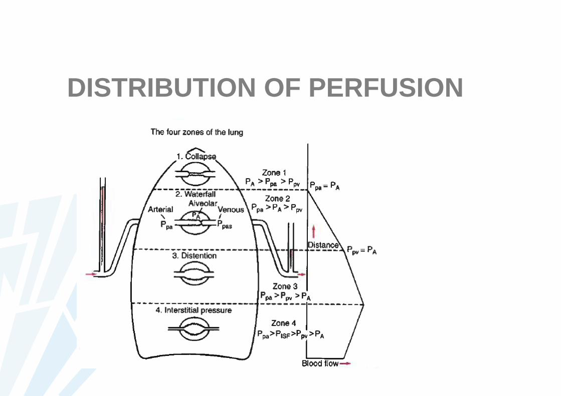
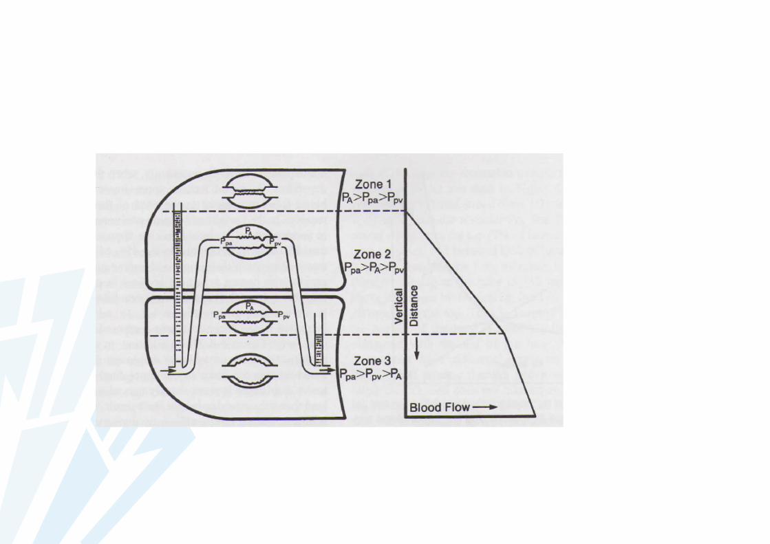
DISTRIBUTION OF PERFUSION
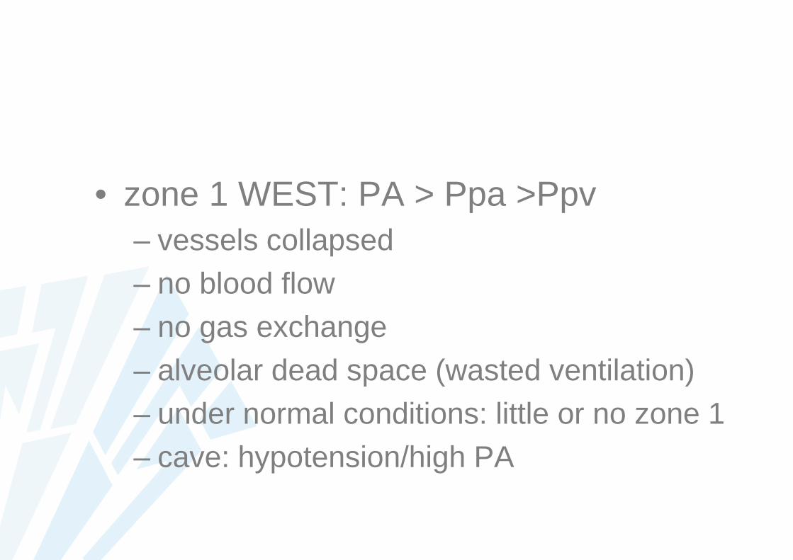
• zone 1 WEST: PA > Ppa >Ppv– vessels collapsed– no blood flow– no gas exchange– alveolar dead space (wasted ventilation)– under normal conditions: little or no zone 1– cave: hypotension/high PA

• zone 2 WEST: Ppa > PA >Ppv– blood flow determined by mean Ppa – PA
difference– waterfall over a dam (waterfall, starling
resistor, sluice effect)– mean blood flow increases linearly
downwards zone 2 (PA=constant)– cave: cyclic variation of the pressures

• zone 3 WEST: Ppa > Ppv >PA– blood flow determined by pulmonary
arteriovenous pressure difference– capillary systems permanently open– gravity causes increase of Ppa and Ppv at
same rate: perfusion pressure unchanged– vessel radii increase because of increase in
transmural distending pressures

• zone 4 WEST: Ppa > PISF > Ppv >PA– in case of accumulation of excessive fluid
into interstitial connective tissue compartment
– in case of very low lung volume– positive interstitial pressure, causing extra-
alveolar vessel compression, increasedextra-alveolar vascular resistance and decreased regional blood flow.

VENTILATION-PERFUSION RATIO

awake – closed chest

aneshtesia – spontaneous ventilation –closed chest
daling FRC because of anesthesia

anesthesia – mechanical ventilation –closed chest
•dependent lung volume further reduced•compression of abdominal organs•compression of mediastinum

anesthesia – mechanical ventilation –open chest
•further overventilation of non-dependent lung

AVOID HYPOXEMIA AVOID LUNG INJURYOPTIMIZE COLLAPSE
ONE-LUNG VENTILATION
PROTOCOLIZED APPROACH

PREVENTION OF HYPOXEMIA

PEEP - CPAP
Dependent lung-peep: ventilation
Non-dependent CPAP:oxygenation
Differential CPAP/PEEP
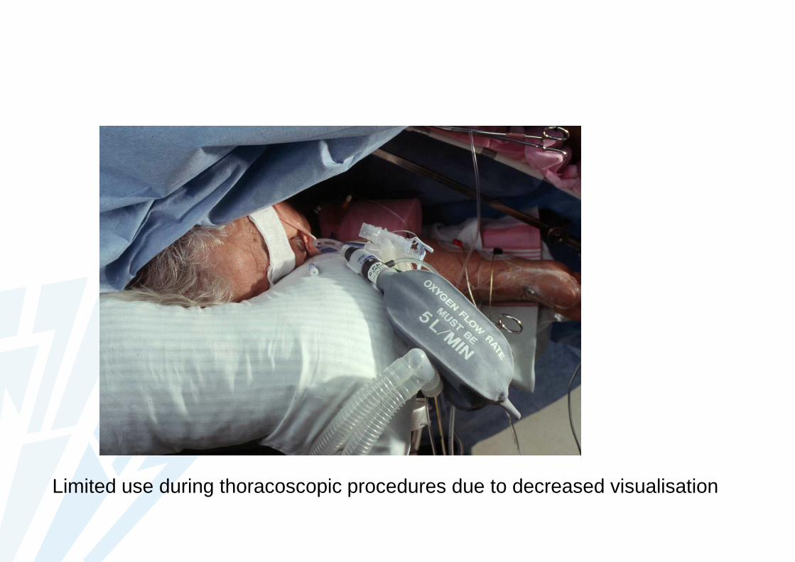
Limited use during thoracoscopic procedures due to decreased visualisation

HFJV non -dependent lungone-lung ventilationair-trappingimproved RV function
as alternative for CPAPoptimal exposure
to avoid one-lung ventilation (2-lung HFJV)lower peak pressures
Settings:frequency +/- 180pressure 1.8 – 2.2 barhigher PCO2 levels
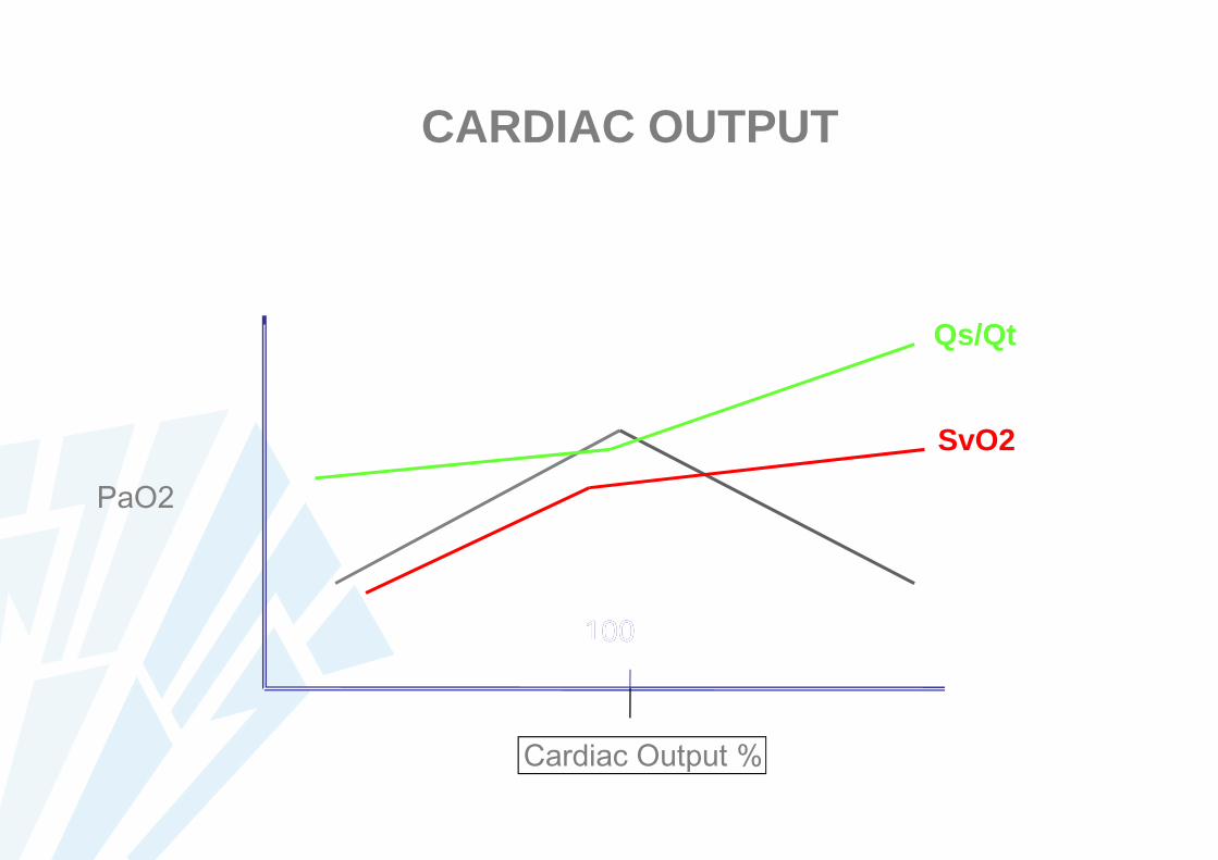
PaO2
Cardiac Output %
100
Qs/Qt
SvO2
100
CARDIAC OUTPUT

• optimizes V/Q (reduction 40%)
• contraction smooth muscle
• PAO2 40-100 mmHg
• determinants: PAO2 and PvO2
• early response 15 min
• maximal response 4 h
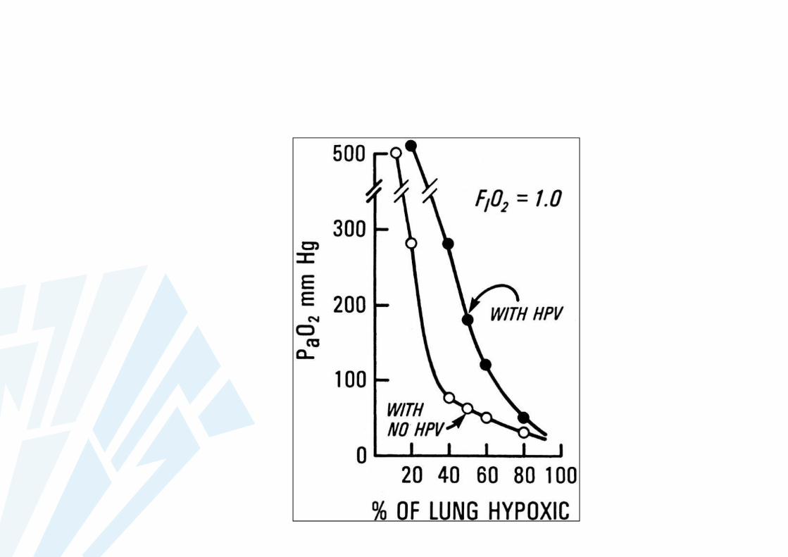
REDISTRIBUTION OF PERFUSIONhypoxic pulmonary vasoconstriction


CONDITION INCREASE DECREASE NO EFFECT
COPD +
cirrhosis +
sepsis +
pregnancy +
female sex +
exercise +
systemichypertension
+
ethanol +

CONDITION INCREASE DECREASE NO EFFECT
lateral decubitus +
supine position +
surgical lungretraction
+
hemodilution +
epidural +
almitrine +
inhaled NO +

CONDITION INCREASE DECREASE NO EFFECT
acidosis +
alkalosis +
hypercapnia +
hypocapnia +
hyperthermia +
hypothermia +
increased LAP +
increased PvO2 +
decreased PvO2 +

CONDITION INCREASE DECREASE NO EFFECT
nitrous oxide +
halothane +
isoflurane +
sevoflurane +
propofol + +
ketamine +
opioids +
epidural +
almitrine +
nitric oxide + +

CONDITION INCREASE DECREASE NO EFFECT
verapamil +
diltiazem +
propranolol +
phenoxybenzamine
+
phentolamine +
clonidine +
hydralazine +
nitroglycerin +
nitroprusside +
sildenafil +

CONDITION INCREASE DECREASE NO EFFECT
dopamine +
isoproterenol +
norepinephrine +
phenylephrine +
salbutamol +
lidocaine +
acetyl cysteïne +

AVOIDING LUNG INJURY

Pinhu et Al. Lancet 2003; 361: 332-340
Atelectrauma (open lung concept)•Repetitive opening and closure of atelectatic zones•recruitment and PEEP
Overdistention (baby lung)•volutrauma in functional reduced lung volume•reduction in tidal volume
PATHOPHYSIOLOGY OF LUNG INJURY:volutrauma versus atelectrauma

MECHANICAL FORCES
INFLAMMATION / BIOTRAUMABARRIER DYSFUNCTION
LUNG INJURYIncreased permeability
DISTENTION / VOLUTRAUMA
ATELECTRAUMA
UNDERLYING LUNG INJURYARDS/ALI MULTIPLE HIT
VILI (ventilation induced lung injury)
VALI (ventilation associated lung injury)
PATHOPHYSIOLOGY OF LUNG INJURY:multiple hits
• high tidal volumes 10 – 15 ml/kg– oxygenation– “end-inspiratory alveolar recruitment”
• PPE (postpneumonectomy pulmonary edema)
• low tidal volume in ARDS is beneficial
• outcome? surrogate markers
• effect of protective lung ventilation on healthy lungs
LUNG INJURY DURING OLVhistorical facts and pitfalls

• PATIENT– poor postoperative predicted lung function– preexisting lung injury
• trauma• infection• chemotherapy
– ethanol abuse– female gender
• PROCEDURE– lung transplantation– major resection (pneumonectomy > lobectomy)– esophagectomy – fluid administration– transfusion– prolonged OLV (>100 min) Peak pressure > 35-40 cmH2O– plateau pressure > 25 cmH2O
LUNG INJURY DURING OLVrisk factors
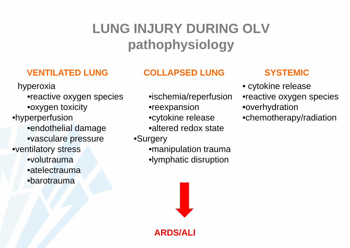
LUNG INJURY DURING OLVpathophysiology
VENTILATED LUNG COLLAPSED LUNG SYSTEMIC
• hyperoxia•reactive oxygen species•oxygen toxicity
•hyperperfusion•endothelial damage•vasculare pressure
•ventilatory stress•volutrauma•atelectrauma•barotrauma
• OLV•ischemia/reperfusion•reexpansion•cytokine release•altered redox state
•Surgery•manipulation trauma•lymphatic disruption
• cytokine release•reactive oxygen species•overhydration•chemotherapy/radiation
ARDS/ALI
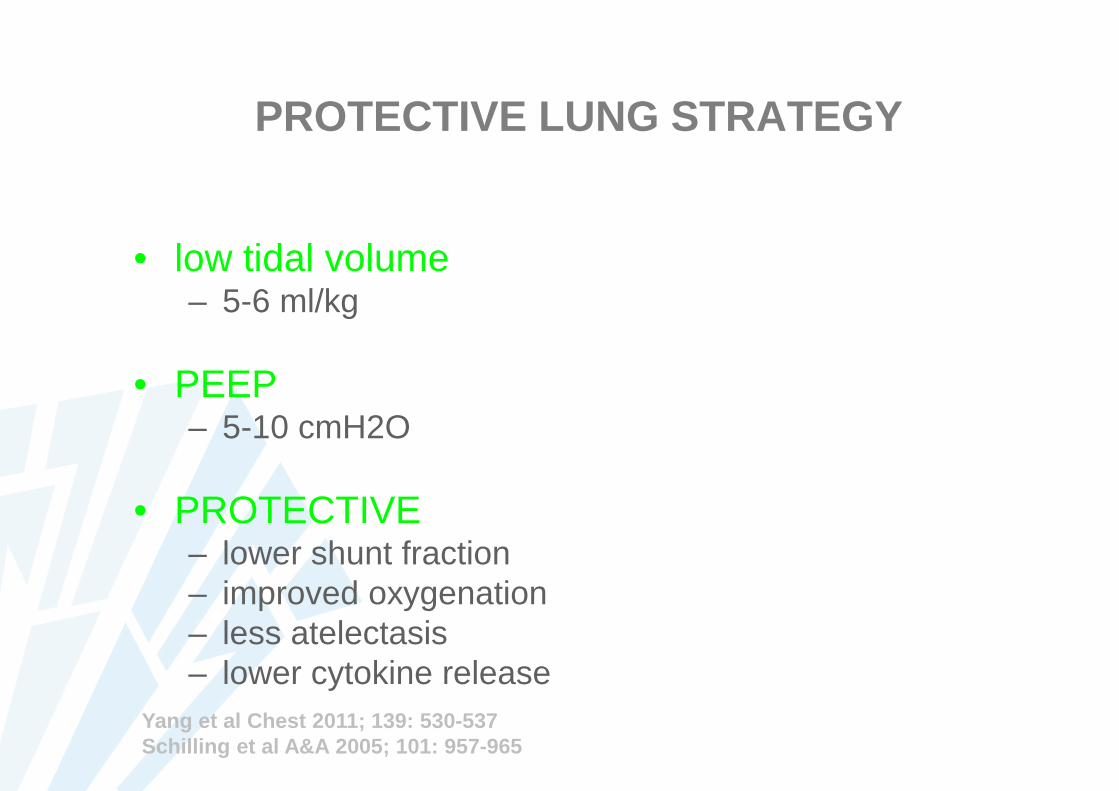
• low tidal volume– 5-6 ml/kg
• PEEP– 5-10 cmH2O
• PROTECTIVE– lower shunt fraction– improved oxygenation– less atelectasis– lower cytokine release
PROTECTIVE LUNG STRATEGY
Yang et al Chest 2011; 139: 530-537Schilling et al A&A 2005; 101: 957-965
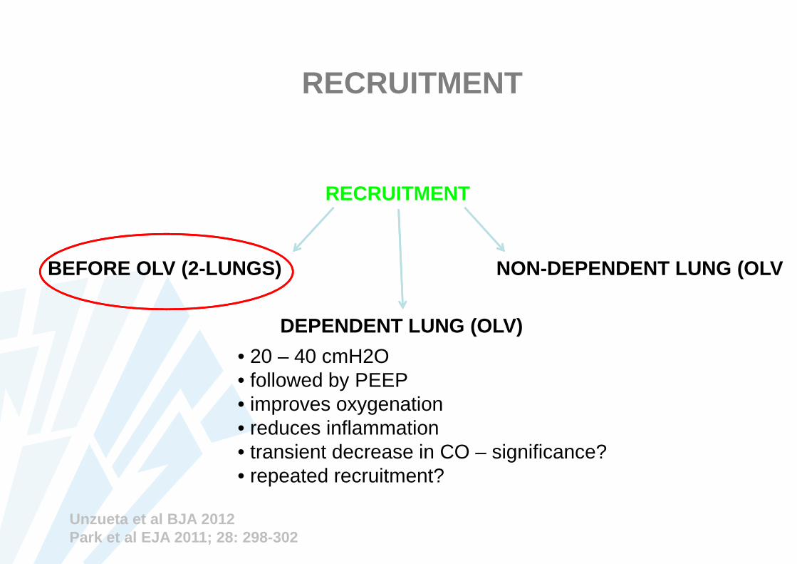
RECRUITMENT
RECRUITMENT
• 20 – 40 cmH2O• followed by PEEP• improves oxygenation• reduces inflammation• transient decrease in CO – significance?• repeated recruitment?
BEFORE OLV (2-LUNGS)
DEPENDENT LUNG (OLV)
NON-DEPENDENT LUNG (OLV
Unzueta et al BJA 2012Park et al EJA 2011; 28: 298-302

• reexpansion pulmonary edema
• with longer intervals of OLV
• gradual opening of non-dependent lung
• low FiO2 (ROS)
• Cave stapler lines
REEXPANSION INJURY

• up to date no clear benefit for PCV orVCV
• more homogeneous distribution withPCV?
• historical impact of limited AwP
MODE OF VENTILATION
TAILORED APPROACHONE SIZE DOES NOT FIT ALL

VENTILATORY MANAGEMENT OF OLV
GOAL
AVOID-THREAT HYPOXIA (CHECK BLOOD GASES REGULARLY!)
OPTIMAL LUNG COLLAPS FOR SURGICAL EXPOSURE (LOOK IN SURGICAL FIELD!)
AVOID ALI – PROTECTIVE VENTILATION (RELATED TO OUTCO ME!)
parameter target remark
Fi02 0.9 - reduce to 0.5 if possible(after onset of HPV)
•adjust 5 min prior to OLV •less inflammation with lower FiO2•re-inflation of non-dependent lung with air + recruitment
Tidal Volume 4-6 ml/kg •reduce stretch
Respir Rate increase to maintain MV •cave: increased Vd: higher RR necessary to maintain Va•cave: airtrapping if inadequate E-time•obstructive: I:E = 1:3 / restrictive: I:E = 1:1
Pplat AwP limit to 25-30 cmH20 •allow hypercapnia if necessary•air leak with BB when higher than 25 cmH20
PEEP 5-10 cmH20 •titrate to oxygenation (LIP)•reduces atelectasis – shear-stress•reduces auto-PEEP
PCO2 40 – 60 mmHg •permissive hypercapnia is protective•permissive hypercapnia in case of airtrapping or high Pplat AwP
ventilatory mode PCV -VCV •until now, no evidence for beneficial effect of a specific mode. allowPpeakAwP to be higher during VCV•cave: pressure in circuit is higher than alveolar pressure.

VENTILATORY MANAGEMENT OF OLV
• RESUMING VENTILATION OF NON-DEPENDENT LUNG– re-inflate with low FiO2 to limit oxidative stress (lung and right ventricle) (air – FiO2 0.3)– gradually re-inflate lung
• recruitment of collapsed alveoli• AwP 20-30 cmH20 during 30 sec• followed by PEEP• cave: reduction in C.I.
• avoid hyperinflation with large tidal volumes• consider use of CPAP device to re-inflate lung
• EMERGENCE OF ANESTHESIA FOR THORACIC PROCEDURE
• NEVER SPONTANEOUS BREATHING WITH DLT/SLT IN SITU• PRESSURE-SUPPORT VENTILATION ALLOWED TO MAINTAIN RECRUITMENT - MV
• ALWAYS MAINTAIN PEEP/CPAP• CONSIDER RECRUITMENT• OPTIMIZE ANALGESIA BEFORE EXTUBATION• AVOID HYPERCAPNIA• ASPIRATION OF SECRETIONS• CHECK FOR REMOVAL OF NEUROMUSCULAR BLOCKADE• CHECK CHEST X-RAY BEFORE EXTUBATION (EXCLUDE ATELECTASIS)• MOST COMMON PROBLEM AFTER THORACIC SURGERY IS ATELECTASIS: OUTCOME!

MANAGEMENT OF HYPOXIA DURING OLV
HYPOXIA: Sat < 91% (incidence 1%)
1. increase FiO2 to 1.02. check position DLT/BB3. optimize cardiac output
• preload: 250-500 CC colloids• contractility• arrhytmias
4. recruitment dependent lung• AwP 20-30 cmH20 during 30 sec• followed by PEEP• cave: reduction in C.I.
5. optimize PEEP dependent lung towards LIP (� or �)6. CPAP to non-dependent lung
• recruitment first• 5-10 cmH20• NOT during VATS (surgical exposure)
7. intermittent inflate non-dependent lung (communicate with surgeon)8. partial ventilation of non-ventilated lung
• lobar re-inflation• selective lobar collapse (BB)• oxygen insufflation (consider insufflation in surgical field, cave combustion)• (high frequency ventilation)
9. reduce blood flow to non-ventilated lung• clamping of pulmonary artery (cave increased afterload to right ventricle)
10. maintain oxygen carrying capacity11. (ECMO as rescue)
SHUNT
MALPOSITION DLT/BB
MILD/GRADUAL SEVERE (<85%)
1. resume 2-lung ventilation2. (communicate with surgeon)3. increase FiO2 to 1.04. check position DLT/BB

PREDICTION OF HYPOXIA DURING OLV
– INCREASED RISK• low PO2 preoperatively and during two-lung ventilation• distribution of perfusion on V/Q scan• operative side (R>L)• dorsal decubitus > lateral decubitus• (FEV1)• compliant pulmonary vascular bed (younger patients)• use capnography:
– change in ETCO2 during TLV and OLV– large change predicts low oxygenation!
– DECREASED RISK• obstructive lung disease (airtrapping and AUTO-PEEP)• large central tumors have less perfusion to non-dependent lung

RISK FACTORS FOR ALI POST-OLV
• PATIENT RELATED– poor postoperative predicted lung function– preexisting lung injury
• trauma• infection• chemotherapy
– ethanol abuse– female gender
• PROCEDURE RELATED– lung transplantation– major resection (pneumonectomy > lobectomy– esophagectomy with large perioperative fluid load– transfusion– prolonged OLV (>100 min)– peak AwP > 35 – 40 cmH20– plateau AwP > 25 cmH20
• re-inflation of non-dependent lung with air (limits inflammation)

HEMODYNAMIC OPTIMIZATION DURING OLV
• AFTERLOAD to right ventricle is increased– pneumonectomy– hypoxic pulmonary vasoconstriction– less compensation of right ventricle to increased afterload– optimize reduction in PVR (recruitment to FRC, normal pH, avoid hypoxia, avoid hypercapnia, avoid
surgical stress, consider use of selective pulmonary vasodilators: inhaled NO / inhaled Prostacyclins)– check with TEE if necessary– consider use of PA-catheter
• CONTRACTILITY– cave sympathetic blockade due to epidural catheter– less sympathetic blockade when continuous infusion of local anesthestic– consider inotropic support if necessary (dobutamine / corotrope)– consider reduction in afterload with inhaled NO / inhaled prostacyclines when RV dysfunction– re-inflation of non-dependent lung with air to minimize oxidative stress– check with TEE if necessary– consider use of PA-catheter
• PERFUSION PRESSURE– perfusion of right ventricle dependent on systolic pressure– maintain adequate perfusion pressure with levophed (start at 0.05 gamma)
• PRELOAD-CARDIAC OUTPUT– optimal preload and cardiac ouput necessary for oxygenation– consider adequate preload (250-500 cc fluid challenge)– consider use of inotropics (ephedrine>dobutamine>corotrope)