Embed Size (px)
Citation preview
Experimental Hematology 2012;-:-–-
Nicotinamide, a SIRT1 inhibitor, inhibits differentiation and facilitatesexpansion of hematopoietic progenitor cells with enhanced bone
marrow homing and engraftment
Tony Peleda, Hadas Shohama, Dorit Aschengraua, Dima Yackoubova, Gabi Freia, Noga Rosenheimer Ga,Batya Lerrerb, Haim Y. Cohenb, Arnon Naglerc, Eitan Fibachd, and Amnon Pelede
aGamida Cell Ltd., Jerusalem, Israel; bThe Mina and Everard Goodman Faculty of Life Sciences, Bar-Ilan University, Ramat-Gan, Israel;cHematology Division, BMT&CBB, Chaim Sheba Medical Center, Tel-Hashomer, Israel; dDepartment of Hematology;
eGoldyne Savad Institute of Gene Therapy, Hadassah-Hebrew University Medical Center, Jerusalem, Israel
(Received 17 August 2011; revised 5 December 2011; accepted 11 December 2011)
Offprint requests
Therapy Technologies
Supplementary data
1016/j.exphem.2011.
0301-472X/$ - see fro
doi: 10.1016/j.exph
Strategies that increase homing to the bone marrow and engraftment efficacy of ex vivo ex-pended CD34+ cells are expected to enhance their clinical utility. Here we report that nicotin-amide (NAM), a form of vitamin B-3, delayed differentiation and increased engraftmentefficacy of cord blood–derived human CD34+ cells cultured with cytokines. In the presenceof NAM, the fraction of CD34+CD38L cells increased and the fraction of differentiated cells(CD14+, CD11b+, and CD11c+) decreased. CD34+ cells cultured with NAM displayedincreased migration toward stromal cell derived factor–1 and homed to the bone marrowwith higher efficacy, thus contributing to their increased engraftment efficacy, which wasmaintained in competitive transplants with noncultured competitor cells. NAM is a knownpotent inhibitor of several classes of ribosylase enzymes that require NAD for their activity,as well as sirtuin (SIRT1), class III NAD+-dependent-histone-deacetylase. We demonstratedthat EX-527, a specific inhibitor of SIRT1 catalytic activity, inhibited differentiation ofCD34+ cells similar to NAM, while specific inhibitors of NAD-ribosylase enzymes didnot inhibit differentiation, suggesting that the NAM effect is SIRT1-specific. Our findingssuggest a critical function of SIRT1 in the regulation of hematopoietic stem cell activityand imply the clinical utility of NAM for ex vivo expansion of functional CD34+ cells. � 2012ISEH - Society for Hematology and Stem Cells. Published by Elsevier Inc.
Strategies to expand hematopoietic progenitor cells (HPC)in vitro are of clinical importance to improve the outcomeof cord blood transplantations. Exposure of HPC todifferent combinations of cytokines promotes their exitfrom the G0 phase of the cell cycle and enables extensiveproliferation. Nonetheless, in vitro proliferation is tightlycoupled with a commitment to differentiation and reducedself-renewal [1]. Although early-acting cytokines inducerobust in vitro expansion of CD34D cells, expansion of en-graftable progenitors is modest [2]. This phenomenon couldbe explained, at least in part, by an acquired defect in thebone marrow (BM) homing capacity of ex vivo–expanded
EXPHEM2839_proof ■ 18
to: Tony Peled, Ph.D., Gamida Cell Ltd., Cell
, P.O. Box 34670, Jerusalem 91340, Israel; E-mail:
m; http://www.gamida-cell.com
related to this article can be found online at doi:10.
12.005
nt matter. Copyright � 2012 ISEH - Society for Hematolo
em.2011.12.005
HPC [3,4], which is primarily attributed to their activecycling [5], accompanied by alterations in adhesion andchemokine receptor expression or functionality [6]. There-fore, strategies to augment BM homing and engraftmentefficacy are particularly important to increase clinical appli-cability of ex vivo–expanded CD34D cells [7].
Nicotinamide (NAM), a form of vitamin B-3, serves asa precursor of nicotinamide adenine dinucleotide (NADD)[8]. NAM is also a potent inhibitor of enzymes that requireNADD for their activities [9,10], such as mono-ADP-ribosyltransferases, poly-ADP-ribose polymerases, CD38,and cyclic ADP ribose/NADase [11]. In addition, NAM isa well-established potent inhibitor of the sirtuin family ofhistone/protein deacetylases, the NAD-dependent class IIIhistone deacetylase (HDAC) [12]. SIRT1, one of themammalian sirtuins, catalyzes the deacetylation of acetyl-lysine residues by a mechanism whereby NADþ is cleaved.The reaction results in the release of NAM, which acts as anend-product noncompetitive inhibitor of SIRT1 by binding
-1-2012 15-27-3
gy and Stem Cells. Published by Elsevier Inc.
2 T. Peled et al./ Experimental Hematology 2012;-:-–-
to a conserved pocket adjacent to NAD(þ), thereby block-ing NAD(þ) hydrolysis [13]. It was reported that hemato-poietic cells derived from SIRT1-deficient mice (SIRT1�/�)display increased in vitro proliferation activity, althoughtheir self-renewal and in vivo function were not addressed[14]. Having multiple effects on numerous cells, NAM isimplicated in the regulation of cell adhesion, polarity,migration, proliferation, and differentiation [15]. NAMwas shown to modulate the fate of embryonic stem cells[16] and primary nonhematopoietic cells [17–19]. With re-gard to hematopoietic cell lines, NAM was reported toinhibit HL-60 cell differentiation mediated by retinoicacid [20], and other studies reported that NAM enhancesHL-60 cell differentiation [21].
Here we studied the effect of NAM on primary culturesof umbilical cord blood (UCB) CD34þ cells. Our resultsdemonstrate that NAM delayed differentiation and enhancedmigration, homing, and engraftment of CD34þ cellsexpanded ex vivo with cytokines. The SIRT1-specific inhib-itor, EX-527 [22,23], exhibited an effect similar to that ofNAM on cultured CD34þ cells, while NAM-related[24–26] and nonrelated [27–29] NAD-dependent ADP-ribosyltransferase inhibitors were not effective. Based onthese findings, we propose SIRT1 deacetylase as a targetaccountable for NAM modulating CD34þ cell differentia-tion in ex vivo cultures.
Materials and methods
CB samplesCells were obtained from UCB samples harvested from consentingmothers after normal full-term deliveries (Sheba Medical Center,Tel-Hashomer, Israel). Samples were collected and frozen accord-ing to Rubinstein et al. [30]. Before use, cells were thawed, themononuclear cells purified on a Ficoll-Hypaque gradient, andCD34þ cells were isolated using a MidiMACS CD34 ProgenitorCell Isolation Kit (Miltenyi Biotec, Bergisch, Gladbach, Germany)as described previously [31].
Ex vivo expansion culturesPurified CD34þ cells were cultured in culture bags (AmericanFluoroseal Co., Gaithersburg, MD, USA) at 1 �104 cells/mL (atleast 8 mL/bag) in minimum essential medium–a, 10% fetal bovineserum (FBS), and cytokines: thrombopoietin, interleukin (IL)-6,fms-like tyrosine kinase–3 ligand, and stemcell factor, each at a finalconcentration of 50 ng/mL (Pepro Tech, Inc., RockyHill, NJ, USA),with or without NAM (Sigma Aldrich, Milwaukee, WI, USA;catalog number N5535 and Vertellus, Indianapolis IN, USA;catalog number 100547) and incubated at 37�C in a humidifiedatmosphere of 5%CO2 in air. Until week 3, the cultures were toppedweekly with the same volume of fresh medium. For long-termexperiments, the cultures were weekly demi-depopulated. Thenumber of total nucleated cells (TNC) in culture was determinedafter dilution (1:10) with phosphate-buffered saline (PBS) byCEDEX, an automatic cell counter (Innovatis AG, Bielefeld,Germany). The CEDEX is an automated cell counting system basedon the well-established Trypan Blue exclusion method for
EXPHEM2839_proof ■ 1
determining cell viability. The number of cells in culture was deter-mined by multiplying the number of cells/mL by the culturevolume. Fold-expansion was calculated by dividing total numberof cells in culture with the culture input number of cells. Numberof CD34þ cells andCD34þ38� cells were calculated bymultiplyingpercentages with total number of cells in culture/100. Fold-expansion was calculated by dividing the calculated number ofCD34þ and CD34þCD38� cells obtained following culture withthe number of culture seeded CD34þ and CD34þCD38– cells.For the colony-forming unit (CFUc) assay, 1000CD34þ cells beforeculture and 1500 cells after culture were added per 3 mLMethoCult(MethoCult GFþH4435 complete methylcellulose medium withrecombinant cytokine and erythropoietin for colony assays ofhuman cells; StemCell Technologies, Vancouver, BC, Canada).After stirring, the mixture was divided into two 35-mm dishes andincubated for 14 days at 37�C in a humidified atmosphere of 5%CO2 in air. At the end of the incubation period, colonies (bothmyeloid and erythroid) were counted under an inverted microscopeat 40� magnification. The CFUc content/mL culture was calcu-lated as follows: number of scored colonies per two dishes � totalcell number/1500 or 1000. Fold-expansion was calculated bydividing total number of colonies in culture (number/mL � culturevolume) with the culture input number of colonies.
ImmunophenotypingCultured and noncultured cells were washed with PBS containing1% bovine serum albumin and double-stained (at 4�C for 30minutes) with phycoerythrin (PE) or fluorescein isothiocyanate–conjugated antibodies to CD45, CD34, CXCR4, very late antigen4 and lymphocyte function–associated antigen 1 (BectonDickinson,Erembodegem, Belgium) or with differentiation antigens CD38,CD33, CD14, CD15, CD11b, and CD11c (DAKO, Glostrup,Denmark). Cells were then washed in the buffer and analyzedusing a flow cytometry (Becton Dickinson, San Jose, CA,USA). The emission of 104 cells was measured using logarith-mic amplification and analyzed using CellQuest software (Bec-ton Dickinson, Belgium).
PKH26 labelingFreshly purified CD34þ cells (2 � 106) were incubated at roomtemperature for 5 minutes with 4 mM PKH26 (PKH26–PE-CellLinker Kit; Sigma Aldrich, Milwaukee, WI, USA) according tomanufacturer’s instructions. Then, an equal volume of 1% FBSwas added for 1 minute, and the labeled cells were washed threetimes in PBS supplemented with 5% human serum albumin.
20, 70-Bis-(carboxyethyl)-5(60)-carboxyfluoresceinacetoxymethyl ester (BCECF/AM) labelingBriefly, cells were washed and resuspended at !107 cells/mL inserum-free medium, and BCECF/AM at a final concentration of5 mg/mL was added for 10 minutes at 37�C. Uptake of the dyewas stopped by the addition of FBS (to give a final concentrationof 10%). After labeling, cells were washed three times in PBSwith 10% FBS and analyzed by flow cytometry for fluorescenceintensity [32].
Tracking of cell-division historyPurified CD34þ cells were labeled with PKH2. An aliquot wasanalyzed with flow cytometry for PKH2 intensity (t 5 0) and therest was cultured with cytokines (i.e., stem cell factor, thrombo-poietin, fms-like tyrosine kinase–3, and IL-6), with or without
8-1-2012 15-27-3

3T. Peled et al./ Experimental Hematology 2012;-:-–-
5 mM NAM. On day 7, cells were harvested and CD34þ cells werereisolated using the MiniMACS CD34 progenitor cell isolation kitand double-stained for CD34 and CD38. CD34 cells and the gatedCD34þCD38� cells were analyzed with flow cytometry for PKH2fluorescence intensity.
In vitro migration assayMinimum essential medium–a plus 1% FBS and 100 ng/mLstromal cell derived factor–1 (SDF-1; R&D Systems Inc,Minneapolis, MN, USA) was placed into the lower chamber ofa Costar 24-well Transwell (Corning, Corning, NY, USA). Cells(2 � 105) in 100 mL medium were placed into the upper chamberover a porous membrane (pore size, 5 mm). After 4 hours, cellswere collected from the lower chamber and counted. Spontaneousmigration was evaluated without SDF-1 in the lower chamber.
In vivo homing assayNOD/LtSz- (nonobese diabetic/severe combined immunodeficient[NOD/SCID]) mice (8–10 weeks old) (Harlan Biotech Israel Ltd.,Rehovot, Israel) were sublethally irradiated (with 375 cGy at 67cGy/min) and 24 hours later inoculated via the tail vein with 10to 20 million cells stained with BCECF/AM (Calbiochem,Darmstadt, Germany). The experiments were approved by theAnimal Care Committee of the Hadassah, Hebrew UniversityMedical Center. Mice were sacrificed at 24 hours post injection.BM samples were obtained by flushing their femurs and tibiaswith PBS at 4�C. Homing of human cells was detected by flow cy-tometry. The bright fluorescence of BCECF/AM was sufficient toseparate labeled human cells from unlabeled murine cells by atleast 1 log. To quantify homing of human progenitor cells, BMcells were stained with allophycocyanin-conjugated anti-humanCD34 monoclonal antibodies (Becton Dickinson, Belgium) andBCECF/AMþCD34þ cells were enumerated. For each sample,100,000 events were acquired and analyzed. The Animal CareCommittee of Hadassah, Hebrew University Medical Center,Jerusalem, Israel approved these experiments. To evaluate BMhoming after cotransplantation, noncultured cells were stainedwith BCECF/AM (fluorescein isothiocyanate) and cultured cellsstained with PKH (PE), as described.
Transplantation of human CD34þ cells into NOD/SCID miceand quantification of SCID repopulating cells (SRC)NOD/SCID mice were bred and maintained at the Weizmann Insti-tute, Rehovot, Israel in sterile intraventilated cages (Techniplast,Bugugiatte, Italy) or at Harlan Biotech. The experiments wereapproved by the Animal Care Committee of theWeizmann Instituteand of Harlan Biotech. Eight-week-old mice were sublethally irra-diated as described and transplanted with human CB-derived cells.Mice were sacrificed on week 6, and the BM cells were immuno-phenotyped as described here [31,33]. To compare engraftmentbefore and after culture, each CB unit was frozen into two portions.CD34 cells purified from one portion were cultured for 3 weeks asdescribed. The second portion was kept frozen. On the day of trans-plantation, this portion was thawed and TNC or purified CD34þ
cells were transplanted. In some experiments, to avoid donor vari-ability, this procedure was carried out with CD34þ cells pooledfrom several CB units. Single units were used in experiments wherecultured and noncultured TNC were cotransplanted in the samemouse. In these experiments, to avoid clamping of nonculturedcells, the two cell populations were mixed just before cell injection.
EXPHEM2839_proof ■ 18
The frequencies of SRC were quantified by a limiting dilution anal-ysis and applying Poisson statistics to the single-hit model asdescribed previously [34]. Micewere scored as positively engraftedif at least 0.5% of their marrow cells expressed human CD45.Frequencies of SRC and statistical comparison between individualpopulations were calculated by maximum likelihood estimatorusing L-Calc software (StemCell Technologies) [34,35].
Western blottingCD34þ cells were cultured with cytokines, with and without 5 mMNAM, 18 hours before the addition of lysis Tris/saline/azide (1 mM).For the immunoprecipitation, cells were lysed in lysis buffer con-taining (50 mM Tris-HCl [pH 7.5], 150 mM NaCl, 1 mM EDTA ,1% NP-40, and a cocktail of protease inhibitor). HDAC inhibitorswere supplemented in the lysis buffer at the following final concen-trations: 5 mM NAM and 1 mM Tris/saline/azide. Cellular lysateswere incubated with agarose-conjugated anti-acetyllysine antibody(-Ac-K) (ImmuneChem Pharmaceuticals Inc., Burnaby, BC,Canada) overnight at 4�C on a rotation wheel. Immunocomplexeswere washed four times with lysis buffer, boiled, and resolved bysodiumdodecyl sulfate polyacrylamide gel electrophoresis.Westernblotting analysis of preimmunoprecipitation (input) and imm-unoprecipitated samples (-Ac-K) were performed with an anti-Ku70 antibody (Santa Cruz Biotechnology, Inc., Santa Cruz, CA,USA).
StatisticsThe nonparametric Wilcoxon rank test was applied for testingdifferences between the study groups. All the tests applied weretwo-tailed, and a p value of #0.05 was considered statisticallysignificant. Data were analyzed using SAS software (SAS Insti-tute, Cary, NC, USA).
Results
Characterization of NAM effect on ex vivo CD34þ cellculturesThe effect of NAM was determined in CD34þ cells derivedfrom UCB during 3 weeks in cultures supplemented withcytokines (i.e., fms-like tyrosine kinase–3, IL-6, thrombo-poietin, and stem cell factor). Analysis included the numberof TNCs, CFUc, and phenotypic characterization of hem-atopoietic progenitors, CD34þ and CD34þCD38� cells(Fig. 1A–L). As early as 1 week post seeding, the numberof TNCs (Fig. 1A) and CD34þ cells (Fig. 1B) was substan-tially lower, while percentages (Fig. 1C, D) and absolutenumber (Fig. 1E) of CD34þCD38� cells were significantlyhigher in cultures treated with 2.5 and 5 mM NAM ascompared with control cultures treated with cytokinesonly. The divisional history of seeded CD34þ cells stainedwith PKH indicated that during the first week in culture,the vast majority of CD34þ cells underwent several cyclesunder both culture conditions (Fig. 1F, G), with consistentlesser divisions (higher fluorescence intensity) of cellscultured with NAM (Fig. 1H). Slower cycling was particu-larly prominent in the CD34þCD38� subset (Fig. 1G),which, nevertheless, increased within the expanded cell
-1-2012 15-27-3

print&
web
4C=FPO
Figure 1. Effect of NAM on CD34þ cell cultures. CB-derived purified CD34þ cells were cultured for 3 weeks with cytokines or with cytokines and NAM at
2.5 and 5 mM. The culture, analysis procedures and calculations are described in Materials and Methods. Each bar represents the mean 6 standard error of
four independent experiments. (A–E) Analysis of cultured cells 1 week post seeding. (A) Number of TNCs (*p! 0.01 vs NAM, 2.5 and 5 mM). (B) Number
of CD34þ cells (*p ! 0.008 vs NAM). (C) Representative flow cytometry analysis dot plots of cells double stained with CD34 PE and CD38 fluorescein
isothiocyanate. (D) Percentages of CD34þCD38� cells (*p ! 0.01 vs NAM-nontreated cultures). (E) Numbers of CD34þCD38� cells (*p ! 0.03 vs NAM-
nontreated cultures). (F–H) To track cell-division history, freshly purified CD34þ cells were labeled with PKH2, cultured, and analyzed 7 days post seeding,
as described in Material and Methods. Histograms of PKH fluorescence intensity of CD34þ (F) and CD34þCD38� (G) cells are shown. The histograms
present the same number of cells for both control and NAM-treated cells in a representative experiment out of three experiments performed. (H) The median
fluorescence intensities of NAM expanded cells on day 7 cultures of three separate experiments are shown as percentages of control cultures treated with
cytokines alone. (I) Numbers of CD34þCD38� cells, 1 week (*p # 0.03 vs NAM-nontreated), 2 weeks (*p # 0.02 and **p # 0.02 vs NAM-
nontreated) and 3 weeks (*p # 0.01 vs NAM-nontreated) post seeding. (J–M) Fold expansion (FE) of TNC (*p ! 0.01 vs NAM; **p !0.03 vs NAM
5 mM) (J), CFUc (*p ! 0.03 vs NAM; **p ! 0.02 (K), CD34þ cells (*p # 0.01 vs NAM) (L), and CD34þ38� cells 1 week (*p # 0.04 vs NAM-
nontreated), 2 weeks (*p # 0.03 and **p # 0.02 vs NAM-nontreated), and 3 weeks (*p # 0.01 vs NAM-nontreated) post seeding (M), are shown.
4 T. Peled et al./ Experimental Hematology 2012;-:-–-
population from week 1 through week –3 (Fig. 1I). After 3weeks in culture, fold-expansion of TNCs (Fig. 1J), CFUc(Fig. 1K), and CD34þ cells (Fig. 1L) in NAM-treatedcultures reached the values in NAM-nontreated cultures,while fold-expansion of CD34þCD38� cells was superiorin cultures treated with NAM throughout the 3-week cultureduration (Fig. 1M). Higher concentrations of NAM (10 mM)deteriorated TNC, CD34, and CFU proliferation throughoutthe culture duration, while a lower concentration of NAM (1mM) had a slight effect on the expansion of CD34þCD38�
cells (Supplementary Figure E1; online only, available atwww.exphem.org). Phenotype characterization of lineage
EXPHEM2839_proof ■ 1
differentiated cells in 3-week cultures revealed lesseningof differentiation in cultures treated with NAM (2.5 and5 mM) than in cultures treated with cytokines alone, asdemonstrated by significantly lower percentages of CD14,CD11b, CD11c, and CD15þ cells (Supplementary Figure E2;online only, available at www.exphem.org).
NAM attenuates differentiation and promotes long-termexpansion of cultured CD34þ cellsIn order to test the long-term potential of short-term NAM-treated cultures, CD34þ cells from 3 UCB units werecultured for 3 weeks with and without NAM, and
8-1-2012 15-27-3

Figure 2. The long-term expansion potential of NAM-treated cells. (A, B) CD34þ cells were cultured in medium supplemented with cytokines with or
without NAM (5 mM). After 3 weeks, both cultures were supplemented with cytokines only. On the indicated weeks, CD34þ cells (A) and CFUc (B)
were determined. Each data point represents the mean count of two duplicates. The average number of three experiments initiated with cells derived
from different donors 6 standard error and the p values of the difference between cultures with and without NAM are indicated. (C–F) CD34þ cells
were cultured for 3 weeks with the four early-acting cytokines (fms-like tyrosine kinase–3 [FLT3], stem cell factor, thrombopoietin, and IL-6) and IL-3
at 20 ng/mL (Pepro Tech, Inc.), with or without NAM (2.5–7.5 mM). Number of 3-week CFUc (C) and TNC (D) are shown. After 3 weeks, all cultures
were supplemented with four early-acting cytokines alone (without IL-3 and NAM) for an additional 3 weeks. Number of 6-week CFUc (E) and TNC
(F) are shown. (G) Phase contrast images (�20) of 3- to 6-week cultures (n 5 3, *p ! 0.03; **p ! 0.04; #p ! 0.001, ##p ! 0.002 vs NAM-
nontreated cultures).
5T. Peled et al./ Experimental Hematology 2012;-:-–-
subsequently monitored throughout an additional 10 weeksof culture in the absence of NAM. Regardless of NAM treat-ment, the number of CFUc in long-term expansion culturesare exceeding the number of CD34þ cells, suggesting thatthe decline in CD34þ cells precedes the decline in CFUc.However, after 9 and 13 weeks in culture, both the numbersof CD34þ cells and their clonogenic activity (CFUc) weresignificantly increased by the initial treatment of the culturesfor 3 weeks with NAM over control cultures not treated withNAM.Moreover, in two out of three cultures initially treatedwith NAM, numbers of CD34þ cells and CFUc considerablyincreased throughout the culture duration (from week 7 toweek 13) (Fig. 2A, B), suggesting that the 3-week treatmentwith NAM not only increased the number of cells displayingan early progenitor cell phenotype, but also preserved theirpotential for long-term expansion.
To further test the effect of NAM on in vitro differenti-ation, IL-3, a cytokine that hastens myeloid differentiation
EXPHEM2839_proof ■ 18
of ex vivo–expanded CD34þ cells [36], was added to themedium of cultures supplemented with the four early-acting cytokines and increasing concentrations of NAM(2.5–7.5 mM). After 3 and 6 weeks, both clonogenicactivity (Fig. 2C, E) and total cellularity (Fig. 2D, F)were increased, in a dose response, in cultures treatedwith NAM over control, NAM-nontreated cultures. Further-more, phase-contrast images (Fig. 2G) visualize the differ-ences in cell morphology between short and long-termcultures treated with and without NAM. Taken together,treatment with NAM delayed differentiation and promotedexpansion of progenitors with enhanced self-renewalcapacity.
Short-term SRC potential of NAM-treated cellsThe short-term SCID reconstituting capacity of NAM-treated cells was evaluated in transplantation experimentsusing human anti-CD45 at a threshold level of 0.5% of
-1-2012 15-27-9

print&
web
4C=FPO
Figure 3. The short-term SCID-repopulating potential of NAM-treated cells. Noncultured CD34þ cells from two independent experiments, 3 � 103 (A), 6 �103 (B), or their entire progeny following 3-week culture with and without NAM, as described in Materials and Methods, were transplanted into NOD/SCID
mice. Six weeks later, human CD45 cells in the mouse marrows were scored and presented as percentage (%) of the total nuclear cells. (C–E) SRCs were
calculated by plotting the engraftment frequencies at each dose. The resultant curve indicates the estimated frequency of short-term SRC within noncultured
CD34þ cells (C) and the increase in short-term SRC number after culturing without (D) or with NAM (E) was determined. The number shown in each box
indicates the calculated frequency of SRCs using the maximum likelihood estimator. (F) BM of grafted mice derived from the groups transplanted with 6 �103 noncultured cells or their total progeny after culturing with NAM were dually stained with antibodies to human CD45 and with antibodies to human
differentiation antigens as indicated. Percentages of dual-positive cells are shown. (G) NOD/SCID mice were inoculated with 2 � 106 noncultured cells
(a) or 2 � 106 NAM-treated cultured cells (b) or with a mixture of cultured (2 � 106) and noncultured (2 � 106) cells (aþb) as described in Material
and Methods. Each experiment was initiated with cells derived from a single CB unit. Percent (mean 6 standard error) of engrafted human CD45 cells
is shown (n 5 10/experiment; *p ! 0.05 and **p ! 0.05 vs noncultured cells).
6 T. Peled et al./ Experimental Hematology 2012;-:-–-
marrow cellularity. Sublethally irradiated NOD/SCIDmice were grafted with 3 � 103 (Fig. 3A) or 6 � 103
(Fig. 3B) purified CD34þ cells , or the entire progeny ofthe same number of cells was cultured for 3 weeks with orwithout NAM. Evidence of engraftment was found at 6weeks post transplantation in 8% of mice grafted with 3 �103 noncultured CD34þ cells (Fig. 3A). During 3 weeks ofculture, a total number of 1 � 106 cells was obtained, irre-spective of the presence of NAM. However, while none ofthe mice grafted with cells cultured with cytokines showedengraftment, exposure to NAM resulted in engraftment in50% of the mice (Fig. 3A). A donor inoculum of 6 � 103
EXPHEM2839_proof ■ 1
cells and their entire progeny (2 � 106 cells) resulted inengraftment in 33%, 46%, and 93% of recipients of freshCD34þ cells, cytokine-cultured cells, and NAM-treatedcells, respectively (Fig. 3B). These data suggested a signifi-cant advantage in engraftment of cells upon exposure toNAM, over both cultured and fresh CD34þ cells. Short-term SRC frequency was 1 in 18,764 cells (95% confidenceinterval, 1/44,982–1/7828) and 1 in 16,013 cells (95%confidence interval, 1/38,347–1/6687) for nonculturedand cytokine cultured cells, respectively (Fig. 3C, D).NAM-supplemented cultures yielded a short-termSRC frequency of 1 in 2203 cells (95% confidence interval,
8-1-2012 15-27-10

7T. Peled et al./ Experimental Hematology 2012;-:-–-
1/3964–1/1224) (Fig. 3E). Applying the maximum likeli-hood estimator test [34,35], the short-term SRC frequencycalculated within NAM-cultured cells was significantlyhigher than the short-term SRC frequency in either noncul-tured or cytokine-only cultured cells (p 5 0.05 andp 5 0.045, respectively). These data correspond to a netninefold increase in short-term SRC activity of NAM-treated cells compared to fresh CD34þ cells and a 7.6-foldincrease over cells cultured without NAM. The in vivo differ-entiation potential to myeloid and lymphoid lineages afterengraftment of noncultured and NAM-cultured cells isshown in Figure 3F.
Prior competitive assays reported that fresh (noncul-tured) CD34þ cells had a significant engraftment advantageand therefore outcompeted engraftment of cytokine-cultured cells when cotransplanted in the same mouse[37–39]. We transplanted a similar number of culturedand noncultured cells (TNC) derived from the same CBunit (Fig. 3G), either separately or together (cotransp-lantation) in the same mouse. Mean engraftment (thepercentage of human CD45 cells) of mice transplantedwith 2 � 106 noncultured cells containing 0.5 � 104
CD34þ cells (0.25% of 2 � 106 TNC) and that of 2 �106 NAM cultured cells, the progeny of 0.5 � 104 cultureseeded CD34þ cells (representing a 400-fold TNC expan-sion throughout the 3-week culture) was 1.2 6 0.3 and8.7 6 1.4, respectively (Fig. 3G, Experiment 1) and 3.46 1.8 and 27 6 2.6, respectively (Fig. 3G, Experiment2). Based on these experiments, the net increase in the levelof engraftment after a 3-week culture with NAM was 7.4-(Experiment 1) and 8.0- (Experiment 2) fold, respectively.Cotransplantation of equal numbers (2 � 106) of freshand NAM-cultured cells resulted in levels of human cellengraftment of 12.2 6 2.1 (Experiment 1) and 33.2 6 4.1(Experiment 2). The additive effect on engraftmentsuggests that these grafts did not compete for engraftment,but rather yielded distinct contribution to human short-termSRC function.
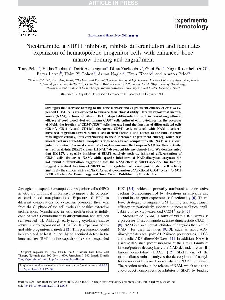
BM homing of NAM-treated cellsEarlier studies have attributed, in part, reduced engraftabil-ity of ex vivo expanded cells to defective homing to theBM caused by exposure to cytokines in culture [3]. Inview of the superior engraftment of NAM-cultured cells(Fig. 3), we tested whether their homing ability wasimproved. NOD/SCID mice were grafted with 10 � 106
fresh (noncultured) UCB cells or with their total progenyafter 3 weeks of culture, 10 � 106 cells (calculated asdescribed here). Cells were labeled with BCECF/AM andhoming to the mouse BM was assessed 24 hours after tra-nsplantation (Fig. 4A–C). Representative flow cytometryanalysis of the BM of grafted mice is shown inFigure 4D–J, allowing the evaluation of BM-homedCD45þ (BCECF/AMþ) (Fig. 4D–G) and CD34þ (BCECF/AMþCD34þ) cells (Fig. 4H–J) in the respective gates of
EXPHEM2839_proof ■ 18
labeled cells. Even though the same number of cells andCD34þ cells were transplanted from both cultured groups(due to a similar fold expansion of TNC and CD34þ cellsfollowing 3-week culture 6 NAM, Figure 1J, L, respec-tively), the homing capacity of cells cultured with NAMincreased compared to nontreated cultures (Fig. 4A).The number of CD34þ cells that home to the BM afterexpansion with NAM was sixfold higher, while that ofNAM-nontreated cultures was only twofold higher overthe homing of culture input CD34þ cells (Fig. 4B). In spiteof the substantial advantage obtained following expansionwith NAM, calculation of homing efficacy based on numberof CD34þ cells infused demonstrate higher efficacy of non-cultured CD34þ cells over the two cultured groups(Fig. 4C). The homing efficacy of NAM-cultured CD34þ
cells was significantly higher over the homing efficacy ofCD34þ cells cultured without NAM.
Therefore, although there is a significant improvement inhoming, NAM treatment does not completely overcomehoming defect resulting from in vitro culture. The increasednumbers of CD34þ cells being injected contribute to thesubstantial increase in the absolute number of CD34þ cellsthat home to the BM after expansion with NAM over non-cultured cells.
Next, we sought to determine the homing capacity ofNAM-treated cultured cells when transplanted along orwith noncultured competitor cells. To this end, 20 � 106
fresh cells (noncultured) labeled with BCECF/AM were co-transplanted with an equal number of NAM-treated cellslabeled with PKH membrane linkers (Fig. 4K). Thecapacity of NAM-treated cells was not affected, indicatingthat NAM-cultured and fresh cells do not compete witheach other during homing.
Dependency of migration and homing of NAM-treatedcells on CXCR4Next, we tested the migration capacity of CD34þ cellstoward SDF-1 gradient in vitro in a Transwell migrationassay [40]. Background spontaneous migration of cells ina Transwell was very low in all groups of cells, in particularafter 3 weeks of culture (Fig. 5A). Similar to their in vivohoming capacity, migration of NAM-treated cells inresponse to SDF-1 also increased compared to that ofcytokine-cultured and freshly isolated cells (Fig. 5B).However, treatment of cells with NAM did not changethe number of cells expressing CXCR4 or the intensity(mean fluorescence intensity) of CXCR4 cell surfaceexpression. Number of cells expressing the cell adhesionmolecules, very late antigen 4 and lymphocyte function–associated antigen 1, and their mean fluorescence intensitywas increased in both cultured groups (Fig. 5C, D). There-fore, quantitative expression of these relevant cell surfacemolecules cannot explain the increased migration andhoming potential of NAM-treated cultured cells.
-1-2012 15-27-17
print&
web
4C=FPO
Figure 4. BM homing of cultured and noncultured cells. NOD/SCID mice were transplanted with either 10 � 106 noncultured TNC (containing 5 � 104
CD34þ cells), or with the total progeny of their purified CD34þ cells following 3-week culture with or without NAM (both containing 200 � 104 CD34þ
cells). Before transplantation, cells were labeled with BCECF/AM, and 24 hours later, BCECF/AM-labeled CD45þ cells (A) and CD34þ cells (B) in the BM
were quantified by flow cytometry, as detailed in Materials and Methods. Homing efficacy of CD34þ cells (number of events normalized to relative number of
CD34þ cells infused) is shown in (C). Homing of human cells is presented as the number of positive events per 100,000 marrow cells analyzed (each bar
represents the mean 6 standard error of three independent experiments, n 5 8/experiment; *p ! 0.05 vs cytokines only; **p ! 0.001 vs cytokines only and
noncultured cells). (D–J) Representative dot plots (side scatter vs BCECF/AM fluorescence) of BM cells from noninjected mice (D), mice injected with
noncultured cells (E), mice injected with cells cultured without NAM (F), and mice injected with cells cultured with NAM (G). (F–J) BCECF/AM-
positive cells were gated and analyzed for BCECF/AM (x axis) and CD34 (y axis). The upper and lower right quadrants represent homing of total human
cells, and the upper right quadrant represents homing of human CD34þ cells. (K) BCECF/AM-labeled noncultured cells (20 � 106) (a) and PKH-labeled
NAM-cultured cells (20 � 106) (b) were transplanted separately or cotransplanted (aDb) in the same mouse (n5 10/experimental group). Twenty four hours
later, BCECF/AM-positive cells (a), PKH-positive cells (b), BCECF/AM-positive and PKH-negative or PKH-positive and BCECF/AM-negative cells (aDb)
were gated and quantified by flow cytometry as described (*p ! 0.04 vs noncultured cells).
8 T. Peled et al./ Experimental Hematology 2012;-:-–-
Cellular target of NAM on cultured CD34þ cellsNAM was reported as a noncompetitive inhibitor of SIRTI[19], the class III HDAC. Inhibition of SIRT1 deacetylaseactivity by NAM in our cultures was tested by Western blot-ting with an antibody specific for acetylated Ku70, a deace-tylation target of SIRT1 [41]. Figure 6A shows that Ku70was highly acetylated in cells cultured with NAM, whilethe level of acetylation was substantially lower in cellscultured with cytokines only, indicating inhibition ofSIRT1 deacetylase activity by NAM in our cultures.
To study the causal role of SIRT1, CD34þ cultures weretreated with EX-527, a selective inhibitor of SIRT1 that
EXPHEM2839_proof ■ 1
does not inhibit class I and II HDAC or other sirtuin dea-cetylase family members (IC50 values are 98; 19,600;48,700; O100,000, and O100,000 nM for SIRT1, SIRT2,SIRT3, HDAC, and NADase, respectively) [22,23]. Similarto NAM, but at substantially lower concentrations, EX-527increased the fraction of CD34þCD38� cells and decreasedthe fraction of more differentiated, monocytic (CD14),dendritic (CD11c), and common myeloid (CD11b) cells(Fig. 6B, C). Both molecules also similarly attenuated theaccelerated differentiation imposed by IL-3, as demon-strated by the increase in number of CFUc in 5-weekcultures compared to cytokine-treated cultures (Fig. 6D).
8-1-2012 15-27-17

Figure 5. Dependency ofmigration and homing onCXCR4. Spontaneous (*p! 0.03 vs cultured cells) (A) andSDF-1 (100 ng/mL) inducedTranswellmigration
(B) of CD34þ cells either before or after 3-week culture are shown (*p! 0.05 vs cytokines and noncultured). CD34þ cells, either before or after culture with or
without NAM, were stained with antibodies to CXCR4, very late antigen 4 (VLA4), lymphocyte function–associated antigen 1 (LFA1), and flow cytometry
analyzed. The percentages of positive cells (C) and the mean fluorescence intensity (MFI) (CXCR4: *p ! 0.02 vs cultured cells, VLA4: *p ! 0.01; **p !0.02, LFA1: **p! 0.02 vs noncultured cells) (D) are shown. Each bar represents the mean6 standard error of four independent experiments.
9T. Peled et al./ Experimental Hematology 2012;-:-–-
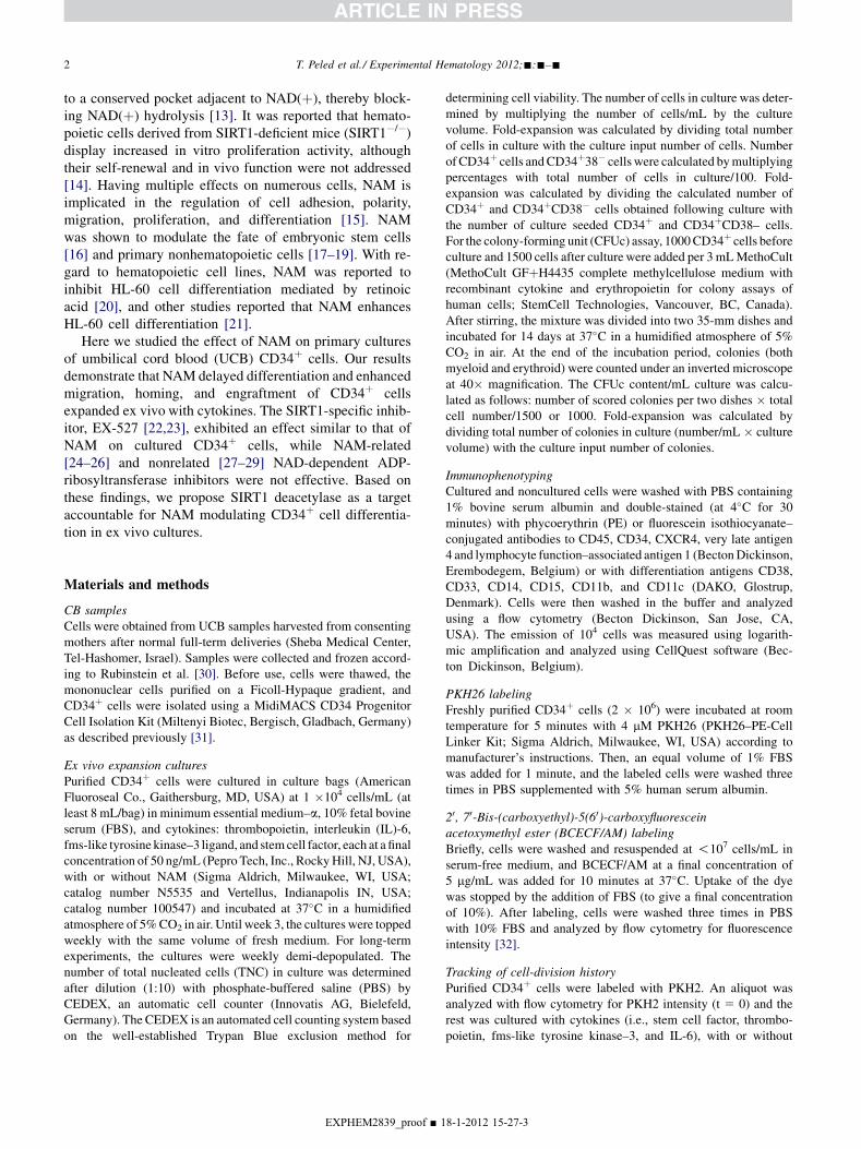
To find out whether inhibition of ADP-ribosylation mightalso be mechanistically relevant to the effect of NAM, weused positional NAM isomers [26] and NAM-relatedmolecules possessing [25] and lacking [24] the inhibitoryactivity over NAD(þ)-dependent ADP-ribosyltransferase,NAM structurally unrelated inhibitors of ADP ribosylationincluding mono-ADP-ribosyltransferases [29] and poly-ADP-ribose polymerases [28] -ADP-ribosyltransferases anda CD38 antagonist [27] (Table 1). All of these moleculesfailed to enrich CD34þCD38� and to decrease the fractionof differentiated CD14þ cells, indicating that the effect ofNAMon culturedCD34þ cellswas notmechanisticallymedi-ated by the inhibition of ADP-ribosylation. Taken together,these results imply that the NAM effect on CB-derivedcultured CD34þ cells is mediated by the inhibition of SIRT1.
DiscussionHere we show that NAM, a potent inhibitor of enzymes thatrequire NADþ for their activity such as ribosylase enzymes[9–11] and class III HDAC [12], modulates in vitro expan-sion and differentiation and in vivo engraftability ofex vivo–cultured, CB-derived CD34þ cells.
Treatment of CD34þ cells with NAM together withcytokines resulted in about a 400-fold increase in total
EXPHEM2839_proof ■ 18
cellularity, a 80-fold increase in CD34þ cells, and a 60-fold increase in clonogenic activity over input numbersthat were not substantially different from cultures treatedwith cytokines only. However, in the cultures treated withNAM, the number of CD34þCD38� cells was substantiallyincreased compared to NAM-nontreated cultures. Further-more, the number of differentiated cells was significantlyreduced, the cycling of CD34þ cells was delayed, andresponsiveness to differentiation stimuli was attenuated.In vitro, the chemotactic migration toward SDF-1 wasimproved and, in parallel, the in vivo homing of NAM-treated cells to murine BM was increased by threefoldcompared to cultures without NAM. Importantly, the inci-dence of short-term SRC resulted in a 7.6-fold increaseover cultures without NAM and 9-fold over culture inputnumber of short-term SRC (noncultured cells). Finally,the advantage of NAM-cultured cells was evident when co-transplanted with noncultured competitor cells. Longerexperiments will be required to demonstrate effects onlong-term SRCs.
Phenotype analysis of cultures treated with NAMshowed an increase in CD34þ cell numbers during long-term cultures, which was primarily attributed to phenotypicstability of cycling CD34þCD38� cells. In parallel, cellscultured with NAM displayed increased long-term
-1-2012 15-27-23

print&
web
4C=FPO
Figure 6. The cellular target of NAM in CD34þ cell cultures. (A) CD34þ cells were cultured for 3 weeks with NAM (5 mM) or without NAM. Western
blotting analysis of preimmunoprecipitation (input) and immunoprecipitated (IP) samples ( -Ac-K) were performed with an anti-Ku70 antibody, as described
in Material and Methods. (B, C) CD34þ cells were cultured with cytokines, cytokines þ NAM (5 mM) or cytokines and EX527 (25–50 mM). After 3 weeks,
cells were dually stained with antibodies to human CD34 and with antibodies to human differentiation antigens as indicated. Representative flow cytometry
analysis dot plots (B) and percentages of CD34þCD38� cells and of cells positive for differentiation Ags (C) (calculated from the upper and lower right
quadrants) are shown (n 5 3; *p ! 0.01 NAM vs cytokines; **p ! 0.004 EX527 vs cytokines and #p ! 0.02; ##p ! 0.03; ###p ! 0.01 cytokines vs
NAM and EX527). (D) CD34þ cells were cultured with IL-3, as previously described, and treated without or with NAM and EX527 as indicated. Number
of CFUc/mL culture, 4 weeks post seeding, are shown (n 5 3; *p ! 0.05; **p ! 0.05 vs cytokines).
10 T. Peled et al./ Experimental Hematology 2012;-:-–-
expansion potential of colony-forming cells. The relevanceof these findings to the engraftability of NAM-culturedcells is difficult to interpret because it has already beenshown that there is a dissociation between hematopoieticactivity of expanded cells in vitro, such as long-term cultureinitiating cell, and their SCID-repopulating potential [2].
Furthermore, the CD34þCD38� phenotype has beenshown to be an unpredictable surrogate marker for cultureexpansion of in vivo repopulating cells [42], such as thephenotype is dissociated from their in vivo repopulat-ing ability [43]. However, it was shown that SRCs follo-wing in vitro expansion reside exclusively within theCD34þCD38� cell compartment, although their repo-pulating efficacy is significantly lower than that of nonc-ultured cells displaying a similar phenotype [44]. The
EXPHEM2839_proof ■ 1
engraftment defect was attributed to cytokine-inducedaccelerated cycling of the cells in culture [5]. Hematopoieticprogenitors usually cycle slower than their more differen-tiated progeny. Alteration of the rate of cell cycle transi-tion, such as by deactivation of the G1 check-pointregulator and the cyclin-dependent kinase inhibitor p21,has been reported to accelerate HPC expansion and causea defect in their engraftment [45]. We showed that CD34þ
cells cultured with NAM displayed an initial slowerproliferation rate than their counterparts cultured withoutNAM. This was most pronounced in the putative HPCcompartment defined as CD34þCD38� cells, along withan increase in their numbers in the presence of NAM.Our results suggest that slower proliferation rates causedby NAM impacted positively on the preservation of the
8-1-2012 15-27-25
Table 1. Screening for molecules with NAM-like activity on CD34þ cell cultures
Tested compounds Concentrations (mM) ADPR inhibitory activity (published data) NAM-like activity
NAM positional isomers Isonicotinamide 1–10 þ �Picolinamide 1–10 þ �
NAM-related compounds N-methylnicotinamide 1–10 þ �Nicotinic acid 1–10 � �
NAM-nonrelated ADPR inhibitors Meta-iodobenzylguanidine 2–1 þ �3-aminobenzamide 20–500 þ �
CD38 cADPR antagonist) 8- amino-cADPR 10–500 � �NAD 10–100 � �
ADP ribosylation (ADPR), as indicated (N-methylnicotinamide was purchased from ABCR (Karlsruhe, Germany), 8-amino-cADPR from Invitrogen
Corporation (Carlsbad, CA, USA), while the others were from Sigma Aldrich). Inhibition of differentiation (NAM-like activity) by the tested compounds
was determined based on percentages of CD34þCD38� and CD14þ cells in 2- or 3-week cultures, relative to their percentages in cytokines only and
NAM- treated cultures.
11T. Peled et al./ Experimental Hematology 2012;-:-–-
premature phenotype of cycling progenitor cells, as wellas their functionality.
Engraftment is a multistep process involving directedmigration of the inoculated cells, homing to the BM, reten-tion within the BM niche, followed by self-renewal anddifferentiation [46]. One of the major causes of deficientengraftment of CD34þ cells cultured with cytokines is inef-fective homing to the murine BM as compared to freshCD34þ cells [3,6,47]. The homing deficiency acquired inculture explains, at least in part, the discrepancy betweenin vitro functional HPC assays and in vivo SRC activity.The superior engraftment obtained after culture of CD34þ
cells with NAM prompted us to evaluate whether these cellshomed to the BM with higher efficacy than cells culturedwith cytokine only. In our experiments, SCID mice weregrafted with similar numbers of noncultured and culturedcells. Despite higher numbers of CD34þ cells used fortransplant after culture, homing of CD34þ cells to theBM was increased by twofold in mice inoculated withcytokine-cultured cells and sixfold after treatment withNAM over homing of noncultured CD34þ cells. Thus,NAM increases the homing potential of ex vivo–expandedCD34þ cells. Interestingly, the effect of NAM on homingof cytokine-cultured cells required continuous exposure,as short-term treatment (1–48 hours) before transplantationwas ineffective (data not shown). Hence, reversal ofacquired deficiencies in cultured CD34þ cells caused byNAM occurred through modulation of cytokine signalingin early stages of expansion.
The homing defect of cultured HPC was ascribed toa sustained increase in adhesion receptor expression alongwith a decrease in CXCR4 resulting in nonspecific bindingof cultured cells to extramedullary endothelial surfaces[48]. Consistently, we found a substantial increase in thelevels of the adhesion molecules expression, includingvery late antigen 4 and lymphocyte function–associatedantigen 1, and a decrease in CXCR4 mean fluorescentintensity on cultured CD34þ cells, irrespective of the pres-ence of NAM. Despite that, CD34þ cells cultured withNAM displayed increased migratory activity over
EXPHEM2839_proof ■ 18
cytokine-cultured cells, which is likely associated withsuperior homing and engraftment, suggesting modulationof CXCR4 downstream signaling by NAM.
NAM has been shown as a noncompetitive inhibitor ofSIRT1 deacetylase [13]. In lower eukaryotes, Sir2 (themammalian SIRT1 homolog) has been strongly implicatedin the modulation of replicative life span and promotion oflongevity in response to stress, while NAM strongly inhibitssilencing and shortens replicative life span [13]. In contrastto its function in lower eukaryotes, SIRT1 function inhigher eukaryotes is still debatable [16]. In higher eukary-otes, SIRT1 binds to and deacetylates a number of impor-tant transcription factors, such as nuclear factor–kB,Ku70, peroxisome proliferator-activated receptor–g, perox-isome proliferator-activated receptor–a, peroxisomeproliferator-activated receptor–g coactivator 1 a, theFOXOs family of transcription factors, and others, thuslinking SIRT1 activity to oxidative stress, cell survival,tumorigenesis, metabolism, and cell differentiation [49].In neural progenitor cells, SIRT1 activator, resveratrol,was shown to suppress proliferation and directs their differ-entiation toward the astroglial lineage, thus mimicking theeffect of oxidative conditions. This effect was blocked bysmall interfering RNAs against SIRT1 messenger RNA[49]. NAM, through inhibition of SIRT1, was shown toinhibit neural cells differentiation. In addition, NAM wasshown to inhibit all-trans retinoic acid–induced differentia-tion of neuroblastoma cells, while class I and II HDACinhibitors had no effect. Thus, SIRT1 was suggested asa novel regulator of neuronal differentiation [50]. In normalhuman keratinocytes, NAM was shown to inhibit theexpression of keratinocyte differentiation markers, whereasthe SIRT1 activator, resveratrol, to enhance the expressionof differentiation markers. Similar results were obtainedin keratinocytes manipulated to overexpress or underex-press SIRT1. It was concluded that SIRT1 functions innormal human keratinocytes to inhibit proliferation and topromote differentiation [51]. In contrast to SIRT1 effecton the differentiation of neural and keratinocyte progenitorcells, its overexpression in muscle and fat cells was shown
-1-2012 15-27-29
12 T. Peled et al./ Experimental Hematology 2012;-:-–-
to inhibit differentiation whilst NAM greatly stimulateddifferentiation [52,53]. Therefore, the conflicting reportsin higher eukaryotes demonstrating promotion or inhibitionof differentiation depended on cell or tissue type [16].
Here we show that, similar to NAM, EX-527, a specificSIRT1 inhibitor [23], reduced significantly the expressionof granulomonocytic (CD11b, CD11C, and CD14) markers,increased the fraction of CD34þCD38� cells, and inhibitedthe differentiation forced by IL-3 on CB-derived, CD34þ
cell cultures. NAM positional isomers [26], NAM-relatedmolecules [25], or NAM-nonrelated poly-ADP-ribose poly-merases [28] and mono-ADP-ribosyltransferases [29]inhibitors did not inhibit differentiation, suggesting thatthe effect of NAM on cultured CD34þ cells is probablythrough regulation of SIRT1 activity. Although Ex-527has been reported to be a selective inhibitor of SIRT1,NAM inhibits most sirtuins. Our preliminary results suggestthat although inhibition of SIRT2 by its specific inhibitor,AGK2, attenuates differentiation, its effect is less signifi-cant than that obtained with EX527 or NAM (data notshown). Current studies are designed to appraise the roleof SIRT1 and to define the signaling pathways upstreamand downstream of SIRT1 and characterize the role thateach plays in the regulation of self-renewal and differenti-ation of CB-derived HPCs.
Several strategies have been reported to modulate thefate of HPCs cultured with different combinations of cyto-kines, including the immobilized form of the Notch ligandd-1 [54], copper chelators [31,33,55], Wnt3a [56], prosta-glandin E2 [57], enforced expression of the HOXB4 tran-scription factor [58], and coculture of HPCs withsupportive stromal cells [59] . Even though all of thesestrategies promoted expansion in ex vivo cultures, eachdistinctly contributes and advances our understanding ofthe biology of HPCs and the pathways involved in the regu-lation of self-renewal and differentiation. However, thesuccessful development of epigenetic strategies capable ofinducing human HPC expansion ex vivo, while preservingtheir in vivo function after transplantation, remains a chal-lenge. Several of these strategies are presently being evalu-ated in the clinic [54,55,59]; however, the optimalexpansion conditions are still not known.
ConclusionsThe results of the present study uncover SIRT1, class IIINADþ-dependent-histone-deacetylase, as a novel pathwayinvolved in regulation of self-renewal and differentiation,as well as additional fundamental functions of CB-derivedHPCs, such as directed migration, homing to the BM niche,and engraftment.
This novel expansion strategy based on downregulation ofSIRT1 by NAM is currently being evaluated in a pilot studyfor CB transplantation in patients with hematological malig-nancies (ClinicalTrials.gov Identifier: NCT01221857).
EXPHEM2839_proof ■ 1
Conflict of Interest DisclosureThe following authors may have a financial interest in thepresentwork:DrsT. Peled, Shoham,Aschengrau,Yackoubov,Frei, and Rosenheimer G are employed by Gamida Cell Ltd.,and Drs. T. Peled, Shoham, and Rosenheimer G haveownership interest. Drs. Nagler and A. Peled are consultantsfor Gamida Cell Ltd. For the remaining authors, no financialinterest/relationships with financial interest relating to thetopic of this article have been declared.
AcknowledgmentsWe thank Dr. Nadir Askenasy of the Department of PediatricHematology-Oncology, Schneider Children’s Medical Center ofIsrael for the critical reading of the manuscript and Mary Clausen,Ido D. Weiss, Katia Beider the Gene Therapy Institute, HadassahUniversity Hospital, Jerusalem, Israel for technical assistance.
Author contributionsTony Peled: Conception and design, data analysis and interpreta-tion, manuscript writing, final approval of manuscript; HadasShoham: Performed research, collection and/or assembly ofdata, data analysis and interpretation; Gabi Frei: Performedresearch, collection and/or assembly of data, data analysis andinterpretation; Dorit Aschengrau: Performed research, collectionand/or assembly of data, data analysis and interpretation; DimaYackoubov: Performed research, collection and/or assembly ofdata, data analysis and interpretation; Noga Rosenheimer G:Performed research, data analysis and interpretation; Haim Y.Cohen: Data analysis and interpretation; Batya Lerrer: Performedresearch, Data analysis and interpretation; Arnon Nagler: Dataanalysis and interpretation; Eitan Fibach: Data analysis and inter-pretation, manuscript writing; Amnon Peled: Data analysis andinterpretation, manuscript writing.
References1. Von Drygalski A, Alespeiti G, Ren L, Adamson JW. Murine bone
marrow cells cultured ex vivo in the presence of multiple cytokine
combinations lose radioprotective and long-term engraftment poten-
tial. Stem Cells Dev. 2004;13:101–111.
2. Xu R, Reems JA. Umbilical cord blood progeny cells that retain
a CD34þ phenotype after ex vivo expansion have less engraftment
potential than unexpanded CD34þ cells. Transfusion. 2001;41:
213–218.
3. Szilvassy SJ, Bass MJ, Van Zant G, Grimes B. Organ-selective homing
defines engraftment kinetics of murine hematopoietic stem cells and is
compromised by ex vivo expansion. Blood. 1999;93:1557–1566.
4. Szilvassy SJ, Meyerrose TE, Grimes B. Effects of cell cycle activation
on the short-term engraftment properties of ex vivo expanded murine
hematopoietic cells. Blood. 2000;95:2829–2837.
5. Takatoku M, Sellers S, Agricola BA, et al. Avoidance of stimulation
improves engraftment of cultured and retrovirally transduced hemato-
poietic cells in primates. J Clin Invest. 2001;108:447–455.
6. Ahmed F, Ings SJ, Pizzey AR, et al. Impaired bone marrow homing of
cytokine-activated CD34þ cells in the NOD/SCID model. Blood.
2004;103:2079–2087.
7. Hofmeister CC, Zhang J, Knight KL, Le P, Stiff PJ. Ex vivo expansion
of umbilical cord blood stem cells for transplantation: growing
8-1-2012 15-27-29

13T. Peled et al./ Experimental Hematology 2012;-:-–-
knowledge from the hematopoietic niche. Bone Marrow Transplant.
2007;39:11–23.
8. Berger F, Ramirez-Hernandez MH, Ziegler M. The new life of a cente-
narian: signalling functions of NAD(P). Trends Biochem Sci. 2004;29:
111–118.
9. Banasik M, Komura H, Shimoyama M, Ueda K. Specific inhibitors of
poly(ADP-ribose) synthetase and mono(ADP-ribosyl)transferase. J
Biol Chem. 1992;267:1569–1575.
10. Corda D, Di Girolamo M. Functional aspects of protein mono-ADP-
ribosylation. EMBO J. 2003;22:1953–1958.
11. Krebs C, Adriouch S, Braasch F, et al. CD38 controls ADP-
ribosyltransferase-2-catalyzed ADP-ribosylation of T cell surface
proteins. J Immunol. 2005;174:3298–3305.
12. Denu JM. Vitamin B3 and sirtuin function. Trends Biochem Sci. 2005;
30:479–483.
13. Bitterman KJ, Anderson RM, Cohen HY, Latorre-Esteves M, Sinclair
DA. Inhibition of silencing and accelerated aging by nicotinamide,
a putative negative regulator of yeast sir2 and human SIRT1. J Biol
Chem. 2002;277:45099–45107.
14. Narala SR, Allsopp RC, Wells TB, et al. SIRT1 acts as a nutrient-
sensitive growth suppressor and its loss is associated with increased
AMPK and telomerase activity. Mol Biol Cell. 2008;19:1210–1219.
15. Glowacki G, Braren R, Firner K, et al. The family of toxin-related
ecto-ADP-ribosyltransferases in humans and the mouse. Protein Sci.
2002;11:1657–1670.
16. Vaca P, Berna G, Martin F, Soria B. Nicotinamide induces both prolif-
eration and differentiation of embryonic stem cells into insulin-
producing cells. Transplant Proc. 2003;35:2021–2023.
17. Miura M, Kameda Y. Nicotinamide promotes long-term survival and
extensive neurite outgrowth in ultimobranchial C cells cultured from
chick embryos. J Comp Neurol. 2005;492:334–348.
18. Sato F, Mitaka T, Mizuguchi T, Mochizuki Y, Hirata K. Effects
of nicotinamide-related agents on the growth of primary rat hepato-
cytes and formation of small hepatocyte colonies. Liver. 1999;19:
481–488.
19. Papaccio G, Ammendola E, Pisanti FA. Nicotinamide decreases MHC
class II but not MHC class I expression and increases intercellular
adhesion molecule-1 structures in non-obese diabetic mouse pancreas.
J Endocrinol. 1999;160:389–400.
20. Munshi CB, Graeff R, Lee HC. Evidence for a causal role of CD38
expression in granulocytic differentiation of human HL-60 cells. J
Biol Chem. 2002;277:49453–49458.
21. Iwata K, Ogata S, Okumura K, Taguchi H. Induction of differentiation
in human promyelocytic leukemia HL-60 cell line by niacin-related
compounds. Biosci Biotechnol Biochem. 2003;67:1132–1135.
22. Solomon JM, Pasupuleti R, Xu L, et al. Inhibition of SIRT1 catalytic
activity increases p53 acetylation but does not alter cell survival
following DNA damage. Mol Cell Biol. 2006;26:28–38.
23. Napper AD, Hixon J, McDonagh T, et al. Discovery of indoles as
potent and selective inhibitors of the deacetylase SIRT1. J Med
Chem. 2005;48:8045–8054.
24. Colon-Otero G, Sando JJ, Sims JL, McGrath E, Jensen DE,
Quesenberry PJ. Inhibition of hemopoietic growth factor-induced
proliferation by adenosine diphosphate-ribosylation inhibitors. Blood.
1987;70:686–693.
25. Sanchez-Pacheco A, Perez P, Villa A, Pascual A, Aranda A. Nicotin-
amide analogs and DNA-damaging agents deplete thyroid hormone
receptor and c-erbA mRNA levels in pituitary GH1 cells. Mol Cell
Endocrinol. 1993;91:127–134.
26. Itoh H, Okajima F, Ui M. Conversion of adrenergic mechanism from
an alpha- to a beta-type during primary culture of rat hepatocytes.
Accompanying decreases in the function of the inhibitory guanine
nucleotide regulatory component of adenylate cyclase identified as
the substrate of islet-activating protein. J Biol Chem. 1984;259:
15464–15473.
EXPHEM2839_proof ■ 18
27. Aarhus R, Gee K, Lee HC. Caged cyclic ADP-ribose. Synthesis and
use. J Biol Chem. 1995;270:7745–7749.
28. Songin M, Jesko H, Czapski G, Adamczyk A, Strosznajder RP. GSK-
3beta and oxidative stress in aged brain. Role of poly(ADP- ribose)
polymerase-1. Folia Neuropathol. 2007;45:220–229.
29. Smets LA, Loesberg C, Janssen M, Van Rooij H. Intracellular
inhibition of mono(ADP-ribosylation) by meta-iodobenzylguanidine:
specificity, intracellular concentration and effects on glucocorticoid-
mediated cell lysis. Biochim Biophys Acta. 1990;1054:49–55.
30. Rubinstein P, Dobrila L, Rosenfield RE, et al. Processing and
cryopreservation of placental/umbilical cord blood for unrelated bone
marrow reconstitution. Proc Natl Acad Sci USA. 1995;92:10119–10122.
31. Peled T, Landau E, Mandel J, et al. Linear polyamine copper chelator
tetraethylenepentamine augments long-term ex vivo expansion of cord
blood-derived CD34(þ) cells and increases their engraftment potential
in NOD/SCID mice. Exp Hematol. 2004;32:547–555.
32. Lyons AB, Parish CR. Determination of lymphocyte division by flow
cytometry. J Immunol Methods. 1994;171:131–137.
33. Peled T, Mandel J, Goudsmid RN, et al. Pre-clinical development of
cord blood-derived progenitor cell graft expanded ex vivo with cyto-
kines and the polyamine copper chelator tetraethylenepentamine.
Cytotherapy. 2004;6:344–355.
34. Wang JC, Doedens M, Dick JE. Primitive human hematopoietic cells
are enriched in cord blood compared with adult bone marrow or mobi-
lized peripheral blood as measured by the quantitative in vivo SCID-
repopulating cell assay. Blood. 1997;89:3919–3924.
35. Taswell C. Limiting dilution assays for the determination of immuno-
competent cell frequencies. I. Data analysis. J Immunol. 1981;126:
1614–1619.
36. Nteliopoulos G, Marley SB, Gordon MY. Influence of PI-3K/Akt
pathway on Wnt signalling in regulating myeloid progenitor cell
proliferation. Evidence for a role of autocrine/paracrine Wnt regula-
tion. Br J Haematol. 2009;146:637–651.
37. Wyss BK, Meyers JL, Sinn AL, Cai S, Pollok KE, Goebel WS. A
novel competitive repopulation strategy to quantitate engraftment of
ex vivo manipulated murine marrow cells in submyeloablated hosts.
Exp Hematol. 2008;36:513–521.
38. Tatekawa T, Ogawa H, Kawakami M, et al. A novel direct competitive
repopulation assay for human hematopoietic stem cells using
NOD/SCID mice. Cytotherapy. 2006;8:390–398.
39. Rosler ES, Brandt JE, Chute J, Hoffman R. An in vivo competitive re-
population assay for various sources of human hematopoietic stem
cells. Blood. 2000;96:3414–3421.
40. Plett PA, Frankovitz SM, Wolber FM, Abonour R, Orschell-Traycoff
CM. Treatment of circulating CD34(þ) cells with SDF-1alpha or
anti-CXCR4 antibody enhances migration and NOD/SCID repopulat-
ing potential. Exp Hematol. 2002;30:1061–1069.
41. Cohen HY, Miller C, Bitterman KJ, et al. Calorie restriction promotes
mammalian cell survival by inducing the SIRT1 deacetylase. Science.
2004;305:390–392.
42. Danet GH, Lee HW, Luongo JL, Simon MC, Bonnet DA. Dissociation
between stem cell phenotype and NOD/SCID repopulating activity in
human peripheral blood CD34(þ) cells after ex vivo expansion. Exp
Hematol. 2001;29:1465–1473.
43. Dorrell C, Gan OI, Pereira DS, Hawley RG, Dick JE. Expansion of
human cord blood CD34(þ)CD38(-) cells in ex vivo culture during
retroviral transduction without a corresponding increase in SCID repo-
pulating cell (SRC) frequency: dissociation of SRC phenotype and
function. Blood. 2000;95:102–110.
44. Vanheusden K, Van Coppernolle S, De Smedt M, Plum J,
Vandekerckhove B. In vitro expanded cells contributing to rapid scid
repopulation activity are Cd34þ38-33þ90þ45ra. Stem Cells. 2007;
25:107–714.
45. Cheng T, Rodrigues N, Shen H, et al. Hematopoietic stem cell quies-
cence maintained by p21cip1/waf1. Science. 2000;287:1804–1808.
-1-2012 15-27-30

14 T. Peled et al./ Experimental Hematology 2012;-:-–-
46. Lapidot T, Dar A, Kollet O. How do stem cells find their way home?
Blood. 2005;106:1901–1910.
47. Foguenne J, Huygen S, Greimers R, Beguin Y, Gothot A. Modulation
of homing properties of primitive progenitor cells generated by
ex vivo expansion. Haematologica. 2005;90:445–451.
48. Denning-Kendall P, Singha S, Bradley B, Hows J. Cytokine expansion
culture of cord blood CD34þ cells induces marked and sustained changes
in adhesion receptor andCXCR4 expressions. StemCells. 2003;21:61–70.
49. Haigis MC, Sinclair DA. Mammalian sirtuins: biological insights and
disease relevance. Annu Rev Pathol. 2010;5:253–295.
50. Kim MJ, Ahn K, Park SH, et al. SIRT1 regulates tyrosine hydroxylase
expression and differentiation of neuroblastoma cells via FOXO3a.
FEBS Lett. 2009;583:1183–1188.
51. Blander G, Bhimavarapu A, Mammone T, et al. SIRT1 promotes
differentiation of normal human keratinocytes. J Invest Dermatol.
2009;129:41–49.
52. Pardo PS, Boriek AM. The physiological roles of Sirt1 in skeletal
muscle. Aging. 2011;3:430–437.
53. Backesjo CM, Li Y, Lindgren U, Haldosen LA. Activation of
Sirt1 decreases adipocyte formation during osteoblast differenti-
EXPHEM2839_proof ■ 1
ation of mesenchymal stem cells. Cells Tissues Organs. 2009;
189:93–97.
54. Delaney C, Varnum-Finney B, Aoyama K, Brashem-Stein C, Bernstein
ID. Dose-dependent effects of the Notch ligand Delta1 on ex vivo
differentiation and in vivo marrow repopulating ability of cord blood
cells. Blood. 2005;106:2693–2699.
55. Peled T, Glukhman E, Hasson N, et al. Chelatable cellular copper
modulates differentiation and self-renewal of cord blood-derived
hematopoietic progenitor cells. Exp Hematol. 2005;33:1092–1100.
56. Sato N, Meijer L, Skaltsounis L, Greengard P, Brivanlou AH. Mainte-
nance of pluripotency in human and mouse embryonic stem cells
through activation of Wnt signaling by a pharmacological GSK-3-
specific inhibitor. Nat Med. 2004;10:55–63.
57. North TE, Goessling W, Walkley CR, et al. Prostaglandin E2 regulates
vertebrate haematopoietic stem cell homeostasis. Nature. 2007;447:
1007–1011.
58. Antonchuk J, Sauvageau G, Humphries RK. HOXB4-induced expan-
sion of adult hematopoietic stem cells ex vivo. Cell. 2002;109:39–45.
59. Kelly SS, Sola CB, de Lima M, Shpall E. Ex vivo expansion of cord
blood. Bone Marrow Transplant. 2009;44:673–681.
8-1-2012 15-27-30

EXPHEM2839_proof ■ 18-1-2012 15-27-30
Supplementary Figure E1. The effect of increasing concentrations of NAM on cultured CD34þ cells. CB–derived purified CD34þ cells were cultured for 3
weeks with cytokines or with cytokines and increasing concentrations of NAM as indicated. The culture, analysis procedures and calculations are described in
Materials and Methods. Each bar represents the mean 6 standard error of three independent experiments. Results show the fold-expansion TNC (A) and of
CFUc (B). Percentages in culture of CD34þ and CD34þCD38� cells are shown in (C) and (D), respectively.
Supplementary Figure E2. Phenotype characterization of lineage-differentiated cells in cultures treated with or without NAM. CB-derived purified CD34þ
cells were cultured with cytokines or with cytokines and 2.5 or 5 mM NAM. After 3 weeks in culture, cells were dually stained with antibodies to human
CD45 or CD34 and with antibodies to human differentiation antigens as indicated. Percentages of dual-positive cells 6 standard error are shown (n 5 4).
14.e1T. Peled et al./ Experimental Hematology 2012;-:-–-