Embed Size (px)
Citation preview
1
Fluorine-induced J-aggregation enhances emissive properties of a new NLO push-pull chromophore
Chiara Botta,*a Elena Cariati,*b Gabriella Cavallo,c Valentina Dichiarante,c Alessandra Forni,*d Pierangelo Metrangolo,*c Tullio Pilati,c Giuseppe Resnati,*c Stefania Righetto,b Giancarlo Terraneoc and Elisa Tordinb
a ISMAC-CNR, via Bassini 15, 20133 Milan, Italy;b Dept. Chemistry and INSTM UdR Milano, University of Milan, via Golgi 19, I-20133 Milan,
Italy; c NFMLab, DCMIC “Giulio Natta”, Politecnico di Milano, via Mancinelli 7, I-20131 Milan, Italy;d ISTM-CNR, University of Milan, via Golgi 19, I-20133 Milan, Italy;
*:
- Chiara Botta, ISMAC-CNR, via Bassini 15, 20133 Milan, Italy. Phone: +39 02 2369 9734, Fax:
+39 02 7063 6400, e-mail: [email protected]
- Elena Cariati, Dept. Chemistry and INSTM UdR Milano, University of Milan, via Golgi 19, I-
20133 Milan, Italy. Phone: +39 02 50314370, Fax: +39 02 50314405, e-mail: [email protected]
- Alessandra Forni, ISTM-CNR, University of Milan, via Golgi 19, I-20133 Milan, Italy. Phone:
+39 02 50314273, Fax: +39 02 50314300, e-mail: [email protected]
- Pierangelo Metrangolo, Giuseppe Resnati, NFMLab, Department of Chemistry, Materials, and
Chemical Engineering ‘‘Giulio Natta’’, Politecnico di Milano, via Mancinelli 7, 20131 Milan, Italy.
Phone: +39 02 23993041 (P. M.), +39 02 23993032 (G. R.), Fax: +39 02 23993180, E-mail:
[email protected] (P.M.); [email protected] (G. R.).
Electronic Supplementary Material (ESI) for Journal of Materials Chemistry C.This journal is © The Royal Society of Chemistry 2014
2
Electronic Supplementary Information
Experimental procedures
Materials and methods
Commercial HPLC-grade solvents were used without further purification. Starting materials were
purchased from Sigma Aldrich, Acros Organics and Apollo Scientific. Reactions were carried out in
oven-dried glassware under a nitrogen atmosphere.
1H NMR and 19F NMR spectra were recorded at room temperature with a Bruker AV500
spectrometer. Unless otherwise stated, CDCl3 was used as both solvent and internal standard in 1H
NMR spectra. For 19F NMR spectra, CDCl3 was used as solvent and CFCl3 as internal standard.
Spectra were acquired on sample with concentration ranging between 10-5 M to 5 x 10⁻³ M in order
to study the intermolecular interactions occurring at different concentrations. No shift was detected
in 19F signals, proving the absence of significant intermolecular interactions.
Mass spectra were performed on a Finnigan MAT TSQ70 apparatus for EI, on a Agilent 6890 series
GC / Agilent 5973 mass selective detector network for GC-MS, and on Bruker Esquire 3000 Plus
spectrometer for ESI.
Dynamic light scattering (DLS) measurements were performed at 25°C on a Malvern Nanozetasizer
instrument with a 633 nm He-Ne laser, on chloroform solutions with concentration ranging from 10-
5 M to 5 x 10⁻³ M.
Attenuated total reflectance FTIR (ATR-FTIR) spectra were recorded on a Nicolet Nexus FTIR
spectrometer equipped with a UATR unit. The values were given in wavenumbers and were
rounded to 1 cm−1 upon automatic assignment.
Melting points were assigned through an Olympus BX51 polarized-light optical microscope
equipped with a Linkam Scientific LTS 350 heating/cooling stage and a Sony CCD-IRIS/RGB
color video camera connected to a Sony video monitor CMA-D2 (heating rate 10 °C/min).
Deoxycholic acid (DCA) was purchased from Sigma Aldrich and used without further purification.
Films of DCA inclusion compound 1b-DCA were obtained by casting 1b/DCA mixtures with a 1:4
molar ratio, from THF solutions, following the procedure described in ref. S1.
Optical absorption measurements were performed with a Perkin-Elmer Lambda-9 spectrometer.
Photoluminescence and photoluminescence excitation profiles were obtained with a SPEX 270 M
3
monochromator equipped with a N2 cooled charge-coupled device, exciting with a monochromatic
Xe lamp. All spectra were corrected for the instrument response. Solid state PL QY measurements
were carried out using a home-made integrating sphere, according to a previously reported
procedure.S1
EFISH NLO measurements were carried out in CHCl3 and DMF solutions at different
concentrations, at a nonresonant fundamental wavelength of 1907 nm, using a Q-switched, mode-
locked Nd3+:YAG laser. The 1064 nm initial wavelength was shifted to 1907 nm by a Raman shifter
with a high-pressure H2 cell.
Single crystal X-ray diffraction data were collected on a Bruker KAPPA APEX II diffractometer
with Mo-Kα radiation and CCD detector. The structures were solved by SIR2002S2 and refined by
SHELXL-97S3 programs, respectively. The refinement was carried on by full-matrix least-squares
on F2. Hydrogen atoms were placed using standard geometric models and with their thermal
parameters riding on those of their parent atoms.
X-Ray Powder Diffraction (XRPD) measurements were performed with a Bruker AXS D8 powder
diffractometer, using the following experimental parameters: Cu-Kα radiation (λ = 1.54056 Å);
scanning interval: 5 - 40° 2θ; step size: 0.016°; exposure time: 1.5 s per step. The crystallite average
sizes were then estimated for both samples through the Scherrer equation integrating the area
underneath the XRD peak at 2 = 12,84°. The peak position and the full-width at half maximum
height (FWHM) of the peak were obtained using TOPAS software.
The milling was carried out in a high speed ball-milling apparatus for 10 min at 30 Hz.
4
Synthesis of 1b
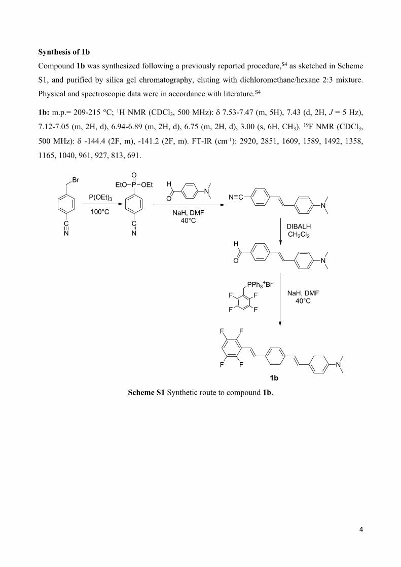
Compound 1b was synthesized following a previously reported procedure,S4 as sketched in Scheme
S1, and purified by silica gel chromatography, eluting with dichloromethane/hexane 2:3 mixture.
Physical and spectroscopic data were in accordance with literature.S4
1b: m.p.= 209-215 °C; 1H NMR (CDCl3, 500 MHz): 7.53-7.47 (m, 5H), 7.43 (d, 2H, J = 5 Hz),
7.12-7.05 (m, 2H, d), 6.94-6.89 (m, 2H, d), 6.75 (m, 2H, d), 3.00 (s, 6H, CH3). 19F NMR (CDCl3,
500 MHz): -144.4 (2F, m), -141.2 (2F, m). FT-IR (cm-1): 2920, 2851, 1609, 1589, 1492, 1358,
1165, 1040, 961, 927, 813, 691.
Br
CN
P(OEt)3
100°C
P OEt
CN
EtOO
NO
H
NaH, DMF40°C
NCN
DIBALHCH2Cl2
NO
H
FF
F F
PPh3+Br-NaH, DMF40°C
N
FF
F F
1Scheme S1 Synthetic route to compound 1b.
1b
5
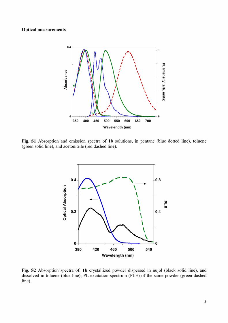
Optical measurements
0
0.4
0
1
350 400 450 500 550 600 650 700
Abs
orba
nce
PL Intensity (arb. units)
Wavelength (nm)
Fig. S1 Absorption and emission spectra of 1b solutions, in pentane (blue dotted line), toluene (green solid line), and acetonitrile (red dashed line).
0
0.2
0.4
0
0.4
0.8
380 420 460 500 540
Opt
ical
Abs
orpt
ion
PLE
Wavelength (nm)
Fig. S2 Absorption spectra of: 1b crystallized powder dispersed in nujol (black solid line), and dissolved in toluene (blue line); PL excitation spectrum (PLE) of the same powder (green dashed line).
6
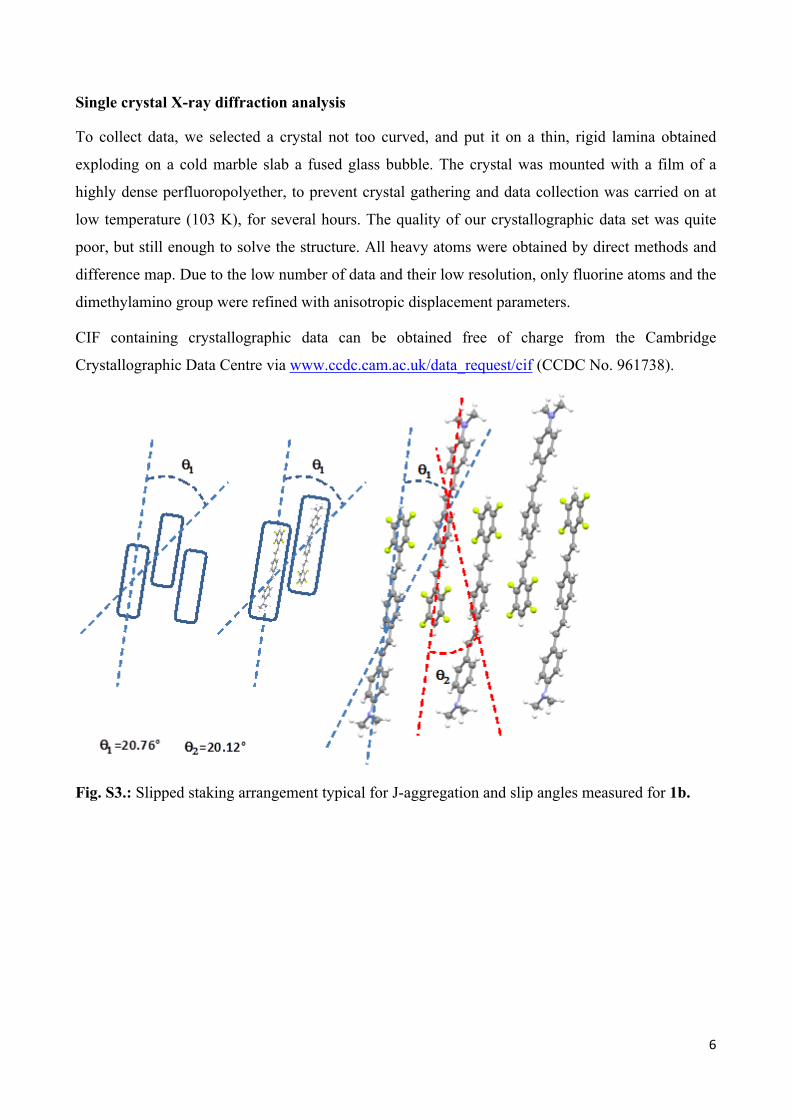
Single crystal X-ray diffraction analysis
To collect data, we selected a crystal not too curved, and put it on a thin, rigid lamina obtained
exploding on a cold marble slab a fused glass bubble. The crystal was mounted with a film of a
highly dense perfluoropolyether, to prevent crystal gathering and data collection was carried on at
low temperature (103 K), for several hours. The quality of our crystallographic data set was quite
poor, but still enough to solve the structure. All heavy atoms were obtained by direct methods and
difference map. Due to the low number of data and their low resolution, only fluorine atoms and the
dimethylamino group were refined with anisotropic displacement parameters.
CIF containing crystallographic data can be obtained free of charge from the Cambridge
Crystallographic Data Centre via www.ccdc.cam.ac.uk/data_request/cif (CCDC No. 961738).
Fig. S3.: Slipped staking arrangement typical for J-aggregation and slip angles measured for 1b.
7
Table S1. Crystallographic data for 1b crystals.
Chemical Formula C24H19F4NMr 397.40Crystal system, space group Monoclinic, P21/cTemperature (K) 103a, b, c (Å) 7.594 (5), 5.874 (4), 41.51 (3)β (°) 94.302 (8)V (Å3) 1846 (2)Z 4Radiation type Mo Kα (mm−1) 0.11Crystal size 0.28 × 0.14 × 0.01Data collection
Diffractometer Bruker APEX-II CCD area detector diffractometer
Absorption correction -No. of measured, independent and observed [I > 2σ(I)] reflections 5727, 1814, 1100
Rint 0.083θmax (°)a 20.8RefinamentR[F2 > 2σ(F2)], wR(F2), S 0.113, 0.294, 1.07No. of reflections 1814No. of parameters 155No. of restraints 0No. of H-atom treatment H-atom parameters constrained
w = 1/[σ2(Fo2) + (0.1036P)2 +
19.5133P] where P = (Fo
2 + 2Fc2)/3
Δρmax, Δρmin (e Å−3) 0.50, −0.37
a The crystals were always twinned and/or curved. The sample chosen was apparently the best for dimensions and diffraction quality. It was put on a very thin, rigid lamina obtained exploding a bubble of fused glass; the crystal was fixed on the lamina through a film of a highly dense perfluorinated compound; this technique assured a good adhesion between glass and crystal and prevented the crystal curvature increasing during the freezing process. In spite of the long time of data collection (120"/frame), it was impossible to collect data at > 21°. Moreover, due to the very anisotropic form of the spot, probably caused by the curvature of the crystal, a number of reflections were not integrated, so that the data quality was very poor and the resolution was low. Nethertheless, the structure solution was easy, leaving no doubt about its correctness.
X-Ray Powder Diffraction (XRPD) analysis
a)

8
5 10 15 20 25 30 35 400
2000
4000
6000
8000
10000
2-Theta (°)
Coun
ts
b)
5 10 15 20 25 30 35 400
2000
4000
6000
8000
10000
2-Theta (°)
Coun
ts
9
c)
5 10 15 20 25 30 35 400
2000
4000
6000
8000
10000
2-Theta (°)
Coun
ts
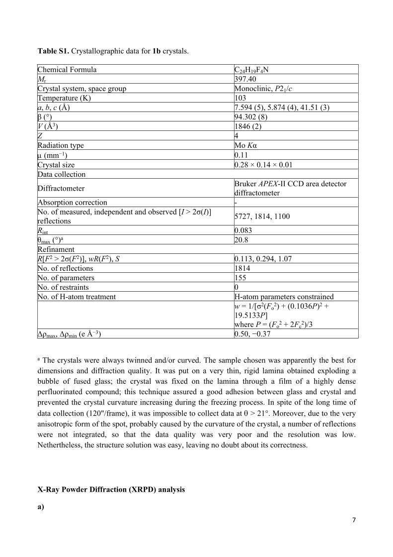
Fig. S4 PXRD patterns of: (a) 1b, simulated from single crystal X-ray structure; (b) 1b, native powder; (c) 1b, powder obtained by fast precipitation from chloroform solution.
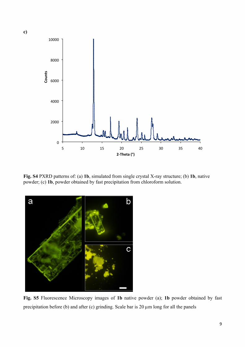
Fig. S5 Fluorescence Microscopy images of 1b native powder (a); 1b powder obtained by fast
precipitation before (b) and after (c) grinding. Scale bar is 20 m long for all the panels
10
Computational details
All calculations were performed with the Gaussian suite of programs.S5
The molecular structure of compound 1b has been optimized in vacuo within the DFT approach,
using the 6-311++G(d,p) basis set and the PBE0 functional,S6,S7 which was previously judged well
suited for describing the electronic and optical features of a series of organic dyes.S8 The X-ray
diffraction structure was used as starting point for geometry optimization. Using the PBE0/6-
311++G(d,p) optimized geometry, standard vertical TD-PBE0/6-311++G(d,p) calculationsS9-S11
were carried out to determine the absorption wavelengths. In Table S1 we report the values of
computed electronic and optical properties of 1b compared with those of 1a, as determined at the
same level of theory.
Geometry optimization of dimers of compound 1b in different arrangements were carried out in
vacuo with the 6-31++G(d,p) basis set, using the B97D functional.S12 Such functional has been
chosen owing to the dispersive nature of the - interactions implied in these dimers, as denoted by
the short C∙∙∙C intermolecular distances observed in crystal structure (shortest intermolecular
contacts: C2∙∙∙C12-x,1-y,2-z, 3.326 Å, and C9∙∙∙C51-x,1-y,2-z, 3.379 Å). Preliminary test calculations on
the dimers were also performed with the same functional used for the monomer, PBE0, but in all
cases convergence problems were encountered. The computed interaction energies were corrected
for basis set superposition error by the counterpoise technique.S13
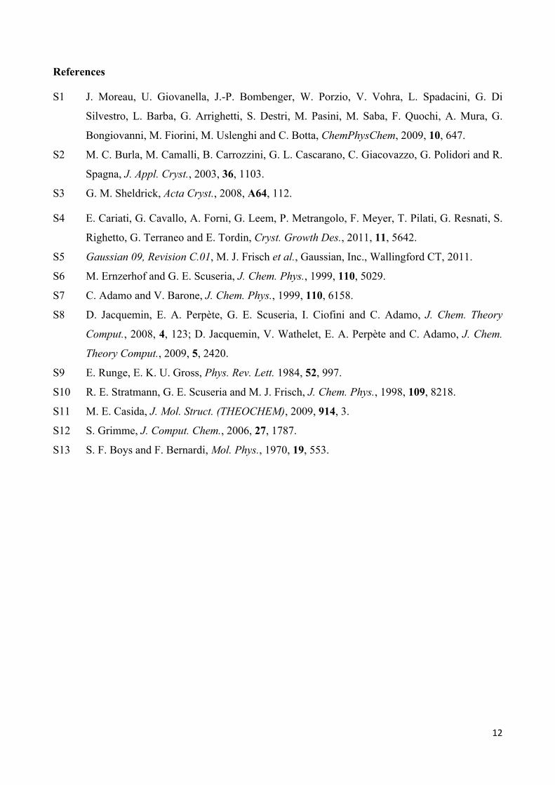
Figure S5 shows the optimized dimer in the antiparallel J arrangement, mimicking that ‘extracted’
from the crystal structure. In such optimized dimer, the molecules, while shifted according to the J
arrangement, are essentially overlapped along their long axis allowing to optimize both aryl-
fluoroaryl quadrupolar electrostatic interactions and dispersive contributions. On the other hand, in
the crystal structure, adjacent molecules within a π-π overlapped pile show as well a lateral shift as
a consequence of stabilizing side-to-side interactions, which are not taken into account in DFT
calculations. Such lateral shift explains the quite large values observed for the distances between
centroids of fluorinated and hydrogenated rings, equal to 3.772 and 3.829 Å, if compared with the
shortest C∙∙∙C intermolecular contacts (see above). As a result, such centroids distances are
reproduced by calculations with accuracy somewhat lower than that related to the shortest C∙∙∙C
contacts (about 0.26 against 0.15 Å, respectively. See Figure S5 for the relevant intermolecular
distances within the computed dimer).
11
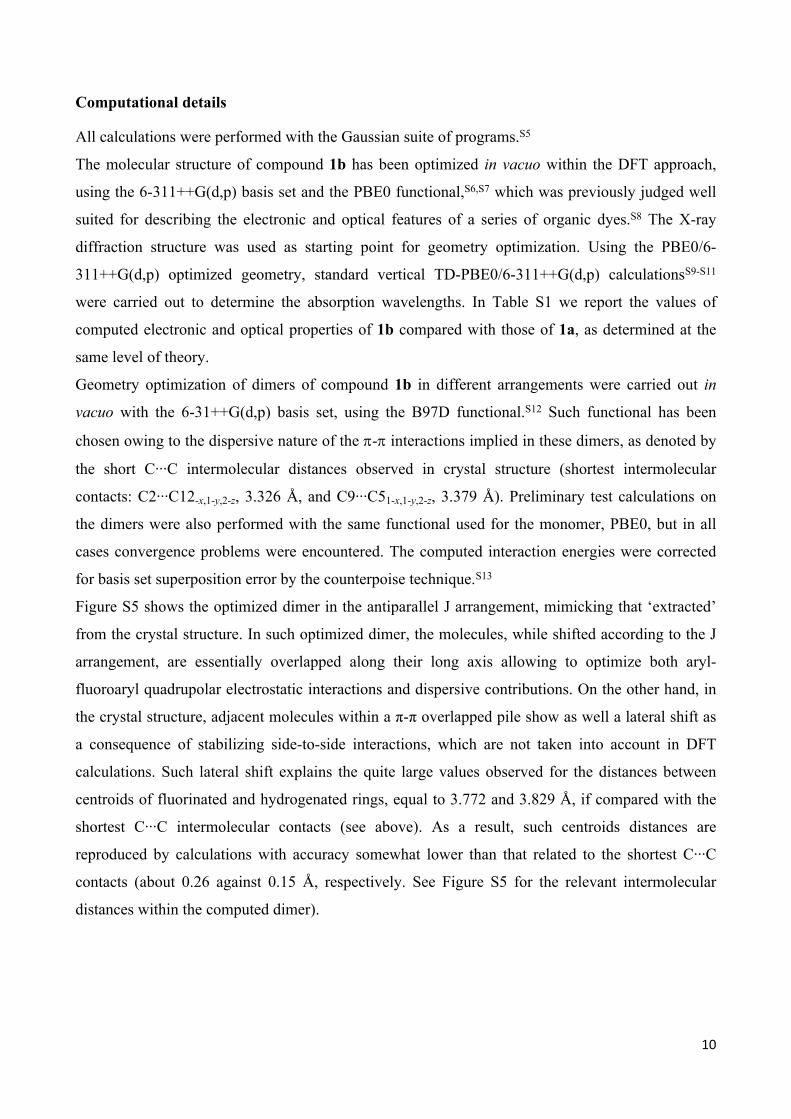
Table S1. Computed ground state dipole moments (μ, D) and electronic transitions (λmax, nm), along with the associated excited state dipole moments (μe, D), the transition dipole moments (μeg, D) and the oscillator strengths (f), for compounds 1a and 1ba
Compound max e eg f
1a 7.62 460 26.61 12.70 1.65
1b 6.54 449 24.23 12.03 1.52
aGround state properties and electronic transitions computed in vacuo at respectively PBE0/6-311++G(d,p) and TD PBE0/6-311++G(d,p) level with Gaussian09, rev. C.01.
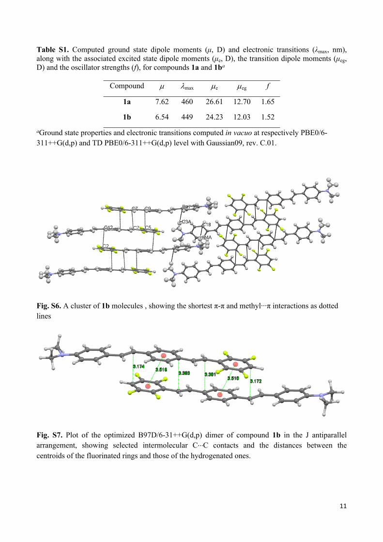
Fig. S6. A cluster of 1b molecules , showing the shortest π-π and methyl∙∙∙π interactions as dotted lines
Fig. S7. Plot of the optimized B97D/6-31++G(d,p) dimer of compound 1b in the J antiparallel arrangement, showing selected intermolecular C∙∙∙C contacts and the distances between the centroids of the fluorinated rings and those of the hydrogenated ones.
12
References
S1 J. Moreau, U. Giovanella, J.-P. Bombenger, W. Porzio, V. Vohra, L. Spadacini, G. Di
Silvestro, L. Barba, G. Arrighetti, S. Destri, M. Pasini, M. Saba, F. Quochi, A. Mura, G.
Bongiovanni, M. Fiorini, M. Uslenghi and C. Botta, ChemPhysChem, 2009, 10, 647.
S2 M. C. Burla, M. Camalli, B. Carrozzini, G. L. Cascarano, C. Giacovazzo, G. Polidori and R.
Spagna, J. Appl. Cryst., 2003, 36, 1103.
S3 G. M. Sheldrick, Acta Cryst., 2008, A64, 112.
S4 E. Cariati, G. Cavallo, A. Forni, G. Leem, P. Metrangolo, F. Meyer, T. Pilati, G. Resnati, S.
Righetto, G. Terraneo and E. Tordin, Cryst. Growth Des., 2011, 11, 5642.
S5 Gaussian 09, Revision C.01, M. J. Frisch et al., Gaussian, Inc., Wallingford CT, 2011.
S6 M. Ernzerhof and G. E. Scuseria, J. Chem. Phys., 1999, 110, 5029.
S7 C. Adamo and V. Barone, J. Chem. Phys., 1999, 110, 6158.
S8 D. Jacquemin, E. A. Perpète, G. E. Scuseria, I. Ciofini and C. Adamo, J. Chem. Theory
Comput., 2008, 4, 123; D. Jacquemin, V. Wathelet, E. A. Perpète and C. Adamo, J. Chem.
Theory Comput., 2009, 5, 2420.
S9 E. Runge, E. K. U. Gross, Phys. Rev. Lett. 1984, 52, 997.
S10 R. E. Stratmann, G. E. Scuseria and M. J. Frisch, J. Chem. Phys., 1998, 109, 8218.
S11 M. E. Casida, J. Mol. Struct. (THEOCHEM), 2009, 914, 3.
S12 S. Grimme, J. Comput. Chem., 2006, 27, 1787.
S13 S. F. Boys and F. Bernardi, Mol. Phys., 1970, 19, 553.