Embed Size (px)
Citation preview
Introduction
Nocardiosis is a granulomatous and suppurative infec-tion. It may be localized or disseminated and usually re-sults from the inhalation of the Nocardia asteroides soilspores and rarely from skin lesion contamination [1]. Itusually affects immunocompromised hosts and mani-fests as a chronic pneumopathy. However, it may alsoaffect healthy patients [2]. The central nervous system(CNS) is the most common extrapulmonary, metastaticsite of involvement. Nevertheless, almost every organcan be involved as a secondary site of infection.
Incidental discovery of previously unsuspected adre-nal masses has been reported in 0.6±1.0 % of patientsundergoing abdominal CT for other reasons [3].
We report a case of a patient with a Nocardial adre-nal abscess originating from a focus of pulmonary nocar-diosis.
Case report
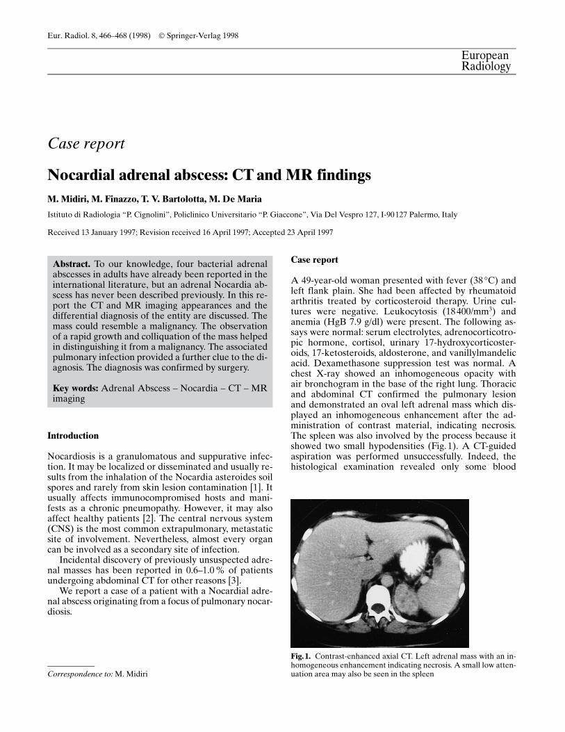
A 49-year-old woman presented with fever (38 °C) andleft flank plain. She had been affected by rheumatoidarthritis treated by corticosteroid therapy. Urine cul-tures were negative. Leukocytosis (18 400/mm3) andanemia (HgB 7.9 g/dl) were present. The following as-says were normal: serum electrolytes, adrenocorticotro-pic hormone, cortisol, urinary 17-hydroxycorticoster-oids, 17-ketosteroids, aldosterone, and vanillylmandelicacid. Dexamethasone suppression test was normal. Achest X-ray showed an inhomogeneous opacity withair bronchogram in the base of the right lung. Thoracicand abdominal CT confirmed the pulmonary lesionand demonstrated an oval left adrenal mass which dis-played an inhomogeneous enhancement after the ad-ministration of contrast material, indicating necrosis.The spleen was also involved by the process because itshowed two small hypodensities (Fig. 1). A CT-guidedaspiration was performed unsuccessfully. Indeed, thehistological examination revealed only some blood
Eur. Radiol. 8, 466±468 (1998) Ó Springer-Verlag 1998
EuropeanRadiology
Case report
Nocardial adrenal abscess: CT and MR findings
M. Midiri, M. Finazzo, T. V. Bartolotta, M. De Maria
Istituto di Radiologia ªP. Cignoliniº, Policlinico Universitario ªP. Giacconeº, Via Del Vespro 127, I-90127 Palermo, Italy
Received 13 January 1997; Revision received 16 April 1997; Accepted 23 April 1997
Abstract. To our knowledge, four bacterial adrenalabscesses in adults have already been reported in theinternational literature, but an adrenal Nocardia ab-scess has never been described previously. In this re-port the CT and MR imaging appearances and thedifferential diagnosis of the entity are discussed. Themass could resemble a malignancy. The observationof a rapid growth and colliquation of the mass helpedin distinguishing it from a malignancy. The associatedpulmonary infection provided a further clue to the di-agnosis. The diagnosis was confirmed by surgery.
Key words: Adrenal Abscess ± Nocardia ± CT ± MRimaging
Correspondence to: M. Midiri
Fig.1. Contrast-enhanced axial CT. Left adrenal mass with an in-homogeneous enhancement indicating necrosis. A small low atten-uation area may also be seen in the spleen
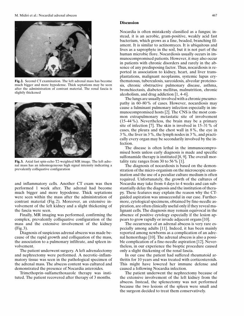
and inflammatory cells. Another CT exam was thenperformed 1 week after. The adrenal had becomemuch bigger and more hypodense. Thick septationswere seen within the mass after the administration ofcontrast material (Fig. 2). Moreover, an extensive in-volvement of the left kidney and a slight thickening ofthe fascia were seen.
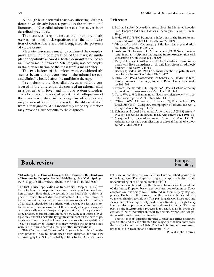
Finally, MR imaging was performed, confirming thecomplex, prevalently colliquative configuration of themass and the extensive involvement of the kidney(Fig.3).
Diagnosis of suspicious adrenal abscess was made be-cause of the rapid growth and colliquation of the mass,the association to a pulmonary infiltrate, and spleen in-volvement.
The patient underwent surgery. A left adrenalectomyand nephrectomy were performed. A necrotic±inflam-matory tissue was seen in the pathological specimen ofthe adrenal mass. The abscess content was cultured anddemonstrated the presence of Nocardia asteroides.
Trimethoprin±sulfamethoxazole therapy was insti-tuted. The patient recovered after therapy of 3 months.
Discussion
Nocardia is often mistakenly classified as a fungus; in-stead, it is an aerobic, gram-positive, weakly acid fastbacterium, which grows as a fine, beaded, branching fil-ament. It is similar to actinomyces. It is ubiquitous andlives as a saprophyte in the soil, but it is not part of thehuman microbic flore. Nocardiosis usually occurs in im-munocompromised patients. However, it may also occurin patients with chronic disorders and rarely in the ab-sence of any predisposing factor. Thus, nocardiosis is re-ported in association to kidney, heart, and liver trans-plantations, malignant neoplasms, systemic lupus ery-thematosus, tuberculosis, sarcoidosis, alveolar proteino-sis, chronic obstructive pulmonary disease, asthma,bronchiectasis, diabetes mellitus, malnutrition, chronicalcoholism, and drug addiction [1, 4±6].
The lungs are usually involved with a chronic pneumo-pathy in 60±80% of cases. However, nocardiosis maycause a fulminant pulmonary infection especially in im-munocompromised hosts [2]. The CNS is the most com-mon extrapulmonary metastatic site of involvement(15±44 %). Nevertheless, the brain may be a primarysite of infection [7]. The skin is involved in 15±31% ofcases, the pleura and the chest wall in 8 %, the eye in3 %, the liver in 3 %, the lymph nodes in 3 %, and practi-cally every organ may be secondarily involved by the in-fection.
The disease is often lethal in the immunocompro-mised hosts unless early diagnosis is made and specificsulfonamide therapy is instituted [8, 9]. The overall mor-tality rate ranges from 30 to 56% [1].
The diagnosis of nocardiosis is based on the demon-stration of the micro-organism on the microscopic exam-ination and the use of a peculiar culture medium is oftenrequired. Unfortunately, the growth of the cultures ofNocardia may take from 4 days to 4 weeks and can sub-stantially delay the diagnosis and the institution of thera-py. These features may explain the reason why the CT-guided aspiration was unsuccessful in our case. Further-more, cytological specimens, obtained by fine-needle as-piration, are often clinically useful only if they reveal ma-lignant cells. The diagnosis may remain equivocal in theabsence of positive cytology especially if the lesion ap-pears to grow rapidly or invade adjacent organs [10].
The occurrence of an adrenal abscess is very rare es-pecially among adults [11]. Indeed, it has been mainlyreported among newborns as a complication of an adre-nal hemorrhage [10]. The adrenal abscess is also a possi-ble complication of a fine-needle aspiration [12]. Never-theless, in our experience the bioptic procedure causedonly a slight thickening of the renal fascia.
In our case the patient had suffered rheumatoid ar-thritis for 10 years and was treated with corticosteroids.This might have lowered her immune defense andcaused a following Nocardia infection.
The patient underwent the nephrectomy because ofthe extensive involvement of the left kidney from theabscess. Instead, the splenectomy was not performedbecause the two lesions of the spleen were small andthe surgeon decided to treat them conservatively.
M. Midiri et al.: Nocardial adrenal abscess 467
Fig.2. Second CT examination. The left adrenal mass has becomemuch bigger and more hypodense. Thick septations may be seenafter the administration of contrast material. The renal fascia isslightly thickened
Fig.3. Axial fast spin-echo T2-weighted MR image. The left adre-nal mass has an inhomogeneous high signal intensity indicating aprevalently colliquative configuration
Although four bacterial abscesses affecting adult pa-tients have already been reported in the internationalliterature, a Nocardial adrenal abscess has never beendescribed previously.
The mass was as hypodense as the other adrenal ab-scesses, but it had thick septations after the administra-tion of contrast material, which suggested the presenceof viable tissue.
Magnetic resonance imaging confirmed the complex,prevalently liquid configuration of the mass; its multi-planar capability allowed a better demonstration of re-nal involvement; however, MR imaging was not helpfulin the differentiation of the mass from a malignancy.
The two lesions of the spleen were considered ab-scesses because they were next to the adrenal abscessand clinically healed after the antibiotic therapy.
In conclusion, the Nocardial abscess should be con-sidered in the differential diagnosis of an adrenal massin a patient with fever and immune system disorders.The observation of a rapid growth and colliquation ofthe mass was critical in the diagnosis of abscess andmay represent a useful criterion for the differentiationfrom a malignancy. An associated pulmonary infectionmay provide a further clue to the diagnosis.
References
1. Boiron P (1994) Nocardia et nocardiose. In: Maladies infectie-uses. Encycl Med Chir. Editions Techniques, Paris, 8±037-K-10, p. 5
2. McLoud TC (1989) Pulmonary infections in the immunocom-promised host. Radiol Clin North Am 27: 1059
3. Glazer GM (1988) MR imaging of the liver, kidneys and adre-nal glands. Radiology 166: 303
4. Arduino RC, Johnson PC, Miranda AG (1993) Nocardiosis inrenal trasplant recipients undergoing immunosuppression withcyclosporine. Clin Infect Dis 16: 505
5. Raby N, Forbes G, Williams R (1990) Nocardia infection in pa-tients with liver transplants or chronic liver disease: radiologicfindings. Radiology 174: 713
6. Berkey P, Bodey GP (1989) Nocardial infection in patients withneoplastic disease. Rev Infect Dis 11: 407
7. Filice GA (1993) Nocardiosis. In: Sarosi GA, Davies SF (eds)Fungal diseases of the lung, 2nd edn. Raven Press, New York,pp 191±204
8. Presant CA, Wirnik PH, Serpick AA (1973) Factors affectingsurvival nocardiosis Am Rev Resp Dis 108: 1444
9. Curry WA (1980) Human nocardiosis: a clinical review with se-lected case reports. Arch Intern Med 140: 818
10. O'Brien WM, Choyke PL, Copeland CJ, Klappenbach RS,Lynch JH (1987) Computed tomography of adrenal abscess. JComput Assist Tomogr 11: 550
11. Echaniz A, Miguel J de, Arnal A, Pedreira JD (1985) Escheri-chia coli abscess as an adrenal mass. Ann Intern Med 103: 481
12. Masquimel L, Hernandez-Pascual C, Simo R, Mase J (1993)Adrenal abscess as a complication of adrenal fine needle biop-sy. Am J Med 95: 244
M. Midiri et al.: Nocardial adrenal abscess468
Book review EuropeanRadiology
McCartney, J.P., Thomas-Lukes, K.M., Gomez, C.R.: Handbookof Transcranial Doppler. Berlin, Heidelberg, New York: Springer,1997. 92 pp., 88 illustrations, (ISBN 0-387-94693-4), DM 58.00.
The first clinical application of transcranial Doppler (TCD) wasthe detection of vasospasm in victims of aneurysmal subarachnoidhemorrhage. Since then, the technique has been able to show as-pects of other clinical disorders: detection of stenotic lesions ofthe arteries at the base of the brain and assessment of the patternsof collateral circulation in patients with obstructive lesions in ex-tracranial arteries, assessment of flow velocity changes in suspect-ed brain death, study of major supply arteries and flow patterns tolarge arteriovenous malformations. A new subject of intense inves-tigation ± one with potentially significant impact on the care of pa-tients who have suffered ischemic brain events ± is the capability ofTCD to detect embolic arteries as they traverse the cerebral bloodvessels, e. g. during carotid surgery or other interventions.
This Handbook of Transcranial Doppler is introduced as theonly practical `how-to' book specifically designed for the newultrasonographer. `Only' probably relates to the American mar-
ket; similar booklets are available in Europe, albeit possibly inother languages. The simplistic progressive approach aims to aidunderstanding of the fundamentals of TCD.
The first chapters address the classical basics: vascular anatomyof the brain, Doppler basics and cerebral hemodynamics. Thesechapters are extremely well illustrated in their step-by-step ap-proach. The bulk of the booklet (one third of the volume) is devot-ed to examination techniques. This part is again well illustrated andshows multiple examples of typical curves. Reading through it mayleave a false impression of an easy-to-learn technique. The finalpart, on the interpretation process, is too short as an in-depth dis-cussion to be of potential interest to doctors responsible for pa-tients with cerebrovascular disorders.
The text is short and not referenced. Selected further reading isgiven at the end of each chapter, the majority of which dates fromthe late 1980s and early 1990s. This book is first and foremost apractical aid in learning and performing TCD.
R. Verhaeghe, Leuven

![Adrenal Imaging - University of Floridaxray.ufl.edu/files/2010/02/Adrenal-Imaging.pdfadrenal glands [3], and a metastasis might ... CT, adrenal imaging, adrenal lymphoma imaging, adrenal](https://img.pdfslide.net/doc/110x75/5b26814c7f8b9a8c0f8b4820/adrenal-imaging-university-of-glands-3-and-a-metastasis-might-ct-adrenal.jpg)