Embed Size (px)
Citation preview
Malaysian Journal of Dermatology
26 MJD 2014 Jul Vol 32
GENERAL DERMATOLOGY - Short Case
NODULAR MELANOMA PRESENTING WITH PARAPLEGIA:A CASE REPORT
Nur Ashikin Ahmad, MRCP, Nor Salmah Bakar, MPath(UKM), Tarita Taib, MAdvDerm, Yap Shean Huet, MMed (Rad)(UKM)
Keywords: skin cancer, bone metastasis, spinal cord compression
IntroductionNodular melanoma (NM) is clinically a distinct subtype of melanoma. It is the predominant contributor to melanoma-related deaths1. Clinically lesion of a NM is most often symmetric, elevated, uniform in colour, and non-pigmented in contrast to superficial spreading melanoma (SSM)2. It is an aggressive form of skin cancer and it can metastasize quickly from the primary site to other sites in the body3-4. Bone metastasis is very rare, affecting mainly the axial skeleton, represents the end stage of the disease and carries a poor prognosis5. We here describe a young man with metastatic nodular melanoma with features of spinal cord compression. To date, there are very few reported cases of nodular melanoma with bone metastasis presented in young age group4.
CorrespondenceNur Ashikin Ahmad, MRCP (UK)Universiti Teknologi MARA, Selangor, MalaysiaEmail: [email protected]
Case reportA 33-year-old Burmese man presented with sudden onset of paraplegia and altered bowel habit for 3 days prior to presentation. He also had a 2-month history of constant back pain irrespective of type of activities. He also had multiple hyperpigmented painful nodules and papules for the past 2 months. The nodular lesions were predominantly located on the left lower limb and were rapidly increasing in size and numbers. He reported no recent trauma, fever, chills or difficulty with gait. He lost 10kg in 2 months prior to presentation. There was no family history of malignant melanoma or congenital naevus and he was not immunosuppressed.
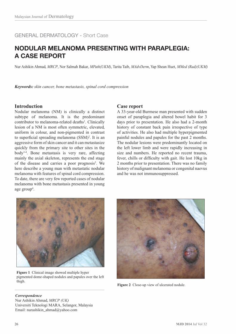
Figure 1 Clinical image showed multiple hyper pigmented dome-shaped nodules and papules over the left thigh.
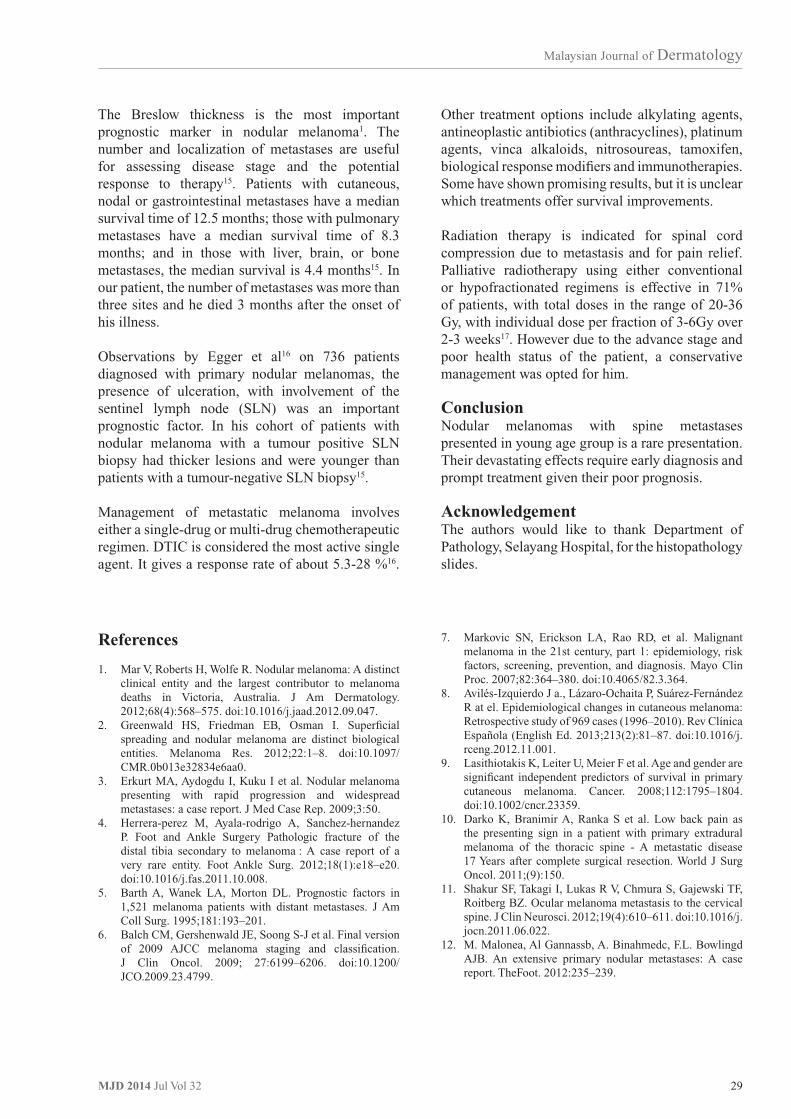
Figure 2 Close-up view of ulcerated nodule.

Malaysian Journal of Dermatology
27MJD 2014 Jul Vol 32
Clinically on the left thigh there were multiple hyperpigmented, ulcerated small papules and nodules at the periphery of the large nodules. Some of the bigger nodules were ulcerated with hemorrhagic discharge (Figure 1 & 2). There were multiple non-matted bilateral inguinal lymph nodes, with largest nodes measured 2 x 2 cm.
The neurological examination of the lower limbs revealed bilateral lower limb weakness with muscle power of 1/5 and sensory loss from L1 onwards. His blood investigations revealed a raised lactate dehydrogenase at 1138U/L. Other investigations including blood culture were normal.
MRI of the spine revealed heterogeneous enhancing lesions involving bilateral pedicles and posterior arch of T10 vertebra (with necrotic centre) and spinous process of T1. There was intraspinal extradural extension of the lesion from the level of T7 until T12 level with high T2 signal intensity, resulting in narrowing and compression of the spinal cord at the level T8 to T12. There was also extension of the lesion in the right T8, T12 neural foramen and bilateral T9-11 neural foramen, which encases the exiting nerve roots. Nodules were seen in the lung and liver base from ultrasound of the abdomen.
The patient underwent excisional biopsy of a small, hyperpigmented papule over the dorsal-medial aspect of the left thigh. The histology revealed a diffuse dermis infiltration by malignant cells which extends into the subcutaneous tissue. There were occasional abnormal mitotic figures. The cells have mildly pleomorphic, hyperchromatic nuclei with ample clear to pale cytoplasm (Figure 3a-d). The histology finding is consistent with nodular melanoma.
Given the clinicopathologic characteristics and investigations, the patient was diagnosed to have nodular melanoma with multiple distant metastases of the skeletal system, lymph nodes, liver and lung. He was staged IV (N2b, M1c) according to the American Joint Commission on Cancer6. Due to the extremely poor prognosis, he was managed conservatively by the palliative care team. The patient succumbed to his disease a month after the diagnosis was made.
DiscussionMalignant melanoma accounts for 1% to 3% of all cancers. The reported incidence and mortality of melanoma is rapidly rising and is second only to lung cancer7. Currently, melanoma comprises
65% to 75% of all skin cancer deaths. Melanoma has been classified based on histologic and clinical appearances, with superficial spreading melanoma (SSM) as the most common followed by nodular melanoma (NM), and lentigo maligna melanoma (LMM)8. NM often presents with atypical clinical features that mimic other benign and malignant tumours and some inflammatory lesions.
Nodular melanomas are typically encountered in the fourth through sixth decades of life8. However our patient is in the younger age group without pre-existing pigmented macule. It has been suggested that the incidence of melanoma in younger age group is also increasing but with more favourable prognosis9.
Nodular melanoma is an aggressive subtype of cutaneous melanoma with evidence of metastases upon presentation1,3. Initial metastases are most commonly found in the skin, subcutaneous tissue, lymph nodes, and lungs Metastases to the bone is very rare and usually represents an end-stage situation of the disease. The axial and peripheral bones are effected and lesion is predominantly lytic lesion4,10-12. In our patient, metastatic lesions were found in the spine, lung, lymph node and liver at the time of diagnosis.
One of the malignancy that has similar clinical, histopathological and immunochemistry presentation is clear cell sarcoma (CCS) or melanoma of the soft part. It occurs more common in the young age group (30 - 40 year old)13,14. Clear cell sarcomas are rarely aggressive, soft tissue tumour which shared similar clinical, pathologic, and molecular properties with melanoma13,14. The tumours are located almost exclusively on the extremities; the foot and ankle are the most common locations. Clear cell sarcoma is a slowly growing tumour, commonly presented with subcutaneous mass with associated tenderness13.
In the present case, the rapid progression and metastases of the painless nodules within 2 months support nodular melanoma. In contrast to melanoma, CCS is characterized by a recurrent chromosomal translocation t (12; 22), which results in fusion of the EWS gene on 22q with the ATF1 gene on 12q. This genomic abnormality may represent a good marker for identifying these tumours14 . This differentiation is clinically important due to significant differences in patient management. However the molecular test was not available to this patient at the time of diagnosis.

Malaysian Journal of Dermatology
28 MJD 2014 Jul Vol 32
Figure 3a Low-power magnification showed diffuse infiltration by malignant cells which extends up to the subcutaneous fat. H&E x20.
Figure 3c This high-power image of malignant melanoma cells showed nuclear hyperchromatism and pleomorphism with mitotic figure (arrow). H&Ex400.
Figure 3b Higher magnification of the deep portion lesion seen in image (a) illustrating the involvement of the subcutaneous fat. H&E x40.
Figure 3d The cells were diffusely positive with HMB-45 (Human Melanoma Black). HMB45 x40.
Malaysian Journal of Dermatology
29MJD 2014 Jul Vol 32
References
1. Mar V, Roberts H, Wolfe R. Nodular melanoma: A distinct clinical entity and the largest contributor to melanoma deaths in Victoria, Australia. J Am Dermatology. 2012;68(4):568–575. doi:10.1016/j.jaad.2012.09.047.
2. Greenwald HS, Friedman EB, Osman I. Superficial spreading and nodular melanoma are distinct biological entities. Melanoma Res. 2012;22:1–8. doi:10.1097/CMR.0b013e32834e6aa0.
3. Erkurt MA, Aydogdu I, Kuku I et al. Nodular melanoma presenting with rapid progression and widespread metastases: a case report. J Med Case Rep. 2009;3:50.
4. Herrera-perez M, Ayala-rodrigo A, Sanchez-hernandez P. Foot and Ankle Surgery Pathologic fracture of the distal tibia secondary to melanoma : A case report of a very rare entity. Foot Ankle Surg. 2012;18(1):e18–e20. doi:10.1016/j.fas.2011.10.008.
5. Barth A, Wanek LA, Morton DL. Prognostic factors in 1,521 melanoma patients with distant metastases. J Am Coll Surg. 1995;181:193–201.
6. Balch CM, Gershenwald JE, Soong S-J et al. Final version of 2009 AJCC melanoma staging and classification. J Clin Oncol. 2009; 27:6199–6206. doi:10.1200/JCO.2009.23.4799.
7. Markovic SN, Erickson LA, Rao RD, et al. Malignant melanoma in the 21st century, part 1: epidemiology, risk factors, screening, prevention, and diagnosis. Mayo Clin Proc. 2007;82:364–380. doi:10.4065/82.3.364.
8. Avilés-Izquierdo J a., Lázaro-Ochaita P, Suárez-Fernández R at el. Epidemiological changes in cutaneous melanoma: Retrospective study of 969 cases (1996–2010). Rev Clínica Española (English Ed. 2013;213(2):81–87. doi:10.1016/j.rceng.2012.11.001.
9. Lasithiotakis K, Leiter U, Meier F et al. Age and gender are significant independent predictors of survival in primary cutaneous melanoma. Cancer. 2008;112:1795–1804. doi:10.1002/cncr.23359.
10. Darko K, Branimir A, Ranka S et al. Low back pain as the presenting sign in a patient with primary extradural melanoma of the thoracic spine - A metastatic disease 17 Years after complete surgical resection. World J Surg Oncol. 2011;(9):150.
11. Shakur SF, Takagi I, Lukas R V, Chmura S, Gajewski TF, Roitberg BZ. Ocular melanoma metastasis to the cervical spine. J Clin Neurosci. 2012;19(4):610–611. doi:10.1016/j.jocn.2011.06.022.
12. M. Malonea, Al Gannassb, A. Binahmedc, F.L. Bowlingd AJB. An extensive primary nodular metastases: A case report. TheFoot. 2012:235–239.
The Breslow thickness is the most important prognostic marker in nodular melanoma1. The number and localization of metastases are useful for assessing disease stage and the potential response to therapy15. Patients with cutaneous, nodal or gastrointestinal metastases have a median survival time of 12.5 months; those with pulmonary metastases have a median survival time of 8.3 months; and in those with liver, brain, or bone metastases, the median survival is 4.4 months15. In our patient, the number of metastases was more than three sites and he died 3 months after the onset of his illness.
Observations by Egger et al16 on 736 patients diagnosed with primary nodular melanomas, the presence of ulceration, with involvement of the sentinel lymph node (SLN) was an important prognostic factor. In his cohort of patients with nodular melanoma with a tumour positive SLN biopsy had thicker lesions and were younger than patients with a tumour-negative SLN biopsy15.
Management of metastatic melanoma involves either a single-drug or multi-drug chemotherapeutic regimen. DTIC is considered the most active single agent. It gives a response rate of about 5.3-28 %16.
Other treatment options include alkylating agents, antineoplastic antibiotics (anthracyclines), platinum agents, vinca alkaloids, nitrosoureas, tamoxifen, biological response modifiers and immunotherapies. Some have shown promising results, but it is unclear which treatments offer survival improvements.
Radiation therapy is indicated for spinal cord compression due to metastasis and for pain relief. Palliative radiotherapy using either conventional or hypofractionated regimens is effective in 71% of patients, with total doses in the range of 20-36 Gy, with individual dose per fraction of 3-6Gy over 2-3 weeks17. However due to the advance stage and poor health status of the patient, a conservative management was opted for him.
Conclusion Nodular melanomas with spine metastases presented in young age group is a rare presentation. Their devastating effects require early diagnosis and prompt treatment given their poor prognosis.
AcknowledgementThe authors would like to thank Department of Pathology, Selayang Hospital, for the histopathology slides.
Malaysian Journal of Dermatology
30 MJD 2014 Jul Vol 32
13. Mackey SL, Hebel J, Mar W. Melanoma of the soft parts ( clear cell sarcoma ) : A case report and r eview of the literature. J Am Dermatology. 1998;(38):815–819.
14. Hocar O, Le Cesne a, Berissi S et al. Clear cell sarcoma (malignant melanoma) of soft parts: a clinicopathologic study of 52 cases. Dermatol Res Pract. 2012;2012:984096. doi:10.1155/2012/984096.
15. Egger ME, Dunki-Jacobs EM, Callender GG et al. Outcomes and prognostic factors in nodular melanomas. Surgery. 2012;152(4):652–9; discussion 659–60. doi:10.1016/j.surg.2012.07.006.
16. Lui P, Cashin R, Machado M et al. Treatments for metastatic melanoma: synthesis of evidence from randomized trials. Cancer Treat Rev. 2007;33(8):665–80. doi:10.1016/j.ctrv.2007.06.004.
17. Forschner A, Heinrich V, Pflugfelder A et al. The role of radiotherapy in the overall treatment of melanoma. Clin Dermatol. 2013;31(3):282–9. doi:10.1016/j.clindermatol.2012.08.009.

Malaysian Journal of Dermatology
31MJD 2014 Jul Vol 32
GENERAL DERMATOLOGY - Original Article
A RETROSPECTIVE REVIEW OF TINEA CAPITIS INFECTION
Tan SS, MBBS, Chan LC, MMed
Abstract
Background: Tinea capitis (TC), a fungal infection of the scalp, hair follicles and hair shafts, is common among the paediatric population especially under tropical conditions1. The etiological factors vary between different regions of the world. Clinical presentation of tinea capitis varies widely from non-inflammatory to severe, painful inflammatory lesions.
Aim: To look into the clinical manifestations, causative agents and the treatment pattern for tinea capitis in Penang Hospital.
Methods: A retrospective study of all patients who were treated clinically for tinea capitis in Penang Hospital from January 2011 to June 2013.
Results: There were a total of thirty nine patients treated for tinea capitis during this period. Tinea capitis was found to be most common in the 7-12 year age group (44%) with a male to female ratio of 2:1. Non-inflammatory type (54%) was more common then the inflammatory type. Twenty seven of them (69%) had positive fungal culture of their pluck hair roots. The most common dermatophyte detected was Microsporum canis (92%) followed by Trichophyton rubrum (4%) and Trichophyton metagraphyte (4%). Thirty-one (80%) of them were treated with griseofulvin at a dose of 10-15mg /kg /day. The rest were treated with itraconazole, terbinafine or fluconazole. All of them responded well to the treatment. In this cohort only one patient, has a second episode of infection a year later. He is a child who was concomitantly undergoing chemotherapy for acute lymphoblastic leukaemia.
Conclusion: Tinea capitis is predominantly an infection of pre-adolescent children and M. canis was the most common dermatophyte isolated.
Keywords: Fungal infection, Microsporum canis, scalp lesion, Malaysia
CorrespondenceTan Sam SiewDepartment of Dermatology, Hospital Penang,10990, Georgetown, Penang, Malaysia Email: [email protected]
Microsporum(M) canis is the commonest cause of zoophilic infections transmitted by cats and dogs2. Infection could be ectothrix (infection occurs outside hair shaft with cuticle destruction) or endothrix (infection occurs within hair shaft without cuticle destruction).
Clinical presentation of TC varied widely from non-inflammatory to severe, painful inflammatory lesions3. Non-inflammatory type includes “black dot” or “gray patch”. Inflammatory lesions consist of kerion, flavus and arginate folliculitis4.
IntroductionTinea capitis (TC), a fungal infection of the scalp, hair follicles and hair shafts, is common among the paediatric population especially under tropical conditions1. The etiological factors vary between different regions of the world. Infections can spread from child to child (anthropophilic) or from animals to children (zoophilic).

Malaysian Journal of Dermatology
32 MJD 2014 Jul Vol 32
AimThis study was performed to look into the clinical manifestations, causative agents and the treatment pattern of tinea capitis in our centre.
MethodsThis is a retrospective study carried out in the Dermatology Department, Penang Hospital from January 2011 to June 2013. Clinic notes of all the patients who were treated clinically for TC during this period were reviewed. Patients were categorized into pre-school (< 7year-old), school going (7 to <13 year-old) and adolescents and adults (≥13 year-old). All of them have their involved hair plucked and sent for fungal culture to identify the causative dermatophyte. The samples were then inoculated on Sabouraud′s Dextrose Agar (SDA) plate, SDA with chloramphenicol plate and SDA with
chloramphenicol with cycloheximicle plate. The cultures were examined twice a week for up to three weeks. No growth after 21 days was considered as negative culture.
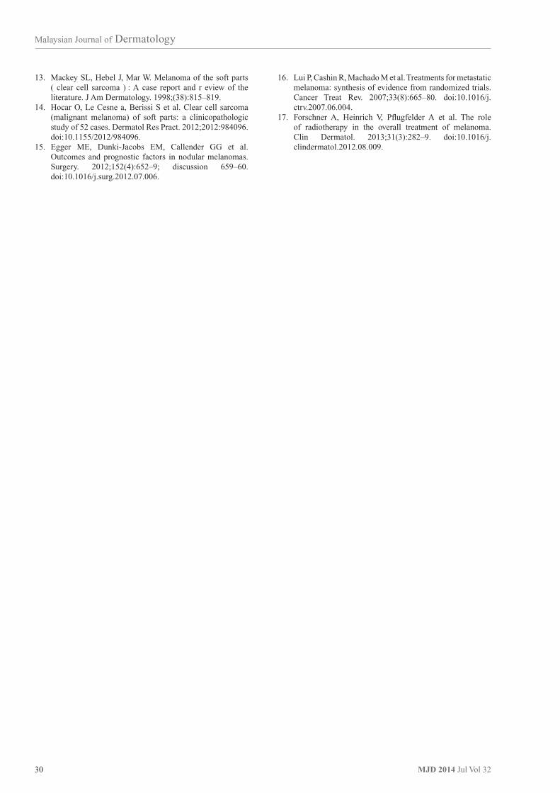
ResultsA total of 39 patients were treated for TC. The male to female ratio was 2:1. Majority of them were Malays (87%), Indians (8%) and Chinese (5%). TC was found to be most common in the 7-12 year age group (44%) (Figure 1).
Non-inflammatory type (54%) was more common than the inflammatory type. Twenty seven of patients (69%) had positive fungal culture of their plucked hair roots. Wood’s lamp examination was done in 15 patients. Ten patients had positive bluish-green fluorescence of their hair shaft and in 9 of them, the cultures grew M. canis.
Figure 1 Patients gender and age group distribution.
n = 39
Female
Male
Number
< 7 7 to 12 > 13 Age Group (years)
Malaysian Journal of Dermatology
33MJD 2014 Jul Vol 32
Figure 2 Types of lesions.
Figure 3 Types of dermatophyte isolated.
Thirty-one (80%) of them were treated with griseofulvin at a dose of 10-15mg /kg /day for the duration of 3 to 12 weeks. Majority of them (15, 48%) were treated for 8 to 10 weeks. Only one patient required up to 12 weeks of therapy. The rest were treated with itraconazole, terbinafine or fluconazole. All of them responded well to the treatment. In this cohort only a child with an acute lymphoblastic leukaemia undergoing chemotherapy, had a second episode of tinea capitis infection a year later.
DiscussionIn our cohort, the rate of infection was significantly higher among the Malay population as the racial distribution of Penang state was 45.5% Chinese, 43.7% Malay, 10.4% Indian and others 0.4% (base on 2010 population census). TC was found to be most common in the school going group. They were at a higher risk of transmission probably due to close contact with each other. Beyond this age group, the incidence declines because of the onset of puberty
Malaysian Journal of Dermatology
34 MJD 2014 Jul Vol 32
and seborrhoea. The infection was more common in boys. Similar finding was also reported by Friedlander et al5 and other studies done in West Bengal6, Rajasthan7 and Kenya8. It may be due to shorter hair, allowing easy access for circulating spores5. However, two studies reported by Singal et al9 and Chander et al10 respectively from north India showed no sex difference.
Clinical presentation of the patients to the clinic was also consistent with other studies done in Rajasthan7, North India9,10, Eastern Nepal11, Lahore(Pakistan)12 and Tirupati13.
The most common dermatophyte detected was M. canis followed by Trichophyton rubrum and Trichophyton metagraphyte. This might due to the stray cats or dogs in the residential area or even pets at home. None of the specimen here grew Trichophyton violaceum which is the most common dermatophyte reported in India7, Nepal11, Pakistan14, South Africa and the UK15.
Treatment was not a challenge in this cohort. A meta-analysis was done by Tey et al16 showed that griseofulvin was more efficacious than terbinafine in treating TC caused by Microsporum species, whereas terbinafine was more efficacious than griseofulvin in treating TC caused by Trichophyton species.
Conclusion TC is predominantly an infection of pre-adolescent children and M. canis was the most common dermatophyte isolated.
AcknowledgementThe authors would like to thank Miss Keah Kwee Chu, microbiologist, for her help in providing the laboratory support.
References
1. Ghannoum M, Isham N, Hajjeh R et al. Tinea capitis in Cleveland: Survey of elementary school students. J Am Acad Dermatol 2003;48:189-93.
2. Roderick Hay, Sandra E. Bendeck, Suephy Chen et al. Chapter 37, Skin Diseases: Disease Control Priorities in Developing Countries. 2nd edition.
3. Hay RJ, Moore MK. Mycology. In: Burns. Rook’s Textbook of Dermatology. 7th ed. Massachusetts: Blackwell Publishing; 2004; 31.1-31.101.
4. Section 23, Cutaneous Fungal Infection, Fitzpatricks Color Atlas and Synopsis of Clinical Dermatology, 5th edition: 707 - 712.
5. Friedlander SF, Rueda M, Chen BK, Caceros-Rios HW. Fungal, protozoal and helminthic infections. Pediatric Dermatology. 3 rd ed. Mosby 2003; 1093-140.
6. Kundu et al. Prevalence of Tinea capitis in school going children in Kolkata, West Bengal. J Nat SciBiol Med. 2012 Jul-Dec; 3(2): 152–155.
7. Kalla G, Begra B, Solanki A, Goyal A, Batra A. Clinico-Mycological study of Tinea capitis in Desert district of Rajasthan. Indian J DermatolVenereolLeprol.1995;61:342–5.
8. Ayaya SO, Kamar KK, Kakai R. Aetiology of Tinea capitis in school children. East Afr Med J. 2001; 78:531–5.
9. Singal A, Rawat S, Bhattacharya S, Mohanty S, Baruah MC. Clinico- mycological profile of tinea capitis in North India and response to griseofulvin. J Dermatol 2001; 28:22-6.
10. Chander Grover, Pooja Arora, Vikas Manchanda. Tinea capitis in the Pediatric Population: A Study In North India. Indian Journal of Dermatology, Venereology, and Leprology September-October, 2010;76:527-532.
11. Jha BN, Garg VK, Agrawal S et al. Tinea capitis in Eastern Nepal. Int J Dermatol 2006;45:100-2.
12. Hussain I, Aman S, Haroon TS et al. Tinea capitis in Lahore, Pakistan. Int J Dermatol 1994;33:255-7.
13. Kumar AG, Lakshmi N. Tinea capitis in Tirupati. Ind J Pathol Microbiol 1990;33:360-3.
14. Jahangir M, Hussain I, Khurshid K, Haroon TS. A clinico-etiologic correlation in Tinea capitis. Int J Dermatol 1999;38:275-8.
15. Mills CM, Philpot CM. Tinea capitis in south Wales-observations in change of causative fungi. Clin Exp Dermatol 1994;19:473-5.
16. Hong Liang Tey, Andy Soon Leong Tan, Yuin Chew Chan. Meta-analysis of randomized, controlled trials comparing griseofulvin and terbinafine in the treatment of tinea capitis J Am Acad Dermatol 2011; 64:663-70).

Malaysian Journal of Dermatology
35MJD 2014 Jul Vol 32
GENERAL DERMATOLOGY - Short Case
VARICELLA ZOSTER-ASSOCIATED VASCULITIS INAN IMMUNOCOMPETENT HOST
Evelyn Tay Yuxin, Chuah Sai Yee, Joyce Lee Siong See, Pan Jiun Yit
Keywords: herpes virus, leukocytoclastic vasculitis, haemorrhagic vesicles
IntroductionVaricella zoster virus (VZV) infection can present with a myriad of cutaneous and extracutaneous signs. It can cause vasculitis, most commonly vasculitis of the central nervous system where it spreads to the arteries via ganglionic afferent fibres1. However, VZV can also affect renal, coronary, retinal, choriodal and cutaneous arteries2-11. We report an unusual case of varicella zoster-associated vasculitis occuring in an immunocompetent 83-year-old Chinese female.
Case reportAn 83-year-old Chinese female presented with a one week history of left lower limb pruritic rash. This was preceded by left knee and calf pain one day before onset of the rash. The rash then spread to involve her right lower limb and hip. She had a past medical history of ischaemic heart disease and Child’s A cryptogenic liver cirrhosis. She was otherwise well with no fever or prior injury, new medications or contactants.
On examination, there were purpuric macules and papules, some with central haemorrhagic vesiculation more prominent on the left lower limbs. There were also some scattered purpuric macules on the right shin and hip. Our initial differential diagnoses were bullous vasculitis, vasculitis secondary to infections. Routine blood tests including vasculitic screening and a skin biopsy for histology and direct immunofluorescence (DIF) were done.
CorrespondenceDr. Evelyn Yuxin TayNational Skin Centre1 Mandalay Road, Singapore 308205Email: [email protected]
She was treated with topical corticosteroids and oral analgesia, pending the results of investigations. On review two days later, her rash had progressed. There were more clustered haemorrhagic vesicles and bullae along the L3 and L4 dermatomal distribution of the left lower limb (Figure 1). The diagnosis of varicella zoster-associated leukocytoclastic vasculitis was considered.
This was supported by the histology results showing an intraepidermal blister with necrotic and acantholytic keratinocytes exhibiting steel-gray nuclear inclusions and chromatin margination. Multinucleated giant cells with moulded, steel-grey nuclei were present. In addition, there was fibrinoid necrosis of the upper dermal blood vessels in association with red cell extravasation, neutrophils and nuclear dust. There was a superficial and deep perivascular infiltrate of lymphocytes and neutrophils. This was consistent with herpes infection and concomitant leukocytoclastic vasculitis (Figure 2). Direct immunofluorescence was negative for any immunoreactants.
Hepatitis B and C serologies, anti-neutrophil cytoplasmic antibodies (ANCA), anti-streptolysin-O titres (ASOT) were negative. Erythrocyte sedimentation rate was 12mm/hour (Normal range: 0-10mm/hour). Retroviral screen was negative. Of note, antinuclear antibody (ANA) titres were high at 1:640 in a speckled pattern. However, the patient did not have any systemic symptoms suggestive of an underlying connective tissue disease and her anti-double stranded DNA was also negative.
The patient was initiated on oral acyclovir 800mg five times a day for 10 days with complete resolution of the rash. She experienced post herpetic neuralgia that was controlled with oral analgesia and has not experienced recurrence of vasculitic lesions one month after receiving treatment.

Malaysian Journal of Dermatology
36 MJD 2014 Jul Vol 32
Figure 1 A) Purpuric macules, some with central vesiculation, along the distribution of the L3 and L4 dermatome of the left lower limb. B) Posterior and C) Anterior views of the left lower limb one week after completion of antiviral therapy showing post inflammatory hyperpigmentation in a dermatomal distribution.
Figure 2 An intraepidermal blister containing acantholytic keratinocytes and multinucleated giant cells with steel-gray nuclear inclusions was seen. In addition, there was fibrinoid necrosis of the upper dermal blood vessels in association with red cell extravasation and nuclear dust (H&E, x 200).
Malaysian Journal of Dermatology
37MJD 2014 Jul Vol 32
DiscussionThis case highlights an atypical presentation of VZV in the skin. VZV can be an infectious complication of primary vasculitis4. It can also be responsible for a myriad of vascular disorders that range from leukocytoclastic vasculitis5,6,7, granulomatous vasculitis8 to obliterative angiitis9,10. The most widely reported is granulomatous vasculitis, which is induced by the persistence of viral glycoproteins in arterioles post-infection8.
Given that i) the rash was preceded by pain, ii) was predominantly along the distribution of the L3 and L4 dermatome of the left lower limb and iii) resolved completely with the administration of oral antivirals; we believe that this is a case of segmental leukocytoclastic vasculitis induced by herpes zoster. On the other hand, VZV reactivation in a primary systemic vasculitis cannot be ruled out as this presentation was peculiar in that there were vasculitic lesions on the right hip and right shin of the patient.
We postulate that there could be a low level of viremia with deposition of immune complexes in the blood vessels away from the site of zoster reactivation. Direct immunofluorescence could have been negative due to the specimen being taken six days after the onset of the rash, and usually has a sensitivity of 60 to 80 percent for cutaneous vasculitis12. Notably, the inflammatory markers were not raised and work-up for an underlying cause for a systemic vasculitis was negative.
Impairment of cell-mediated immunity is often associated with unusual presentations of VZV infection such as verrucous, hyperkeratotic,
pox-like or ecchymotic lesions9. Previous case reports of acute herpes-related vasculitis have centred around individuals on methotrexate or combination chemotherapy for underlying malignancies5,6,9 and on corticosteroids for pulmonary sarcoidosis and systemic lupus erythematosus7,11.
We also seek to highlight that unusual manifestations of herpes zoster can occur in immunocompetent hosts, especially in elderly patients like ours. In addition to our case occurring in an immunocompetent elderly female, there was another report of a healthy 90-year-old patient who developed acute herpetic folliculitis and lymphocytic obliterative arteritis of the face and neck10.
Our patient responded to a 10-day course of acyclovir without the need for any systemic immunosuppressants. This is similar to previous reports in the literature in which patients responded to oral or acyclovir6,9, brivudine7 and valacyclovir10 without the need for any additional immunosupression.
SummaryWe present an unusual case of leukocytoclastic vasculitis secondary to concomitant VZV infection in an immunocompetent patient. Clues to the diagnosis included significant pain prior to the onset of the rash and the dermatomal distribution of the vesicles. When the diagnosis of VZV related vasculitis is suspected, a skin biopsy is required to confirm the diagnosis and tests for underlying immunosuppression should be performed. This entity is usually responsive to antivirals without the need for concomitant use of immunosuppressants.
References
1. Nagel MA, Traktinskiy I, Azarkh Y et al. Varicella zoster virus vasculopathy. Neurology 2011; 77: 364-70.
2. Lidar M, Lipschitz N, Langevitz P et al. The infectious etiology of vasculitis. Autoimmunity 2009; 42: 432-8.
3. Shimizu J, Inatsu A, Oshima S et al. Unique angiopathy after herpes virus infection. J Rheumatol 2004; 31: 925-30.
4. Moosig F, Holle JU, Gross WL. Value of anti-infective chemoprophylaxis in primary systemic vasculitis: what is the evidence? Arthritis Res Ther 2009; 11: 253.
5. Cohen C, Trapuckd S. Leukocytoclastic vasculitis associated with cutaneous infection by herpesvirus. Am J Dermatopath 1984; 6: 561-5.
6. Uhoda I, Piérard- Franchimont C, Piérard GE. Varicella-Zoster Virus Vasculitis: A Case of Recurrent Varicella without Epidermal Involvement. Dermatology 2000; 200: 173-5.
7. Wollina U, Schönlebe J. Segmental leukocytoclastic vasculitis in herpes zoster. Int J Dermatol 2012; 51 : 1351-2.
8. Carlson JA, Chen K-R. Cutaneous Vasculitis Update: Neutrophilic Muscular Vessel and Eosinophilic, Granulomatous, Lymphocytic Vasculitis Syndromes. Am J Dermatopath; 29: 32-43.
Malaysian Journal of Dermatology
38 MJD 2014 Jul Vol 32
9. Erhard H, Rünger TM, Kreienkamp M et al. Atypical varicella-zoster virus infection in an immunocompromised patient: Result of a virus-induced vasculitis. J Am Acad Dermatol 1995; 32: 908-11.
10. Aram G, Rohwedder A, Nazeer T et al. Varicella-Zoster-Virus Folliculitis Promoted Cutaneous Lymphoid Hyperplasia. Am J Dermatopathol 2005; 27: 411-7.
11. Chi Y-W, Osinbowale O. Herpes vasculitis in systemic lupus erythematosus. Vasc Med 2009; 14: 401.
12. Boom BW, Mommaas AM, Vermeer BJ. Presence and interpretation of vascular immune deposits in human skin: The value of direct immunofluorescence. J Dermatol Sci 1992; 3: 26-34.
Malaysian Journal of Dermatology
39MJD 2014 Jul Vol 32
ABSTRACTS OF MASTERSIN ADVANCE DERMATOLOGY (UKM) DISSERTATION
ACNEACNE VULGARIS: QUALITY OF LIFE AND COST OF ILLNESSIN GOVERNMENT DERMATOLOGY CLINICS IN SARAWAK Felix Yap Boon Bin
Background: Acne vulgaris is a common disorder of the pilosebaceous units with significant impact on the quality of life. However, little is known about this impact and the cost of illness in Malaysia albeit their importance in the optimal management of acne vulgaris.
Objective: The aim of this study is to determine the quality of life impairment in patients with acne vulgaris attending government Dermatology Clinics in Sarawak and to estimate the cost of illness.
Method: This cross sectional study involved 200 patients with acne vulgaris attending the government Dermatology Clinics in Sarawak. The severity of acne was assessed using subjective visual analog scale and objective facial lesional counting and Global Acne Grading System (GAGS). The quality of life was measured using Dermatology Life Quality Index (DLQI), Children Dermatology Life Quality Index (CDLQI) and Cardiff Acne Disability Index (CADI). The cost of illness involved estimates of direct medical and direct non medical costs.
Results: Majority of patients had mild to moderate quality of life impairment. The median DLQI, CDLQI and CADI scores were 3, 1 and 4 respectively. Symptoms and feelings was the main domain affected. The quality of life impairment was significantly more severe in patients with specialist care < 6 months and family income < RM 3000 (p < 0.05). There were significant positive linear correlations between acne severity and quality of life impairment (p < 0.001). The correlation between GAGS and DLQI (Spearman’s coefficient 0.268) and CADI (Spearman’s coefficient 0.422) was low. The correlation between subjective patient’s assessment and DLQ1 was low (Spearman’s coefficient 0.425) and CADI was moderate (Spearman’s coefficient 0.649). The median cost of illness was RM 957.87. The cost of illness was higher in patients with tertiary education, working, > 18 years, specialist care < 6 months, severe acne using objective scoring and severe quality of life impairment using CADI (p < 0.05).
Conclusion: Acne vulgaris has considerable impact on the quality of life in Sarawak, with a low to moderate correlation with the acne severity. The cost of treating patients with acne in government Dermatology Clinics in Sarawak is high.
Malaysian Journal of Dermatology
40 MJD 2014 Jul Vol 32
ACNEANTIBIOTIC SENSITIVITY OF PROPIONIBACTERIUM ACNES ISOLATED FROM PATIENTS WITH ACNE VULGARIS INKUALA LUMPUR HOSPITAL, MALAYSIA Tang Jyh Jong
Background: Antibiotic therapy directed against Propionibacterium acnes has been a mainstay of treatment in acne vulgaris for more than 40 years. Prolonged antibiotic usage has been associated with emergence of antibiotic-resistant P. acnes and is linked to treatment failure. Little work has been done in Malaysia on drug resistance in P. acnes and there is no surveillance data on this aspect to guide the clinical decision.
Objective: This study aims to evaluate antibiotic sensitivity of P. acnes isolated from patients with acne vulgaris in Kuala Lumpur Hospital, Malaysia.
Method: This is a non interventional, single centered, cross-sectional hospital-based survey of antibiotic sensitivity of P. acnes isolated from patients with acne vulgaris in Kuala Lumpur Hospital from January 2010 to June 2010.
Results: A total of 100 patients were recruited in our study. P. acnes was isolated in 53% of patients and 11% had gram negative organism. Antibiotic resistant P. acnes was found in 15.1% of positive isolates. Clindamycin resistance was most common (15.1%) followed by erythromycin (7.5%), doxycycline (5.7%), tetracycline (1.9%) and minocycline (0%). Isolates of antibiotic resistant P. acnes was significantly higher in patients treated with antibiotics within the last 6 months (29%) as compared with non antibiotic treated patients (0%) (p<0.05).The mean duration of prior antibiotic treatment was significantly longer in the group of antibiotic resistant P. acnes as compared with antibiotic sensitive P. acnes (17.13 weeks vs 5.74 weeks, p<0.05).
Conclusion: Antibiotic resistant P. acnes is present locally with clindamycin and erythromycin conferring the highest resistance. Longer duration of antibiotic treatment predisposes to antibiotic resistant P. acnes and may also induce emergence of gram negative organisms. Strategies to reduce antibiotic resistance should be emphasized when prescribing antibiotic for acne vulgaris in order to achieve optimal therapeutic results while reducing the potential for antibiotic resistance.

Malaysian Journal of Dermatology
41MJD 2014 Jul Vol 32
ADVERSE DRUG REACTIONSHUMAN LEUKOCYTE ANTIGEN (HLA) IN TOXIC EPIDERMAL NECROLYSIS (TEN) AND STEVENS JOHNSON SYNDROME Chang Choong Chor
Background: Severe adverse cutaneous drug reactions (ACDR) such as toxic epidermal necrolysis (TEN) and Stevens-Johnson syndrome (SJS) are potentially life-threatening hazards of health care that have largely been unpredictable. Previous studies in other countries have identified a number of HLA associations which may be exploited in the future development of a screening test for these reactions.
Objectives: This study aims to investigate the association between HLA and TEN/SJS in general and the association between HLA and TEN/SJS caused by specific drugs in a population consisting mainly of Malays, Chinese and Indians in Malaysia.
Method: 95 unrelated patients who had been diagnosed to have TEN or SJS caused by drugs, and race-matched healthy controls (300 Malays, 300 Chinese, and 150 Indians) were recruited and genotyped for HLA-A, B and DR with polymerase chain reaction (PCR) technique. Woolf and Haldane Odds ratio (OR) and corrected P-value (P′) obtained from two-tailed Fisher’s exact test were used in the analysis of the HLA associations.
Results: The allele frequencies of HLA-B*1502 were significantly higher in Malay patients with carbamazepine-induced TEN/SJS than in normal controls [Odds Ratio (OR)=16.15, Pc<0.0001]. Whereas frequencies of HLA-B58, A33 and DR17 were higher in allopurinol-induced TEN/SJS than in normal controls in both Malays (OR=64.7, Pc<0.0001; OR=11.9, Pc=0.0054; and OR=23.0. Pc=0.00025 respectively), and Chinese patients (OR=89.9, Pc<0.0001: OR=28.9. Pc=0.00025; and OR=49.1, Pc<0.0001 respectively). HLA-B*1513 was more frequently present in Malay patients with phenytoin-induced TEN/SJS compared to controls (0R=12.2, Pc=0.011). These HLA associations seemed to be specific to the drugs causing the reactions. Frequencies of the associated alleles were generally higher in Asian populations (Malaysia, Singapore, Thailand and Taiwan) than Caucasians in the US and Ireland.
Conclusion: HLA-B*1502 for carbamazepine-induced TEN/SJS, and HLA-B58. A33 and DR17 for allopurinol-induced TEN/SJS, and HLA-B*1513 for phenytoin-induced TEN/SJS are potentially useful genetic markers for these ACDR. More studies with larger number of patients are required to elucidate the genetic susceptibility of TEN and SJS induced by other drugs and in other races.

Malaysian Journal of Dermatology
42 MJD 2014 Jul Vol 32
DERMATITISFINGERPRINT BIOMETRICS CHANGES IN HAND DERMATITIS Lee Chew Kek
Background: Hand dermatitis is common and causes fingerprint changes that interfere with fingerprint biometric verification processes.
Objectives: The objectives of the study were to determine the prevalence of fingerprint verification failure, the effectiveness of betamethasone valerate in recovering verification with treatment and to develop fingerprint verification failure prediction model for patients with hand dermatitis.
Method: A case - control observational study involving one hundred patients and one hundred controls was conducted. Data on demographics, history of a verification failure, disease severity and quality of life impairment were collected. All recruits underwent fingerprint verification with My Kad. Patient which failed verification were treated with betamethasone valerate 0.1% ointment and followed up monthly for 2 months.
Results: Compared to the controls, hand eczema patients had higher prevalence of failed fingerprint verification history (36.0% vs 6.0%, p<0.001) and failed verification at enrolment (27.0% vs 2.0%, p<0.001). The main fingerprint abnormalities were areas of dystrophy (42.0%) and abnormal white lines (79.5%). Risk factors of verification failure were disease severity, presence of ≥25.0% area of of dystrophy, abnormal long white lines and abnormal braod white lines. Fingerprint verification prediction model comprising of one major criterion (area of dystrophy ≥25%) and two minor criteria (long horizontal lines and long vertical lines) were validated. The positive predictive value with one major criterion were 77.6%, two minor criteria were 77.6% and 1 minor criterion were 20.0%. in the absence of these criteria, the negative predictive value was 98.7%. Following treatment, 25.3% of patients had successful recovery from verification failure. Presence long fine horizontal lines was associated with higher verification recovery (p=0.023).
Conclusion: Fingerprint verification failure is prevalent in hand dermatitis. The proposed verification failure prediction model is highly predictive, specific and reliable. Betamethasone valerate 0.1% ointment is effective in recovering fingerprint verification in some patients.

Malaysian Journal of Dermatology
43MJD 2014 Jul Vol 32
DERMATITISPREVALENCE OF POSITIVE PATCH TEST AMONGHEALTHCARE WORKERS TO RUBBER ADDITIVES, NATURAL RUBBER LATEX AND SYNTHETIC RUBBER GLOVES AND RISK FACTORS ASSOCIATED WITH POSITIVE PATCH TEST Peter Ch’ng Wee Beng
Background: Occupational hand eczema is a major burden to healthcare workers. Rubber gloves and their contents are known to cause allergic contact hand eczema among healthcare workers. Malaysia is the world’s leading producer and exporter of medical gloves in 2011.
Objectives: The aim of this study is to determine the prevalence of positive patch test among healthcare workers to rubber additives, natural rubber latex and synthetic rubber gloves and risk factors associated with positive patch test.
Method: This is a cross-sectional study conducted in Selayang Hospital, Malaysia from 1st July to 31st December 2011. An announcement and explanation about the study was communicated through the hospital intranet to all doctors, assistant medical officer and nurses. All participants (symptomatic or asymptomatic) were patch tested to extended Chemotechnique rubber additive series including thiuram and mercapto mix and 6 types of rubber gloves.
Results: Out of the 174 subjects who completed the patch test, 77 (44.3%) subjects were positive to at least 1 rubber additive or rubber glove. Seventy-five (43.1%) subjects were positive to at least 1 rubber additive while 18 (10.3%) subjects had positive patch test to at least 1 type of rubber glove. Two (2.6%) subjects who had tested negative to rubber additives had a positive patch test to rubber gloves. Testing solely against the Chemotechnique European Standard Series and omitting the rubber additive series and gloves, 64/174 (36.8%) subjects who were sensitized to rubber additive would have been missed. Seven subjects (4.0%) were clinically relevant while 28/174(16.1%) had past relevance. Males [OR (95%CI): 4.0 (1.601, 10.080); p-value = 0.003] and those who worked for more than 10 years in healthcare service [OR (95%CI): 3.3 (1.514, 7.288); p-value = 0.003] were at higher risk of getting positive results.
Conclusion: There is a high prevalence of sensitization to rubber gloves and its additives among healthcare workers though they may be asymptomatic. Healthcare workers with glove related hand eczema should undergo patch test to extended rubber additive series in addition to European Standard series and suspected rubber gloves.

Malaysian Journal of Dermatology
44 MJD 2014 Jul Vol 32
DERMATITISEFFICACY AND SAFETY OF SODIUM HYPOCHLORITE (BLEACH) BATHS IN PATIENTS WITH MODERATE TO SEVERE ATOPIC DERMATITIS Wong Su-Ming
Background: Atopic dermatitis (AD) is a pruritic, chronically relapsing inflammatory skin condition characterized by papules, excoriations and lichenification. It has been shown that individuals with AD have an increased susceptibility to colonization with Staphylococcus aureus (S.aureus), contributing to the exacerbation of the disease. Sodium hypochlorite (bleach) has both in vitro and in vivo antimicrobial activity against S.aureus. We therefore sought to determine if the addition of sodium hypochlorite (bleach) baths as an adjunctive treatment could result in improvement of the clinical severity of atopic dermatitis and a reduction of S.auretts density in patients with atopic dermatitis in our Malaysian population.
Objectives: This study aims to evaluate the efficacy and safety of diluted sodium hypochlorite (bleach) baths in patients with moderate to severe atopic dermatitis.
Method: This was a prospective randomized, investigator-blinded, placebo-controlled study. Patients were randomly assigned to treatment (bleach baths) or placebo (distilled water baths). Patients were instructed to soak in the baths neck down for 10 minutes, twice a week for 2 months. They were maintained on a stable topical regimen throughout the study. The efficacy outcome measures were the Eczema Area and Severity Index (EASI) score, percentage body surface area involved, quantitative S.aureus counts and patient’s assessment of overall response (including itch scores). Safety outcomes were also assessed.
Results: A total of 36 patients completed the study. EASI scores showed significant improvement between treatment and placebo groups at 2 months (p=0.02). In the treatment group, there was also significant improvement at 1 month and 2 months (p < 0.001) from baseline. Body surface area also had significant improvement at 2 months compared to placebo (p=0.002) and in the treatment group, there was significant improvement of the itch scores at 2 months (p=0.02). At baseline, 88% of patients S.aureus from lesional skin. Although most cultures in the treatment group at 1 month and 2 months continued to yield S.aureus, there was a reduction in the density over time, although not statistically significant. Five patients reported burning/stinging and dry skin in the treatment arm which did not differ significantly compared to placebo. No patient in the treatment group withdrew from the study because of intolerance to the baths.
Conclusion: Diluted sodium hypochlorite baths as an adjunctive treatment decreased the clinical severity of patients with moderate to severe atopic dermatitis and may reduce S.aureus density. This treatment was well tolerated with minimal adverse effects.

Malaysian Journal of Dermatology
45MJD 2014 Jul Vol 32
DERMATITISTHE EFFICACY AND SAFETY OF TACROLIMUS OINTMENT IN PATIENTS WITH MODERATE TO SEVERE ATOPIC ECZEMA Ng Ting Guan
Background: Atopic dermatitis is an intensely pruritic, chronic relapsing, inflammatory, immunologically mediated skin disease which can significantly impact the physical and psychosocial health of a patient. Topical immunomodulator like tacrolimus ointment and pimecrolimus cream has recently been found to be effective in the treatment of atopic dermatitis. This study represents the first clinical experience in the use of topical tacrolimus with atopic dermatitis in Hospital Kuala Lumpur.
Objective: To evaluate the safety and efficacy of tacrolimus ointment 0.1% in adults and 0.03% in paediatric patients with moderate to severe atopic dermatitis.
Method: This is an open-label, single arm study with topical tacrolimus ointment twice daily for four weeks. Patients enrolled were divided into 2 groups of the paediatric (2 -15 years old) and the adult (16 years and older).11 adult patients and 10 paediatric patients were enrolled with statistical analysis performed separately.
Results: Based on a primary efficacy endpoint, the Physician’s Global Evaluation of Clinical Response, 80% and 100% of paediatric and adult patients had moderate improvement respectively. The improvement in secondary endpoints: Patient’s Assessment of Overall Response, Eczema Area and Severity Index (EASI), percentage of body surface area ( BSA) involved, and patients’s assessment of pruritus were significant (p< 0.003).Safety profile was based on adverse events reported by patients or observed by the physician. 60% paediatric and 100% adult patients reported adverse events. The most common adverse events reported were transient skin burning and/or pruritus at the application site. One patient discontinued therapy because of the adverse event.
Conclusion: Tacrolimus ointment is effective and safe for treatment of moderate to severe atopic dermatitis in both the paediatric and adult patients.

Malaysian Journal of Dermatology
46 MJD 2014 Jul Vol 32
ERYTHRODERMACOMPARISON OF TRANSEPIDERMAL WATER LOSS (TEWL)BETWEEN NORMAL AND ERYTHRODERMIC PATIENTS OF VARIOUS AETIOLOGY Noorlaily Mohd Noor
Background: Barrier to water loss at the stratum corneum is one of the critical skin barrier functions. The passive movement of water through the stratum corneum into the atmosphere is known as transepidermal water loss (TEWL) and is used as a measure of skin barrier function. Erythroderma is a condition associated with a significant breakdown of skin functions due to extensive skin involvement, resulting in skin failure and its complications. Knowing the TEWL in these patient will enable us to be more precise in fluid managment and skin directed therapy of this potential dermatological emergency.
Objectives: This study aims to compare TEWL between the skin of healthy subjects and erythrodermic patients of various aetiologies and at different anatomical sites. TEWL between patients with acute and chronic erythroderma were also compared.
Method: 25 erythrodermic patients of various causes and 26 age, race and sex-matched healthy controls were recruites. TEWL measurements were perfomed on each patient and control at 5 sites, namely the right cheek, left volar forearm, abdomen, upper back and right calf according to the guidelines of the Standardization Group of the European Society of Contact Dermatitis using Tawameter TM 210 under a controlled room temperature of 20-22′C and humidity of 50%.
Results: The mean TEWL (in g/m²h) in erythrodermic patients were: right cheek 34.01±17.59, left volar forearm 31.68±11.26, abdomen 38.77±13.23, upper back 29.36±12.85 and right calf 31.60±12.11; whereas TEWL in the control group were: right cheek 15.13±4.17, left volar forearm 6.28±1.21, abdomen 7.92±1.90, upper back 7.53±1.49 and right and right calf 6.12±1.04. TEWL values in erythrodermic patients were significantly higher compared to healthy individuals at all the five sites (p<0.001). There were significant differences in TEWL between different anatomical sites in the controls groups were that of the abdomen (38.77±13.23 g/m ²h) and the right cheek (15.13±4.17 g/m ²h) respectively. The mean TEWL in patients with acute arythroderma were: right cheek 41.37±22.41, left volar forearm 32.07±10.46, abdomen 36.15±11.20, upper back 31.58±8.01 and right calf 35.88±11.65; whereas TEWL in patients with chronic erythroderma were: right cheek 29.87±13.28, left volar forearm 31.46±12.01, abdomen 40.25±14.38,upper back 28.11±15.01 and right calf 29.20±12.04. Acute erythrodermic patients (n=9) seemed to have a higher TEWL than chronic erythrodermic patients (n=16) although the difference were not statistically significant. There was no significant different aetiologies and between acute and chronic erythroderma were difficult to achieve, even if larger number of patients have been recruited.
Conclusion: Erythrodermic skin regardless of aetiology has much higher TEWL compared to normal skin. Difference in TEWL among various anatomical sites observed in normal skin were not abserved in erythroderma. TEWL measurements performed in a larger number of patients under local temperature and humadity may be more reflective of the actual clinical condition.

Malaysian Journal of Dermatology
47MJD 2014 Jul Vol 32
PEMPHIGUSIMPACT OF SYSTEMIC GLUCOCORTICOIDS ON BONEMINERAL DENSITY IN PATIENTS WITH PEMPHIGUS Mazlin Baseri
Background: Glucocorticoid-induced osteoporosis (GIOP) is a well-known complication of prolonged systemic glucocorticoid (GC) therapy. GC is the cornerstone of treatment for pemphigus but data on its impact on bone mineral density (BMD) in patients with pemphigus is scarce.
Objectives: To describe the changes of BMD over time in patients with pemphigus, with reference toDEXAscanresultsavailableinthemedicalrecordsandtocompareourresultswiththenormalpopulation data.
Method: This cross sectional study was conducted at the Department of Dermatology Hospital Kuala LumpurfromJanuaryuntilJune2011.Demographicdata,GCdosesanddurationandDEXAscanresults were obtained from medical notes. Patients were interviewed about level of daily activity, menopausalstatus,smokinghabitsandalcoholintake.AsecondDEXAscanwasperformedforthosewho have not had a scan at least a year apart.
Results: 29 patients with pemphigus were recruited. Twelve patients were males and 17 were females with a mean age of 52.2 ± 12.04 years. 20 patients had pemphigus vulgaris and 9 had pemphigus foliaceous.AllpatientsreceivedelementalcalciumandvitaminDsupplementation.OninitialDEXAscan, 3 (10.3%) patients were osteoporotic and 11 (37.9%) were osteopenic. Reduction in BMD was seen in 20 (69%) patients; increase BMD in 8 (27.6%) and BMD remained stable in 1 (3.4%) patient. Cumulative GC dose was 24980 mg (8457- 99735 mg) and median daily dose of GC was 23.42 ± 17.61 mg/day (4.98-75.96) over 41 months. Among the patients with reduced BMD, the rate of reduction in BMD was 0.02 ± 0.02 g/cm2/year in the lumbar spine and 0.01% ± 0.03 g/cm2/year in the left hip. Percentage reduction of BMD was 4.22% ± 3.08 and 2.44% ± 6.44 in the lumbar spine and left hip respectively. Our patients’ BMD was lower than the normal population across all age groups.
Conclusion: There appears to be a varied BMD response to GC but the BMD of our patients are generally lower than normal population. Elemental calcium and activated vitamin D started concurrently with GC appear to be adequate in preventing GIOP in some but not all patients. Bisphosphonates should be considered early in high risk patients.

PHOTOBIOLOGYNARROWBAND UVB PHOTOTHERAPY: COMPARISON OF TWO STARTING DOSES USING 50%MED AND SKIN PHOTOTYPE Kartini Farah Abd. Rahim
Background: Narrow-band Ultraviolet B (NB-UVB) phototherapy is an established treatment option for psoriasis. There are two methods of starting the treatment, either based on Fitzpatrick skin phototype (SPT) or minimal erythema dose (MED). The SPT classification, initially proposed for Caucasian skin, had failed in predicting cutaneous response to UV radiation in Asian population. Objective method by determining individual MED using phototest might be a better alternative. To date, there is still debate on the best method in determining the starting dose of NB-UVB in Asians.
Objectives: This study aims to determine the optimal NB-UVB starting dose for the treatment of psoriasis, by comparing 50% MED and SPT based regimen. Our primary objective is to look at the number of treatments to 50%, 75% and 90% reduction in PASI score. Our secondary objective is to study the MED of these patients from multi-ethnic population and to correlate it with SPT classification.
Method: This was a randomized, double-blind, controlled study comparing two different starting doses in patients referred to the phototherapy unit of Hospital Kuala Lumpur for chronic plaque psoriasis. Assessment of SPT and MED were performed prior to treatment. Treatment was given three times weekly as per protocol. Clinical efficacy using Psoriasis Area and Severity Index (PASI) scoring was evaluated in a blinded manner at baseline, every 6th treatment and clearance. Dermatology Life Quality Index (DLQI) was used to measure disease-related quality of life.
Results: There were 37 patients, of which 65% were males and 35% were females. They were made up of Malays, 62%, followed by Indians, 22% and Chinese 16%. The mean age was 38.7, ranging from 13 to 64 years. SPT ranged from SPT III, 48.6% to SPT IV, 48.6% and SPT V, 2.8%.Majority of Malays had SPT III and IV. All Chinese had SPT III. Most Indians had SPT IV and one had SPT V. There was significant overlap of MEDs for all the different skin phototypes. Mean MED for SPT HI was 714.7 ± 283.26 mJ/cm2 and SPT IV was 952.9 ± 285.30 mJ/cm2.
The number of visits to reach PASI 50 was lower in the 50% MED arm. The median number of visits were 10 in the 50% MED arm compared to 16.5 in the SPT arm (p = 0.03). There were no significant differences in reaching PAST 75 and PAST 90 in both arms. Despite higher doses used in 50% MED arm, there were no increase in adverse effects and protocol adjustments compared to SPT arm.
Conclusion: Starting NB-UVB treatment with 50% MED resulted in clearance of psoriatic lesions faster than using SPT regime. Thus, MED should form the basis of starting NB-UVB phototherapy in our multi-ethnic patients.
48 MJD 2014 Jul Vol 32

Malaysian Journal of Dermatology
49MJD 2014 Jul Vol 32
PSORIASISCARDIAC ABNORMALITIES IN PSORIASIS Priya Gill
Background: Psoriasis is considered as an independent cardiovascular risk factor due to its chronic systemic inflammatory nature. It is also associated with other cardiac abnormalities like abnormal cardiac conduction, valvular heart disease, pulmonary hypertension and myocardial disease.
Objectives: This study aims to determine and describe the cardiac abnormalities using echocardiography and electrocardiography in patients with plaque psoriasis compared to healthy controls.
Method: This is a case control study of psoriasis patients with no previous history of cardiac disease. One hundred and thirty five consecutive patients with chronic plaque psoriasis attending Dermatology Clinic, Hospital Kuala Lumpur were recruited. A full history, physical examination, echocardiogram and electrocardiogram were done. The controls group were 135 age and sex matched healthy individuals.
Results: The psoriasis group had a significantly higher body mass index and blood pressure. The echocardiogram results showed that the mean left ventricular wall diastolic thickness, aortic annulus diameter and the isovolumetric relaxation time of the left ventricle was significantly prolonged in the psoriasis group. The prevalence of tricuspid regurgitation was higher in the psoriasis group. On the electrocardiogram, more psoriasis patients had left ventricular hypertrophy, ischaemia and right bundle branch block. The QRS interval was significantly shorter in the psoriasis group. The tricuspid valve E/A ratio was significantly lower in patients with arthropathy. The mitral valve early filling velocity deceleration time, tricuspid valve E/A ratio and QRS interval were significantly higher among patients who had never received systemic therapy. The mean mitral valve and tricuspid valve E/A ratio were significantly lower in patients with disease duration of more than 10 years. This group also had a significantly larger mean ascending aorta diameter.
Conclusion: Psoriasis may be associated with an increased risk of cardiac abnormalities suggesting diastolic dysfunction, pulmonary hypertension and conduction disturbances; demonstrated on the echocardiogram and electrocardiogram. These abnormalities appear to be related to disease duration.

Malaysian Journal of Dermatology
50 MJD 2014 Jul Vol 32
PSORIASISTHE EFFECT OF SMOKING CESSATION ON SEVERITYOF PSORIASIS Adawiyah Jamil
Background: Smoking is a risk factor in the onset and exacerbations of psoriasis. Smoking in psoriasis patients is important due to the association of psoriasis with cardiovascular disease. It may be an essential modifiable aspect in psoriasis management.
Objectives: We investigated the association between smoking and psoriasis severity, and concomitant cardiovascular risk factors in our psoriasis patients.
Method: This was a case control study involving patients with chronic plaque psoriasis. Smokers were identified, and age, gender and ethnic matched non-smokers were recruited. Psoriasis severity was evaluated using Psoriasis Severity Index (PASI) and body surface area (BSA) affected by psoriasis.
Results: A total of 89 patients were screened. 24 were active smokers, 55 non smokers and 10 ex-smokers. 24 smokers and 24 matched non-smokers were included in the study. There were no significant differences in the presence of medical co-morbidities, blood pressure, body mass index (BMI), age of psoriasis onset and duration of disease in both groups. The mean age patients started smoking was 20.2±5.6 years. The mean duration of smoking was 16.3±11.1 years, the number of cigarette per day 11.9±6.1 sticks and the number of cigarette pack per year was 10.7±9.2. BSA affected by psoriasis and PASI scores were significantly higher in the subjects who smoked compared to the non smokers.
Conclusion: Smoking is associated with more severe psoriasis. Psoriasis patients (smokers and non-smokers) with multiple cardiovascular risk factors require intervention in risk reduction. Cessation of smoking is relevant as psoriasis is increasingly recognized as an independent cardiovascular risk.
Malaysian Journal of Dermatology
51MJD 2014 Jul Vol 32
PSORIASISDEVELOPMENT OF A COMPUTERIZED OBJECTIVE ASSESSMENT OF AREA AND ERYTHEMA FOR PASI SCORING OF SEVERITY OF PSORIASIS AND COMPARING WITH THE CONVENTIONAL VISUAL ASSESSMENT OF PASI BY DERMATOLOGIST Azura Afandi
Background: Psoriasis Area and Severity Index (PASI) is the gold standard method used in clinical trials for evaluating the severity of psoriasis. However, PASI is not frequently used in daily practice, due to the complexity of the assessments and calculations. Furthermore, it is subjective, and has marked inter-individual variations. Therefore, an objective method is needed, which is reliable, valid and consistent from investigator to investigator.
Objectives: The aim of this study is to develop an objective and computerized image analysis system to assess the surface area and erythema of psoriasis for use in PASI scoring. We also want to compare the scores from the computerized image analysis with the conventional visual assessment of PASI by dermatologists.
Method: 41 patients with plaque psoriasis participated in this study. Photographs of the patients (head, upper limbs, trunk and lower limbs) were taken using a digital camera and Konica Minolta Non Contact 3D Digitizer VIVID-910. The colour of the normal skin and representative plaques were assessed using Konica Minolta Chroma Meter CR-400. Two dermatologists evaluated independently the surface area and erythema for PASI score by visual inspection.
Results: There were moderate agreements in PASI score for area in the head region between Dermatologists 1 and Computerized Image Analysis (Kappa 0.54) and Dermatologist 2 and Computerized Image Analysis (Kappa 0.69). For the upper limb, trunk and lower limb, there were no agreement in the surface area scores given between Dermatologist 1 & 2 and Computerized Image Analysis. There were moderate agreements in PASI scores between Dermatologist 1 and Computerized Image Analysis for the assessment of erythema in both the head and trunk regions (Kappa 0.56 and 0.53 respectively). However, there were minimal agreements in the erythema scores between Dermatologist 1 and Computerized Image Analysis in the upper and lower limbs regions. There was substantial agreement in PASI scores for erythema between Dermatologist 2 and Computerized Image Analysis in the head region (Kappa 0.79). For the upper limb, trunk and lower limbs, there was no agreement in the erythema scores between Dermatologist 2 and Computerized Image Analysis. There were more agreements between the two dermatologists in the assessments of surface area. For erythema assessment, apart from the head and lower limb regions, there were minimal agreements between the two dermatologists.
Conclusion: In this study, we have developed an objective assessment of the surface area and erythema of psoriasis using a computerized image analysis system. There were marked variations in the PASI area and erythema scores obtained using the newly developed computerized method, compared to the conventional visual assessment of PASI by dermatologists. There was more inter-rater variability noted between the two dermatologists in the assessment of erythema, compared to area of psoriasis involvement. Work is currently in progress to evaluate the thickness and scaliness scores objectively.
Malaysian Journal of Dermatology
52 MJD 2014 Jul Vol 32
PSORIASISQUALITY OF LIFE AND COST OF ILLNESS IN PATIENTS WITH PSORIASIS IN MALAYSIA: A MULTICENTRE STUDY Tang Min Moon
Background: Psoriasis is an immune mediated chronic inflammatory skin disease which affects approximately 2% of the world’s population. It has a major impact on patients’ quality of life, influencing their career, social activities, family and all other aspects of life. Many studies have described the various ways in which psoriasis can affects patients’ life. Very little is known however about the impact of psoriasis on the quality of life of patients treated in Malaysia and the cost of illness in this region.
Objectives: This study aims to describe the extent of psoriasis affecting the quality of life of patients treated in the government dermatology clinics in Malaysia and to estimate the cost of illness.
Method: A total of two hundred and fifty patients with psoriasis treated at 8 dermatology clinics in the government hospitals Malaysia were studied. The severity of psoriasis was assessed by dermatologists. Quality of life was evaluated using the Dermatology Life Quality Index (DLQI) and the second version of 12-Item Short Form Health Survey (SF12v2). The SF12v2 of healthy subjects and patients with other medical conditions such as depression, diabetes mellitus, hypertension and ischaemic heart disease were also assessed for comparison. The costs of dermatology outpatient consultant fees, medications, investigations, procedures, transportation fees, over-the-counter medications and hospitalizations were retrospectively estimated using questionnaires.
Results: The studied cohort had median PASI of 9.9 and a median DLQI of 10.0. The average SF12v2 were 43.68 (SD 9.23) and 42.25 (SD 10.7) for physical health summary and mental health summary respectively. The impact of disease on the quality of life is greater in those with more extensive psoriatic lesion involvement, in younger patients and in those with psoriatic arthropathy. Psoriasis affects the quality of life in both genders equally. Body mass index has no effect on the severity of psoriasis and the quality of life. Patients with psoriasis had a significantly lower SF12v2 score than healthy subjects. The comparison with other chronic medical conditions demonstrated that psoriasis imparts a negative health related quality of life similar to the impact of other chronic conditions. The estimated cost of illness for psoriasis in the current cohort was RM1,307.47 per person per year excluding hospitalization. Patients were noted to spend a large amount of money on over-the counter products without doctors’ prescriptions.
Conclusion: The quality of life of patients with psoriasis was significantly impaired compared to healthy subjects and was comparable to patients with other chronic medical illnesses. The estimated cost of illness for psoriasis in the current study was lower than other countries mainly because the public hospitals healthcare cost was heavily subsidized by government and also low usage of newer but more expensive treatment options.
Malaysian Journal of Dermatology
53MJD 2014 Jul Vol 32
PSORIASISCOMPARISON OF TWO DOSING REGIMENS FOR ADMINISTERING ORAL METHOTRAXATE IN PATIENTS WITH MODERATE TO SEVERE PLAQUE PSORIASIS(THE CO-TROMP STUDY) Chong Yew Thong
Background: Methotrexate has been widely used as an effective systemic therapy for psoriasis for more than 50 years. Quality data on its efficacy and side effects is sparse. Retrospective data showed an efficacy rate of 70-80% but recent RCTs using PASI 75 as primary endpoint showed wide variations in efficacy. Different dosing regimens for methotrexate may explain this variation
Objective: The aim of this study was to compare the efficacy and tolerability of two different dosing rigimes (‘step-up dose’ regime or ‘step-down dose’ regime) of administering oral methotrexate in the treatment of patients with moderate to severe plaque psoriasis.
Method: A prospective, multicentre, randomized, comparative study was conducted from October 2009 to June 2010 at three dermatology clinics in Malaysia. Patients with moderate-to-severe plaque psoriasis were randomized to receive either a ‘step-up dose’ regime (started at 7.5mg) or a ‘step-down dose’ regime (started at 20mg) of oral methotrexate for 16 weeks. The primary efficacy endpoint was at least 75% improvement in Psoriasis Area and Severity Index (PASI 75) at week 16 and analysis was by intent-to-treat. The secondary endpoints were Dermatology Life Quality Index (DLQI) and physician global assessment (PGA). Tolerability and safety were assessed at all visits.
Results: Forty patients received oral methotrexate treatment with equal numbers in each arm. Two patients from ‘step-down dose’ group discontinued study prematurely; one due to adverse effect (raised liver enzyme) and one withdrew due to earlier response achieved. After 16- week treatment with oral methotrexate, 55% (11) of patients in ‘step-up dose’ group and 65% (13) of patients in ‘step-down dose’ group achieved PASI 75 (p > 0.05). A significantly higher number of patients in ‘step-down dose’ group achieved PASI 75 at week 4 and week 8 (p < 0.05) compared to the `step-up dose’ group. There was no significant difference in efficacy between the two groups from week 12 onwards. The physician’s global assessment (PGA) and dermatology life quality index (DLQI) were also similar in the two groups at week 16. The rate of adverse events was similar in both groups. Elevation of liver enzymes more than 1.5 times upper normal limit was noted in 4 patients in ‘step-down dose’ group while none was noted in step-up dose’ group (p > 0.05).
Conclusion: There was no significant difference in efficacy between both regimes at the end of 16 weeks study but significant efficacy was observed in patients on ‘step-down dose’ regime as early as week 4. The side effect profile and tolerability were similar in both groups.

Malaysian Journal of Dermatology
54 MJD 2014 Jul Vol 32
SCABIESCOMPARATIVE STUDY OF THE EFFICACY OF BENZYL BENZOATE AND PERMETHRIN IN THE TREATMENT OF SCABIES Penny Lim Poh Lu
Background: Scabies is highly contagious ectoparasitic infestation caused by sarcoptes scabiei var hominis mite. In Malaysia, it is commonly found in crowded dwellings such as orphanages, boarding schools, dormitories, nursing homes and council flats. It causes intense pruritus especially at night leading to impairment in the quality of life. Mass treatment of patients and contacts is vital in a outbreak. The treatment should be effective, safe, well-tolerated, and preferably requiring a single topical application to encourage compliance. Many available treatments are often irritating to skin, have an odour and are cumbersome to use.
Objective: To compare the therapeutic efficacy and to determine the safety and tolerability between generic topical permetherin (A-scab®, HOE pharmaceuticals) and topical benzyl benzoate (BB) lotion in the treatment of scabies.
Method: A randomized, prospective, investigator-blinded, comparative study was conducted at Dermatology Clinic, Kuala Lumpur Hospital from June 2007 till December 2008. A total of 62 patients with clinically diagnosed scabies were randomized to receive either topical permetherin 5% lotion (A-scabs®) single application or topical benzyl benzoate lotion( 25% for adults and 12.5% for children) daily for three consecutive days. Primary end points were clinical cure and pruritus reduction. Secondary endpoint were clinical evolution of skin lesions and adverse effects. Study outcomes were assessed at baseline and then at 2 and 4 weeks after treatment.
Results: 62 patients (31permetherin, A-scabs®, 31 benzyl benzoate) completed the study. At 2 weeks post treatment, clinical cure rate were 81.5% and 71.4% for the permethrin and benzyl benzoate groups respectively (p=0.4982). Those who did not respond were treated with a repeat dose of the same drug. At 4 weeks post-treatment, the cure rate remained the same in permethrin (A-scabs®) group and increased to 84.2% in the benzyl benzoate groups (p=1.0000). 5 patients in the permethrin (A-scabs®) group and three patients in the benzyl benzoate group had reinfestation. Burning were experienced by 14 out of 27 (51.9%) in the benzyl benzoate group and only one (1.4%) patient in the permethrin (A-scabs®) group (p=0.001).
Conclusion: Permethrin (A-scabs®) was as effective in treating with single dose compared with single dose compared with three 24-hour consentive applications of benzyl benzoate in the treatment of scabies, and better tolerated with less adverse effects
Malaysian Journal of Dermatology
55MJD 2014 Jul Vol 32
VITILIGOEFFICACY AND SAFETY OF TACROLIMUS OINTMENT IN VITILIGO USING BOTH AN OBJECTIVE AND SUBJECTIVE METHOD FOR THE EVALUATION OF REPIGMENTATION PROGRESSIONNorashikin Shamsudin
Background: Vitiligo is an acquired pigmentary disorder characterized by depigmented patches due to loss of epidermal melanocytes. Tacrolimus ointment is a topical calcineurin inhibitor which has recently shown encouraging efficacy and safety profile in treating vitiligo. In this study, we collaborated with researchers from the Universiti Teknologi Petronas to develop a computerized digital imaging analysis technique that could measure changes in lesional vitiligo skin surface area in response to treatment.
Objectives: This study aimed to evaluate the efficacy and safety of tacrolimus ointment in vitiligo patients objectively by using a computerized method and subjectively with the Physician’s Global Assessment. The efficacy results from the two methods were compared.
Methods: This was a prospective, open-labelled study involving 60 adult (>15 years) and 17 paediatric (2-15 years) patients. Upon ethical approval from the National Institute of Health, Ministry of Health Malaysia, tacrolimus ointment 0.1% in adult patients and 0.03% in paediatric patients were applied on identified areas for 24 weeks and patients were followed-up thereafter for 12 weeks. Patients were seen once every six weeks for efficacy and safety assessment.
Photographs were taken at each visit for repigmentation analysis with the digital imaging analysis technique. The primary efficacy endpoint was the mean percentage of repigmentation as assessed by the computerized method (MPR-Digital) and by the Physician’s Global Assessment (MPR-PGA). Response was graded from none (0%) to mild (1-25%), moderate (26-50%), good (51-75%) and excellent to complete (76-100%). Safety was based on adverse events reported by the patient or observed by the physician.
All statistical tests were two-sided, with the significance level of alpha = 0.05 and 95% confidence interval.
Results: A total of 71 patients were eligible for analysis (56 adults, 15 children). MPR-Digital: the mean percentage of repigmentation for adults were 18.5%± 18.8% (95% CI: 13.5-23.5%) and for children, 23.6% ± 38.6% (95% CI: 2.2-45%). 44 (79%) adult and 9(60%) paediatric patients showed some repigmentation to tacrolimus ointment. 3(20%) children had excellent repigmentation (76-100%) and 1 (2%) adult showed good (50-75%) response.
MPR-PGA: 39 (70%) adults and 10(67%) paediatric patients responded to tacrolimus ointment. 2(4%) adults and 2 (14%) children achieved > 50% repigmentation.
Lesions on the head and neck responded the best as assessed by both the computerized method (adult, p<0.0001; paediatric, p=0.028) and the physician. The efficacy results of the computerized method did not correspond well to that of the Physician’s Global Assessment particularly in adults. (children, ′= 0.64; adults, ′=0.18). The most common side-effects were transient burning and pruritus which subsided with continued use. No serious adverse effects were reported.
Conclusion: Tacrolimus ointment demonstrated some efficacy and was safe in the treatment of vitiligo. The computerized method did not produce comparable results with the Physician’s Global Assessment.

Malaysian Journal of Dermatology
56 MJD 2014 Jul Vol 32
39th AnnualGeneral Meeting &Malaysian DermatologyCongress
OrganizersDermatological Society of Malaysia
ThemeChallenges in Skin, Hair & Nail Diseases
VenueG Hotel Penang
Date15-18 September 2014
Abstract DeadlineJuly 31st, 2014
RegistrationWebsite: www.dermatology.org.my

Malaysian Journal of Dermatology
57MJD 2014 Jul Vol 32
Abstract DeadlineAugust 29th, 2014
RegistrationWebsite: www.aad.org/meetings
PARTICIPATE IN THE WORLD’S LARGESTDERMATOLOGY MEETING
The 73rd Annual Meetingin San Francisco

Malaysian Journal of Dermatology
58 MJD 2014 Jul Vol 32
Welcome to the23rd WORLD CONGRESS
OF DERMATOLOGY
IMPORTANT DATES
Abstract DeadlineSeptember 2014
RegistrationWebsite: http://derm2015.org


![Nodular Malignant Melanoma · Valeria, et al.: Nodular Malignant Melanoma 2 Asclepius MedicAl cAse RepoRts • Vol 2 • issue 1 • 2019 disease.[6,7] The most common clinical variant](https://img.pdfslide.net/doc/110x75/5eb847a0aff6407337066b5c/nodular-malignant-melanoma-valeria-et-al-nodular-malignant-melanoma-2-asclepius.jpg)