Embed Size (px)
Citation preview
© 2017 Dental Press Journal of Orthodontics Dental Press J Orthod. 2017 Jan-Feb;22(1):110-25110
special article
Non-surgical treatment of transverse deficiency in
adults using Microimplant-assisted Rapid Palatal
Expansion (MARPE)
Daniel Paludo Brunetto1, Eduardo Franzzotti Sant’Anna2, Andre Wilson Machado3, Won Moon4
1 Post-graduation Professor of Orthodontics, Universidade Federal do Paraná,
Dental School, Department of Restorative Dentistry, Curitiba/PR, Brazil.2 Associate Professor, Universidade Federal do Rio de Janeiro, Dental School,
Department of Pediatric Dentistry and Orthodontics, Rio de Janeiro/RJ,
Brazil.3 Adjunct Professor, Universidade Federal da Bahia, Dental School,
Department of Orthodontics, Salvador/BA, Brazil.4 Associate Professor, University of California, Los Angeles, Dental School,
Orthodontics Area, Los Angeles/CA, EUA.
Contact address: Daniel Paludo Brunetto
Av. Sete de Setembro 4456, Curitiba/PR, Brasil – CEP: 80.250-210
E-mail: daniel_brunetto@hotmail
Introduction: Maxillary transverse deficiency is a highly prevalent malocclusion present in all age groups, from
primary to permanent dentition. If not treated on time, it can aggravate and evolve to a more complex malocclusion,
hindering facial growth and development. Aside from the occlusal consequences, the deficiency can bring about se-
rious respiratory problems as well, due to the consequent nasal constriction usually associated. In growing patients,
this condition can be easily handled with a conventional rapid palatal expansion. However, mature patients are
frequently subjected to a more invasive procedure, the surgically-assisted rapid palatal expansion (SARPE). More
recently, researches have demonstrated that it is possible to expand the maxilla in grown patients without perform-
ing osteotomies, but using microimplants anchorage instead. This novel technique is called microimplant-assisted
rapid palatal expansion (MARPE). Objective: The aim of the present article was to demonstrate and discuss a
MARPE technique developed by Dr. Won Moon and colleagues at University of California – Los Angeles (UCLA).
Methods: All laboratory and clinical steps needed for its correct execution are thoroughly described. For better
comprehension, a mature patient case is reported, detailing all the treatment progress and results obtained. Conclu-
sion: It was concluded that the demonstrated technique could be an interesting alternative to SARPE in the major-
ity of non-growing patients with maxillary transverse deficiency. The present patient showed important occlusal
and respiratory benefits following the procedure, without requiring any surgical intervention.
Keywords: Microimplant-assisted Rapid Palatal Expansion. Palatal expansion technique. Polysomnography. Obstruc-
tive Sleep Apnea Syndrome. Adult patients. Maxillary transverse deficiency. Posterior crossbite.
DOI: http://dx.doi.org/10.1590/2177-6709.22.1.110-125.sar
How to cite this article: Brunetto DP, Sant’Anna EF, Machado AW,
Moon W. Non-surgical treatment of transverse deficiency in adults using
Microimplant-assisted Rapid Palatal Expansion (MARPE). Dental Press J
Orthod. 2017 Jan-Feb;22(1):110-25.
DOI: http://dx.doi.org/10.1590/2176-9451.22.1.110-125.sar
Submitted: September 06, 2016
Revised and accepted: October 10, 2016
» The authors report no commercial, proprietary or financial interest in the products or companies described in this article.
» Patients displayed in this article previously approved the use of their facial and in-traoral photographs.
© 2017 Dental Press Journal of Orthodontics Dental Press J Orthod. 2017 Jan-Feb;22(1):110-25111
Brunetto DP, Sant’Anna EF, Machado AW, Moon W special article
INTRODUCTION
The prevalence of transverse maxillary deiciency,
which afects an important number of patients seek-
ing orthodontic care, may reach 23.3% within the pri-
mary dentition population.1 This type of malocclusion
usually develops during facial growth and development
and, if let untreated, will probably afect the perma-
nent dentition, since the chances of spontaneous cor-
rection are low. Some of the most prevalent factors on
its multifactorial etiology are myofunctional disorders
of the stomatognathic system, usually associated with
deleterious habits such as thumb sucking.2,3 In these
cases, the tongue may be in an abnormally lower po-
sition, which leaves room for the antagonist muscles
(buccinators) to apply dominant forces and conse-
quently constrict the maxillary arch. Intramembranous
maxillary bone formation may be afected by and de-
pends on surrounding muscles activity and individual
breathing pattern along development.4,5
At the same time, genetic and hereditary factors
may determine the development of maxillary trans-
verse deficiencies. Typical cases are those of patients
with Class III malocclusion with mandibular progna-
thism, in which P561T polymorphism in the GHR
candidate gene, responsible for growth hormone
receptors, for instance, determines the excessive
growth on condylar cartilage.6 As a result, maxillary
and mandibular posterior teeth may present with in-
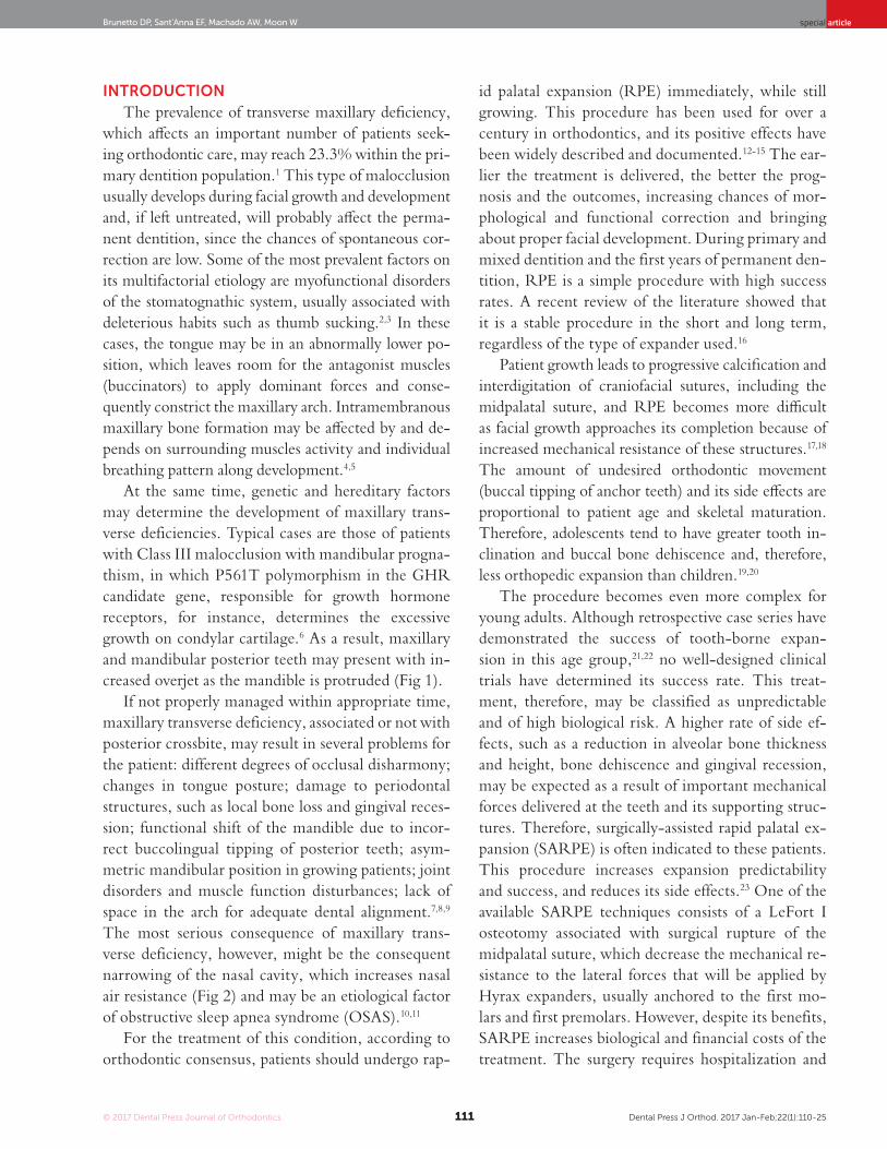
creased overjet as the mandible is protruded (Fig 1).
If not properly managed within appropriate time,
maxillary transverse deficiency, associated or not with
posterior crossbite, may result in several problems for
the patient: different degrees of occlusal disharmony;
changes in tongue posture; damage to periodontal
structures, such as local bone loss and gingival reces-
sion; functional shift of the mandible due to incor-
rect buccolingual tipping of posterior teeth; asym-
metric mandibular position in growing patients; joint
disorders and muscle function disturbances; lack of
space in the arch for adequate dental alignment.7,8,9
The most serious consequence of maxillary trans-
verse deficiency, however, might be the consequent
narrowing of the nasal cavity, which increases nasal
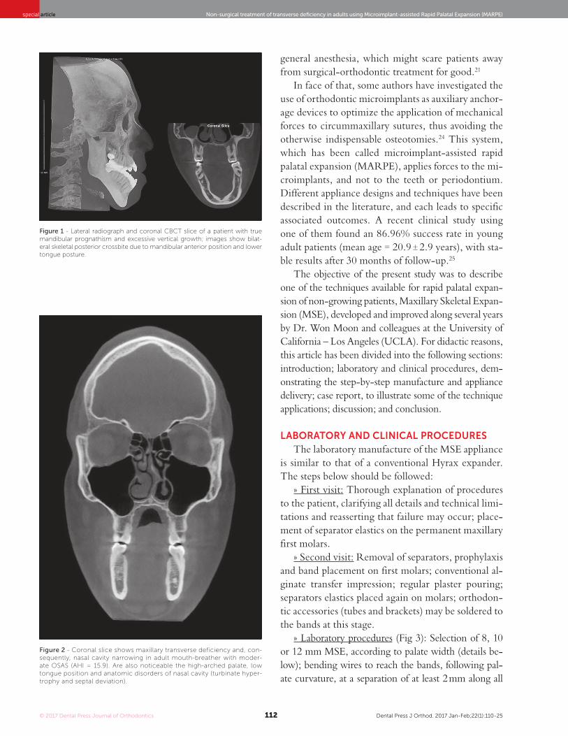
air resistance (Fig 2) and may be an etiological factor
of obstructive sleep apnea syndrome (OSAS).10,11
For the treatment of this condition, according to
orthodontic consensus, patients should undergo rap-
id palatal expansion (RPE) immediately, while still
growing. This procedure has been used for over a
century in orthodontics, and its positive effects have
been widely described and documented.12-15 The ear-
lier the treatment is delivered, the better the prog-
nosis and the outcomes, increasing chances of mor-
phological and functional correction and bringing
about proper facial development. During primary and
mixed dentition and the first years of permanent den-
tition, RPE is a simple procedure with high success
rates. A recent review of the literature showed that
it is a stable procedure in the short and long term,
regardless of the type of expander used.16
Patient growth leads to progressive calciication and
interdigitation of craniofacial sutures, including the
midpalatal suture, and RPE becomes more diicult
as facial growth approaches its completion because of
increased mechanical resistance of these structures.17,18
The amount of undesired orthodontic movement
(buccal tipping of anchor teeth) and its side efects are
proportional to patient age and skeletal maturation.
Therefore, adolescents tend to have greater tooth in-
clination and buccal bone dehiscence and, therefore,
less orthopedic expansion than children.19,20
The procedure becomes even more complex for
young adults. Although retrospective case series have
demonstrated the success of tooth-borne expan-
sion in this age group,21,22 no well-designed clinical
trials have determined its success rate. This treat-
ment, therefore, may be classified as unpredictable
and of high biological risk. A higher rate of side ef-
fects, such as a reduction in alveolar bone thickness
and height, bone dehiscence and gingival recession,
may be expected as a result of important mechanical
forces delivered at the teeth and its supporting struc-
tures. Therefore, surgically-assisted rapid palatal ex-
pansion (SARPE) is often indicated to these patients.
This procedure increases expansion predictability
and success, and reduces its side effects.23 One of the
available SARPE techniques consists of a LeFort I
osteotomy associated with surgical rupture of the
midpalatal suture, which decrease the mechanical re-
sistance to the lateral forces that will be applied by
Hyrax expanders, usually anchored to the first mo-
lars and first premolars. However, despite its benefits,
SARPE increases biological and financial costs of the
treatment. The surgery requires hospitalization and
© 2017 Dental Press Journal of Orthodontics Dental Press J Orthod. 2017 Jan-Feb;22(1):110-25112
Non-surgical treatment of transverse deficiency in adults using Microimplant-assisted Rapid Palatal Expansion (MARPE)special article
general anesthesia, which might scare patients away
from surgical-orthodontic treatment for good.21
In face of that, some authors have investigated the
use of orthodontic microimplants as auxiliary anchor-
age devices to optimize the application of mechanical
forces to circummaxillary sutures, thus avoiding the
otherwise indispensable osteotomies.24 This system,
which has been called microimplant-assisted rapid
palatal expansion (MARPE), applies forces to the mi-
croimplants, and not to the teeth or periodontium.
Different appliance designs and techniques have been
described in the literature, and each leads to specific
associated outcomes. A recent clinical study using
one of them found an 86.96% success rate in young
adult patients (mean age = 20.9 ± 2.9 years), with sta-
ble results after 30 months of follow-up.25
The objective of the present study was to describe
one of the techniques available for rapid palatal expan-
sion of non-growing patients, Maxillary Skeletal Expan-
sion (MSE), developed and improved along several years
by Dr. Won Moon and colleagues at the University of
California – Los Angeles (UCLA). For didactic reasons,
this article has been divided into the following sections:
introduction; laboratory and clinical procedures, dem-
onstrating the step-by-step manufacture and appliance
delivery; case report, to illustrate some of the technique
applications; discussion; and conclusion.
LABORATORY AND CLINICAL PROCEDURES
The laboratory manufacture of the MSE appliance
is similar to that of a conventional Hyrax expander.
The steps below should be followed:
» First visit: Thorough explanation of procedures
to the patient, clarifying all details and technical limi-
tations and reasserting that failure may occur; place-
ment of separator elastics on the permanent maxillary
first molars.
» Second visit: Removal of separators, prophylaxis
and band placement on first molars; conventional al-
ginate transfer impression; regular plaster pouring;
separators elastics placed again on molars; orthodon-
tic accessories (tubes and brackets) may be soldered to
the bands at this stage.
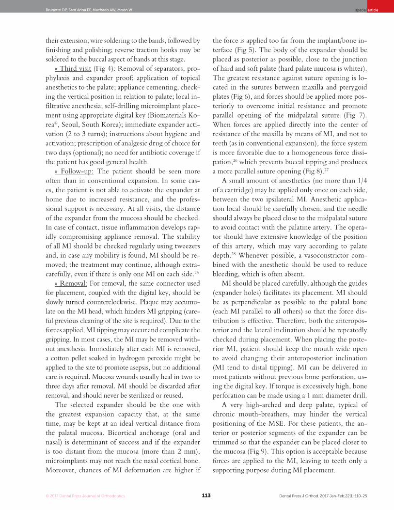
» Laboratory procedures (Fig 3): Selection of 8, 10
or 12 mm MSE, according to palate width (details be-
low); bending wires to reach the bands, following pal-
ate curvature, at a separation of at least 2 mm along all
Figure 1 - Lateral radiograph and coronal CBCT slice of a patient with true
mandibular prognathism and excessive vertical growth; images show bilat-
eral skeletal posterior crossbite due to mandibular anterior position and lower
tongue posture.
Figure 2 - Coronal slice shows maxillary transverse deficiency and, con-
sequently, nasal cavity narrowing in adult mouth-breather with moder-
ate OSAS (AHI = 15.9). Are also noticeable the high-arched palate, low
tongue position and anatomic disorders of nasal cavity (turbinate hyper-
trophy and septal deviation).
© 2017 Dental Press Journal of Orthodontics Dental Press J Orthod. 2017 Jan-Feb;22(1):110-25113
Brunetto DP, Sant’Anna EF, Machado AW, Moon W special article
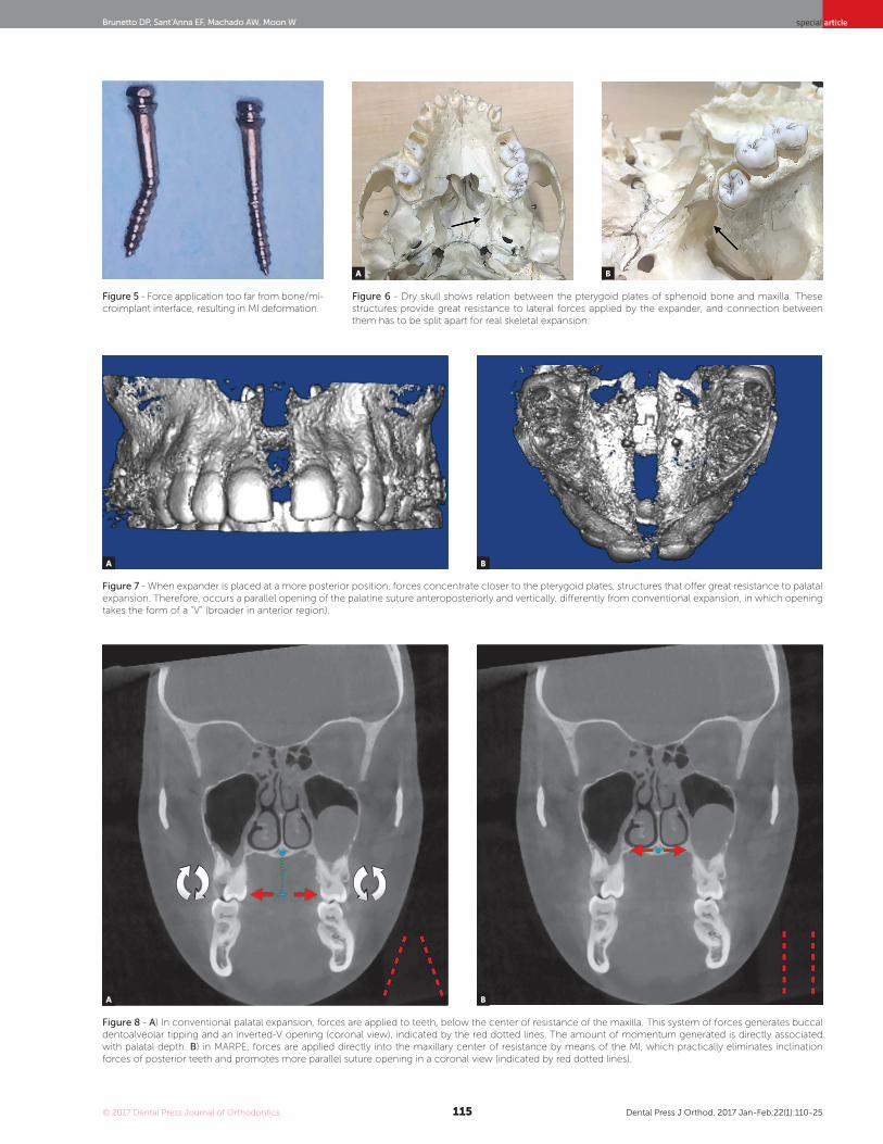
the force is applied too far from the implant/bone in-
terface (Fig 5). The body of the expander should be
placed as posterior as possible, close to the junction
of hard and soft palate (hard palate mucosa is whiter).
The greatest resistance against suture opening is lo-
cated in the sutures between maxilla and pterygoid
plates (Fig 6), and forces should be applied more pos-
teriorly to overcome initial resistance and promote
parallel opening of the midpalatal suture (Fig 7).
When forces are applied directly into the center of
resistance of the maxilla by means of MI, and not to
teeth (as in conventional expansion), the force system
is more favorable due to a homogeneous force dissi-
pation,26 which prevents buccal tipping and produces
a more parallel suture opening (Fig 8).27
A small amount of anesthetics (no more than 1/4
of a cartridge) may be applied only once on each side,
between the two ipsilateral MI. Anesthetic applica-
tion local should be carefully chosen, and the needle
should always be placed close to the midpalatal suture
to avoid contact with the palatine artery. The opera-
tor should have extensive knowledge of the position
of this artery, which may vary according to palate
depth.28 Whenever possible, a vasoconstrictor com-
bined with the anesthetic should be used to reduce
bleeding, which is often absent.
MI should be placed carefully, although the guides
(expander holes) facilitates its placement. MI should
be as perpendicular as possible to the palatal bone
(each MI parallel to all others) so that the force dis-
tribution is effective. Therefore, both the anteropos-
terior and the lateral inclination should be repeatedly
checked during placement. When placing the poste-
rior MI, patient should keep the mouth wide open
to avoid changing their anteroposterior inclination
(MI tend to distal tipping). MI can be delivered in
most patients without previous bone perforation, us-
ing the digital key. If torque is excessively high, bone
perforation can be made using a 1 mm diameter drill.
A very high-arched and deep palate, typical of
chronic mouth-breathers, may hinder the vertical
positioning of the MSE. For these patients, the an-
terior or posterior segments of the expander can be
trimmed so that the expander can be placed closer to
the mucosa (Fig 9). This option is acceptable because
forces are applied to the MI, leaving to teeth only a
supporting purpose during MI placement.
their extension; wire soldering to the bands, followed by
inishing and polishing; reverse traction hooks may be
soldered to the buccal aspect of bands at this stage.
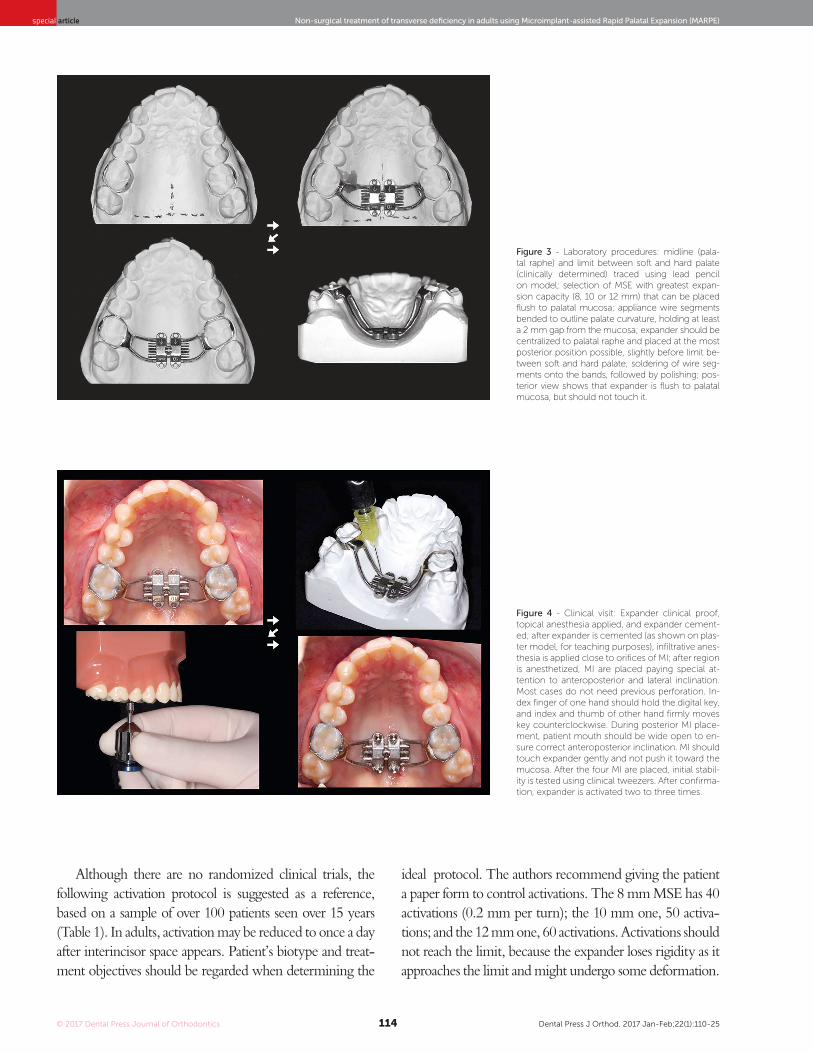
» Third visit (Fig 4): Removal of separators, pro-
phylaxis and expander proof; application of topical
anesthetics to the palate; appliance cementing, check-
ing the vertical position in relation to palate; local in-
filtrative anesthesia; self-drilling microimplant place-
ment using appropriate digital key (Biomaterials Ko-
rea®, Seoul, South Korea); immediate expander acti-
vation (2 to 3 turns); instructions about hygiene and
activation; prescription of analgesic drug of choice for
two days (optional); no need for antibiotic coverage if
the patient has good general health.
» Follow-up: The patient should be seen more
often than in conventional expansion. In some cas-
es, the patient is not able to activate the expander at
home due to increased resistance, and the profes-
sional support is necessary. At all visits, the distance
of the expander from the mucosa should be checked.
In case of contact, tissue inflammation develops rap-
idly compromising appliance removal. The stability
of all MI should be checked regularly using tweezers
and, in case any mobility is found, MI should be re-
moved; the treatment may continue, although extra-
carefully, even if there is only one MI on each side.25
» Removal: For removal, the same connector used
for placement, coupled with the digital key, should be
slowly turned counterclockwise. Plaque may accumu-
late on the MI head, which hinders MI gripping (care-
ful previous cleaning of the site is required). Due to the
forces applied, MI tipping may occur and complicate the
gripping. In most cases, the MI may be removed with-
out anesthesia. Immediately ater each MI is removed,
a cotton pellet soaked in hydrogen peroxide might be
applied to the site to promote asepsis, but no additional
care is required. Mucosa wounds usually heal in two to
three days ater removal. MI should be discarded ater
removal, and should never be sterilized or reused.
The selected expander should be the one with
the greatest expansion capacity that, at the same
time, may be kept at an ideal vertical distance from
the palatal mucosa. Bicortical anchorage (oral and
nasal) is determinant of success and if the expander
is too distant from the mucosa (more than 2 mm),
microimplants may not reach the nasal cortical bone.
Moreover, chances of MI deformation are higher if
© 2017 Dental Press Journal of Orthodontics Dental Press J Orthod. 2017 Jan-Feb;22(1):110-25114
Non-surgical treatment of transverse deficiency in adults using Microimplant-assisted Rapid Palatal Expansion (MARPE)special article
ideal protocol. The authors recommend giving the patient
a paper form to control activations. The 8 mm MSE has 40
activations (0.2 mm per turn); the 10 mm one, 50 activa-
tions; and the 12 mm one, 60 activations. Activations should
not reach the limit, because the expander loses rigidity as it
approaches the limit and might undergo some deformation.
Although there are no randomized clinical trials, the
following activation protocol is suggested as a reference,
based on a sample of over 100 patients seen over 15 years
(Table 1). In adults, activation may be reduced to once a day
ater interincisor space appears. Patient’s biotype and treat-
ment objectives should be regarded when determining the
Figure 3 - Laboratory procedures: midline (pala-
tal raphe) and limit between soft and hard palate
(clinically determined) traced using lead pencil
on model; selection of MSE with greatest expan-
sion capacity (8, 10 or 12 mm) that can be placed
flush to palatal mucosa; appliance wire segments
bended to outline palate curvature, holding at least
a 2 mm gap from the mucosa; expander should be
centralized to palatal raphe and placed at the most
posterior position possible, slightly before limit be-
tween soft and hard palate; soldering of wire seg-
ments onto the bands, followed by polishing; pos-
terior view shows that expander is flush to palatal
mucosa, but should not touch it.
Figure 4 - Clinical visit: Expander clinical proof,
topical anesthesia applied, and expander cement-
ed; after expander is cemented (as shown on plas-
ter model, for teaching purposes), infiltrative anes-
thesia is applied close to orifices of MI; after region
is anesthetized, MI are placed paying special at-
tention to anteroposterior and lateral inclination.
Most cases do not need previous perforation. In-
dex finger of one hand should hold the digital key,
and index and thumb of other hand firmly moves
key counterclockwise. During posterior MI place-
ment, patient mouth should be wide open to en-
sure correct anteroposterior inclination. MI should
touch expander gently and not push it toward the
mucosa. After the four MI are placed, initial stabil-
ity is tested using clinical tweezers. After confirma-
tion, expander is activated two to three times.
© 2017 Dental Press Journal of Orthodontics Dental Press J Orthod. 2017 Jan-Feb;22(1):110-25115
Brunetto DP, Sant’Anna EF, Machado AW, Moon W special article
Figure 5 - Force application too far from bone/mi-
croimplant interface, resulting in MI deformation.
Figure 6 - Dry skull shows relation between the pterygoid plates of sphenoid bone and maxilla. These
structures provide great resistance to lateral forces applied by the expander, and connection between
them has to be split apart for real skeletal expansion.
Figure 8 - A) In conventional palatal expansion, forces are applied to teeth, below the center of resistance of the maxilla. This system of forces generates buccal
dentoalveolar tipping and an inverted-V opening (coronal view), indicated by the red dotted lines. The amount of momentum generated is directly associated
with palatal depth. B) in MARPE, forces are applied directly into the maxillary center of resistance by means of the MI, which practically eliminates inclination
forces of posterior teeth and promotes more parallel suture opening in a coronal view (indicated by red dotted lines).
A B
Figure 7 - When expander is placed at a more posterior position, forces concentrate closer to the pterygoid plates, structures that offer great resistance to palatal
expansion. Therefore, occurs a parallel opening of the palatine suture anteroposteriorly and vertically, differently from conventional expansion, in which opening
takes the form of a "V" (broader in anterior region).
A B
A B
© 2017 Dental Press Journal of Orthodontics Dental Press J Orthod. 2017 Jan-Feb;22(1):110-25116
Non-surgical treatment of transverse deficiency in adults using Microimplant-assisted Rapid Palatal Expansion (MARPE)special article
CASE REPORT
A 22-year and 6-month-old female was seen for
orthodontic treatment at the Orthodontic Clinic of
the Universidade Federal do Paraná, Brazil. Her chief
complaint was posterior crossbite and deficient
breathing, especially during sleep. The patient had
not undergone any orthodontic treatment before, but
had already made up her mind to avoid maxillary ex-
pansion surgery.
Facial examination revealed a harmonic profile and
proportional facial thirds. The smiling photo showed
excessive buccal corridor display and easily noticeable
transverse maxillary deficiency (Fig 10). The man-
dibular arch had moderate anterior and posterior
crowding and left transverse asymmetry due to the
posterior crossbite on that side. In the maxillary arch,
there was mild crowding and transverse asymmetry
(opposite to the mandibular arch) on the left side as
well, due to the same crossbite (Fig 11). It was also
found microdontic maxillary lateral incisors and
right maxillary midline shift. Right molars and ca-
nines displayed a Class I relationship, bearing nor-
mal horizontal and vertical overjet. On the left side,
canines had an edge-to-edge relationship (Class II),
with posterior crossbite (Fig 12). Lateral radiograph
showed a good skeletal relationship, as well as good
inclination and position of maxillary and mandibu-
lar incisors (Fig 13). A coronal CBCT slice revealed
exacerbated inclination of teeth (torque) in posteri-
or crossbite (Fig 14). Sagittal slices of the joints re-
vealed that the condyles were not centrally positioned
in the fossa, which confirmed the clinically present
double-bite (Fig 15). Because of her breathing com-
plaints, it was applied the Epworth Sleepiness Scale
and Quebec Sleep Questionnaire as screening tools,
which revealed a high risk of obstructive sleep apnea
syndrome (OSAS). Therefore, the patient underwent
in-home polysomnography (Nox Medical, Reykja-
vik, Iceland), and results revealed an apnea/hypopnea
index (AHI) of 7.9, classified as mild apnea syndrome
according to the American Association of Sleep Med-
icine guideline,29 associated with moderate snoring
and isolated episodes of bruxism (Fig 16).
The first treatment option was non-surgical
rapid palatal expansion (MARPE) because the pa-
tient refused to have SARPE. We thought skeletal
expansion was necessary because of the patient’s re-
spiratory disorder, reported by the patient herself at
first, and later confirmed by the polysomnography.
Treatment alternative consisted of fixed orthodontic
appliance and microimplants for intrusion and buc-
cal inclination of the left maxillary posterior teeth to
compensate the buccolingual inclination of teeth in
crossbite area, with possible future side effects on its
supporting structures.
Treatment started with the placement of a 10 mm
maxillary skeletal expander (MSE) and three imme-
diate activations (1/4 of a turn, 90 degrees each), fol-
lowed by two daily activations. By the second week,
the patient reported having heard clicks in the region
of the palatal suture and, in the following days, ap-
pearance of the interincisal diastema (Fig 17). There
was a discrete opening of the anterior bite due to con-
tact of the buccal cuspid of the left first maxillary mo-
lar, which moved in the direction of overlapping the
antagonist mandibular molar. Photographs after 34
activations confirmed suture opening and lack of col-
lateral buccal inclination of maxillary molars (Fig 18).
The patient often needed help to perform the activa-
Figure 9 - Maxillary occlusal photograph showing removal of anterior wire seg-
ments of MSE, to improve vertical fit in a very narrow and high-arched palate.
AGE GROUP ACTIVATION
Beginning of adolescence 3 to 4x/week
End of adolescence 1x/day
Young adults 2x/day
Older than 25 years 2x or +/day
Table 1 - Suggested activation protocol.
© 2017 Dental Press Journal of Orthodontics Dental Press J Orthod. 2017 Jan-Feb;22(1):110-25117
Brunetto DP, Sant’Anna EF, Machado AW, Moon W special article
tion because of increased mechanical resistance. Af-
ter 44 activations, at a total of 8.8 mm screw open-
ing, the MSE was removed for the placement of an-
other expander, a common practice depending on
case severity. At this time, crossbite was still present
(Fig 19). However, instead of using another MSE and
continuing with pure skeletal expansion, we decided
to place a conventional tooth-borne Hyrax expander
for two reasons: circummaxillary sutures had already
been mobilized, and, therefore, skeletal gains should
be preserved; and we would like to ensure buccal in-
clination of maxillary left posterior teeth to optimize
future orthodontic treatment. At this point, the pa-
tient had already reported important improvement
of sleep quality, with facilitated nose breathing and
reduction of rhinitis episodes, frequent in the past.
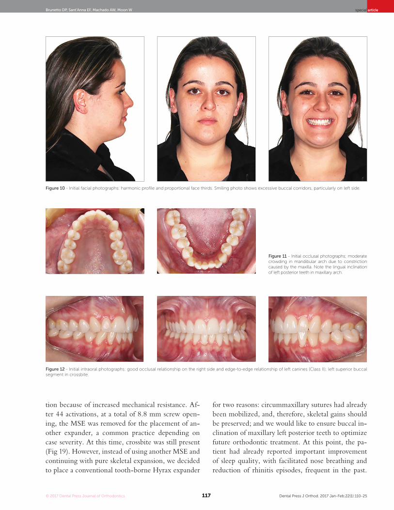
Figure 10 - Initial facial photographs: harmonic profile and proportional face thirds. Smiling photo shows excessive buccal corridors, particularly on left side.
Figure 11 - Initial occlusal photographs; moderate
crowding in mandibular arch due to constriction
caused by the maxilla. Note the lingual inclination
of left posterior teeth in maxillary arch.
Figure 12 - Initial intraoral photographs: good occlusal relationship on the right side and edge-to-edge relationship of left canines (Class II); left superior buccal
segment in crossbite.
© 2017 Dental Press Journal of Orthodontics Dental Press J Orthod. 2017 Jan-Feb;22(1):110-25118
Non-surgical treatment of transverse deficiency in adults using Microimplant-assisted Rapid Palatal Expansion (MARPE)special article
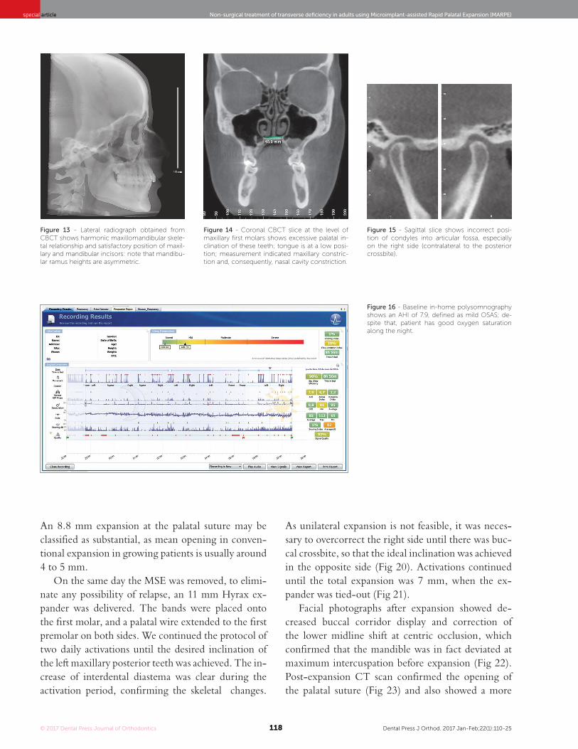
Figure 13 - Lateral radiograph obtained from
CBCT shows harmonic maxillomandibular skele-
tal relationship and satisfactory position of maxil-
lary and mandibular incisors: note that mandibu-
lar ramus heights are asymmetric.
Figure 14 - Coronal CBCT slice at the level of
maxillary first molars shows excessive palatal in-
clination of these teeth; tongue is at a low posi-
tion; measurement indicated maxillary constric-
tion and, consequently, nasal cavity constriction.
Figure 15 - Sagittal slice shows incorrect posi-
tion of condyles into articular fossa, especially
on the right side (contralateral to the posterior
crossbite).
Figure 16 - Baseline in-home polysomnography
shows an AHI of 7.9, defined as mild OSAS; de-
spite that, patient has good oxygen saturation
along the night.
An 8.8 mm expansion at the palatal suture may be
classified as substantial, as mean opening in conven-
tional expansion in growing patients is usually around
4 to 5 mm.
On the same day the MSE was removed, to elimi-
nate any possibility of relapse, an 11 mm Hyrax ex-
pander was delivered. The bands were placed onto
the irst molar, and a palatal wire extended to the irst
premolar on both sides. We continued the protocol of
two daily activations until the desired inclination of
the let maxillary posterior teeth was achieved. The in-
crease of interdental diastema was clear during the
activation period, conirming the skeletal changes.
As unilateral expansion is not feasible, it was neces-
sary to overcorrect the right side until there was buc-
cal crossbite, so that the ideal inclination was achieved
in the opposite side (Fig 20). Activations continued
until the total expansion was 7 mm, when the ex-
pander was tied-out (Fig 21).
Facial photographs after expansion showed de-
creased buccal corridor display and correction of
the lower midline shift at centric occlusion, which
confirmed that the mandible was in fact deviated at
maximum intercuspation before expansion (Fig 22).
Post-expansion CT scan confirmed the opening of
the palatal suture (Fig 23) and also showed a more
© 2017 Dental Press Journal of Orthodontics Dental Press J Orthod. 2017 Jan-Feb;22(1):110-25119
Brunetto DP, Sant’Anna EF, Machado AW, Moon W special article
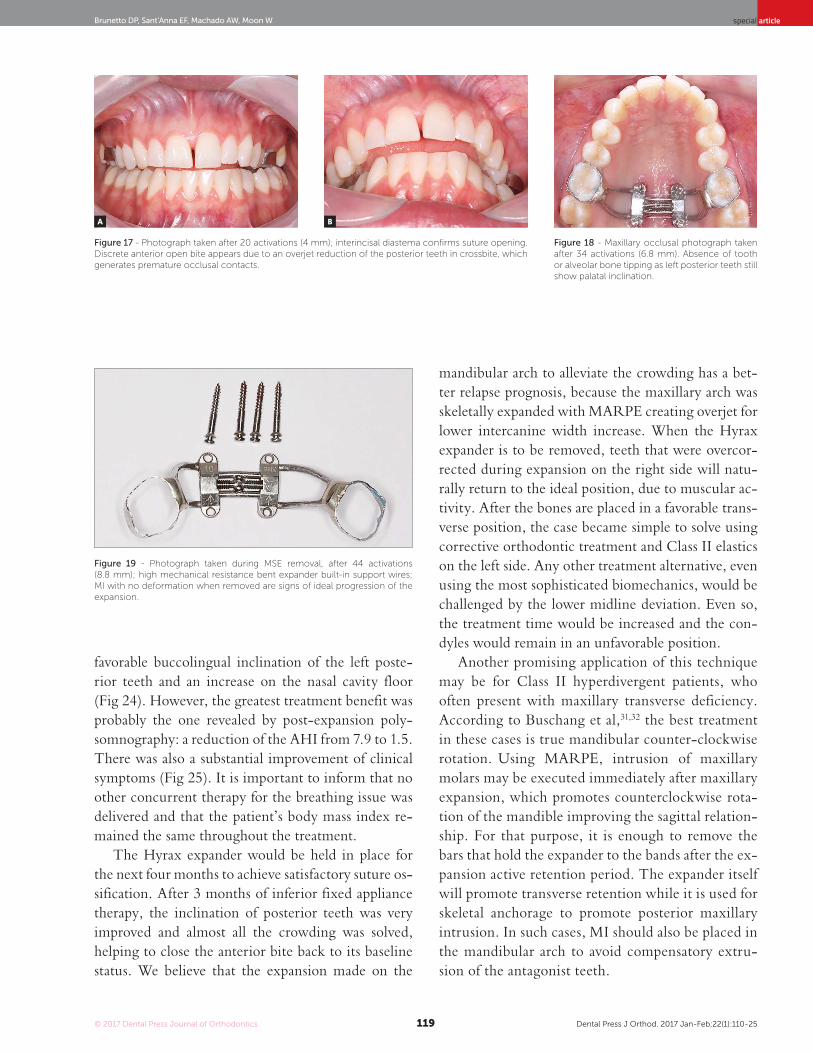
Figure 17 - Photograph taken after 20 activations (4 mm); interincisal diastema confirms suture opening.
Discrete anterior open bite appears due to an overjet reduction of the posterior teeth in crossbite, which
generates premature occlusal contacts.
Figure 18 - Maxillary occlusal photograph taken
after 34 activations (6.8 mm). Absence of tooth
or alveolar bone tipping as left posterior teeth still
show palatal inclination.
Figure 19 - Photograph taken during MSE removal, after 44 activations
(8.8 mm); high mechanical resistance bent expander built-in support wires;
MI with no deformation when removed are signs of ideal progression of the
expansion.
favorable buccolingual inclination of the left poste-
rior teeth and an increase on the nasal cavity floor
(Fig 24). However, the greatest treatment benefit was
probably the one revealed by post-expansion poly-
somnography: a reduction of the AHI from 7.9 to 1.5.
There was also a substantial improvement of clinical
symptoms (Fig 25). It is important to inform that no
other concurrent therapy for the breathing issue was
delivered and that the patient’s body mass index re-
mained the same throughout the treatment.
The Hyrax expander would be held in place for
the next four months to achieve satisfactory suture os-
sification. After 3 months of inferior fixed appliance
therapy, the inclination of posterior teeth was very
improved and almost all the crowding was solved,
helping to close the anterior bite back to its baseline
status. We believe that the expansion made on the
mandibular arch to alleviate the crowding has a bet-
ter relapse prognosis, because the maxillary arch was
skeletally expanded with MARPE creating overjet for
lower intercanine width increase. When the Hyrax
expander is to be removed, teeth that were overcor-
rected during expansion on the right side will natu-
rally return to the ideal position, due to muscular ac-
tivity. After the bones are placed in a favorable trans-
verse position, the case became simple to solve using
corrective orthodontic treatment and Class II elastics
on the left side. Any other treatment alternative, even
using the most sophisticated biomechanics, would be
challenged by the lower midline deviation. Even so,
the treatment time would be increased and the con-
dyles would remain in an unfavorable position.
Another promising application of this technique
may be for Class II hyperdivergent patients, who
often present with maxillary transverse deficiency.
According to Buschang et al,31,32 the best treatment
in these cases is true mandibular counter-clockwise
rotation. Using MARPE, intrusion of maxillary
molars may be executed immediately after maxillary
expansion, which promotes counterclockwise rota-
tion of the mandible improving the sagittal relation-
ship. For that purpose, it is enough to remove the
bars that hold the expander to the bands after the ex-
pansion active retention period. The expander itself
will promote transverse retention while it is used for
skeletal anchorage to promote posterior maxillary
intrusion. In such cases, MI should also be placed in
the mandibular arch to avoid compensatory extru-
sion of the antagonist teeth.
A B
© 2017 Dental Press Journal of Orthodontics Dental Press J Orthod. 2017 Jan-Feb;22(1):110-25120
Non-surgical treatment of transverse deficiency in adults using Microimplant-assisted Rapid Palatal Expansion (MARPE)special article
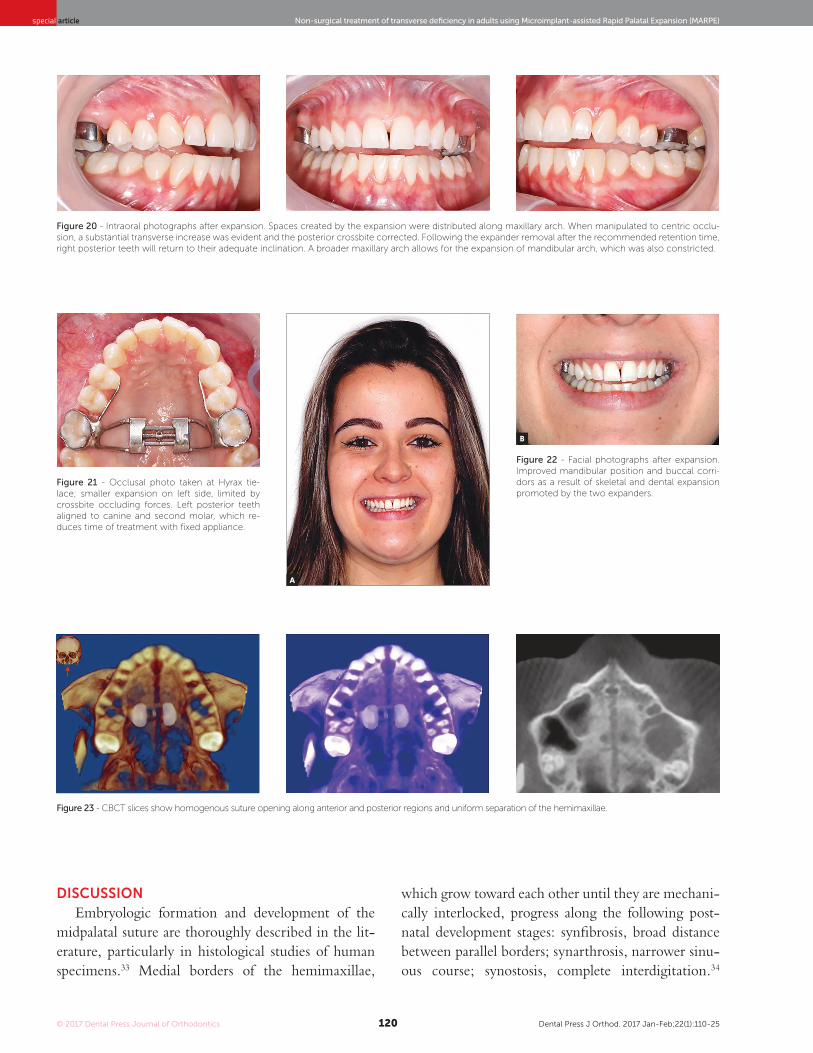
Figure 20 - Intraoral photographs after expansion. Spaces created by the expansion were distributed along maxillary arch. When manipulated to centric occlu-
sion, a substantial transverse increase was evident and the posterior crossbite corrected. Following the expander removal after the recommended retention time,
right posterior teeth will return to their adequate inclination. A broader maxillary arch allows for the expansion of mandibular arch, which was also constricted.
Figure 23 - CBCT slices show homogenous suture opening along anterior and posterior regions and uniform separation of the hemimaxillae.
Figure 21 - Occlusal photo taken at Hyrax tie-
lace; smaller expansion on left side, limited by
crossbite occluding forces. Left posterior teeth
aligned to canine and second molar, which re-
duces time of treatment with fixed appliance.
Figure 22 - Facial photographs after expansion.
Improved mandibular position and buccal corri-
dors as a result of skeletal and dental expansion
promoted by the two expanders.
A
B
DISCUSSION
Embryologic formation and development of the
midpalatal suture are thoroughly described in the lit-
erature, particularly in histological studies of human
specimens.33 Medial borders of the hemimaxillae,
which grow toward each other until they are mechani-
cally interlocked, progress along the following post-
natal development stages: synibrosis, broad distance
between parallel borders; synarthrosis, narrower sinu-
ous course; synostosis, complete interdigitation.34
© 2017 Dental Press Journal of Orthodontics Dental Press J Orthod. 2017 Jan-Feb;22(1):110-25121
Brunetto DP, Sant’Anna EF, Machado AW, Moon W special article
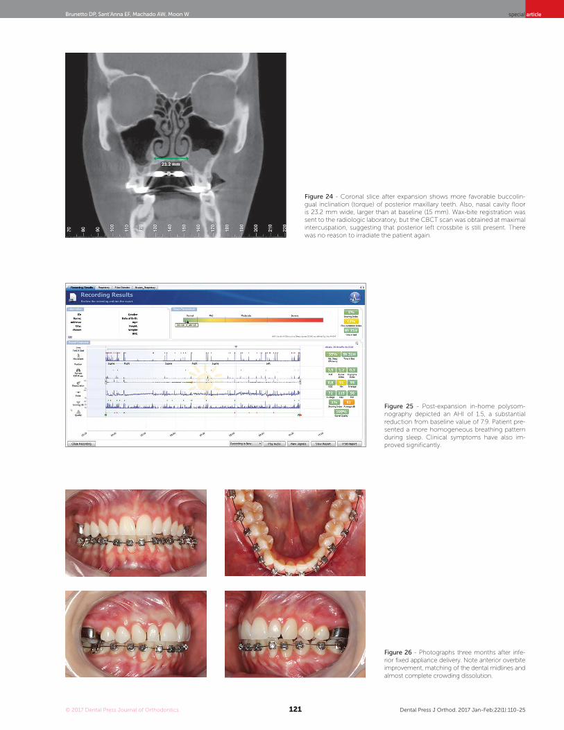
Figure 24 - Coronal slice after expansion shows more favorable buccolin-
gual inclination (torque) of posterior maxillary teeth. Also, nasal cavity floor
is 23.2 mm wide, larger than at baseline (15 mm). Wax-bite registration was
sent to the radiologic laboratory, but the CBCT scan was obtained at maximal
intercuspation, suggesting that posterior left crossbite is still present. There
was no reason to irradiate the patient again.
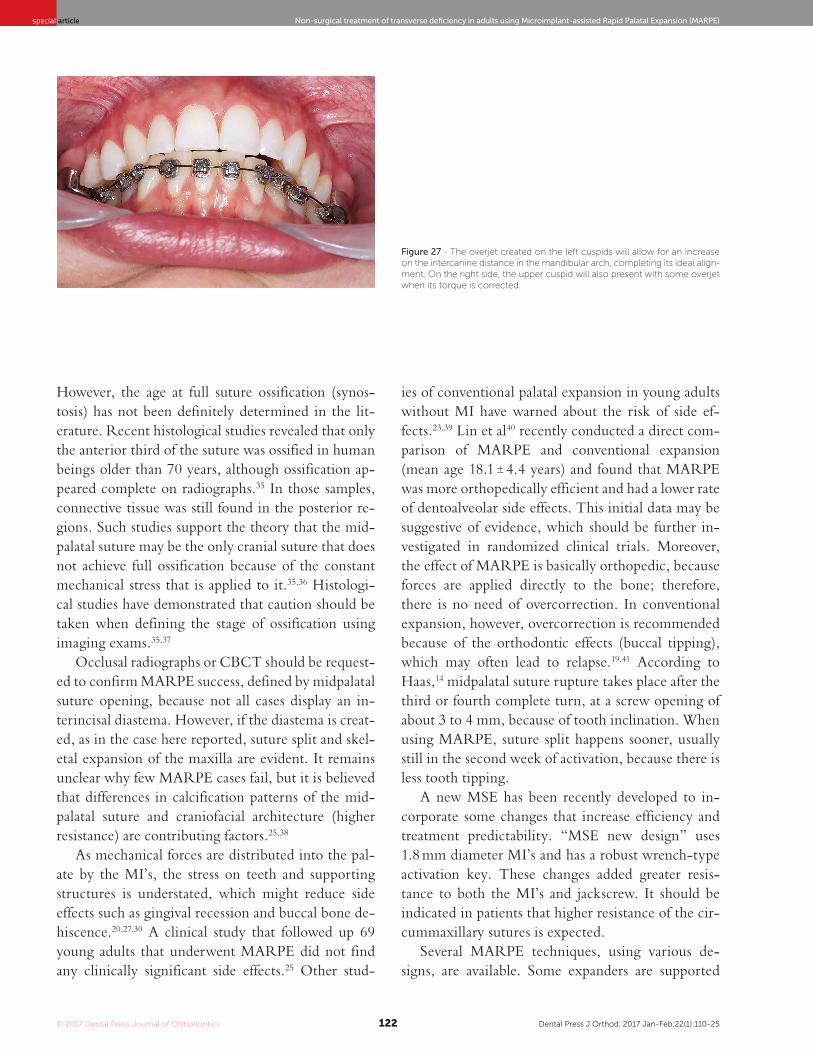
Figure 25 - Post-expansion in-home polysom-
nography depicted an AHI of 1.5, a substantial
reduction from baseline value of 7.9. Patient pre-
sented a more homogeneous breathing pattern
during sleep. Clinical symptoms have also im-
proved significantly.
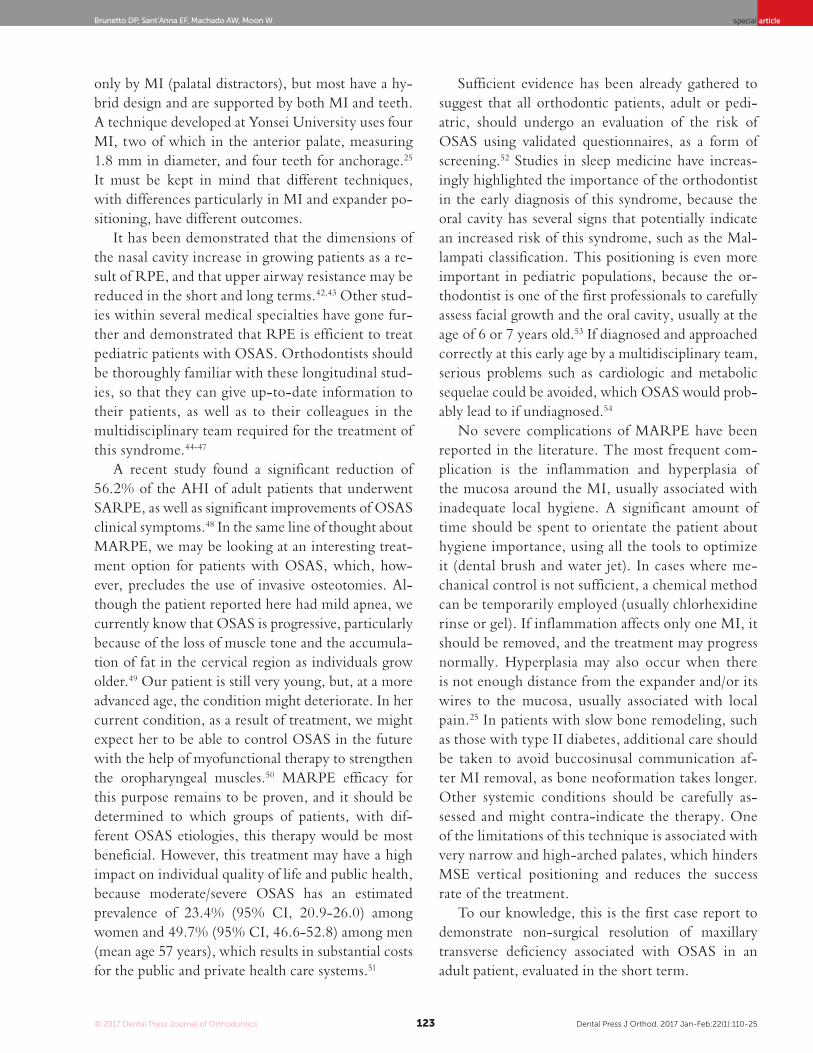
Figure 26 - Photographs three months after infe-
rior fixed appliance delivery. Note anterior overbite
improvement, matching of the dental midlines and
almost complete crowding dissolution.
© 2017 Dental Press Journal of Orthodontics Dental Press J Orthod. 2017 Jan-Feb;22(1):110-25122
Non-surgical treatment of transverse deficiency in adults using Microimplant-assisted Rapid Palatal Expansion (MARPE)special article
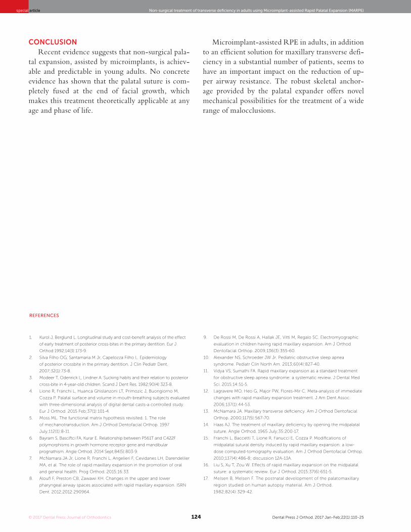
Figure 27 - The overjet created on the left cuspids will allow for an increase
on the intercanine distance in the mandibular arch, completing its ideal align-
ment. On the right side, the upper cuspid will also present with some overjet
when its torque is corrected.
However, the age at full suture ossification (synos-
tosis) has not been definitely determined in the lit-
erature. Recent histological studies revealed that only
the anterior third of the suture was ossified in human
beings older than 70 years, although ossification ap-
peared complete on radiographs.35 In those samples,
connective tissue was still found in the posterior re-
gions. Such studies support the theory that the mid-
palatal suture may be the only cranial suture that does
not achieve full ossification because of the constant
mechanical stress that is applied to it.35,36 Histologi-
cal studies have demonstrated that caution should be
taken when defining the stage of ossification using
imaging exams.35,37
Occlusal radiographs or CBCT should be request-
ed to confirm MARPE success, defined by midpalatal
suture opening, because not all cases display an in-
terincisal diastema. However, if the diastema is creat-
ed, as in the case here reported, suture split and skel-
etal expansion of the maxilla are evident. It remains
unclear why few MARPE cases fail, but it is believed
that differences in calcification patterns of the mid-
palatal suture and craniofacial architecture (higher
resistance) are contributing factors.25,38
As mechanical forces are distributed into the pal-
ate by the MI’s, the stress on teeth and supporting
structures is understated, which might reduce side
effects such as gingival recession and buccal bone de-
hiscence.20,27,30 A clinical study that followed up 69
young adults that underwent MARPE did not find
any clinically significant side effects.25 Other stud-
ies of conventional palatal expansion in young adults
without MI have warned about the risk of side ef-
fects.23,39 Lin et al40 recently conducted a direct com-
parison of MARPE and conventional expansion
(mean age 18.1 ± 4.4 years) and found that MARPE
was more orthopedically efficient and had a lower rate
of dentoalveolar side effects. This initial data may be
suggestive of evidence, which should be further in-
vestigated in randomized clinical trials. Moreover,
the effect of MARPE is basically orthopedic, because
forces are applied directly to the bone; therefore,
there is no need of overcorrection. In conventional
expansion, however, overcorrection is recommended
because of the orthodontic effects (buccal tipping),
which may often lead to relapse.19,41 According to
Haas,14 midpalatal suture rupture takes place after the
third or fourth complete turn, at a screw opening of
about 3 to 4 mm, because of tooth inclination. When
using MARPE, suture split happens sooner, usually
still in the second week of activation, because there is
less tooth tipping.
A new MSE has been recently developed to in-
corporate some changes that increase efficiency and
treatment predictability. “MSE new design” uses
1.8 mm diameter MI’s and has a robust wrench-type
activation key. These changes added greater resis-
tance to both the MI’s and jackscrew. It should be
indicated in patients that higher resistance of the cir-
cummaxillary sutures is expected.
Several MARPE techniques, using various de-
signs, are available. Some expanders are supported
© 2017 Dental Press Journal of Orthodontics Dental Press J Orthod. 2017 Jan-Feb;22(1):110-25123
Brunetto DP, Sant’Anna EF, Machado AW, Moon W special article
only by MI (palatal distractors), but most have a hy-
brid design and are supported by both MI and teeth.
A technique developed at Yonsei University uses four
MI, two of which in the anterior palate, measuring
1.8 mm in diameter, and four teeth for anchorage.25
It must be kept in mind that different techniques,
with differences particularly in MI and expander po-
sitioning, have different outcomes.
It has been demonstrated that the dimensions of
the nasal cavity increase in growing patients as a re-
sult of RPE, and that upper airway resistance may be
reduced in the short and long terms.42,43 Other stud-
ies within several medical specialties have gone fur-
ther and demonstrated that RPE is efficient to treat
pediatric patients with OSAS. Orthodontists should
be thoroughly familiar with these longitudinal stud-
ies, so that they can give up-to-date information to
their patients, as well as to their colleagues in the
multidisciplinary team required for the treatment of
this syndrome.44-47
A recent study found a significant reduction of
56.2% of the AHI of adult patients that underwent
SARPE, as well as significant improvements of OSAS
clinical symptoms.48 In the same line of thought about
MARPE, we may be looking at an interesting treat-
ment option for patients with OSAS, which, how-
ever, precludes the use of invasive osteotomies. Al-
though the patient reported here had mild apnea, we
currently know that OSAS is progressive, particularly
because of the loss of muscle tone and the accumula-
tion of fat in the cervical region as individuals grow
older.49 Our patient is still very young, but, at a more
advanced age, the condition might deteriorate. In her
current condition, as a result of treatment, we might
expect her to be able to control OSAS in the future
with the help of myofunctional therapy to strengthen
the oropharyngeal muscles.50 MARPE efficacy for
this purpose remains to be proven, and it should be
determined to which groups of patients, with dif-
ferent OSAS etiologies, this therapy would be most
beneficial. However, this treatment may have a high
impact on individual quality of life and public health,
because moderate/severe OSAS has an estimated
prevalence of 23.4% (95% CI, 20.9-26.0) among
women and 49.7% (95% CI, 46.6-52.8) among men
(mean age 57 years), which results in substantial costs
for the public and private health care systems.51
Sufficient evidence has been already gathered to
suggest that all orthodontic patients, adult or pedi-
atric, should undergo an evaluation of the risk of
OSAS using validated questionnaires, as a form of
screening.52 Studies in sleep medicine have increas-
ingly highlighted the importance of the orthodontist
in the early diagnosis of this syndrome, because the
oral cavity has several signs that potentially indicate
an increased risk of this syndrome, such as the Mal-
lampati classification. This positioning is even more
important in pediatric populations, because the or-
thodontist is one of the first professionals to carefully
assess facial growth and the oral cavity, usually at the
age of 6 or 7 years old.53 If diagnosed and approached
correctly at this early age by a multidisciplinary team,
serious problems such as cardiologic and metabolic
sequelae could be avoided, which OSAS would prob-
ably lead to if undiagnosed.54
No severe complications of MARPE have been
reported in the literature. The most frequent com-
plication is the inflammation and hyperplasia of
the mucosa around the MI, usually associated with
inadequate local hygiene. A significant amount of
time should be spent to orientate the patient about
hygiene importance, using all the tools to optimize
it (dental brush and water jet). In cases where me-
chanical control is not sufficient, a chemical method
can be temporarily employed (usually chlorhexidine
rinse or gel). If inflammation affects only one MI, it
should be removed, and the treatment may progress
normally. Hyperplasia may also occur when there
is not enough distance from the expander and/or its
wires to the mucosa, usually associated with local
pain.25 In patients with slow bone remodeling, such
as those with type II diabetes, additional care should
be taken to avoid buccosinusal communication af-
ter MI removal, as bone neoformation takes longer.
Other systemic conditions should be carefully as-
sessed and might contra-indicate the therapy. One
of the limitations of this technique is associated with
very narrow and high-arched palates, which hinders
MSE vertical positioning and reduces the success
rate of the treatment.
To our knowledge, this is the first case report to
demonstrate non-surgical resolution of maxillary
transverse deficiency associated with OSAS in an
adult patient, evaluated in the short term.
© 2017 Dental Press Journal of Orthodontics Dental Press J Orthod. 2017 Jan-Feb;22(1):110-25124
Non-surgical treatment of transverse deficiency in adults using Microimplant-assisted Rapid Palatal Expansion (MARPE)special article
1. Kurol J, Berglund L. Longitudinal study and cost-beneit analysis of the efect
of early treatment of posterior cross-bites in the primary dentition. Eur J
Orthod 1992;14(3):173-9.
2. Silva Filho OG, Santamaria M Jr, Capelozza Filho L. Epidemiology
of posterior crossbite in the primary dentition. J Clin Pediatr Dent.
2007;32(1):73-8.
3. Modeer T, Odenrick L, Lindner A. Sucking habits and their relation to posterior
cross-bite in 4-year-old children. Scand J Dent Res. 1982;90(4):323-8.
4. Lione R, Franchi L, Huanca Ghislanzoni LT, Primozic J, Buongiorno M,
Cozza P. Palatal surface and volume in mouth-breathing subjects evaluated
with three-dimensional analysis of digital dental casts-a controlled study.
Eur J Orthod. 2015 Feb;37(1):101-4.
5. Moss ML. The functional matrix hypothesis revisited. 1. The role
of mechanotransduction. Am J Orthod Dentofacial Orthop. 1997
July;112(1):8-11.
6. Bayram S, Basciftci FA, Kurar E. Relationship between P561T and C422F
polymorphisms in growth hormone receptor gene and mandibular
prognathism. Angle Orthod. 2014 Sept;84(5):803-9.
7. McNamara JA Jr, Lione R, Franchi L, Angelieri F, Cevidanes LH, Darendeliler
MA, et al. The role of rapid maxillary expansion in the promotion of oral
and general health. Prog Orthod. 2015;16:33.
8. Aloui F, Preston CB, Zawawi KH. Changes in the upper and lower
pharyngeal airway spaces associated with rapid maxillary expansion. ISRN
Dent. 2012;2012:290964.
9. De Rossi M, De Rossi A, Hallak JE, Vitti M, Regalo SC. Electromyographic
evaluation in children having rapid maxillary expansion. Am J Orthod
Dentofacial Orthop. 2009;136(3):355-60.
10. Alexander NS, Schroeder JW Jr. Pediatric obstructive sleep apnea
syndrome. Pediatr Clin North Am. 2013;60(4):827-40.
11. Vidya VS, Sumathi FA. Rapid maxillary expansion as a standard treatment
for obstructive sleep apnea syndrome: a systematic review. J Dental Med
Sci. 2015;14:51-5.
12. Lagravere MO, Heo G, Major PW, Flores-Mir C. Meta-analysis of immediate
changes with rapid maxillary expansion treatment. J Am Dent Assoc.
2006;137(1):44-53.
13. McNamara JA. Maxillary transverse deiciency. Am J Orthod Dentofacial
Orthop. 2000;117(5):567-70.
14. Haas AJ. The treatment of maxillary deiciency by opening the midpalatal
suture. Angle Orthod. 1965 July;35:200-17.
15. Franchi L, Baccetti T, Lione R, Fanucci E, Cozza P. Modiications of
midpalatal sutural density induced by rapid maxillary expansion: a low-
dose computed-tomography evaluation. Am J Orthod Dentofacial Orthop.
2010;137(4):486-8; discussion 12A-13A.
16. Liu S, Xu T, Zou W. Efects of rapid maxillary expansion on the midpalatal
suture: a systematic review. Eur J Orthod. 2015;37(6):651-5.
17. Melsen B, Melsen F. The postnatal development of the palatomaxillary
region studied on human autopsy material. Am J Orthod.
1982;82(4):329-42.
REFERENCES
CONCLUSION
Recent evidence suggests that non-surgical pala-
tal expansion, assisted by microimplants, is achiev-
able and predictable in young adults. No concrete
evidence has shown that the palatal suture is com-
pletely fused at the end of facial growth, which
makes this treatment theoretically applicable at any
age and phase of life.
Microimplant-assisted RPE in adults, in addition
to an efficient solution for maxillary transverse defi-
ciency in a substantial number of patients, seems to
have an important impact on the reduction of up-
per airway resistance. The robust skeletal anchor-
age provided by the palatal expander offers novel
mechanical possibilities for the treatment of a wide
range of malocclusions.
© 2017 Dental Press Journal of Orthodontics Dental Press J Orthod. 2017 Jan-Feb;22(1):110-25125
Brunetto DP, Sant’Anna EF, Machado AW, Moon W special article
18. Persson M, Thilander B. Palatal suture closure in man from 15 to 35 years
of age. Am J Orthod. 1977;72(1):42-52.
19. Garrett BJ, Caruso JM, Rungcharassaeng K, Farrage JR, Kim JS, Taylor GD.
Skeletal efects to the maxilla after rapid maxillary expansion assessed with
cone-beam computed tomography. Am J Orthod Dentofacial Orthop.
2008 July;134(1):8-9.
20. Garib DG, Henriques JF, Janson G, de Freitas MR, Fernandes AY.
Periodontal efects of rapid maxillary expansion with tooth-tissue-borne
and tooth-borne expanders: a computed tomography evaluation. Am J
Orthod Dentofacial Orthop. 2006;129(6):749-58.
21. Stuart DA, Wiltshire WA. Rapid palatal expansion in the young adult: time
for a paradigm shift? J Can Dent Assoc. 2003;69(6):374-7.
22. Handelman CS, Wang L, BeGole EA, Haas AJ. Nonsurgical rapid maxillary
expansion in adults: report on 47 cases using the Haas expander. Angle
Orthod. 2000;70(2):129-44.
23. Northway WM, Meade JB Jr. Surgically assisted rapid maxillary expansion:
a comparison of technique, response, and stability. Angle Orthod.
1997;67(4):309-20.
24. Carlson C, Sung J, McComb RW, Machado AW, Moon W. Microimplant-
assisted rapid palatal expansion appliance to orthopedically correct
transverse maxillary deiciency in an adult. Am J Orthod Dentofacial
Orthop. 2016;149(5):716-28.
25. Choi SH, Shi KK, Cha JY, Park YC, Lee KJ. Nonsurgical miniscrew-assisted
rapid maxillary expansion results in acceptable stability in young adults.
Angle Orthod. 2016 Sept;86(5):713-20.
26. Lee HK, Bayome M, Ahn CS, Kim SH, Kim KB, Mo SS, et al. Stress
distribution and displacement by diferent bone-borne palatal expanders
with micro-implants: a three-dimensional inite-element analysis. Eur J
Orthod 2014;36(5):531-40.
27. MacGinnis M, Chu H, Youssef G, Wu KW, Machado AW, Moon W. The
efects of micro-implant assisted rapid palatal expansion (MARPE) on the
nasomaxillary complex--a inite element method (FEM) analysis. Prog
Orthod. 2014 Aug 29;15:52.
28. Reiser GM, Bruno JF, Mahan PE, Larkin LH. The subepithelial connective
tissue graft palatal donor site: anatomic considerations for surgeons. Int J
Periodontics Restorative Dent. 1996 Apr;16(2):130-7.
29. Berry RB, Budhiraja R, Gottlieb DJ, Gozal D, Iber C, Kapur VK, et al. Rules
for scoring respiratory events in sleep: update of the 2007 AASM Manual
for the Scoring of Sleep and Associated Events. Deliberations of the Sleep
Apnea Deinitions Task Force of the American Academy of Sleep Medicine.
J Clin Sleep Med. 2012 Oct 15;8(5):597-619.
30. Wilmes B, Nienkemper M, Drescher D. Application and efectiveness of a
mini-implant- and tooth-borne rapid palatal expansion device: the hybrid
hyrax. World J Orthod. 2010 Winter;11(4):323-30.
31. Buschang PH. An interview with Peter H. Buschang. Dental Press J Orthod.
2014 Nov-Dec;19(6):26-36.
32. Buschang PH, Carrillo R, Rossouw PE. Orthopedic correction of growing
hyperdivergent, retrognathic patients with miniscrew implants. J Oral
Maxillofac Surg. 2011 Mar;69(3):754-62.
33. Latham RA. The development, structure and growth pattern of the human
mid-palatal suture. J Anat. 1971 Jan;108(Pt 1):31-41.
34. Melsen B. Palatal growth studied on human autopsy material. A histologic
microradiographic study. Am J Orthod. 1975 July;68(1):42-54.
35. N'Guyen T, Ayral X, Vacher C. Radiographic and microscopic anatomy of
the mid-palatal suture in the elderly. Surg Radiol Anat. 2008 Feb;30(1):65-8.
36. Poorsattar Bejeh Mir K, Poorsattar Bejeh Mir A, Bejeh Mir MP, Haghanifar S.
A unique functional craniofacial suture that may normally never ossify:
a cone-beam computed tomography-based report of two cases. Indian J
Dent. 2016 Jan-Mar;7(1):48-50.
37. Wehrbein H, Yildizhan F. The mid-palatal suture in young adults. A radiological-
histological investigation. Eur J Orthod. 2001 Apr;23(2):105-14.
38. Lee KJ, Park YC, Park JY, Hwang WS. Miniscrew-assisted nonsurgical
palatal expansion before orthognathic surgery for a patient with
severe mandibular prognathism. Am J Orthod Dentofacial Orthop.
2010;137(6):830-9.
39. Vanarsdall RL Jr. Transverse dimension and long-term stability. Semin
Orthod 1999 Sept;5(3):171-80.
40. Lin L, Ahn HW, Kim SJ, Moon SC, Kim SH, Nelson G. Tooth-borne vs bone-
borne rapid maxillary expanders in late adolescence. Angle Orthod. 2015
Mar;85(2):253-62.
41. Basdra EK, Zoller JE, Komposch G. Surgically assisted rapid palatal
expansion. J Clin Orthod. 1995;29(12):762-6.
42. Felippe NLO, Silveira AC, Viana G, Kusnoto B, Smith B, Evans CA.
Relationship between rapid maxillary expansion and nasal cavity size and
airway resistance: short- and long-term efects. Am J Orthod Dentofacial
Orthop. 2008 Sept;134(3):370-82.
43. Palaisa J, Ngan P, Martin C, Razmus T. Use of conventional tomography
to evaluate changes in the nasal cavity with rapid palatal expansion. Am J
Orthod Dentofacial Orthop. 2007 Oct;132(4):458-66.
44. Pirelli P, Saponara M, Guilleminault C. Rapid maxillary expansion (RME) for
pediatric obstructive sleep apnea: a 12-year follow-up. Sleep Med. 2015
Aug;16(8):933-5.
45. Villa MP, Rizzoli A, Miano S, Malagola C. Eicacy of rapid maxillary
expansion in children with obstructive sleep apnea syndrome: 36 months
of follow-up. Sleep Breath. 2011 May;15(2):179-84.
46. Guilleminault C, Monteyrol PJ, Huynh NT, Pirelli P, Quo S, Li K. Adeno-
tonsillectomy and rapid maxillary distraction in pre-pubertal children, a
pilot study. Sleep Breath. 2011 May;15(2):173-7.
47. Machado-Junior AJ, Zancanella E, Crespo AN. Rapid maxillary expansion
and obstructive sleep apnea: a review and meta-analysis. Med Oral Patol
Oral Cir Bucal. 2016;21(4):e465-9.
48. Vinha PP, Eckeli AL, Faria AC, Xavier SP, Mello-Filho FV. Efects of surgically
assisted rapid maxillary expansion on obstructive sleep apnea and daytime
sleepiness. Sleep Breath. 2016 May;20(2):501-8.
49. Marcus CL, Brooks LJ, Draper KA, Gozal D, Halbower AC, Jones J,
et al. Diagnosis and management of childhood obstructive sleep apnea
syndrome. Pediatrics. 2012;130(3):e714-55.
50. Camacho M, Certal V, Abdullatif J, Zaghi S, Ruof CM, Capasso R, et al.
Myofunctional therapy to treat obstructive sleep apnea: a systematic
review and meta-analysis. Sleep. 2015 May 1;38(5):669-75.
51. Heinzer R, Vat S, Marques-Vidal P, Marti-Soler H, Andries D, Tobback N,
et al. Prevalence of sleep-disordered breathing in the general population:
the HypnoLaus study. Lancet Respir Med. 2015 Apr;3(4):310-8
52. De Luca Canto G, Singh V, Major MP, Witmans M, El-Hakim H, Major PW,
et al. Diagnostic capability of questionnaires and clinical examinations to
assess sleep-disordered breathing in children: a systematic review and
meta-analysis. J Am Dent Assoc. 2014 Feb;145(2):165-78.
53. Graf I, Schumann U, Neuschulz J, Höfer K, Ritter L, Braumann B, et al.
Sleep-disordered breathing in orthodontic practice: Prevalence of
snoring in children and morphological indings. J Orofac Orthop. 2016
Mar;77(2):129-37.
54. Baldassari CM, Mitchell RB, Schubert C, Rudnick EF. Pediatric obstructive
sleep apnea and quality of life: a meta-analysis. Otolaryngol Head Neck
Surg. 2008 Mar;138(3):265-73.


![Transverse-Spin and Transverse-Momentum Effects in High ... · arXiv:1011.0909v1 [hep-ph] 3 Nov 2010 Transverse-Spin and Transverse-Momentum Effects in High-Energy Processes Vincenzo](https://img.pdfslide.net/doc/110x75/5fe72148dd320764757b53e4/transverse-spin-and-transverse-momentum-eiects-in-high-arxiv10110909v1-hep-ph.jpg)