Embed Size (px)
Citation preview
Radiología. 2019;61(1):16---25
www.elsevier.es/rx
RADIOLOGY THROUGH IMAGES
Non-traumatic spleen disorders in children.Assessment by imaging!
C. Sangüesa Nebota,∗, E. Carazo Palaciosb, R.L. Lorens Salvadora, S. Picó Aliagaa
a Área de Imagen Médica, Sección Pediatría, Hospital La Fe, Valencia, Spainb Cirugía Pediátrica, Hospital La Fe, Valencia, Spain
Received 25 January 2018; accepted 29 July 2018
KEYWORDSSpleen;Ultrasound;Computedtomography;Magnetic resonanceimaging
Abstract The spleen is considered a ‘‘forgotten organ’’ by most radiologists and paediatriciansdespite being affected in many clinical paediatric situations. While it is the organ most oftenaffected in paediatric abdominal trauma, non-traumatic spleen disorders are less well known.
The spleen is well visualised by any imaging technique: ultrasound, computed tomogra-phy (CT) and magnetic resonance imaging (MRI); the former is used most often in children.Using imaging techniques to determine the features of splenic anomalies, both congenital andacquired, enables a correct diagnostic approach, avoids unnecessary surgical procedures orbiopsies, and helps the clinician to prescribe appropriate treatment.
Our aim was to show the behaviour of the spleen in children using the different imagingtechniques: its normal anatomy, the principal anatomical variants and the most common spleendisorder correlating with clinical symptoms, serology and histology.© 2018 SERAM. Published by Elsevier Espana, S.L.U. All rights reserved.
PALABRAS CLAVEBazo;Ecografía;Tomografíacomputarizada;Resonanciamagnética
El bazo pediátrico no traumático. Valoración por imagen
Resumen Para la mayoría de radiólogos y pediatras, el bazo es el ‘‘órgano olvidado’’, a pesarde estar afectado en múltiples situaciones clínicas de la infancia. Mientras que en el trauma-tismo abdominal pediátrico es el órgano más implicado, la patología esplénica no traumáticaes menos conocida.
El bazo se visualiza bien mediante cualquier técnica de imagen: ecografía, tomografía com-putarizada, resonancia magnética, y de ellas, la primera es la más utilizada en ninos. Conocer
! Please cite this article as: Sangüesa Nebot C, Carazo Palacios E, Lorens Salvador RL, Picó Aliaga S. El bazo pediátrico no traumático.Valoración por imagen. Radiología. 2019;61:16---25.
∗ Corresponding author.E-mail address: sanguesa [email protected] (C. Sangüesa Nebot).
2173-5107/© 2018 SERAM. Published by Elsevier Espana, S.L.U. All rights reserved.
Document downloaded from http://www.elsevier.es/, day 08/08/2019. This copy is for personal use. Any transmission of this document by any media or format is strictly prohibited.Document downloaded from http://www.elsevier.es/, day 08/08/2019. This copy is for personal use. Any transmission of this document by any media or format is strictly prohibited.
Non-traumatic spleen disorders in children. Assessment by imaging 17
las características por imagen de las anomalías esplénicas, tanto congénitas como adquiridas,permite realizar una aproximación diagnóstica correcta, evitar procedimientos quirúrgicos obiopsias innecesarias y guiar al clínico hacia un tratamiento adecuado.
Nuestro objetivo es mostrar el comportamiento del bazo en edad pediátrica con las diferentestécnicas de imagen: su anatomía normal, las principales variantes anatómicas y la patologíaesplénica no traumática más frecuente, correlacionando con clínica, serología o histología.© 2018 SERAM. Publicado por Elsevier Espana, S.L.U. Todos los derechos reservados.
Introduction
The spleen is an intraperitoneal lymphatic organ com-posed of lymphoid tissue, red pulp and reticuloendothelialcells with two main functions: defensive, through immuneresponse, and blood filtering. It is often affected bygeneralised infectious, inflammatory, metabolic or neoplas-tic processes; isolated splenic pathology is rare. Table 1shows the most common non-traumatic spleen disease inchildren.
The diagnostic approach to splenic lesions is carried outthrough a concordant clinical history, a guided serology andthe image findings. Ultrasound is the technique of choice inthe detection of splenic congenital anomalies, both in theinitial study of the pathology and in its follow-up. Computedtomography (CT) and magnetic resonance imaging (MRI) help
Table 1 Non-traumatic splenic pathology in children.
Congenital malformations
• Accessory spleen• Wandering spleen• Splenogonadal fusion• Polysplenia/aspleniaSplenic cysts• Primary or true--- Non-parasitic--- Parasitic• Secondary or pseudocystsTumour pathology• Benign--- Hamartoma--- Haemangioma--- Littoral cell angioma• Malignant--- Lymphomas/leukaemias--- AngiosarcomaInfectious pathologySplenic infarctionMiscellaneous• Sickle cell anaemia• Deposit diseases (Gaucher)• Spontaneous rupture• Peliosis
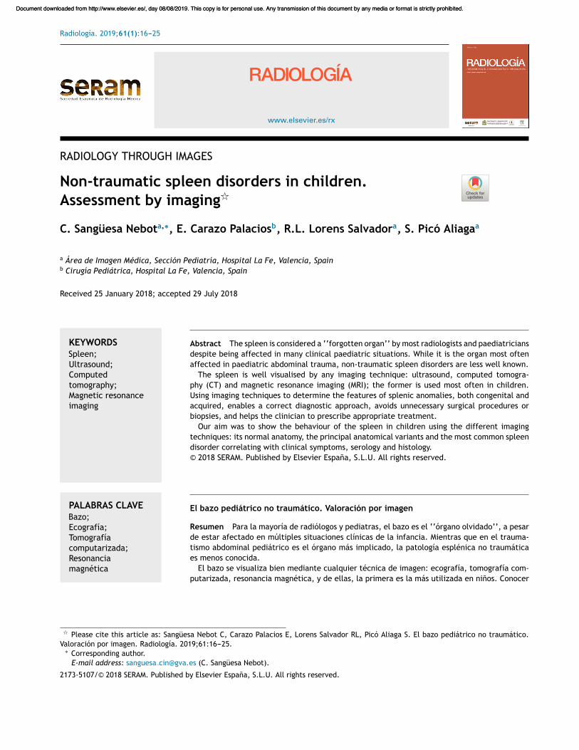
Figure 1 Normal spleen of a 5-year-old child. Abdomi-nal ultrasound performed at a hospital admission for acutepyelonephritis. Longitudinal image of the spleen (B), made witha high-frequency linear transducer, showing diffuse reticulon-odular pattern of the splenic parenchyma. LK: left kidney.
to characterise the lesions and to plan a conservative surgeryof the spleen when required.
Our aim is to show the behaviour of the spleen in thepaediatric age group with the different imaging techniques:its normal anatomy, the main anatomical variants and themost common non-traumatic splenic pathology, correlatingwith clinical, serology or histology.
Normal spleen
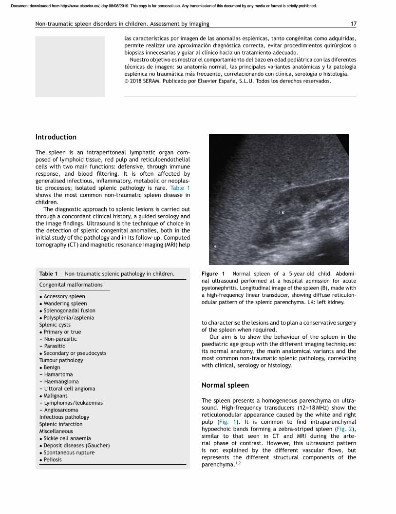
The spleen presents a homogeneous parenchyma on ultra-sound. High-frequency transducers (12---18 MHz) show thereticulonodular appearance caused by the white and rightpulp (Fig. 1). It is common to find intraparenchymalhypoechoic bands forming a zebra-striped spleen (Fig. 2),similar to that seen in CT and MRI during the arte-rial phase of contrast. However, this ultrasound patternis not explained by the different vascular flows, butrepresents the different structural components of theparenchyma.1,2
Document downloaded from http://www.elsevier.es/, day 08/08/2019. This copy is for personal use. Any transmission of this document by any media or format is strictly prohibited.Document downloaded from http://www.elsevier.es/, day 08/08/2019. This copy is for personal use. Any transmission of this document by any media or format is strictly prohibited.
18 C. Sangüesa Nebot et al.
Figure 2 ‘‘Zebra-striped’’ spleen. A 5-month-old girl whounderwent an abdominal ultrasound due to a urine infection.Longitudinal image of the spleen with convex transducer, show-ing hypoechoic bands crossing the splenic parenchyma. H:splenic hilum.
It must be remembered that: the ultrasound zebra-stripe pattern is normal in children, it should not beconfused with masses and does not require furtherstudies.
In CT, the spleen shows a mottled enhancement in thearterial phase due to the variability of the red pulp flows,and is homogenised in the portal phase. In MRI, the spleenof infants is hypointense in T2 and hyperintense in T1 inrelation to the liver, given its predominant composition ofred pulp. From 8 months of age, when the lymphoid tissueexpands and matures, it acquires the signal pattern similarto that of an adult: moderately hyperintense in T2 sequencesand hypointense in T1 with respect to the liver. The patternof enhancement in MRI is the same as that observed withCT.2---4
Congenital malformations
Accessory spleen
This consists of the presence of normal splenic tissue sepa-rated from the body of the spleen, produced by failure of thefusion of splenic embryonic buds in the dorsal mesogastrium.Usually less than 3 cm, it can be multiple. The most commonlocation is in the splenic hilum (75%), tail of the pancreas(20%) and splenorenal, splenogastric and splenophrenic lig-aments (5%).5 It has the same behaviour as the main spleenin all imaging techniques. After a splenectomy, the acces-sory spleen may increase in size and resume the functionof the removed spleen.5,6 When splenectomy is secondaryto haematologic disease (haemolytic anaemia or thrombo-cytopenic purpura), the presence and location of accessoryspleens is essential.
Wandering spleen
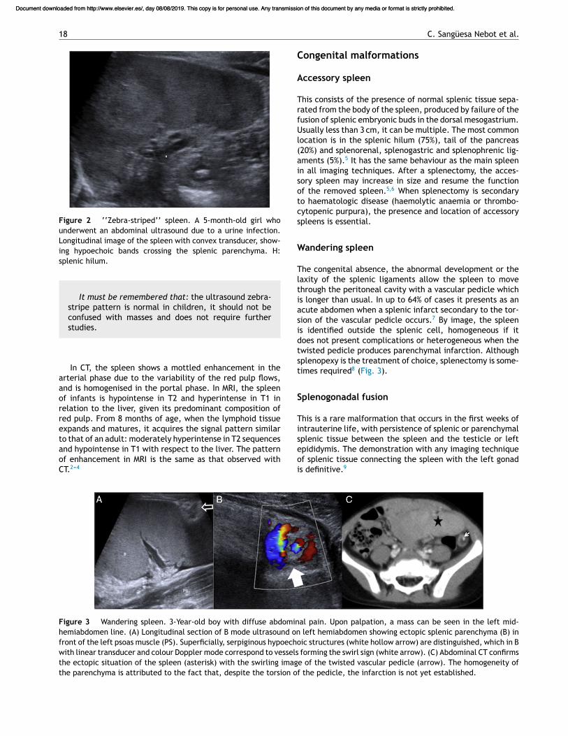
The congenital absence, the abnormal development or thelaxity of the splenic ligaments allow the spleen to movethrough the peritoneal cavity with a vascular pedicle whichis longer than usual. In up to 64% of cases it presents as anacute abdomen when a splenic infarct secondary to the tor-sion of the vascular pedicle occurs.7 By image, the spleenis identified outside the splenic cell, homogeneous if itdoes not present complications or heterogeneous when thetwisted pedicle produces parenchymal infarction. Althoughsplenopexy is the treatment of choice, splenectomy is some-times required8 (Fig. 3).
Splenogonadal fusion
This is a rare malformation that occurs in the first weeks ofintrauterine life, with persistence of splenic or parenchymalsplenic tissue between the spleen and the testicle or leftepididymis. The demonstration with any imaging techniqueof splenic tissue connecting the spleen with the left gonadis definitive.9
Figure 3 Wandering spleen. 3-Year-old boy with diffuse abdominal pain. Upon palpation, a mass can be seen in the left mid-hemiabdomen line. (A) Longitudinal section of B mode ultrasound on left hemiabdomen showing ectopic splenic parenchyma (B) infront of the left psoas muscle (PS). Superficially, serpiginous hypoechoic structures (white hollow arrow) are distinguished, which in Bwith linear transducer and colour Doppler mode correspond to vessels forming the swirl sign (white arrow). (C) Abdominal CT confirmsthe ectopic situation of the spleen (asterisk) with the swirling image of the twisted vascular pedicle (arrow). The homogeneity ofthe parenchyma is attributed to the fact that, despite the torsion of the pedicle, the infarction is not yet established.
Document downloaded from http://www.elsevier.es/, day 08/08/2019. This copy is for personal use. Any transmission of this document by any media or format is strictly prohibited.Document downloaded from http://www.elsevier.es/, day 08/08/2019. This copy is for personal use. Any transmission of this document by any media or format is strictly prohibited.
Non-traumatic spleen disorders in children. Assessment by imaging 19
Asplenia/polysplenia
They are part of the spectrum of anomalies known as het-erotaxy or cardiosplenic syndromes. Polysplenia presentsmultiple splenic nodules located in the right or left upperhypochondrium and is associated with abnormalities such asinterruption of the vena cava, atresia of the biliary tractor intestinal malrotation. Asplenia is the absence of splenictissue and is associated with serious cardiac anomalies. Chil-dren with asplenia suffer high mortality in the first year oflife.5
Splenic cysts
The prevalence of splenic cysts has increased with thewidespread use of ultrasound. Between 30% and 60% of casesare asymptomatic, and the most common complications arehaemorrhage, rupture and superinfection, most likely whenthe size exceeds 8 cm.10,11
Classically, they are divided into: type I, true or pri-mary, covered by epithelial cells, and type II, pseudocysts orsecondary, not covered by epithelium. Other classificationssuggest doing so according to their pathogenesis.10---12
Type I, true or primary
• Parasitic (hydatid): the most common in absolute num-bers, but not in our environment. Single or multiple, theirwall can calcify.
• Non-parasitic: they are more common in the paediatricage group. They can be congenital (epithelial, dermoid,epidermoid) and malformative (lymphatic malformation).
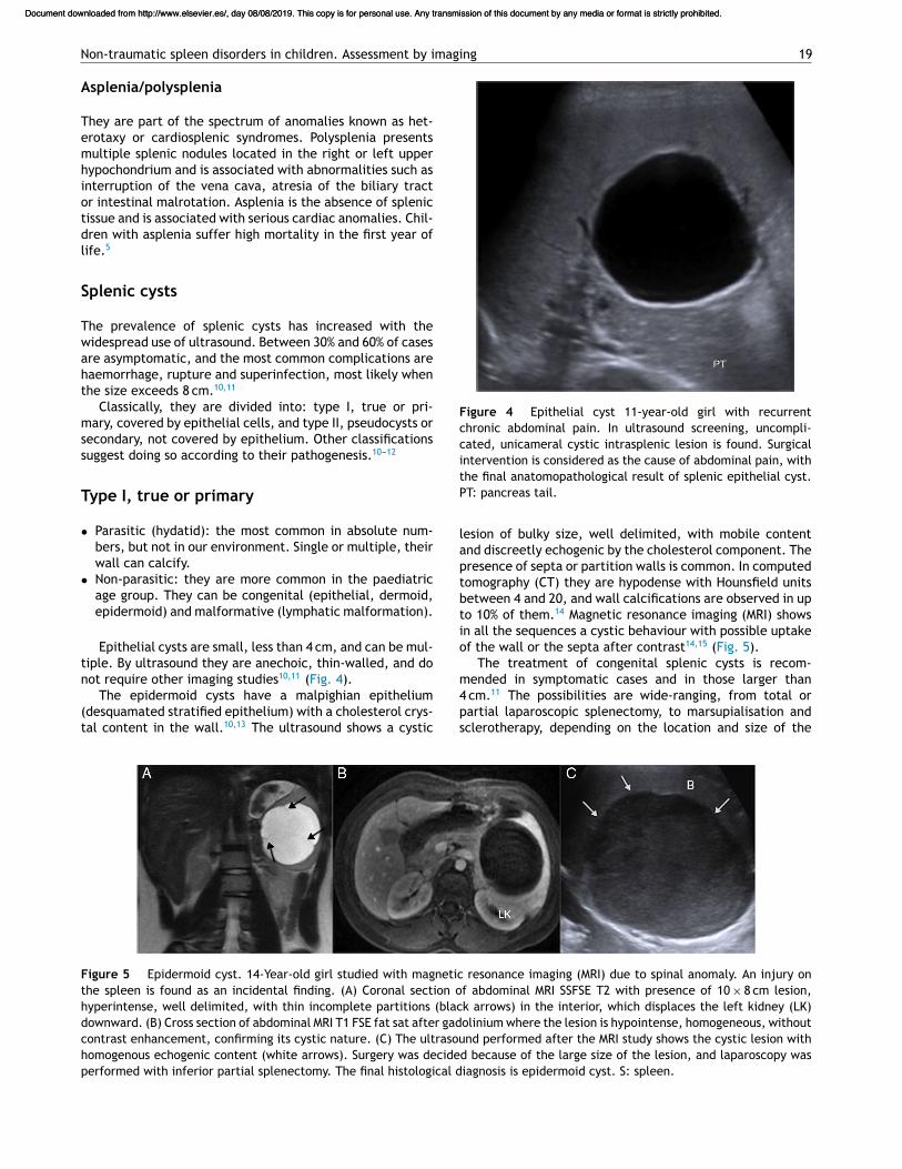
Epithelial cysts are small, less than 4 cm, and can be mul-tiple. By ultrasound they are anechoic, thin-walled, and donot require other imaging studies10,11 (Fig. 4).
The epidermoid cysts have a malpighian epithelium(desquamated stratified epithelium) with a cholesterol crys-tal content in the wall.10,13 The ultrasound shows a cystic
Figure 4 Epithelial cyst 11-year-old girl with recurrentchronic abdominal pain. In ultrasound screening, uncompli-cated, unicameral cystic intrasplenic lesion is found. Surgicalintervention is considered as the cause of abdominal pain, withthe final anatomopathological result of splenic epithelial cyst.PT: pancreas tail.
lesion of bulky size, well delimited, with mobile contentand discreetly echogenic by the cholesterol component. Thepresence of septa or partition walls is common. In computedtomography (CT) they are hypodense with Hounsfield unitsbetween 4 and 20, and wall calcifications are observed in upto 10% of them.14 Magnetic resonance imaging (MRI) showsin all the sequences a cystic behaviour with possible uptakeof the wall or the septa after contrast14,15 (Fig. 5).
The treatment of congenital splenic cysts is recom-mended in symptomatic cases and in those larger than4 cm.11 The possibilities are wide-ranging, from total orpartial laparoscopic splenectomy, to marsupialisation andsclerotherapy, depending on the location and size of the
Figure 5 Epidermoid cyst. 14-Year-old girl studied with magnetic resonance imaging (MRI) due to spinal anomaly. An injury onthe spleen is found as an incidental finding. (A) Coronal section of abdominal MRI SSFSE T2 with presence of 10 × 8 cm lesion,hyperintense, well delimited, with thin incomplete partitions (black arrows) in the interior, which displaces the left kidney (LK)downward. (B) Cross section of abdominal MRI T1 FSE fat sat after gadolinium where the lesion is hypointense, homogeneous, withoutcontrast enhancement, confirming its cystic nature. (C) The ultrasound performed after the MRI study shows the cystic lesion withhomogenous echogenic content (white arrows). Surgery was decided because of the large size of the lesion, and laparoscopy wasperformed with inferior partial splenectomy. The final histological diagnosis is epidermoid cyst. S: spleen.
Document downloaded from http://www.elsevier.es/, day 08/08/2019. This copy is for personal use. Any transmission of this document by any media or format is strictly prohibited.Document downloaded from http://www.elsevier.es/, day 08/08/2019. This copy is for personal use. Any transmission of this document by any media or format is strictly prohibited.
20 C. Sangüesa Nebot et al.
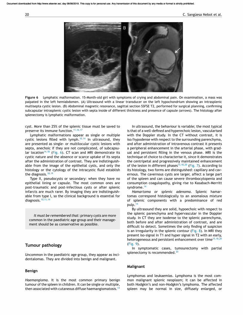
Figure 6 Lymphatic malformation. 15-Month-old girl with symptoms of crying and abdominal pain. On examination, a mass waspalpated in the left hemiabdomen. (A) Ultrasound with a linear transducer on the left hypochondrium showing an intrasplenicmultisepta cystic lesion. (B) Abdominal magnetic resonance, sagittal section SSFSE T2, performed for surgical planning, confirmingsubcapsular intrasplenic cystic lesion with septa inside of different thickness and presence of capsule (arrows). The histology aftersplenectomy is lymphatic malformation.
cyst. More than 25% of the splenic tissue must be saved topreserve its immune function.11,16,17
Lymphatic malformations appear as single or multiplecystic lesions filled with lymph.10,14 In ultrasound, theyare presented as single- or multilocular cystic lesions withsepta, anechoic if they are not complicated, of subcapsu-lar location14,18 (Fig. 6). CT scan and MRI demonstrate itscystic nature and the absence or scarce uptake of its septaafter the administration of contrast. They are indistinguish-able from the image of the epithelial cysts, and only thehistology or the cytology of the intracystic fluid establishthe diagnosis.18,19
Type II, pseudocysts or secondary: when they have noepithelial lining or capsule. The most common ones arepost-traumatic and post-infectious cysts or after splenicinfarcts are much rarer. By imaging they are indistinguish-able from type I, so the clinical background is essential fordiagnosis.10,11,14
It must be remembered that: primary cysts are morecommon in the paediatric age group and their manage-ment should be as conservative as possible.
Tumour pathology
Uncommon in the paediatric age group, they appear as inci-dentalomas. They are divided into benign and malignant.
Benign
Haemangioma. It is the most common primary benigntumour of the spleen in children. It can be single or multiple,then associated with cutaneous diffuse haemangiomatosis.14
In ultrasound, the behaviour is variable; the most typicalis that of a well-defined and hyperechoic lesion, vascularisedwith the Doppler study. In the CT without contrast, it isiso/hypodense with respect to the surrounding parenchyma,and after administration of intravenous contrast it presentsa peripheral enhancement in the arterial phase, with grad-ual and persistent filling in the venous phase. MRI is thetechnique of choice to characterise it, since it demonstratesthe centripetal and progressively maintained enhancementof the lesion in different phases15,18,20 (Fig. 7). According toits histology, two forms are distinguished: capillary and cav-ernous. The cavernous cysts are larger, affect a large partof the spleen and can cause severe thrombocytopenia andconsumption coagulopathy, giving rise to Kasabach-Merrittsyndrome.14
Hamartoma or splenic adenoma. Splenic hamar-tomas correspond histologically to an anomalous mixtureof splenic components with a predominance of redpulp.14
By ultrasound they are solid, hypoechoic with respect tothe splenic parenchyma and hypervascular in the Dopplerstudy. In CT they are isodense to the splenic parenchyma,both before and after administration of contrast, and aredifficult to detect. Sometimes the only finding of suspicionis an irregularity in the splenic contour (Fig. 8). In MRI theypresent iso-signal in T1 and hyper signal in T2 with an early,heterogeneous and persistent enhancement over time15,18,20
(Fig. 9).In symptomatic cases, tumourectomy with partial
splenectomy is recommended.20
Malignant
Lymphomas and leukaemias. Lymphoma is the most com-mon malignant splenic neoplasm; it can be affected inboth Hodgkin’s and non-Hodgkin’s lymphoma. The affectedspleen may be normal in size, diffusely enlarged, or
Document downloaded from http://www.elsevier.es/, day 08/08/2019. This copy is for personal use. Any transmission of this document by any media or format is strictly prohibited.Document downloaded from http://www.elsevier.es/, day 08/08/2019. This copy is for personal use. Any transmission of this document by any media or format is strictly prohibited.
Non-traumatic spleen disorders in children. Assessment by imaging 21
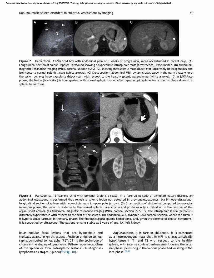
Figure 7 Hamartoma. 11-Year-old boy with abdominal pain of 3 weeks of progression, more accentuated in recent days. (A)Longitudinal section of colour Doppler ultrasound showing a hypoechoic intrasplenic mass (arrowheads), vascularised. (B) Abdominalmagnetic resonance imaging (MRI), coronal section SSFSE T2, showing intrasplenic mass (black star) discretely heterogeneous andisointense to normal splenic tissue (white arrows). (C) Cross section, abdominal MRI, dynamic LAVA study in the early phase wherethe lesion behaves hypervascularly (black star) with respect to the healthy splenic parenchyma (white arrows). (D) In LAVA latephase, the lesion (black star) is homogenised with normal splenic tissue. After laparoscopic splenectomy, the histological result issplenic hamartoma.
Figure 8 Hamartoma. 12-Year-old child with perianal Crohn’s disease. In a flare-up episode of an inflammatory disease, anabdominal ultrasound is performed that reveals a splenic lesion not detected in previous ultrasounds. (A) B-mode ultrasound;longitudinal section of spleen with hypoechoic mass in upper pole (arrow). (B) Cross section of abdominal computed tomographyin venous phase; the lesion is isodense to the normal splenic parenchyma and produces only a distortion in the contour of theorgan (short arrow). (C) Abdominal magnetic resonance imaging (MRI), coronal section SSFSE T2; the intrasplenic lesion (arrows) isdiscretely hyperintense with respect to the rest of the spleen. (D) Abdominal MRI, dynamic LAVA coronal section, where the tumouris hypervascular (arrows) in the early phase. The findings suggest splenic hamartoma, and, given the absence of clinical symptoms,it is controlled by ultrasound. The patient remains stable at 5 years of age. LK: left kidney.
have nodular focal lesions that are hypoechoic andtypically avascular on ultrasound. Positron emission tomog-raphy/computed tomography (PET/CT) is the technique ofchoice in the staging of lymphoma. Diffuse hypermetabolismof the spleen or focal intrasplenic lesions subcategoriseslymphomas as stages (Spleen)18 (Fig. 10).
Angiosarcoma. It is rare in childhood. It is presentedas a heterogeneous mass that in MRI is characteristicallyhypointense in T1 and T2 with respect to the healthyspleen, with intense contrast enhancement during the arte-rial phase, persisting in the venous phase and washing in thelate phase.20---22
Document downloaded from http://www.elsevier.es/, day 08/08/2019. This copy is for personal use. Any transmission of this document by any media or format is strictly prohibited.Document downloaded from http://www.elsevier.es/, day 08/08/2019. This copy is for personal use. Any transmission of this document by any media or format is strictly prohibited.
22 C. Sangüesa Nebot et al.
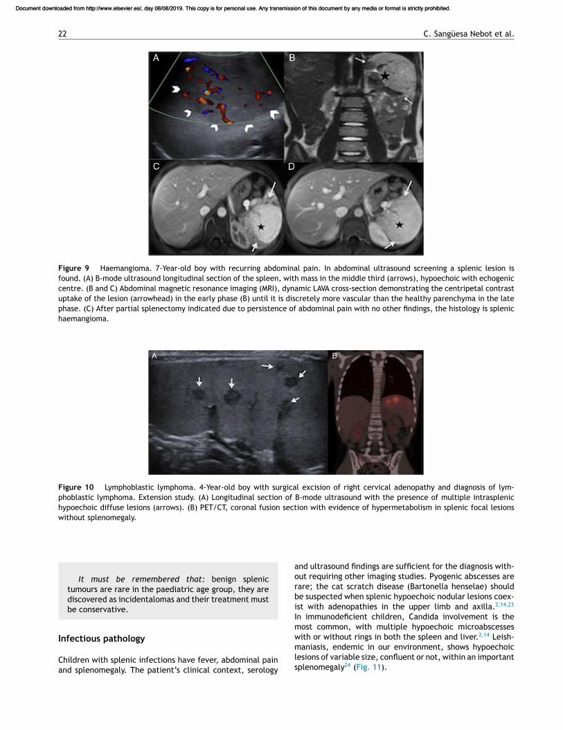
Figure 9 Haemangioma. 7-Year-old boy with recurring abdominal pain. In abdominal ultrasound screening a splenic lesion isfound. (A) B-mode ultrasound longitudinal section of the spleen, with mass in the middle third (arrows), hypoechoic with echogeniccentre. (B and C) Abdominal magnetic resonance imaging (MRI), dynamic LAVA cross-section demonstrating the centripetal contrastuptake of the lesion (arrowhead) in the early phase (B) until it is discretely more vascular than the healthy parenchyma in the latephase. (C) After partial splenectomy indicated due to persistence of abdominal pain with no other findings, the histology is splenichaemangioma.
Figure 10 Lymphoblastic lymphoma. 4-Year-old boy with surgical excision of right cervical adenopathy and diagnosis of lym-phoblastic lymphoma. Extension study. (A) Longitudinal section of B-mode ultrasound with the presence of multiple intrasplenichypoechoic diffuse lesions (arrows). (B) PET/CT, coronal fusion section with evidence of hypermetabolism in splenic focal lesionswithout splenomegaly.
It must be remembered that: benign splenictumours are rare in the paediatric age group, they arediscovered as incidentalomas and their treatment mustbe conservative.
Infectious pathology
Children with splenic infections have fever, abdominal painand splenomegaly. The patient’s clinical context, serology
and ultrasound findings are sufficient for the diagnosis with-out requiring other imaging studies. Pyogenic abscesses arerare; the cat scratch disease (Bartonella henselae) shouldbe suspected when splenic hypoechoic nodular lesions coex-ist with adenopathies in the upper limb and axilla.2,14,23
In immunodeficient children, Candida involvement is themost common, with multiple hypoechoic microabscesseswith or without rings in both the spleen and liver.2,14 Leish-maniasis, endemic in our environment, shows hypoechoiclesions of variable size, confluent or not, within an importantsplenomegaly24 (Fig. 11).
Document downloaded from http://www.elsevier.es/, day 08/08/2019. This copy is for personal use. Any transmission of this document by any media or format is strictly prohibited.Document downloaded from http://www.elsevier.es/, day 08/08/2019. This copy is for personal use. Any transmission of this document by any media or format is strictly prohibited.
Non-traumatic spleen disorders in children. Assessment by imaging 23
Figure 11 Infectious pathology. (A) Candidiasis. 3-Year-old girl with pancytopenia and fever. Longitudinal section of colour Dopplerultrasound on the spleen showing splenomegaly with the presence of diffuse hypoechoic millimetric lesions maintaining the vas-cular architecture. (B) Leishmaniasis. A 5-month-old boy with a fever lasting 7 days and splenomegaly on physical examination.Longitudinal section of B-mode ultrasound of the spleen with the presence of multiple confluent hypoechoic lesions of differentsizes, with involvement of even the accessory spleen (asterisk). IgM serology positive for leishmania. LK: left kidney.
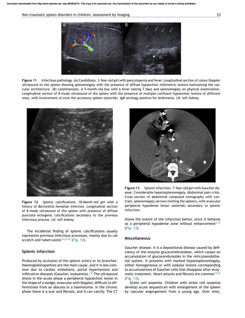
Figure 12 Splenic calcifications. 18-Month-old girl with ahistory of Bartonella henselae infection. Longitudinal sectionof B-mode ultrasound of the spleen with presence of diffusepunctate echogenic calcifications secondary to the previousinfectious process. LK: left kidney.
The incidental finding of splenic calcifications usuallyrepresents previous infectious processes, mainly due to catscratch and tuberculosis14,23,25 (Fig. 12).
Splenic infarction
Produced by occlusion of the splenic artery or its branches.Haemoglobinopathies are the main cause, and it is less com-mon due to cardiac embolisms, portal hypertension andinfiltrative diseases (Gaucher, leukaemia).2,8 The ultrasoundshows in the acute phase a peripheral hypoechoic lesion inthe shape of a wedge, avascular with Doppler, difficult to dif-ferentiate from an abscess or a haematoma. In the chronicphase there is a scar and fibrosis, and it can calcify. The CT
Figure 13 Splenic infarction. 7-Year-old girl with Gaucher dis-ease. Considerable hepatosplenomegaly. Abdominal pain crisis.Cross section of abdominal computed tomography with con-trast, splenomegaly (arrows limiting the spleen), with avascularperipheral hypodense lesion (asterisk) secondary to splenicinfarction.
shows the extent of the infarction better, since it behavesas a peripheral hypodense zone without enhancement2,14
(Fig. 13).
Miscellaneous
Gaucher disease. It is a depositional disease caused by defi-ciency of the enzyme glucocerebrosidase, which causes anaccumulation of glucocerebrosides in the reticuloendothe-lial system. It presents with marked hepatosplenomegaly,either homogeneous or with nodular lesions correspondingto accumulations of Gaucher cells that disappear after enzy-matic treatment. Heart attacks and fibrosis are common14,26
(Fig. 13).Sickle cell anaemia. Children with sickle cell anaemia
develop acute sequestrum with enlargement of the spleenby vascular engorgement from a young age. Over time,
Document downloaded from http://www.elsevier.es/, day 08/08/2019. This copy is for personal use. Any transmission of this document by any media or format is strictly prohibited.Document downloaded from http://www.elsevier.es/, day 08/08/2019. This copy is for personal use. Any transmission of this document by any media or format is strictly prohibited.
24 C. Sangüesa Nebot et al.
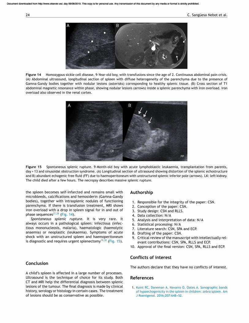
Figure 14 Homozygous sickle cell disease. 9-Year-old boy, with transfusions since the age of 2. Continuous abdominal pain crisis.(A) Abdominal ultrasound, longitudinal section of spleen with diffuse heterogeneity of the parenchyma due to the presence ofGamna-Gandy bodies together with nodular lesions (asterisks) corresponding to healthy splenic tissue. (B) Cross section of T1abdominal magnetic resonance within phase, showing nodular lesions (arrows) inside a splenic parenchyma with iron overload. Ironoverload also observed in the renal cortex.
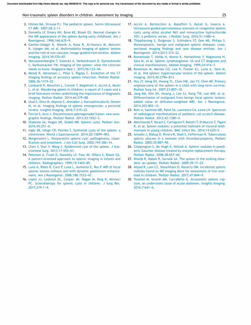
Figure 15 Spontaneous splenic rupture. 9-Month-old boy with acute lymphoblastic leukaemia, transplantation from parents,day + 13 and sinusoidal obstruction syndrome. (A) Longitudinal section of ultrasound showing distortion of the splenic echostructureand B) abundant echogenic free fluid (FF) due to haemoperitoneum with unstructured splenic inferior pole (arrows). LK: left kidney.The child died after a few hours. The necropsy describes massive splenic rupture.
the spleen becomes self-infarcted and remains small withmicrobleeds, calcifications and hemosiderin (Gamna-Gandybodies), together with intrasplenic nodules of functioningparenchyma. If there is transfusion treatment, MRI showsiron overload with a drop in spleen signal for in and out ofphase sequences27,28 (Fig. 14).
Spontaneous splenic rupture. It is very rare. Italways occurs in a pathological spleen: infectious (infec-tious mononucleosis, malaria), haematologic (haemolyticanaemia) or neoplastic (leukaemia). Symptoms of acuteshock with an unstructured spleen and haemoperitoneumis diagnostic and requires urgent splenectomy19,30 (Fig. 15).
Conclusion
A child’s spleen is affected in a large number of processes.Ultrasound is the technique of choice for its study. BothCT and MRI help the differential diagnosis between spleniclesions of the tumour. The final diagnosis is made by clinicalhistory, serology or histology in certain cases. The treatmentof lesions should be as conservative as possible.
Authorship
1. Responsible for the integrity of the paper: CSN.2. Conception of the paper: CSN.3. Study design: CSN and RLLS.4. Data collection: N/A5. Analysis and interpretation of data: N/A6. Statistical processing: N/A7. Literature search: CSN, SPA and ECP.8. Drafting of the paper: CSN.9. Critical review of the manuscript with intellectually rel-
evant contributions: CSN, SPA, RLLS and ECP.10. Approval of the final version: CSN, SPA, RLLS and ECP.
Conflicts of interest
The authors declare that they have no conflicts of interest.
References
1. Kuint RC, Daneman A, Navarro O, Oates A. Sonographic bandsof hypoechogenicity in the spleen in children: zebra spleen. AmJ Roentgenol. 2016;207:648---52.
Document downloaded from http://www.elsevier.es/, day 08/08/2019. This copy is for personal use. Any transmission of this document by any media or format is strictly prohibited.Document downloaded from http://www.elsevier.es/, day 08/08/2019. This copy is for personal use. Any transmission of this document by any media or format is strictly prohibited.
Non-traumatic spleen disorders in children. Assessment by imaging 25
2. Hilmes MA, Strouse PJ. The pediatric spleen. Semin UltrasoundCT MRI. 2007;28:3---11.
3. Donnelly LF, Emery KH, Bove KE, Bisset GS. Normal changes inthe MR appearance of the spleen during early childhood. Am JRoentgenol. 1996;166:635---9.
4. Gaetke-Udager K, Wasnik A, Kaza R, Al-Hawary M, MaturenK, Udager AM, et al. Multimodality Imaging of splenic lesionsand the role of non-vascular, image-guided intervention. AbdomImaging. 2014;39:570---87.
5. Vancauwenberghe T, Snoeckx A, Vanbeckvoort D, DymarkowskiS, Vanhoenacker FM. Imaging of the spleen: what the clinicianneeds to know. Singapore Med J. 2015;56:133---44.
6. Mendi R, Abramson L, Pillai S, Rigsby C. Evolution of the CTImaging findings of accesory spleen infarction. Pediatr Radiol.2006;36:1319---22.
7. Lombardi R, Menchini L, Corneli T, Magistreli A, Accini A, MontiL, et al. Wandering spleen in children: a report of 3 cases and abrief literature review underlining the importance of diagnosticimaging. Pediatr Radiol. 2014;44:279---88.
8. Unal E, Onur M, Akpinar E, Ahmadov J, Karcaaltincaba M, OzmenM, et al. Imaging findings of splenic emergencias: a pictorialreview. Insights Imaging. 2016;7:215---22.
9. Ferron S, Arce J. Discontinuous splenogonadal fusion: new sono-graphic findings. Pediatr Radiol. 2013;43:1652---5.
10. Shabtaie SA, Hogan AR, Slidell MB. Splenic cysts. Pediatr Ann.2016;45:251---6.
11. Ingle SB, Hinge CR, Patrike S. Epithelial cysts of the spleen: aminireview. World J Gastroenterol. 2014;20:13899---903.
12. Morgenstern L. Nonparasitic splenic cyst: pathogenesis, classi-fication and treatment. J Am Coll Surg. 2002;194:306---14.
13. Chen Y, Shyr Y, Wang S. Epidermoid cyst of the spleen. J Gas-trointest Surg. 2013;17:555---61.
14. Paterson A, Frush D, Donnelly LF, Foss JN, OHara S, Bisset GS.A pattern-oriented approach to splenic imaging in infants andchildren. Radiographics. 1999;19:1465---85.
15. Luna A, Ribes R, Caro P, Luna L, Aumente E, Ros P. MRI of focalsplenic lesions without and with dynamic gadolinium enhance-ment. Am J Roentgenol. 2006;186:1533---47.
16. Lopez JJ, Lodwick DL, Cooper JN, Hogan M, King D, MinneciPC. Sclerotherapy for splenic cysts in children. J Surg Res.2017;219:1---4.
17. Accini A, Bertocchini A, Masafferi S, Natali G, Inserra A.Ultrasound-guided percutaneous sclerosis of congenital spleniccysts using ethyl alcohol 96% and minocycline hydrochloride10%: a pediatric series. J Pediatr Surg. 2016;51:1480---4.
18. Thipphavong S, Duigenan S, Schindera ST, Gee MS, Philips S.Nonneoplastic, benign and malignant splenic diseases: cross-sectional imaging findings and rare disease entities. Am JRoentgenol. 2014;2013:315---22.
19. Komatsuda T, Ishida H, Konno K, Hamashima Y, Naganuma H,Sato M, et al. Splenic lymphangioma: US and CT diagnosis andclinical manifestations. Abdom Imaging. 1999;24:414---7.
20. Bowerson M, Menias CO, Lee K, Fowler KJ, Luna A, Yano M,et al. Hot spleen: hypervascular lesions of the spleen. AbdomImaging. 2015;40:2796---813.
21. Hsu JT, Ueng SH, Hwang TL, Chen HM, Jan YY, Chen MF. Primaryangiosarcoma of the spleen in a child with long-term survival.Pediatr Surg Int. 2007;23:807---10.
22. Jang KM, Kim SH, Hwang J, Lee SJ, Kang TW, Lee MW, et al.Differentiation of malignant from benign focal splenic lesions:added value of diffusion-weighted MRI. Am J Roentgenol.2014;203:803---12.
23. Rohr A, Saettele MR, Patel SA, Lawrence CA, Lowe LH. Spectrumof radiological manifestations of pediatric cat-scratch disease.Pediatr Radiol. 2012;42:1380---4.
24. Melchionda F, Varani S, Carfagnini F, Belotti T, Di Muccio T, TiganiR, et al. Spleen nodules: a potential hallmark of visceral leish-maniasis in young children. BMC Infect Dis. 2014;14:620---5.
25. Amodio J, Biskup D, Rivera R, Shah S, Fefferman N. Tuberculosissplenic abscess in a neonate with thrombocytopenia. PediatrRadiol. 2005;35:887---90.
26. Chippington S, Mc Hugh K, Vellodi A. Splenic nodules in paedi-atric Gaucher disease treated by enzyme replacement therapy.Pediatr Radiol. 2008;38:657---60.
27. Khatib R, Rabah R, Sarnaik SA. The spleen in the sickling disor-ders: an update. Pediatr Radiol. 2009;39:17---22.
28. Ahyad R, Lam CZ, Shearkhani O, Navarro OM. Incidental splenicnodules found on MR imaging done for assessment of iron over-load in children. Pediatr Radiol. 2017;47:844---9.
30. Tonolini M, Ierardi AM, Carrafiello G. Atraumatic splenic rup-ture, an underrated cause of acute abdomen. Insights Imaging.2016;7:641---6.
Document downloaded from http://www.elsevier.es/, day 08/08/2019. This copy is for personal use. Any transmission of this document by any media or format is strictly prohibited.Document downloaded from http://www.elsevier.es/, day 08/08/2019. This copy is for personal use. Any transmission of this document by any media or format is strictly prohibited.