Embed Size (px)
Citation preview

RESEARCH ARTICLE Open Access
Nonhypoxic regulation and role ofhypoxia-inducible factor 1 in aromataseinhibitor resistant breast cancerArmina A Kazi1,5, Rabia A Gilani1, Amanda J Schech1, Saranya Chumsri2,3, Gauri Sabnis1,3, Preeti Shah1,3,Olga Goloubeva3,4, Shari Kronsberg4 and Angela H Brodie1,3,6*
Abstract
Introduction: Although aromatase inhibitors (AIs; for example, letrozole) are highly effective in treating estrogenreceptor positive (ER+) breast cancer, a significant percentage of patients either do not respond to AIs or becomeresistant to them. Previous studies suggest that acquired resistance to AIs involves a switch from dependence onER signaling to dependence on growth factor-mediated pathways, such as human epidermal growth factorreceptor-2 (HER2). However, the role of HER2, and the identity of other relevant factors that may be used asbiomarkers or therapeutic targets remain unknown. This study investigated the potential role of transcriptionfactor hypoxia inducible factor 1 (HIF-1) in acquired AI resistance, and its regulation by HER2.
Methods: In vitro studies using AI (letrozole or exemestane)-resistant and AI-sensitive cells were conducted toinvestigate the regulation and role of HIF-1 in AI resistance. Western blot and RT-PCR analyses were conductedto compare protein and mRNA expression, respectively, of ERα, HER2, and HIF-1α (inducible HIF-1 subunit) inAI-resistant versus AI-sensitive cells. Similar expression analyses were also done, along with chromatinimmunoprecipitation (ChIP), to identify previously known HIF-1 target genes, such as breast cancer resistanceprotein (BCRP), that may also play a role in AI resistance. Letrozole-resistant cells were treated with inhibitors toHER2, kinase pathways, and ERα to elucidate the regulation of HIF-1 and BCRP. Lastly, cells were treated withinhibitors or inducers of HIF-1α to determine its importance.
Results: Basal HIF-1α protein and BCRP mRNA and protein are higher in AI-resistant and HER2-transfected cellsthan in AI-sensitive, HER2- parental cells under nonhypoxic conditions. HIF-1α expression in AI-resistant cells is likelyregulated by HER2 activated-phosphatidylinositide-3-kinase/Akt-protein kinase B/mammalian target of rapamycin(PI3K/Akt/mTOR) pathway, as its expression was inhibited by HER2 inhibitors and kinase pathway inhibitors.Inhibition or upregulation of HIF-1α affects breast cancer cell expression of BCRP; AI responsiveness; andexpression of cancer stem cell characteristics, partially through BCRP.
Conclusions: One of the mechanisms of AI resistance may be through regulation of nonhypoxic HIF-1 target genes,such as BCRP, implicated in chemoresistance. Thus, HIF-1 should be explored further for its potential as a biomarkerof and therapeutic target.
* Correspondence: [email protected] of Pharmacology and Experimental Therapeutics, University ofMaryland, Baltimore, MD 21201, USA3School of Medicine, University of Maryland Marlene and StewartGreenebaum Cancer Center, Baltimore, MD 21201, USAFull list of author information is available at the end of the article
© 2014 Kazi et al.; licensee BioMed Central Ltd. This is an Open Access article distributed under the terms of the CreativeCommons Attribution License (http://creativecommons.org/licenses/by/2.0), which permits unrestricted use, distribution, andreproduction in any medium, provided the original work is properly credited.
Kazi et al. Breast Cancer Research 2014, 16:R15http://breast-cancer-research.com/content/16/1/R15

IntroductionBreast cancer is the most prevalent form of cancer amongwomen in the United States and second leading cause ofcancer related deaths [1]. Approximately 70% to 80% ofbreast cancers express estrogen receptor (ER+) and, con-sequently, are estrogen-dependent in their growth. Endo-crine/hormonal therapies have proven effective in treatingER + breast cancers. Selective estrogen receptor modula-tors (SERMS), such as tamoxifen, inhibit estrogen actionon breast cancer cells by blocking ER + signaling. Alterna-tively, aromatase inhibitors (AIs; for example, letrozole,anastrozole, and exemestane) reduce circulating levels ofestrogen by inhibiting the conversion of androgens to es-trogen by the enzyme aromatase [2,3]. Comparing the effi-cacy of tamoxifen versus AIs, a number of clinical studieshave shown that AIs are superior in terms of disease freesurvival, time to recurrence and prevention of contralat-eral breast cancer [4,5]. In the adjuvant setting, AIs areless toxic with minimal adverse effects compared tochemotherapy and provide protection against develop-ment of contralateral breast cancer. AIs are now first-linetreatments for ER + breast cancer in post-menopausalwomen [6]. However, a significant percentage (range 30%to 65%) of patients either does not respond to AIs [7] orbecomes resistant to them [8-10].Studies from this lab and others suggest that resistance
to AIs occurs after a switch from dependence on ER sig-naling to dependence on growth factor-mediated path-ways, such as human epidermal growth factor receptor-2(HER2), a member of the membrane epidermal growthfactor receptor (EGFR) family of receptor tyrosine ki-nases, and insulin-like growth factor receptor (IGFR)[9-11]. Pre-clinical [12] and clinical [10] studies have ex-plored HER2 inhibitors, trastuzumab and lapatinib, astreatments for letrozole-resistant breast cancer. Pre-clinically, our laboratory has shown that trastuzumabalone or in combination with letrozole decreased HER2expression, restored ERα expression, and inhibitedtumor growth of MCF-7Ca xenografts that became resist-ant to letrozole [13]. Clinically, it has been shown thatlapatinib in combination with letrozole significantly in-creased progression-free survival in patients versus letro-zole alone as first-line therapy for hormone receptor- andHER2-positive postmenopausal metastatic breast cancer[10,14]. However, studies with de novo HER2+ breast can-cer (that is, not HER2+ breast cancer of acquired AI re-sistance) indicate that resistance can develop to HER2inhibitors as well [15,16]. Thus, although it has yet to bestudied, there may be a risk of developing resistance tosecond-line HER2 inhibitor therapy in patients who havealready acquired resistance to first-line AI therapy. As amembrane receptor, HER2 can affect many cellular path-ways, some of which may not be directly involved in thedevelopment of AI resistance. Targeting another factor
downstream of HER2 that more directly mediates effectsspecific and essential to the development of AI resistancemay be as effective as targeting HER2 itself, while not hav-ing the same level of risk of producing second-line ac-quired resistance. Currently, the mechanism by whichHER2 is involved in AI resistance remains unclear. It is,therefore, important to: 1) further elucidate the HER2-mediated pathway that contributes to AI resistance, par-ticularly characteristics associated with AI resistant breastcancer cells; and 2) identify other potential factors in-volved that may serve as novel molecular biomarkers andtherapeutic targets.One factor that may be involved in HER2-mediated AI
resistance is HIF-1, a heterodimeric transcription factormade up of an inducible alpha (α) subunit and a consti-tutively expressed beta (β) subunit [17]. HIF-1α is nor-mally kept low in cells by proteosomal degradation, butlack of sufficient oxygen levels (hypoxia, for example, 1%to 2% O2) prevents this degradation. This leads to in-creased intracellular HIF-1α protein levels, formation ofHIF-1, and activation of HIF-1 target genes important forcell survival, metabolic adaptation and angiogenesis. Inter-estingly, HIF-1 expression and/or activation can also beregulated by growth factors, hormones and cytokines in-dependent of O2 levels. For example, ERα- and HIF-1-mediated signaling pathways are known to interact antag-onistically [18,19] and cooperatively [20-23]. EGFR andHER2, as well as kinase signaling pathways, such as theMAPK and PI3K/Akt pathways, have also been shown toregulate HIF-1α expression and activity [22,24,25].The role of hypoxia-regulated HIF-1 in cancer has
been well studied. This is particularly relevant to sizabletumors whose cancer cells are too distant from existingblood vessels to get enough oxygen and nutrients [26].Hypoxia and/or HIF-1 have been implicated in increasedpatient mortality and disease progression [27]. Their in-volvement in tumor formation and metastasis [28,29],and regulation of cancer stem cells [28,30] and stem cellmarkers, such as breast cancer resistant protein (BCRP)[27,30,31], has also been demonstrated. However, nonhy-poxic regulation of HIF-1 and its importance in cancerremains largely unknown. Specific to this study, theregulation and role of nonhypoxic HIF-1 in breast can-cer cell resistance to AIs, specifically letrozole, has yet tobe explored. Using a letrozole-resistant cell line devel-oped from xenograft tumors in our laboratory, thiscurrent study tested the overall hypothesis that nonhy-poxic HIF-1 is an essential factor in HER2-mediated letro-zole resistance. More specifically, in letrozole-responsivetumors, the switch from ERα to HER2-dependent signal-ing increases HIF-1α expression, independent of nutrientor oxygen availability. HIF-1 then acts as a key transcrip-tion factor activating target genes involved in processesthat promote letrozole resistance.
Kazi et al. Breast Cancer Research 2014, 16:R15 Page 2 of 18http://breast-cancer-research.com/content/16/1/R15

MethodsCells and reagentsCell cultureCell lines (and their ER/HER2 status) used are listed inTable 1. MCF-7Ca cells obtained from the laboratory of Dr.Chen through institutional agreement (City of Hope,Duarte, CA, USA) are MCF-7 cells stably transfected withthe human placental aromatase gene [32,33]. Aromatase isthe enzyme that converts androgens to estrogen. Transfec-tion of the aromatase gene does not affect ER/HER2 status,ER activation, or estrogen-dependence of MCF-7 cells, butit does allow MCF-7 cells to proliferate in response to an-drogens (for example, androstenedione) and be sensitive togrowth inhibitor effects of aromatase inhibitors [33]. MCF-7Ca cells were maintained in (D)MEM 1× high glucose(Invitrogen, Grand Island, NY) supplemented with 5% fetalbovine serum (FBS), 1% penicillin/streptomycin (P-S), and700 μg/mL geneticin selective antibiotic (G418). Long-term letrozole-treated (LTLTCa) cells, developed in ourlaboratory [34], are letrozole-resistant breast cancer cellsisolated from MCF-7Ca mouse xenograft tumors treatedfor 56 weeks with letrozole and that have become resistantto the growth inhibitory effects of letrozole. LTLTCa cellswere maintained in phenol red–free (PRF) modifiedImproved Minimal Essential Media (IMEM) (Invitrogen,Grand Island, NY) supplemented with 5% charcoaldextran-treated FBS (CDT-FBS), 1% P-S, 750 μg/mL G418,and 1 μM letrozole. LTLTCa cells can be compared toMCF-7Ca cells in experiments since they originated fromthe MCF-7Ca cell population and their expression of ERαand HER2 returns to levels similar to that of MCF-7Ca cellsafter letrozole withdrawal [35]. MCF-7 cells obtained fromAmerican Type Culture Collection (ATCC) were main-tained in (D)MEM 1× high glucose (Invitrogen, GrandIsland, NY) supplemented with 5% FBS and 1% P-S. Hc7cells were developed in our laboratory; they are MCF-7cells transfected with the HER2 gene and overexpressHER2 [36]. Hc7 cells were maintained in (D)MEM 1× highglucose (Invitrogen, Grand Island, NY) supplemented with5% FBS, 1% P-S, and 500 μg/ml hygromycin. AC1 cells are
another set of MCF-7 cells stably transfected with the hu-man placental aromatase gene [37]. Similar to MCF-7Cacells, AC1 cells are ER+/HER2-, proliferate in response toestrogen or androstenedione, express the aromatase en-zyme and are sensitive to aromatase inhibitors [38]. AC1cells, however, were created in our laboratory rather thanDr. Chen’s, and they express higher levels of aromatase(data not shown). AC1 cells are maintained in (D)MEM1x high glucose (Invitrogen, Grand Island, NY) supple-mented with 5% FBS, 1% P-S, and 800 μg/mL G418.AC1-exemestane resistant (AC1-ExR) cells, developedin our laboratory, are exemestane-resistant cells isolatedfrom AC1 mouse xenograft tumors treated for approxi-mately 10 weeks with exemestane maintained in PRFmodified IMEM (Invitrogen, Grand Island, NY) supple-mented with 5% CDT-FBS, 1% P-S, 800 μg/mL G418, and5 μM exemestane.For experiments determining the effect of oxygen ten-
sion on protein expression, MCF-7Ca and LTLTCa cellswere plated in passage media and incubated either undernormal (20% O2 at 37°C) or more physiological (5% O2;using a hypoxia chamber) cell culture conditions for24 hours. MCF-7 cells used to generate MC-7Ca andAC1 cells were obtained from the ATCC and, thus, didnot require ethical approval or patient consent.
ReagentsThe following drugs were used: letrozole (Novartis,NY, USA); lapatinib (GlaxoSmithKline Pharmaceutical,Brentford, Middlesex, United Kingdom); trastuzumab(Genentech, San Francisco, CA); exemestane (Pfizer, NY,USA); cycloheximide (#C1988), actinomycin D (#A9415),and cobalt chloride (CoCl2; #C8661) (all from Sigma, St.Louis, MO). The following antibodies were used inwestern blot analyses: HER2 (#04-1127) and BCRP(EMD Millipore, Billerica, MA); HIF-1α (#610959; BDBiosciences); ERα (#8644; Cell Signaling Technology,Danvers, MA); phosphorylated and total ERK1/2, Akt(#4058 and #4685), mTOR (#2971 and #2972) and p70 S6kinase (#9205 and #9202) all from Cell signaling Technology,Danvers, MA); and β-actin (#4970; Cell Signaling Technology,Danvers, MA).
Western blot analysisPlated cells were washed with ice-cold PBS and then lysedwith radioimmunoprecipitation (RIPA) buffer containingprotease (#11836145001) and phosphatase inhibitors(#4906837001) (both from Roche Applied Sciences, Indiana-polis, IN) by sonication and incubation for 20 minutes at4°C. Lysed samples were centrifuged at 14,000 rpm for20 minutes at 4°C to collect protein lysates (supernatant). Atotal of 10 to 40 μg of protein underwent 10% SDS–poly-acrylamide gel electrophoresis (SDS-PAGE) and was trans-ferred to a polyvinylidene difluoride membrane (PVDF;
Table 1 ERα and HER2 status and aromatase inhibitor-sensitivity of cell lines used
Cell line ERα status HER2 status AI-sensitivity
MCF-7Ca + - Yes
LTLT-Ca - + No (to letrozole)
MCF7 + - Yes
MCF7 / HER2 + + ND
AC1 + - Yes
AC1-Ex R + + No (to exemestane)
ERα, estrogen receptor alpha; HER2, human epidermal growth factorreceptor 2; ND: not determined.
Kazi et al. Breast Cancer Research 2014, 16:R15 Page 3 of 18http://breast-cancer-research.com/content/16/1/R15

#IPVH00010 Millipore, Billerica, MA). The resulting blotswere probed with specific mouse or rabbit primary anti-bodies and either goat anti-mouse or –rabbit secondaryantibodies conjugated to horseradish peroxidase (#17 2–1011 and#172-1019; Biorad, Hercules, CA), respectively.Blots were developed using SuperSignal West Pico Chemi-luminescent Substrate (#34080; Thermo Scientific,Waltham, MA). Blots that were to be re-probed werestripped with Restore Western Blot Stripping Buffer(#21059; Thermo Scientific, Waltham, MA) for 40 minutesat room temperature prior to incubation with another pri-mary antibody. Densitometry was performed on each blotusing either ImageQuant or ImageJ.
Reverse transcripase-polymerase chain rectionRNA extraction and reverse transcription (RT)RNA was extracted and purified using the RNeasy MiniKit (#74104; Qiagen, Valencia, CA). RNA was reversetranscribed to complementary DNA (cDNA) using 200Uof Moloney murine leukemia virus reverse transcriptase(#28025013; Invitrogen, Grand Island, NY) and incubat-ing at 37°C for one hour.
Real-time PCRmRNA expression analyses were carried out by real-timePCR using a DNA Opticon system (MJ Research,Waltham, MA) and using DyNAmo SYBR green qPCRmix (New England Biolabs, Ipswich, MA). Standardcurves were generated by serially diluting the sample ex-pected to have the most amount of the PCR product.The yield of product for each unknown sample was cal-culated by applying its threshold cycle, or C(T), value tothe standard curve using the Opticon Monitor analysissoftware (version 1.01, MJ Research, Waltham, MA).Values were normalized to corresponding 18S rRNAvalues and expressed as the fold increase relative to con-trols. Primers for HER2, HIF-1α, BCRP, GAPDH, BMI-1,Nanog and TWIST were obtained from (Sigma, St. Louis,MO or Qiagen Valencia, CA).
Chromatin immunoprecipitation assayFor the in vitro chromatin immunoprecipitation(ChIP) assay, the treated cells were washed withDPBS and fixed with 1% formaldehyde/dulbecco'sphosphate buffered saline (DPBS) for 10 minutes at37°C, after which the cells were washed with ice-coldDPBS containing protease inhibitors. The cells werecollected into 1 mL DPBS and pelleted by centrifuga-tion at 6,000 rpm for five minutes at 4°C. The cellpellet was resuspended in nuclear lysis buffer (ChIPkit, #17–295 Millipore, Billerica, MA) and incubatedon ice for 15 minutes. Samples were sonicated on icefor 7 × 10-second cycles, with 20-second pauses be-tween each cycle. The sonicated samples were
centrifuged at 14,000 rpm for 10 minutes at 4°C. Thesonicated samples were diluted 1:10 with dilution buf-fer (ChIP kit) before being immunocleared in a solu-tion containing protein A- or G-Sepharose slurry(#16-156 and #16-266, respectively; Millipore, Billerica,MA) in Tris/ethylenediaminetetraacetic acid (EDTA) buffer,salmon sperm DNA (#15632011; Invitrogen, GrandIsland, NY), and normal mouse or rabbit serum (#M5905and #R9133; Sigma, St. Louis, MO) for two hours at 4°C.Immunocleared supernatants were incubated overnight at4°C with anti-HIF-1α antibody (#610959; BD Biosciences).Protein A- or G-Sepharose beads and salmon sperm DNAwere then added and incubated for one hour at 4°C.The beads were then washed sequentially with 1 mLeach of wash buffers (ChIP kit). The protein-DNAcomplexes were eluted by twice incubating the beadsin an elution buffer for 10 minutes at roomtemperature with vigorous mixing. To separate immu-noprecipitated protein and DNA, the pooled eluteswere incubated at 65°C overnight. The DNA waspurified using the QIAquick PCR Purification kit(#28106; Qiagen Valencia, CA). The yield of targetregion DNA in each sample after ChIP was analyzedby real-time PCR using primers for a region ofBCRP promoter that contains a HIF-1 responseelement (Invitrogen, Grand Island, NY) or a negativecontrol open reading frame (ORF)-free intergenic re-gion (ChIP-qPCR Human IGX1A Negative Controlprimers (#GPH100001C(−)01A, Qiagen, Valencia,CA).
Mammosphere assayThe mammosphere assay was performed using re-agents from Stem Cell Technologies (Vancouver, CA),according to the manufacturer’s instructions. Singlecells were suspended in complete Mammocult mediaaccording to the manufacturer’s instructions (#05620)and plated in ultra low attachment plates (#CLS3471;Corning, Tewksbury, MA) at a density of 10,000 to20,000 cells/mL. Media were replenished every threedays. Mammospheres were counted after at leastseven days and up to three to four weeks. Sphereswith a colony count of at least 50 cells were consid-ered mammospheres.
Statistical analysisAll experiments were performed two to three times,with multiple replicates at each time point (total of n= 4 to 6 independent samples). Thus, quantifiedvalues are means of n = 4 to 6 independent samples/group with standard deviations (SD). Statistical ana-lyses were performed using Graph Pad Prism softwareand included: 1) a two-sided t-test to compare twogroups (for example, MCF-7Ca versus LTLTCa); 2) a
Kazi et al. Breast Cancer Research 2014, 16:R15 Page 4 of 18http://breast-cancer-research.com/content/16/1/R15

one-way analysis of variance (ANOVA) with Tukey’sadjustment to compare three or more groups (for ex-ample, different treatment types, time points, and soon); and 3) a two-way ANOVA with Bonferroni ad-justment (for example, different cell types and treat-ments or genes). For mRNA stability, the linearmixed-effects models approach was used. To assureapproximate normality, the logarithmic transformationwas applied to the normalized value of mRNA, thatis, for each cell line, mRNA expression at each acti-nomycin D time point was normalized using the cor-responding vehicle-treated samples. Average mRNAexpressions were estimated and compared at the pre-specified time-points (Additional file 1: Table S1.1and Table S1.2), the trends over time were deter-mined for HIF-α mRNA and BCRP mRNA. All re-quired models’ diagnostics were performed. Therewere fixed effects for time, experiment, cell lines andinteractions between time and cell lines. The modelshad hierarchical structure as repeated measurementswere taken within a well, nested within time, cell line,and experiment. The compound symmetry waschosen as appropriate to model the variance structureof the random effects. Statistical tests were two-sided.Analyses were conducted using SAS (v.9.22, SAS Inc.,NC, USA). The alpha level applied in all statisticalanalyses was P <0.05.
ResultsLTLTCa cells have higher HIF-1α protein expression thanMCF-7Ca cells under nonhypoxic conditionsPrevious studies have shown that a decrease in ERα andan increase in HER2 protein expression is associatedwith acquired AI-resistance [9-11], represented in thiscurrent study by LTLTCa cells. To determine whetherHIF-1 expression is also associated with acquired AI-resistance, protein expression of the inducible HIF-1αsubunit in LTLTCa and MCF-7Ca cells was determined.As expected, LTLTCa cells had 0.3 ± 0.02-fold ERα(P <0.0001); and 18.0 ± 5.5-fold HER2 (P = 0.002) proteinlevels compared to letrozole-sensitive parental MCF-7Cacells under normal cell culture conditions (that is, non-hypoxic; 20% O2) (Figure 1A). LTLTCa cells also had15.7 ± 5.9–fold (versus 1.0 ± 1.4 MCF-7Ca at 20% O2)higher basal levels of HIF-1α protein than their parentalMCF-7Ca cells, which expressed little to no HIF-1α(Figure 1A).Since oxygen levels in normal tissue [39,40], including
the breast [26,41], range from 2% to 5%, and HIF-1αprotein is known to be sensitive to O2 levels [17], pro-tein expression at 5% O2 was also determined(Figure 1A). ERα and HER2 levels in both LTLTCa andMCF-7Ca cells remained unchanged when the percentO2 was reduced to more physiological levels. HIF-1α ex-pression, in contrast and as expected, increased in both
Figure 1 Comparison of protein expression in parental MCF-7Ca and LTLTCa cells under different oxygen tension and cell confluency.A) Parental MCF-7Ca and LTLTCa cells were plated and cultured in their respective passage media under either 5% O2 (in vivo normoxic/physiological conditions) or 20% O2 (normal, nonhypoxic cell culture conditions). Total protein was extracted and HER2, ERα, HIF-1α and β-actinwere analyzed by Western blot analysis. Shown are representative blots and overall densitometry results of n = 6 independent cell samples/group.Densitometry results are expressed as mean fold-change in protein levels compared to MCF-7Ca cells in 20% O2 after normalization to β-actin(mean ± SD of n = 6 independent cell samples/group; *versus MCF-7Ca and † versus 20% O2; HER2 effect of cell type P = 0.0002, effect of % O2
P = .5749, interaction between cell type and % O2 P = .7337; ERα effect of cell type P <0.0001, effect of % O2 P = .2879, interaction between celltype and% O2 P = .2016; HIF-1α effect of cell type P = 0.0024, effect of % O2 P = 0.0087, interaction between cell type and% O2 P = 0.0413; two-wayANOVA). B) LTLTCa and parental MCF-7Ca cells were plated and cultured in their respective passage media at 1X or 2X density. Total protein wasextracted when 2X density plates reached approximately 90% to 95% confluency, and, consequently, 1X density plates reached approximately50% to 60% confluency. HIF-1α and β-actin protein were analyzed by Western blot. Densitometry results are expressed as mean fold-change comparedto MCF-7Ca cells after normalization to β-actin. (mean ± SD, n = 6 independent cell samples/group; effect of cell confluency P = 0.0006, effect of celltype P < 0.0001, interaction between cell confluency and cell type P = 0.0006, two-way ANOVA). ANOVA, analysis of variance; ERα, estrogen receptoralpha; HER2, human epidermal growth factor receptor 2; HIF-1α, hypoxia inducible factor 1 α subunit; n, number; SD, standard deviation.
Kazi et al. Breast Cancer Research 2014, 16:R15 Page 5 of 18http://breast-cancer-research.com/content/16/1/R15

MCF-7Ca and LTLTCa cells (8.4 ± 3.1-fold and 57.8 ±2.2-fold versus MCF-7Ca at 20% O2, Figure 1A). Never-theless the fold differences in HIF-1α expression be-tween LTLTCa cells and MCF-7Ca persisted and weresignificant.Lastly, since HIF-1α protein expression can also be
affected by cell density/confluency [42,43], proteinexpression in LTLTCa and MCF-7Ca cells at bothapproximately 50% and 95% confluencies were alsoanalyzed. LTLTCa cells had higher levels of HIF-1αprotein than MCF-7Ca cells under nonhypoxic con-ditions at both cell densities (P <0.0001, Figure 1B).Furthermore, while MCF-7Ca cells still had little orno HIF-1α protein at 95% confluency, LTLTCa cells ex-hibited a significant increase in HIF-1α (P = 0.0006;Figure 1B). These results suggest that: 1) letrozole-resistant LTLTCa cells basally and inherently have higherHIF-1α protein expression than letrozole-sensitive MCF-7Ca cells regardless of O2 levels or cell density; and 2)
LTLTCa cells are more sensitive to inducers of HIF-1α ex-pression, such as decreased O2 levels and cell density/confluency.
HIF-1α expression in LTLTCa cells is due to increasedprotein synthesisElevated HIF-1α protein expression in cells can resultfrom increased protein stability and/or synthesis [44].In LTLTCa cells, higher levels of HIF-1α may be dueto increased protein synthesis (for example, increasedmRNA translation to protein) for several reasons.First, unlike HER2 and ERα mRNA, HIF-1α mRNAexpression was not significantly different betweenLTLTCa cells and MCF-7Ca cells (Figure 2A). Thisrules out increased HIF-1α gene transcription as thebasis for increased HIF-1α protein. Second, overallthrough 16 hours of actinomycin D treatment HIF-1αmRNA was not more stable in LTLTCa cells com-pared to MCF-7Ca cells (Figure 2B and Additional
Figure 2 Comparison of HIF-1α mRNA expression and stability in LTLTCa and MCF-7Ca cells. A) LTLTCa and MCF-7Ca cells were platedand cultured in their respective passage media under normal cell culture (nonhypoxic) conditions. Total RNA was extracted and HER2, ERα, HIF-1αand 18S rRNA were analyzed by real-time RT-PCR analysis. Results shown are expressed as the mean fold-change in mRNA levels compared withMCF-7Ca cells after normalization to 18S rRNA (mean ± SD of n = 6 independent cell samples/group; *versus MCF-7Ca; effect of gene typeP <0.0001, effect of cell type P = 0.1376, interaction between gene type and cell type P <0.0001; *MCF-7Ca versus LTLTCa for specific gene,P <0.0001; two-way ANOVA). B) LTLTCa and MCF-7Ca cells were treated with vehicle or 0.5 μg/ml actinomycin D for 0 to 16 hours. Total RNA wasextracted and HIF-1α mRNA underwent real-time RT-PCR. Results are expressed as least square means of log transformed averages of mRNAexpression at various timepoints (trend over time) after normalization to corresponding vehicle-treated samples and analysis by linear mixed effectmodel, adjusting for experiment, cell line and cell line*time interaction (means ± SD of n = 6 independent samples/group; P <0.001 for effect ofcell line, time, their interaction and experiment). C) LTLTCa cells were treated with vehicle or 100 μM CoCl2 for three hours and then with 100 uMcycloheximide for 0 to 60 minutes. Whole cell protein was extracted and underwent Western blot for HIF-1α and AIB1 protein. Shown arerepresentative blots and overall densitometry results of n = 6 independent cell samples/group. Densitometry results are expressed as meanfold-change in protein levels compared to vehicle-treated-0 minutes cycloheximide cells after normalization to AIB1 (mean ± SD of n = 6 independentcell samples/group; *versus no CoCl2-0 minutes CHX, P <0.0001, one-way ANOVA). ANOVA, analysis of variance; ERα, estrogen receptor alpha; HER2,human epidermal growth factor receptor 2; HIF-1α, hypoxia inducible factor 1 α subunit; n, number; SD, standard deviation.
Kazi et al. Breast Cancer Research 2014, 16:R15 Page 6 of 18http://breast-cancer-research.com/content/16/1/R15

file 1: Table S1.1). HIF-1α mRNA was more abundantin LTLTCa cells than MCF-7Ca cells prior to fourhours of actinomycin D treatment, but it was less bysixteen hours (Figure 2B). Statistical analysis of HIF-1α mRNA expression over time in LTLTCa cells com-pared to MCF-7Ca cells showed significant effects oftime, and cell line, and their interaction (P <0.001;linear mixed effect model of time regression analysis).Third, investigation of HIF-1α protein stability aftertreatment with the protein synthesis inhibitor cyclo-heximide with or without the HIF-1α proteinstabilizer CoCl2 [45], demonstrated that after additionof the protein synthesis inhibitor cycloheximide, HIF-1α protein in vehicle-treated LTLTCa cells rapidly de-graded within 15 minutes (Figure 1C). In contrast,HIF-1α expression in CoCl2-treated LTLTCa cells waselevated by 2.8 ± 0.0-fold compared to 1.0 ± 0.3-fold invehicle-treated cells (P <0.0001), and did not decreasethrough 60 minutes of cycloheximide treatment (2- to2.8-fold at each time point, P <0.001) (Figure 1C).These protein results are consistent with what is knownabout the rapid proteosomal degradation of HIF-1αprotein in nonhypoxic cells [44], and the effect of
CoCl2 on HIF-1α protein stability [46]. These proteinstability results further rule out increased protein sta-bility as the basis for elevated HIF-1α levels in LTLTCacells under nonhypoxic conditions.
HER2-activated PI3K/Akt/mTOR pathway regulates HIF-1αexpression in LTLTCa cellsSince LTLTCa cells have significantly higher HER2 proteinand mRNA expression compared to MCF-7Ca cells ([47],Figures 1A and 2A), this current study sought to deter-mine whether endogenously overexpressed HER2 affectsHIF-1α in LTLTCa cells. To do this, the effects oftwo types of HER2 inhibitors on HIF-1α were studied(Figure 3). Lapatinib is a HER2 kinase inhibitor thatdoes not affect HER2 expression but does decrease HER2activation of downstream kinase pathways (for example,MAPK, PI3K/Akt/mTOR pathway). Trastuzumab is aHER2 monoclonal antibody that decreases HER2 expres-sion and its activation of downstream kinase path-ways. As expected, only trastuzumab significantly reducedHER2 protein expression (0.4 ± 0.05 versus 1 ± 0.2 vehicle-treated), but both lapatinib and trastuzumab inhibitedactivation of the MAPK (0.2 ± 0.1-fold and 0.3 ± 0.01-fold,
Figure 3 Regulation of HIF-1α protein in LTLTCa cells. A) LTLTCa cells were treated with either vehicle, 1 μM lapatinib, 20 μM MAPK pathwayinhibitor U0126, 20 μM LY294002 PI3K pathway inhibitor, 500 μg/ml trastuzumab or 100 nM RAD001 for 24 hours. Total protein was extracted and HER2(P = 0.128), phospho- and total-ERK1/2 (P <0.0001 for p-ERK), phospho- and total-Akt (P <0.0001 for p-Akt), phospho- and total mTOR (P = 0.0071),phospho- and total p70 S6 kinase (P <0.0001), ERα (P <0.0001), HIF-1α (P = 0.0003), and β-actin were analyzed by Western blot. Shown are representativeblots and overall densitometry results of n = 6 independent cell samples/group. Densitometry results are expressed as mean fold-change inprotein levels compared to vehicle-treated cells after normalization to β-actin (mean ± SD, n = 6 independent cell samples/group; *versus ve-hicle, P <0.05; † versus vehicle, P <0.001, one-way ANOVA). ANOVA, analysis of variance; ERα, estrogen receptor alpha; HER2, human epidermalgrowth factor receptor 2; HIF-1α, hypoxia inducible factor 1 α subunit; n, number; SD, standard deviation; mTOR, mammalian target ofrapamycin.
Kazi et al. Breast Cancer Research 2014, 16:R15 Page 7 of 18http://breast-cancer-research.com/content/16/1/R15

respectively, versus 1 ± 0.2-fold vehicle-treated of p-ERK1/2, P <0.05) and PI3K/Akt pathways (0.2 ± 0.2-foldand 0.3 ± 0.08-fold versus 1 ± 0.09-fold vehicle of p-Akt,P <0.01) (Figure 3). Both inhibitors also significantlydecreased HIF-1α protein expression in LTLTCa cells(0.2 ± 0.2-fold and 0.4 ± 0.2-fold, respectively, versusof 1 ± 0.1-fold vehicle-treated, P <0.001) (Figure 3).Since both the MAPK and PI3K/Akt/mTOR path-
ways are activated by HER2 and known to regulateHIF-1α expression and activity [44,48,49], the effectof specific inhibition of each pathway on HIF-1α ex-pression was also studied (Figure 3). As expected,the MAPK inhibitor U0126 effectively decreasedp-ERK1/2 protein expression (0.01 ± 0.01-fold versus1 ± 0.2-fold vehicle-treated, P <0.001), and the PI3Kinhibitor LY294002 decreased p-Akt (0.01 ± 0.01-foldversus 1 ± 0.09-fold vehicle-treated), downstream Akttarget p-mTOR (0.5 ± 0.2-fold versus 1 ± 0.2-foldvehicle-treated), downstream mTOR target p-70 S6kinase (0.02 ± 0.02-fold versus 1 ± 0.1-fold vehicle-treated). Also as expected, the mTOR inhibitor
Rad001 decreased phosphorylation of mTOR (0 ±0.01 versus 1 ± 0.1-fold vehicle) and p70 S6 kinase(0.02 ± 0.02-fold versus 0.1 ± 0.1-fold vehicle) withoutaffecting upstream Akt (Figure 3). HIF-1α proteinexpression was significantly decreased in LTLTCacells with LY294002 (0.1 ± 0.1-fold versus 1 ± 0.1-foldvehicle-treated) and Rad001 (0.1 ± 0.06-fold versus 1± 0.1-fold vehicle-treated), but not by U0126. Over-all, these results indicate that HER2 activation ofthe PI3K/Akt/mTOR pathway induces HIF-1αexpression. They also suggest that the HER2-activated MAPK pathway in LTLTCa cells has dis-tinct functions from that of the PI3K/Akt/mTORpathway.
HIF-1α involvement in HER2 regulation of BCRPexpressionAs a transcription factor, HIF-1 may be mediating the ef-fects of HER2 on target genes that contribute to theLTLTCa cell phenotype. One such gene may be thebreast cancer resistance protein (BCRP), an efflux
Figure 4 Comparison of BCRP protein and mRNA expression and stability in LTLTCa and MCF-7Ca cells. LTLTCa and parental MCF-7Cacells were plated and cultured in their respective passage media under normal cell culture (nonhypoxic) conditions. A) Total protein was extracted andBCRP and β-actin were analyzed by Western blot analysis. Densitometry results are expressed as fold-change in protein levels compared to MCF-7Cacells after normalization to β-actin (mean ± SD, n = 6 independent cell samples/group; *versus MCF-7Ca, P <0.0001, two-sided t test). B) Total RNA wasextracted and BCRP mRNA, VEGF mRNA and 18S rRNA were analyzed by real-time RT-PCR analysis. Results are expressed as the fold-change in mRNAlevels compared with MCF-7Ca cells after normalization to 18S rRNA (mean ± SD, n = 6 independent cell samples/group; *versus MCF-7Ca, P <0.0001,two-tailed t-test). C) LTLTCa and MCF-7Ca cells were treated with vehicle or 0.5 μg/ml actinomycin D for 0 to 16 hours. Total RNA was extracted andBCRP mRNA was analyzed by real-time RT-PCR. Results are expressed as least square means of log transformed averages of mRNA expression at varioustimepoints (trend over time) after normalization to corresponding vehicle-treated samples, and analysis by linear mixed effect model adjusting forexperiment, cell line, and cell line*time interaction (mean ± SD of n = 6 independent cell samples/group; *versus MCF-7Ca; effect of gene typeP = 0.0025, effect of cell type P = .3749, interaction between gene type and cell type P = 0.0025; two-way ANOVA). ANOVA, analysis of variance;BCRP, breast cancer resistant protein; n, number; SD, standard deviation; VEGF, vascular endothelial growth factor.
Kazi et al. Breast Cancer Research 2014, 16:R15 Page 8 of 18http://breast-cancer-research.com/content/16/1/R15
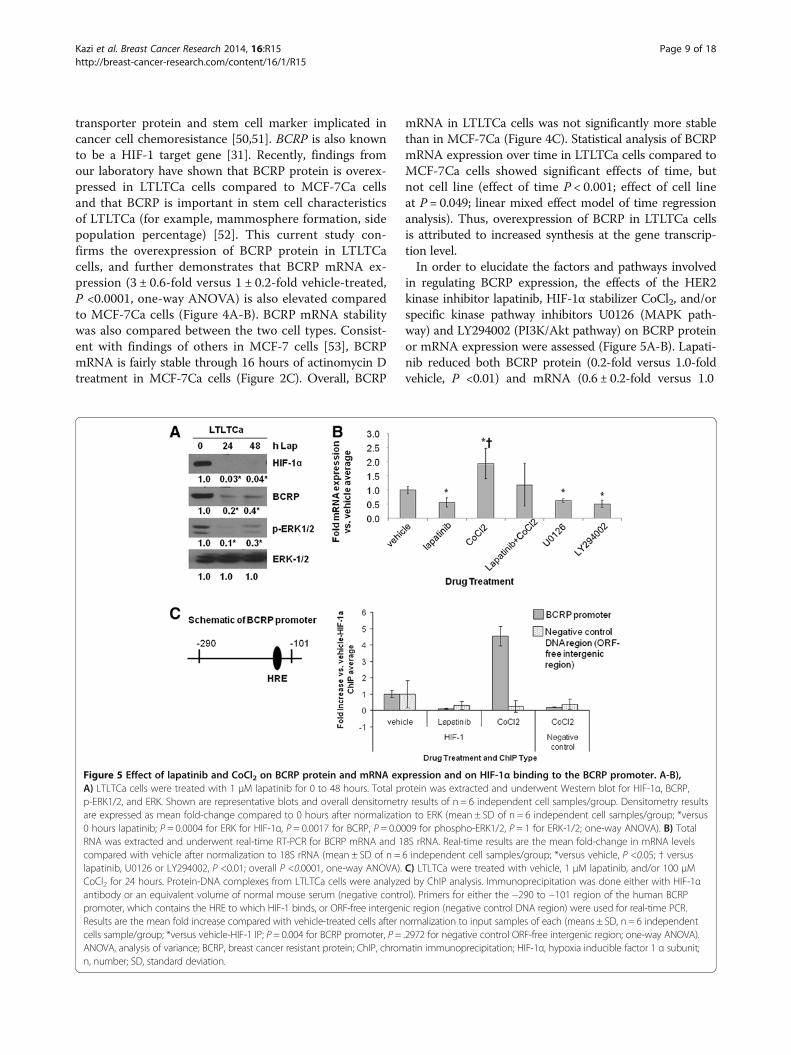
transporter protein and stem cell marker implicated incancer cell chemoresistance [50,51]. BCRP is also knownto be a HIF-1 target gene [31]. Recently, findings fromour laboratory have shown that BCRP protein is overex-pressed in LTLTCa cells compared to MCF-7Ca cellsand that BCRP is important in stem cell characteristicsof LTLTCa (for example, mammosphere formation, sidepopulation percentage) [52]. This current study con-firms the overexpression of BCRP protein in LTLTCacells, and further demonstrates that BCRP mRNA ex-pression (3 ± 0.6-fold versus 1 ± 0.2-fold vehicle-treated,P <0.0001, one-way ANOVA) is also elevated comparedto MCF-7Ca cells (Figure 4A-B). BCRP mRNA stabilitywas also compared between the two cell types. Consist-ent with findings of others in MCF-7 cells [53], BCRPmRNA is fairly stable through 16 hours of actinomycin Dtreatment in MCF-7Ca cells (Figure 2C). Overall, BCRP
mRNA in LTLTCa cells was not significantly more stablethan in MCF-7Ca (Figure 4C). Statistical analysis of BCRPmRNA expression over time in LTLTCa cells compared toMCF-7Ca cells showed significant effects of time, butnot cell line (effect of time P < 0.001; effect of cell lineat P = 0.049; linear mixed effect model of time regressionanalysis). Thus, overexpression of BCRP in LTLTCa cellsis attributed to increased synthesis at the gene transcrip-tion level.In order to elucidate the factors and pathways involved
in regulating BCRP expression, the effects of the HER2kinase inhibitor lapatinib, HIF-1α stabilizer CoCl2, and/orspecific kinase pathway inhibitors U0126 (MAPK path-way) and LY294002 (PI3K/Akt pathway) on BCRP proteinor mRNA expression were assessed (Figure 5A-B). Lapati-nib reduced both BCRP protein (0.2-fold versus 1.0-foldvehicle, P <0.01) and mRNA (0.6 ± 0.2-fold versus 1.0
Figure 5 Effect of lapatinib and CoCl2 on BCRP protein and mRNA expression and on HIF-1α binding to the BCRP promoter. A-B),A) LTLTCa cells were treated with 1 μM lapatinib for 0 to 48 hours. Total protein was extracted and underwent Western blot for HIF-1α, BCRP,p-ERK1/2, and ERK. Shown are representative blots and overall densitometry results of n = 6 independent cell samples/group. Densitometry resultsare expressed as mean fold-change compared to 0 hours after normalization to ERK (mean ± SD of n = 6 independent cell samples/group; *versus0 hours lapatinib; P = 0.0004 for ERK for HIF-1α, P = 0.0017 for BCRP, P = 0.0009 for phospho-ERK1/2, P = 1 for ERK-1/2; one-way ANOVA). B) TotalRNA was extracted and underwent real-time RT-PCR for BCRP mRNA and 18S rRNA. Real-time results are the mean fold-change in mRNA levelscompared with vehicle after normalization to 18S rRNA (mean ± SD of n = 6 independent cell samples/group; *versus vehicle, P <0.05; † versuslapatinib, U0126 or LY294002, P <0.01; overall P <0.0001, one-way ANOVA). C) LTLTCa were treated with vehicle, 1 μM lapatinib, and/or 100 μMCoCl2 for 24 hours. Protein-DNA complexes from LTLTCa cells were analyzed by ChIP analysis. Immunoprecipitation was done either with HIF-1αantibody or an equivalent volume of normal mouse serum (negative control). Primers for either the −290 to −101 region of the human BCRPpromoter, which contains the HRE to which HIF-1 binds, or ORF-free intergenic region (negative control DNA region) were used for real-time PCR.Results are the mean fold increase compared with vehicle-treated cells after normalization to input samples of each (means ± SD, n = 6 independentcells sample/group; *versus vehicle-HIF-1 IP; P = 0.004 for BCRP promoter, P = .2972 for negative control ORF-free intergenic region; one-way ANOVA).ANOVA, analysis of variance; BCRP, breast cancer resistant protein; ChIP, chromatin immunoprecipitation; HIF-1α, hypoxia inducible factor 1 α subunit;n, number; SD, standard deviation.
Kazi et al. Breast Cancer Research 2014, 16:R15 Page 9 of 18http://breast-cancer-research.com/content/16/1/R15

± .0.02-fold vehicle-treated, P <0.01, one-way ANOVA)levels in LTLTCa cells. This decrease correlated withlapatinib’s inhibitory effects on HIF-1α and p-ERK1/2expression. Inhibition of either the MAPK or PI3K/Aktpathways also resulted in decreased BCRP mRNA levels(0.7 ± 0.05-fold and 0.55 ± 0.08-fold versus 1.0 ± .0.02vehicle-treated, P <0.01, respectively; Figure 5B). CoCl2treatment conversely increased BCRP mRNA in LTLTCacells. Co-treatment with lapatinib and CoCl2 resulted inBCRP mRNA levels that tended to be intermediate oflapatinib-inhibited and CoCl2-induced levels, but notsignificantly different from either one and from vehicle(Figure 5B).Although results in Figure 5A-B suggested that HER2,
via the MAPK and PI3K/Akt pathways, and HIF-1α areboth involved in regulating BCRP expression in LTLTCacells, these expression analyses did not test whether HIF-1α actually mediates the effects of HER2 on target genes.ChIP analysis was, therefore, performed to determineHIF-1α binding to the BCRP promoter under basal, non-hypoxic conditions and after lapatinib or CoCl2 treatment.
Real-time PCR analysis of immunoprecipitated DNA afterChIP showed that under basal, nonhypoxic conditionsHIF-1α was bound to a hypoxia-response element (HRE)-containing region of the BCRP promoter in LTLTCa cells(Figure 5C). CoCl2 significantly increased HIF-1α bindingto the BCRP promoter, but lapatinib treatment preventedthis binding (versus 1 ± 1.2-fold vehicle-treated, P = 0.004,one-way ANOVA). Specificity of immunoprecipitation wasconfirmed by the lack of immunoprecipitated DNA inthe negative IP control samples, as well as on the nega-tive control DNA (P = 0.2972, one-way ANOVA). Inaddition, samples in the BCRP promoter PCR (exclud-ing input) amplified at cycles 20 to 30, while samples inthe negative control DNA region PCR (excluding input)amplified at cycles 32 to 40. Correlation between HIF-1αbinding to the BCRP promoter and changes in BCRPmRNA and protein expression in the absence or presenceof lapatinib suggests that HER2-regulated HIF-1 is in-volved in BCRP gene expression in LTLTCa cells.Such regulation, however, does not appear to be relevant
to all known HIF-1 target genes. Vascular endothelial
Figure 6 Effect of YC-1 on HIF-1α and BCRP expression and cell viability in LTLTCa cells. LTLTCa cells were treated with 100 μM YC-1 for 0 to24 hours and effects on HIF-1α (A) and BCRP (B) expression and cell viability (C) were determined. A) After 0 to 4 hours of YC-1 treatment, total proteinwas extracted and HIF-1α and β-actin were analyzed by Western blot analysis. Shown are representative blots and overall densitometry results of n = 6independent cell samples/group. Densitometry results are expressed as mean fold-change in protein levels compared to 0 hours after normalization toβ-actin (mean ± SD of n = 6 independent cell samples/group; *versus 0 hours YC-1, P <0.001; overall P <0.0001, one-way ANOVA). B) After 0 to 8 hoursYC-1 treatment, total RNA was extracted and BCRP mRNA and 18S rRNA were analyzed by real-time RT-PCR analysis. Real-time results are expressed asthe mean fold-change in mRNA levels compared with vehicle after normalization to 18S rRNA (mean ± SD, n = 6 independent cell samples/group;*versus 0 hours YC-1, P <0.001; overall P <0.0001, one-way ANOVA). C) Viability of cells was measured by MTT assay after 0 to 24 hours treatment withYC-1. Results are expressed as mean percent of 0 hours average (mean ± SD, n = 4 independent cell samples/group; *versus 0 hours, P <0.001; overallP <0.0001, one-way ANOVA). ANOVA, analysis of variance; BCRP, breast cancer resistant protein; HIF-1α, hypoxia inducible factor 1 α subunit; MTT,3-[4,5-dimethylthiazol-2-yl]-2,5 diphenyl tetrazolium bromide; n, number; SD, standard deviation.
Kazi et al. Breast Cancer Research 2014, 16:R15 Page 10 of 18http://breast-cancer-research.com/content/16/1/R15

growth factor (VEGF), another known HIF-1 target geneand important therapeutic target in cancer [17], is notupregulated in LTLTCa cells compared to MCF-7Ca cells(Figure 2B). Also, despite being induced by CoCl2, VEGFmRNA expression was not sensitive to lapatinib (data notshown).
Effect of specific inhibition of HIF-1α on BCRPTo further support a connection between HIF-1α andBCRP, HIF-1α expression in LTLTCa cells was specific-ally inhibited by either YC-1, a known pharmacologicalinhibitor of HIF-1α [54,55] or siRNA. Similar to obser-vations with lapatinib treatment, HIF-1α protein andBCRP mRNA expression were significantly decreased(0.1 ± 0.1-fold versus 0.1 ± 0.3-fold vehicle-treated and0.5 ± 0.05-fold versus 1 ± 0.15-fold vehicle treated P<0.0001, respectively) in LTLTCa cells within eight hoursof YC-1 treatment (Figure 6A-B). This correlated with a30% to 40% decrease in LTLTCa cell viability by 16 and24 hours, respectively (Figure 6C). Specific inhibition ofHIF-1α expression by siRNAs also significantly de-creased both HIF-1α mRNA (approximately 0.4 ± 0.04-fold versus 1 ± 0.2-fold negative control siRNA, P =0.0057, one-way ANOVA) and protein (0.3 ± 0.1-fold to0.03 ± 0.1-fold versus 1 ± 0.05-fold negative control, P<0.0001, one-way ANOVA), as well as BCRP mRNA (0.4-to 0.6-fold versus 1 ± 0.2-fold negative control siRNA, P<0.0001, one-way ANOVA) expression after 48 hours(Figure 7A-B).
Correlation between HER2, HIF-1α, and BCRP in HER2-transfected cells and another AI-resistant cell lineTo further confirm the role of HER2 in regulating HIF-1α and BCRP and to determine if ERα is also involved,protein expression in Hc7 cells, ERα +MCF-7 cellstransfected with HER2 gene was also studied. Similar toERα-/HER2+ LTLTCa cells, Hc7 cells overexpressedphospho-ERK, HIF-1α and BCRP protein expressioncompared to ERα+/HER2-parental MCF-7 cells(Figure 8A). Furthermore, HER2 inhibition by lapatinib de-creased HIF-1α protein levels in Hc7 cells (0.1 ± 0.1-foldversus vehicle, P <0.0001; Figure 8B). Interestingly, inhib-ition of ERα alone by the ERα antagonist ICI 182,780 alsoreduced HIF-1α levels, but its effect on the protein levelwas significantly less than that of lapatinib alone or lapati-nib and ICI182,780 in combination (Figure 8C). AnotherAI-resistant cell line, exemestane-resistant AC1-ExRbreast cancer cells, was also analyzed. Despite retainingERα, AC1-ExR cells also showed higher HER2, HIF-1αand BCRP protein levels. Overall, these results further in-dicate that increased HER2 and HER2-activated kinasepathways correlate with increased HIF-1α. They also indi-cate that while ERα can play a role in regulating HIF-1, ashas been suggested by other studies [22], HER2 is likely tobe the more important factor in the cells studied.
Functional importance of HIF-1α in LTLTCa cellsEffect of HIF-1α inhibition on LTLTCa cellsLastly, the functional importance of HIF-1 to the letrozole-resistant cell phenotype was explored. In cancer cells,
Figure 7 Effect of HIF-1α siRNA on mRNA expression in LTLTCa cells. A) LTLTCa cells were plated in passage media and then treated withtwo siRNAs for HIF-1α for 48 hours. Total mRNA was extracted and HIF-1α and BCRP mRNA, and 18S rRNA were analyzed by real-time RT-PCR.Real-time results are expressed as the mean fold-change in mRNA levels compared with negative control after normalization to 18S rRNA (mean± SD, n = 6 independent samples/group; *versus negative control, P = 0.0057 for HIF-1α; P = 0.0026 for BCRP; one-way ANOVA). B) LTLTCa cellswere plated in passage media and then treated with two siRNAs for HIF-1α for 48 hours. Total protein was extracted and HIF-1α and β-actinprotein were analyzed by Western blot. Shown are representative blots and overall densitometry results of n = 6 independent cell samples/group.Densitometry results are expressed as mean fold-change in protein levels compared to negative control after normalization to β-actin (mean ± SD,n = 6 independent cell samples/group; *versus negative control (NC), P <0.0001, one-way ANOVA). ANOVA, analysis of variance; BCRP, breast cancerresistant protein; HIF-1α, hypoxia inducible factor 1 α subunit; n, number; SD, standard deviation.
Kazi et al. Breast Cancer Research 2014, 16:R15 Page 11 of 18http://breast-cancer-research.com/content/16/1/R15

hypoxia and HIF-1 are known to be involved in increasedcell survival, chemoresistance [56,57], resistance to apop-tosis [58] and maintenance of cancer stem cell characteris-tics [59,60]. Previous findings from our laboratory [52] havealready demonstrated that letrozole resistance and cancerstem cell characteristics of LTLTCa cells are reduced by in-hibition of HER2 and/or BCRP. Although Gilani et al. didnot specifically test for the involvement of HIF-1, results ofthis study combined with those of our current study dem-onstrating the HER2-HIF-1-BCRP pathway, supports a rolefor HIF-1 in determining the letrozole-resistant cellphenotype.To determine the functional significance of HIF-1 in
LTLTCa cells, the effect of specific inhibition of HIF-1αexpression by siRNA on mammosphere formation andcell viability was analyzed (Figure 9). Consistent with ourprevious study [52], LTLTCa cells formed mammospheres(306 mammospheres/20,000 cells ± 5) (Figure 9B), and thiswas decreased by BCRP siRNA treatment (64 mammo-spheres/20,000 cells ± 9; P <0.001, one-way ANOVA). HIF-
1α siRNA treatment similarly decreased mammosphereformation in LTLTCa cells (101 mammospheres/20,000cells ± 18), while CoCl2 increased formation (500 mammo-spheres/20,000 cells ± 20) compared to negative control-treated siRNA (P <0.001, one-way ANOVA). These resultscorrelated with the effect of HIF-1α inhibition on othergenes. HIF-1α siRNA treatment decreased BCRP mRNA(P = 0.0377, one-way ANOVA), as well as expression ofGAPDH [61] (P = 0.0058, one-way ANOVA), anotherknown HIF-1 target gene, and BMI-1 (P = 0.0214, one-wayANOVA), another stem cell marker [62] (Figure 9A). HIF-1α siRNA treatment also significantly decreased LTLTCacell viability (P <0.0001, one-way ANOVA) in the presenceof increasing concentrations of letrozole (Figure 9C).
Effect of HIF-1α upregulation on MCF-7Ca cellsTo further confirm the physiological role of HIF-1 andBCRP, converse experiments were done to investigatewhether MCF-7Ca cells could become more letrozole-resistant with increased HIF-1α. CoCl2 was used to
Figure 8 Protein expression in HER2+ cells and exemestane-resistant cells. A) MCF-7Ca (M), Hc7 and LTLTCa (LT) cells were plated in theirrespective passage media. Total protein was extracted and HER2, phosphorylated- and total-ERK, ERα, HIF-1α, BCRP and β-actin protein wereanalyzed by Western blot. Shown are representative blots and overall densitometry results of n = 6 independent cell samples/group. Densitometryresults are expressed as mean fold-change compared to MCF-7Ca after normalization to ERK (mean ± SD of n = 6 independent cell samples/group;*versus MCF-7Ca, P <0.05; † versus MCF-7Ca, P <0.001, one-way ANOVA). Dashed lines indicate omitted lane in between M and Hc7 of the sameblots. B) AC1 (AC1) and AC1-ExR (ExR) cells were plated in their respective passage media. Total protein was extracted and HER2, phosphorylated-and total-ERK, ERα, HIF-1α and β-actin protein were analyzed by Western blot. Shown are representative blots and overall densitometry results ofn = 6 independent cell samples/group. Densitometry results are expressed as mean fold-change compared to vehicle-treated cells afternormalization to β-actin (mean ± SD of n = 6 independent cell samples/group; *versus vehicle, P <0.0001, two-sided t-test). C) MCF-7/HER2 cellswere treated with either vehicle (V), 1 μM lapatinib (Lap), 100 nM ICI 182,780 (ICI) or 1 μM lapatinib + 100 nM ICI 182,780 (Lap + ICI) for 24 hours.Total protein was extracted and HER2, phospho- and total-ERK1/2, ERα, HIF-1α and β-actin were analyzed by Western blot. Shown are representativeblots and overall densitometry results of n = 6 independent cell samples/group. Densitometry results are expressed as mean fold-change compared tovehicle-treated cells after normalization to β-actin (mean ± SD of n = 6 independent cell samples/group; *versus vehicle, P <0.0001, one-way ANOVA).ANOVA, analysis of variance; BCRP, breast cancer resistant protein; ERα, estrogen receptor alpha; HER2, human epidermal growth factor receptor 2;HIF-1α, hypoxia inducible factor 1 α subunit; n, number; SD, standard deviation.
Kazi et al. Breast Cancer Research 2014, 16:R15 Page 12 of 18http://breast-cancer-research.com/content/16/1/R15

increase HIF-1α expression in MCF-7Ca cells and the ef-ficacy of CoCl2 was confirmed by Western blot analysisand RT-PCR. Within 24 hours of CoCl2 treatment, HIF-1α protein and BCRP mRNA and protein expressionwere increased in MCF-7Ca cells (Figure 10A-B). Cellviability experiments in the presence of increasing con-centrations of letrozole were then performed (P <0.0001,one-way ANOVA). Consistent with previous findingsfrom our laboratory [12], MCF-7Ca cells not treated withCoCl2 were sensitive to the growth inhibitory effects ofletrozole (Figure 10C). Additional treatment of MCF-7Cacells with CoCl2 significantly increased their resistance toletrozole. The effects of CoCl2 were attributable to HIF-1,as co-treatment of MCF-7Ca cells with CoCl2 and HIF-1αsiRNA returned their sensitivity to letrozole. Overall, thephysiological experiments on LTLTCa and MCF-7Ca cells
indicate that HIF-1 is likely involved in both cancer stemcell characteristics and cell viability.
DiscussionPrior to this study, AI resistance was associated with in-creased dependence on growth factors and decreased de-pendence on ERα. However, the role that such molecularchanges play in AI resistance and the mechanism by whichthey elicit their effects were not known. Novel results fromthis study demonstrated that nonhypoxic expression ofHIF-1 mediates HER2’s effects on letrozole-resistance. Spe-cifically, the HER2-activated PI3K/Akt pathway increasesHIF-1α protein synthesis in LTLTCa cells. HIF-1α, in turn,upregulates expression of BCRP and other genes and con-tributes to letrozole resistance and stem cell characteristicsof LTLTCa cells.
Figure 9 Effect of HIF-1α and/or BCRP siRNA on mammosphere formation and cell proliferation in LTLTCa cells. A) LTLTCa cells weretreated with either negative control siRNA or HIF-1α siRNA for 48 hours. Total mRNA was extracted and HIF-1α, BCRP, GAPDH, Nanog, BMI-1 andTWIST mRNA, and 18S rRNA were analyzed by real-time RT-PCR. Real-time results are expressed as the fold-change in mRNA levels compared withnegative control after normalization to 18S rRNA (mean ± SD, n = 6 independent cell samples/group; *versus vehicle; P = 0.0.132 HIF-1; P = 0.0058GAPDH, P = 0.0377 BCRP, P = 0.0612 TWIST, P = 0.058 Nanog, P = 0.0214 BMI-1; two-sided t-test). B) LTLTCa cells were plated in passage media andthen treated with negative control siRNA, HIF-1α siRNA, BCRP siRNA, or 100 μM CoCl2 for 48 hours. Cells were then collected and resuspended inmammosphere media on low-attachment cell culture wells. Results are expressed as number of mammospheres counted per 20,000 cells plated(mean ± SD, n = 6 independent cell samples/group; *versus negative control, P <0.001; † versus CoCl2, P < 0.001; overall P <0.0001, one-wayANOVA). BCRP siRNA confirmed to decrease BCRP expression (0.35- and 0.15-fold versus negative control, P <0.01, one-way ANOVA; data not shown).C) Viability of the cells was measured by the MTT assay after 48 hours treatment with negative control or HIF-1 alpha siRNA and subsequently 6 daytreatment with increasing doses of letrozole. Results are expressed as percent of 0 μM letrozole (vehicle) (mean ± SD, n = 4 independent cell samples/group; *versus 0 μM letrozole, P <0.001; overall P <0.0001 one way ANOVA). ANOVA, analysis of variance; BCRP, breast cancer resistant protein; HIF-1α,hypoxia inducible factor 1 α subunit; MTT, 3-[4,5-dimethylthiazol-2-yl]-2,5 diphenyl tetrazolium bromide; n, number; SD, standard deviation.
Kazi et al. Breast Cancer Research 2014, 16:R15 Page 13 of 18http://breast-cancer-research.com/content/16/1/R15

Nonhypoxic regulation of HIF-1 expression and activ-ity in LTLTCa cells is due to the HER2-activated PI3K/Akt/mTOR pathway. This is consistent with findings byothers indicating hypoxia independent upregulation ofHIF-1α in cancer cells by loss of function of tumor sup-pressor genes and gain of function of oncogenes [27]. Theoncogene HER2/neu, in particular, has been previously as-sociated with nonhypoxic HIF-1 [24,25]. Laughner et al.and Li et al. have demonstrated that transfection of HER2into NIH/3 T3 cells or activation of HER2 in MCF-7 cellsled to activation of the PI3K/Akt pathway, and the subse-quent increased HIF-1 expression via protein synthesisand HIF-1 transcriptional activity. Previous studies havealso demonstrated the importance of mTOR [48,49]. Inaddition, mTOR has been explored in two randomized tri-als (BOLERO-2 and TAMRAD) as a potential therapeutictarget for overcoming endocrine therapy resistance [63].Our current study provides evidence that this HER2-
PI3K/Akt-mTOR-HIF-1 signaling mechanism can indeedoccur endogenously in HER2+ cells (Figures 3, 4, and 8),leads to upregulation of BCRP (Figure 2), and has physio-logical relevance as well as potential clinical implications(for example, AI resistance; Figures 9 and 10).Despite providing evidence that HER2 activation of the
PI3K/Akt-mTOR pathway regulates HIF-1α, this studycannot completely exclude the involvement of ERα or theMAPK pathway. In addition to overexpressing HER2,LTLTCa cells also have decreased expression of ERα(Figure 3). It is possible that ERα can also regulate non-hypoxic HIF-1α expression in LTLTCa cells. Indeed, ERα-and HIF-1-mediated signaling pathways are known tointeract antagonistically [18,19] and cooperatively [20-23].Although this current study did not directly investigateERα’s role, the overexpression of HIF-1α observed in bothERα + (Hc7 and Ac1-ExR) and ERα- (LTLTCa) HER2+breast cancer cell lines, suggests that ERα status may not
Figure 10 Effect of CoCl2 on MCF-7Ca protein expression and cell viability. A-B, MCF-7Ca cells were incubated in steroid-free media andthen treated with 100 μM CoCl2 for 0 to 24 hours. A) Total protein was extracted and HIF-1α, BCRP, and β-actin were analyzed by Western blotanalysis. Shown are representative blots and overall densitometry results of n = 6 independent cell samples/group. Densitometry results areexpressed as mean fold-change in protein levels compared to 0 hours after normalization to β-actin (mean ± SD of n = 4 independent cellsamples/group, *versus 0 hours; P = 0.0005 for HIF-1a; P = 0.0065 for BCRP, two-sided t-test). B) Total RNA was extracted and BCRP mRNA and 18SrRNA were analyzed by real-time RT-PCR analysis. Results are expressed as the mean fold-change in mRNA levels compared with 0 hours afternormalization to 18S rRNA (mean ± SD, n = 4 independent cell samples/group; *versus 0 hours, P <0.001; overall P = 0.0002, one-way ANOVA).C) Viability of cells was measured by MTT assay after five days of treatment with increasing doses of letrozole following 48 hours pre-treatmentwith or without 100 μM CoCl2 and HIF-1α siRNA. Results are expressed as mean percent of 0 μM letrozole-without CoCl2-with negative controlsiRNA (mean ± SD of n = 6 independent samples/group; *versus vehicle-negative control siRNA-0 μM letrozole, P <0.001; † versus vehicle-HIF-1αsiRNA and CoCl2-HIF-1α siRNA; effect of letrozole P <0.0001, effect of pre-treatment (vehicle/CoCl2 and negative control siRNA/HIF-1α siRNA)P <0.0001, interaction between letrozole dose and pre-treatment P <0.0001; two-way ANOVA). ANOVA, analysis of variance; BCRP, breast cancerresistant protein; HIF-1α, hypoxia inducible factor 1 α subunit; MTT, 3-[4,5-dimethylthiazol-2-yl]-2,5 diphenyl tetrazolium bromide; n, number;SD, standard deviation.
Kazi et al. Breast Cancer Research 2014, 16:R15 Page 14 of 18http://breast-cancer-research.com/content/16/1/R15

affect HER2 regulation of non-hypoxic HIF-1α levels.With regard to the MAPK pathway, inhibition of this path-way did not affect HIF-1α expression in LTLTCa cells, butit did decrease BCRP mRNA expression under basal, non-hypoxic conditions. It is possible that the MAPK pathway isinvolved in phosphorylation of HIF-1α rather than its syn-thesis. Previous studies have shown that MAPK pathway-mediated phosphorylation of HIF-1α occurs under non-hypoxic conditions and can increase HIF-1α expressionand transcriptional activity [64,65]. These results could alsoindicate the MAPK and PI3K/Akt pathways have very dis-tinct functions in AI-resistant breast cancer cells, regulatingdifferent subsets of genes. For example, the MAPK pathwaymay be involved in HER2 regulation of genes that requireactivation by phosphorylated ERαSer118. In contrast, thePI3K/Akt pathway may be involved in HER2 regulation ofgenes that require HIF-1.Inherent upregulation of HIF-1α protein expression
under nonhypoxic conditions is another novel finding inAI-resistant breast cancer. There is precedence for asso-ciating HIF-1 expression with drug resistance in differ-ent cancer cell types, including chronic myeloidleukemia cells [66], gastric cancer cells [67], non-smallcell lung cancer cells [68], and even breast cancer cells[58]. However, these previous cases involved hypoxia-induced, HIF-1α rather than the non-hypoxic HIF-1.Our findings are also consistent with previous clinicalevidence that HIF-1α is associated with letrozole resist-ance. Generali et al. demonstrated that increased p-MAPK and HIF-1α protein expression were significantdeterminants of primary letrozole resistance in breastcancer patients. In contrast, increased ERα and
decreased p-MAPK were significant determinants of re-sponse to letrozole treatment [56]. The protein expres-sion patterns observed by Generali et al. are similar towhat is observed in letrozole-resistant LTLTCa and –sensitive MCF-7Ca cells, respectively (Figures 3 and 6).Although these clinical findings involve de novo letrozoleresistance, they still correlate with, and likely pertain to,our laboratory’s results on acquired letrozole resistance.These results combined suggest that HIF-1 is involved inboth de novo and acquired AI resistance and, therefore,could be therapeutically targeted to prevent and treat re-sistance to letrozole and the other AIs.Lastly, this study indicates that HIF-1 may contribute
to letrozole resistance by mediating the effects of HER2on target genes, such as BCRP. Previous findings by ourlaboratory had implicated HER2 and BCRP in resistanceto the growth inhibitory effects of letrozole and to main-tenance of stem cell characteristics in letrozole-resistantbreast cancer, and had demonstrated that its expressionwas dependent on HER2 [35,52], but it was unclear untilnow how HER2 regulated BCRP. Moreover, HIF-1 maymediate the effects of HER2 on many other genes. Besides,BCRP, other known HIF-1 target genes that may serve asmarkers of letrozole resistance include: 1) cancer stem cellmaintenance markers (Oct-4, kit ligand, JARID1B); 2)epithelial-mesenchymal-transition (EMT) markers (Snail,vimentin); and 3) invasion markers (c-Met, endothelin 1,fibronectin, MMP-2 and −4) [27,69]. Interestingly, anotherknown HIF-1 target gene,VEGF, was not upregulated inLTLTCa cells compared to MCF-7Ca cells. It is possiblethat nonhypoxic HIF-1 expression has different levels ofinfluence on different HIF-1 target genes, particularly
Figure 11 Proposed model of regulation and role of HIF-1α in AI resistant breast cancer.
Kazi et al. Breast Cancer Research 2014, 16:R15 Page 15 of 18http://breast-cancer-research.com/content/16/1/R15
those that can be regulated by multiple transcriptionfactors. Indeed BCRP and VEGF both are known to beregulated by additional transcription factors, such asERα [22,70].
ConclusionsOverall, this study provides novel evidence that non-hypoxic HIF-1α is inherently expressed in AI-resistantcells, upregulated by HER2-PI3K/Akt-mTOR pathwayand is an important factor in letrozole-resistant breastcancer cells, regulating target genes such as BCRP andregulating AI responsiveness and cancer stem cell char-acteristic expression. Thus, HIF-1α could be used as adiagnostic marker and/or therapeutic target. Based onthis, a proposed model of acquired AI-resistance may in-volve the following scenario: under non-hypoxic condi-tions, when the breast cancer cell population and tumorsize have been reduced by letrozole treatment and prior tosignificant tumor hypoxia, a switch from ERα- to growthfactor (for example, HER2)-mediated signaling occurs viaPI3K/Akt and mTOR, which leads to increased HIF-1αexpression and activation of HIF-1 target genes (for ex-ample, BCRP) that contribute to AI resistance (Figure 11).Consequently, inhibition of HIF-1 expression and/oractivity would prolong cancer cell sensitivity to AIs andprevent recurrence and metastasis. Indeed, a number ofanti-cancer drugs in clinical use are also known to inhibitHIF-1 [27]. They include HER2 inhibitor, trastuzumab[24] and lapatinib. Furthermore, as demonstrated in thisstudy that HIF-1 is regulated mainly via the PI3K/Akt/mTOR pathway, inhibition of the downstream affecter ofthis pathway using mTOR inhibitors, such as rapamycin,temsirolimus/CCI-779 and everolimus/RAD-001, can alsobe considered [24,71-73]. There is also EZN-2968, a spe-cific HIF-1α mRNA inhibitor, shown to reduce cancer cellviability and xenograft tumor growth, which is currentlyunder phase I clinical trial [74]. Any of these anti-cancerdrugs could now potentially, based on the evidence pro-vided by this study, be applied to the prevention and treat-ment of AI-resistant breast cancer.
Additional file
Additional file 1: Table S1.1. Test results conducted to compare meandifferences of normalized, log-transformed HIF-1α mRNA expression atvarious time points of actinomycin D for LTLTCa and MCF-7Ca cells.Shown are estimated mean differences in log transformed, normalizedHIF-1α mRNA expression between different timepoints of actinomycinD treatment. Table results are estimated mean differences in log trans-formed, normalized HIF-1α mRNA expression between different timepointsof actinomycin D treatment calculated from the same data as shownFigure 1B data, which indicates trend over time. Pre-specified timepointscompared within each cell line were 0 versus 2 hours, 2 versus 4 hours, or4 versus 16 hours. Data were analyzed by linear mixed effect modeladjusting for experiment, cell line, and cell line*time interaction mRNA. Fixedeffects for time, experiment, cell lines and interactions between time and
cell lines were determined (means ± SD of n = 6 independent samples/group; P <0.001 for effect of cell line, time, their interaction and experiment).NS, not significant, P >0.05. Table S1.2. Test results conducted to comparemean differences of normalized, log-transformed BCRP mRNA expression atvarious time points of actinomycin D for both LTLTCa and MCF-7Ca cells.Table results are estimated mean differences in log transformed, normalizedBCRP mRNA expression between different timepoints of actinomycinD treatment calculated from the same data as shown in Figure 2C. Pre-specified timepoints compared within each cell line were 0 versus 2 hours,2 versus 4 hours, or 4 versus 16 hours. Data were analyzed by linear mixedeffect model adjusting for experiment, cell line, and cell line*time interactionmRNA. Fixed effects for time, experiment, cell lines and interactionsbetween time and cell lines were determined (means ± SD of n = 6independent samples/group; P <0.001 for effect of time and cell line*timeinteraction). NS, not significant, P >0.05.
AbbreviationsAC1: MCF-7 cells transfected with the aromatase gene by the laboratory ofDr. Brodie; AC1-ExR: long-term exemestane treated AC1 cells; AI: aromataseinhibitor; Akt: also known as protein kinase B; ANOVA: analysis of variance;BCRP: breast cancer resistance protein; CDT: charcoal-dextran treated;ChIP: chromatin immunoprecipitation; (D)MEM: (Dulbecco’s) modified Eagle’smedium; EGFR: epidermal growth factor receptor; ERα: estrogen receptoralpha; FBS: fetal bovine serum; Hc7: MCF-7 cells transfected with HER2 geneby the laboratory of Dr. Brodie; HER2: human epidermal growth factorreceptor 2; HIF-1: hypoxia inducible factor 1; HIF-1α: hypoxia inducible factor1 α subunit; HRE: hypoxia-response element; LTLTCa: long-term letrozoletreated MCF-7Ca cells; MAPK: mitogen-activated protein kinase; MCF-7Ca: MCF-7 cells transfected with the aromatase gene by the laboratory ofDr. Chen; mTOR: mammalian target of rapamycin; PBS: phosphate-bufferedsaline; PI3K: phosphatidylinositide 3-kinase; PRF: phenol red-free; ORF: openreading frame; siRNA: small interfering RNA; RT-PCR: reverse transcriptase-polymerase chain reaction; VEGF: vascular endothelial growth factor.
Competing interestsThe authors declare that they have no competing interests.
Authors’ contributionsAK conceived of and designed the study, performed all experiments, anddrafted the manuscript. RG participated in RT-PCR in Figure 1A and westernblot 1B. AS participated in densitometry analysis of Figure 3A and helped todraft the manuscript. SC coordinated obtaining lapatinib and trastuzumaband participated in discussions of study design. GS participated in the ana-lysis and interpretation of Western blot data for Figures 3, 4, and 8 and statis-tical analyses for Figures 1B and 2C. PS participated in the mammosphere assayand selection of cancer stem cell markers in Figure 9A. OG performed statisticalanalyses for Figures 1B and 2C. SK performed statistical analyses for Figures 1Band 2C. AB participated in the design of the study, interpretation of all results,and the critical review and revision of the manuscript. All authors read and ap-proved final manuscript.
AcknowledgementsFunding for this study was provided by the Department of Defense BreastCancer Research Program Postdoctoral Award (BC1039031).
Author details1Department of Pharmacology and Experimental Therapeutics, University ofMaryland, Baltimore, MD 21201, USA. 2Department of Medicine, University ofMaryland, Baltimore, MD 21201, USA. 3School of Medicine, University ofMaryland Marlene and Stewart Greenebaum Cancer Center, Baltimore, MD21201, USA. 4Division of Biostatistics and Bioinformatics Department ofEpidemiology and Public Health, University of Maryland, Baltimore, MD21201, USA. 5Department of Biology, Loyola University Maryland, Baltimore,MD 21210, USA. 6Department of Pharmacology and ExperimentalTherapeutics, University of Maryland School of Medicine, Health ScienceFacilities, Room 580G, 685 West Baltimore Street, Baltimore, MD 21201, USA.
Received: 23 October 2012 Accepted: 2 January 2014Published: 29 January 2014
Kazi et al. Breast Cancer Research 2014, 16:R15 Page 16 of 18http://breast-cancer-research.com/content/16/1/R15

References1. United States Cancer Statistics: 1999–2008 Incidence and Mortality Web-
based Report. [http://www.cdc.gov/uscs.]2. Chumsri S, Howes T, Bao T, Sabnis G, Brodie A: Aromatase, aromatase
inhibitors, and breast cancer. J Steroid Biochem Mol Biol 2011, 125:13–22.3. Santen RJ, Brodie H, Simpson ER, Siiteri PK, Brodie A: History of aromatase:
saga of an important biological mediator and therapeutic target. EndocrRev 2009, 30:343–375.
4. Nabholtz JM, Mouret-Reynier MA, Durando X, Van Praagh I, Al-Sukhun S,Ferriere JP, Chollet P: Comparative review of anastrozole, letrozole andexemestane in the management of early breast cancer. Expert OpinPharmacother 2009, 10:1435–1447.
5. Dowsett M, Cuzick J, Ingle J, Coates A, Forbes J, Bliss J, Buyse M, Baum M,Buzdar A, Colleoni M, Coombes C, Snowdon C, Gnant M, Jakesz R,Kaufmann M, Boccardo F, Godwin J, Davies C, Peto R: Meta-analysis ofbreast cancer outcomes in adjuvant trials of aromatase inhibitors versustamoxifen. J Clin Oncol 2010, 28:509–518.
6. Winer EP, Hudis C, Burstein HJ, Wolff AC, Pritchard KI, Ingle JN, ChlebowskiRT, Gelber R, Edge SB, Gralow J, Cobleigh MA, Mamounas EP, Goldstein LJ,Whelan TJ, Powles TJ, Bryant J, Perkins C, Perotti J, Braun S, Langer AS,Browman GP, Somerfield MR: American Society of Clinical Oncologytechnology assessment on the use of aromatase inhibitors as adjuvanttherapy for postmenopausal women with hormone receptor-positivebreast cancer: status report 2004. J Clin Oncol 2005, 23:619–629.
7. Miller WR, Larionov AA: Understanding the mechanisms of aromataseinhibitor resistance. Breast Cancer Res 2012, 14:201.
8. Kubo M, Kanaya N, Petrossian K, Ye J, Warden C, Liu Z, Nishimura R, OsakoT, Okido M, Shimada K, Takahashi M, Chu P, Yuan YC, Chen S: Inhibition ofthe proliferation of acquired aromatase inhibitor-resistant breast cancercells by histone deacetylase inhibitor LBH589 (panobinostat). BreastCancer Res Treat 2013, 137:93–107.
9. Sabnis G, Brodie A: Adaptive changes results in activation of alternatesignaling pathways and resistance to aromatase inhibitor resistance.Mol Cell Endocrinol, 340:142–147.
10. Johnston SR, Martin LA, Leary A, Head J, Dowsett M: Clinical strategies forrationale combinations of aromatase inhibitors with novel therapies forbreast cancer. J Steroid Biochem Mol Biol 2007, 106:180–186.
11. Macedo LF, Sabnis GJ, Goloubeva OG, Brodie A: Combination ofanastrozole with fulvestrant in the intratumoral aromatase xenograftmodel. Cancer Res 2008, 68:3516–3522.
12. Sabnis G, Schayowitz A, Goloubeva O, Macedo L, Brodie A: Trastuzumabreverses letrozole resistance and amplifies the sensitivity of breastcancer cells to estrogen. Cancer Res 2009, 69:1416–1428.
13. Brodie A, Macedo L, Sabnis G: Aromatase resistance mechanisms inmodel systems in vivo. J Steroid Biochem Mol Biol 2010, 118:283–287.
14. Schwartzberg LS, Franco SX, Florance A, O'Rourke L, Maltzman J, Johnston S:Lapatinib plus letrozole as first-line therapy for HER-2+ hormonereceptor-positive metastatic breast cancer. Oncologist 2010,15:122–129.
15. Hynes NE, Lane HA: ERBB receptors and cancer: the complexity oftargeted inhibitors. Nat Rev Cancer 2005, 5:341–354.
16. Mukohara T: Mechanisms of resistance to anti-human epidermal growthfactor receptor 2 agents in breast cancer. Cancer Sci 2011, 102:1–8.
17. Semenza GL: Targeting HIF-1 for cancer therapy. Nat Rev Cancer 2003,3:721–732.
18. Cho J, Kim D, Lee S, Lee Y: Cobalt chloride-induced estrogen receptor alphadown-regulation involves hypoxia-inducible factor-1alpha in MCF-7 humanbreast cancer cells. Mol Endocrinol (Baltimore, Md) 2005, 19:1191–1199.
19. Mukundan H, Kanagy NL, Resta TC: 17-beta estradiol attenuates hypoxicinduction of HIF-1alpha and erythropoietin in Hep3B cells. J CardiovascPharmacol 2004, 44:93–100.
20. Kazi AA, Jones JM, Koos RD: Chromatin immunoprecipitation analysis ofgene expression in the rat uterus in vivo: estrogen-induced recruitmentof both estrogen receptor alpha and hypoxia-inducible factor 1 to thevascular endothelial growth factor promoter. Mol Endocrinol (Baltimore,Md) 2005, 19:2006–2019.
21. Cho J, Bahn JJ, Park M, Ahn W, Lee YJ: Hypoxic activation of unoccupiedestrogen-receptor-alpha is mediated by hypoxia-inducible factor-1 alpha.J Steroid Biochem Mol Biol 2006, 100:18–23.
22. Kazi AA, Koos RD: Estrogen-induced activation of hypoxia-induciblefactor-1alpha, vascular endothelial growth factor expression, and edema
in the uterus are mediated by the phosphatidylinositol 3-kinase/Aktpathway. Endocrinology 2007, 148:2363–2374.
23. Hua K, Din J, Cao Q, Feng W, Zhang Y, Yao L, Huang Y, Zhao Y, Feng Y:Estrogen and progestin regulate HIF-1alpha expression in ovarian cancercell lines via the activation of Akt signaling transduction pathway.Oncol Rep 2009, 21:893–898.
24. Laughner E, Taghavi P, Chiles K, Mahon PC, Semenza GL: HER2 (neu)signaling increases the rate of hypoxia-inducible factor 1alpha(HIF-1alpha) synthesis: novel mechanism for HIF-1-mediated vascularendothelial growth factor expression. Mol Cell Biol 2001, 21:3995–4004.
25. Li YM, Zhou BP, Deng J, Pan Y, Hay N, Hung MC: A hypoxia-independenthypoxia-inducible factor-1 activation pathway induced byphosphatidylinositol-3 kinase/Akt in HER2 overexpressing cells.Cancer Res 2005, 65:3257–3263.
26. Vaupel P, Mayer A, Hockel M: Tumor hypoxia and malignant progression.Methods Enzymol 2004, 381:335–354.
27. Semenza GL: Defining the role of hypoxia-inducible factor 1 in cancerbiology and therapeutics. Oncogene 2010, 29:625–634.
28. Mendez O, Zavadil J, Esencay M, Lukyanov Y, Santovasi D, Wang SC,Newcomb EW, Zagzag D: Knock down of HIF-1alpha in glioma cellsreduces migration in vitro and invasion in vivo and impairs their abilityto form tumor spheres. Mol Cancer 2010, 9:133.
29. Lu X, Yan CH, Yuan M, Wei Y, Hu G, Kang Y: In vivo dynamics and distinctfunctions of hypoxia in primary tumor growth and organotropicmetastasis of breast cancer. Cancer Res 2010, 70:3905–3914.
30. Mazumdar J, Dondeti V, Simon MC: Hypoxia-inducible factors in stem cellsand cancer. J Cell Mol Med 2009, 13:4319–4328.
31. Krishnamurthy P, Ross DD, Nakanishi T, Bailey-Dell K, Zhou S, Mercer KE,Sarkadi B, Sorrentino BP, Schuetz JD: The stem cell marker Bcrp/ABCG2enhances hypoxic cell survival through interactions with heme. J BiolChem 2004, 279:24218–24225.
32. Zhou DJ, Pompon D, Chen SA: Stable expression of human aromatasecomplementary DNA in mammalian cells: a useful system for aromataseinhibitor screening. Cancer Res 1990, 50:6949–6954.
33. Yue W, Zhou D, Chen S, Brodie A: A new nude mouse model forpostmenopausal breast cancer using MCF-7 cells transfected with thehuman aromatase gene. Cancer Res 1994, 54:5092–5095.
34. Jelovac D, Sabnis G, Long BJ, Macedo L, Goloubeva OG, Brodie AM:Activation of mitogen-activated protein kinase in xenografts and cellsduring prolonged treatment with aromatase inhibitor letrozole. CancerRes 2005, 65:5380–5389.
35. Sabnis G, Goloubeva O, Gilani R, Macedo L, Brodie A: Sensitivity to thearomatase inhibitor letrozole is prolonged after a “break” in treatment.Mol Cancer Ther 2010, 9:46–56.
36. Burks SR, Macedo LF, Barth ED, Tkaczuk KH, Martin SS, Rosen GM, Halpern HJ,Brodie AM, Kao JP: Anti-HER2 immunoliposomes for selective delivery ofelectron paramagnetic resonance imaging probes to HER2-overexpressingbreast tumor cells. Breast Cancer Res Treat 2010, 124:121–131.
37. Macedo LF, Guo Z, Tilghman SL, Sabnis GJ, Qiu Y, Brodie A: Role ofandrogens on MCF-7 breast cancer cell growth and on the inhibitoryeffect of letrozole. Cancer Res 2006, 66:7775–7782.
38. Schech AJ, Nemieboka BE, Brodie AH: Zoledronic acid inhibits aromataseactivity and phosphorylation: potential mechanism for additivezoledronic acid and letrozole drug interaction. J Steroid Biochem Mol Biol2012, 132:195–202.
39. Stroka DM, Burkhardt T, Desbaillets I, Wenger RH, Neil DA, Bauer C,Gassmann M, Candinas D: HIF-1 is expressed in normoxic tissue anddisplays an organ-specific regulation under systemic hypoxia. FASEB J2001, 15:2445–2453.
40. Jiang BH, Semenza GL, Bauer C, Marti HH: Hypoxia-inducible factor 1 levelsvary exponentially over a physiologically relevant range of O2 tension.Am J Physiol 1996, 271:C1172–C1180.
41. Bergstraesser LM, Weitzman SA: Culture of normal and malignant primaryhuman mammary epithelial cells in a physiological manner simulatesin vivo growth patterns and allows discrimination of cell type. Cancer Res1993, 53:2644–2654.
42. Fang Y, Sullivan R, Graham CH: Confluence-dependent resistance todoxorubicin in human MDA-MB-231 breast carcinoma cells requireshypoxia-inducible factor-1 activity. Exp Cell Res 2007, 313:867–877.
43. Paltoglou SM, Roberts BJ: Role of the von Hippel-Lindau tumoursuppressor protein in the regulation of HIF-1alpha and its oxygen-
Kazi et al. Breast Cancer Research 2014, 16:R15 Page 17 of 18http://breast-cancer-research.com/content/16/1/R15

regulated transactivation domains at high cell density. Oncogene2005, 24:3830–3835.
44. Semenza G: Signal transduction to hypoxia-inducible factor 1. BiochemPharmacol 2002, 64:993–998.
45. Yuan Y, Hilliard G, Ferguson T, Millhorn DE: Cobalt inhibits the interactionbetween hypoxia-inducible factor-alpha and von Hippel-Lindau proteinby direct binding to hypoxia-inducible factor-alpha. J Biol Chem 2003,278:15911–15916.
46. Wang GL, Semenza GL: General involvement of hypoxia-inducible factor1 in transcriptional response to hypoxia. Proc Natl Acad Sci U S A 1993,90:4304–4308.
47. Sabnis G, Brodie A: Understanding resistance to endocrine agents:molecular mechanisms and potential for intervention. Clin Breast Cancer2010, 10:E6–E15.
48. Pore N, Jiang Z, Shu HK, Bernhard E, Kao GD, Maity A: Akt1 activation canaugment hypoxia-inducible factor-1alpha expression by increasingprotein translation through a mammalian target of rapamycin-independent pathway. Mol Cancer Res 2006, 4:471–479.
49. Sudhagar S, Sathya S, Lakshmi BS: Rapid non-genomic signalling by17beta-oestradiol through c-Src involves mTOR-dependent expression ofHIF-1alpha in breast cancer cells. Br J Cancer 2011, 105:953–960.
50. Allen JD, Schinkel AH: Multidrug resistance and pharmacologicalprotection mediated by the breast cancer resistance protein (BCRP/ABCG2). Mol Cancer Ther 2002, 1:427–434.
51. Doyle LA, Ross DD: Multidrug resistance mediated by the breast cancerresistance protein BCRP (ABCG2). Oncogene 2003, 22:7340–7358.
52. Gilani RA, Kazi AA, Shah P, Schech AJ, Chumsri S, Sabnis G, Jaiswal AK,Brodie AH: The importance of HER2 signaling in the tumor-initiating cellpopulation in aromatase inhibitor-resistant breast cancer. Breast CancerRes Treat 2012, 135:681–692.
53. Li X, Pan YZ, Seigel GM, Hu ZH, Huang M, Yu AM: Breast cancer resistanceprotein BCRP/ABCG2 regulatory microRNAs (hsa-miR-328, -519cand −520 h) and their differential expression in stem-like ABCG2+cancer cells. Biochem Pharmacol 2011, 81:783–792.
54. Yeo EJ, Chun YS, Park JW: New anticancer strategies targeting HIF-1.Biochem Pharmacol 2004, 68:1061–1069.
55. Feng Y, Zhu H, Ling T, Hao B, Zhang G, Shi R: Effects of YC-1 targetinghypoxia-inducible factor 1 alpha in oesophageal squamous carcinomacell line Eca109 cells. Cell Biol Int 2011, 35:491–497.
56. Generali D, Buffa FM, Berruti A, Brizzi MP, Campo L, Bonardi S, Bersiga A,Allevi G, Milani M, Aguggini S, Papotti M, Dogliotti L, Bottini A, Harris AL,Fox SB: Phosphorylated ERalpha, HIF-1alpha, and MAPK signaling aspredictors of primary endocrine treatment response and resistance inpatients with breast cancer. J Clin Oncol 2009, 27:227–234.
57. Rohwer N, Cramer T: Hypoxia-mediated drug resistance: novel insights onthe functional interaction of HIFs and cell death pathways. Drug ResistUpdat 2011, 14:191–201.
58. Flamant L, Notte A, Ninane N, Raes M, Michiels C: Anti-apoptotic role ofHIF-1 and AP-1 in paclitaxel exposed breast cancer cells under hypoxia.Mol Cancer 2010, 9:191.
59. Mathieu J, Zhang Z, Zhou W, Wang AJ, Heddleston JM, Pinna CM, HubaudA, Stadler B, Choi M, Bar M, Tewari M, Liu A, Vessella R, Rostomily R, Born D,Horwitz M, Ware C, Blau CA, Cleary MA, Rich JN, Ruohola-Baker H: HIFinduces human embryonic stem cell markers in cancer cells. Cancer Res2011, 71:4640–4652.
60. Oliveira-Costa JP, Zanetti JS, Silveira GG, Soave DF, Oliveira LR, Zorgetto VA,Soares FA, Zucoloto S, Ribeiro-Silva A: Differential expression of HIF-1alphain CD44 + CD24-/low breast ductal carcinomas. Diagn Pathol 2011, 6:73.
61. Higashimura Y, Nakajima Y, Yamaji R, Harada N, Shibasaki F, Nakano Y, InuiH: Up-regulation of glyceraldehyde-3-phosphate dehydrogenase geneexpression by HIF-1 activity depending on Sp1 in hypoxic breast cancercells. Arch Biochem Biophys 2011, 509:1–8.
62. Wu KJ, Yang MH: Epithelial-mesenchymal transition and cancer stemness:the Twist1–Bmi1 connection. Biosci Rep, 31:449–455.
63. Fedele P, Calvani N, Marino A, Orlando L, Schiavone P, Quaranta A, Cinieri S:Targeted agents to reverse resistance to endocrine therapy in metastaticbreast cancer: where are we now and where are we going? Crit RevOncol Hematol 2012, 84:243–251.
64. Berra E, Pages G, Pouyssegur J: MAP kinases and hypoxia in the control ofVEGF expression. Cancer Metastasis Rev 2000, 19:139–145.
65. Minet E, Michel G, Mottet D, Raes M, Michiels C: Transduction pathwaysinvolved in Hypoxia-Inducible Factor-1 phosphorylation and activation.Free Radic Biol Med 2001, 31:847–855.
66. Zhao F, Mancuso A, Bui TV, Tong X, Gruber JJ, Swider CR, Sanchez PV, LumJJ, Sayed N, Melo JV, Perl AE, Carroll M, Tuttle SW, Thompson CB: Imatinibresistance associated with BCR-ABL upregulation is dependent onHIF-1alpha-induced metabolic reprograming. Oncogene 2010,29:2962–2972.
67. Liu L, Ning X, Sun L, Shi Y, Han S, Guo C, Chen Y, Sun S, Yin F, Wu K, Fan D:Involvement of MGr1-Ag/37LRP in the vincristine-induced HIF-1expression in gastric cancer cells. Mol Cell Biochem 2007, 303:151–160.
68. Song X, Liu X, Chi W, Liu Y, Wei L, Wang X, Yu J: Hypoxia-inducedresistance to cisplatin and doxorubicin in non-small cell lung cancer isinhibited by silencing of HIF-1alpha gene. Cancer Chemother Pharmacol2006, 58:776–784.
69. Whipple RA, Matrone MA, Cho EH, Balzer EM, Vitolo MI, Yoon JR, Ioffe OB,Tuttle KC, Yang J, Martin SS: Epithelial-to-mesenchymal transitionpromotes tubulin detyrosination and microtentacles that enhanceendothelial engagement. Cancer Res 2010, 70:8127–8137.
70. Zhang Y, Zhou G, Wang H, Zhang X, Wei F, Cai Y, Yin D: Transcriptionalupregulation of breast cancer resistance protein by 17beta-estradiol inERalpha-positive MCF-7 breast cancer cells. Oncology 2006, 71:446–455.
71. Majumder PK, Febbo PG, Bikoff R, Berger R, Xue Q, McMahon LM, Manola J,Brugarolas J, McDonnell TJ, Golub TR, Loda M, Lane HA, Sellers WR: mTORinhibition reverses Akt-dependent prostate intraepithelial neoplasiathrough regulation of apoptotic and HIF-1-dependent pathways. NatMed 2004, 10:594–601.
72. Faivre S, Kroemer G, Raymond E: Current development of mTOR inhibitorsas anticancer agents. Nat Rev Drug Discov 2006, 5:671–688.
73. Thomas GV, Tran C, Mellinghoff IK, Welsbie DS, Chan E, Fueger B, Czernin J,Sawyers CL: Hypoxia-inducible factor determines sensitivity to inhibitorsof mTOR in kidney cancer. Nat Med 2006, 12:122–127.
74. Greenberger LM, Horak ID, Filpula D, Sapra P, Westergaard M, FrydenlundHF, Albaek C, Schroder H, Orum H: A RNA antagonist of hypoxia-induciblefactor-1alpha, EZN-2968, inhibits tumor cell growth. Mol Cancer Ther 2008,7:3598–3608.
doi:10.1186/bcr3609Cite this article as: Kazi et al.: Nonhypoxic regulation and role ofhypoxia-inducible factor 1 in aromatase inhibitor resistant breast cancer.Breast Cancer Research 2014 16:R15.
Submit your next manuscript to BioMed Centraland take full advantage of:
• Convenient online submission
• Thorough peer review
• No space constraints or color figure charges
• Immediate publication on acceptance
• Inclusion in PubMed, CAS, Scopus and Google Scholar
• Research which is freely available for redistribution
Submit your manuscript at www.biomedcentral.com/submit
Kazi et al. Breast Cancer Research 2014, 16:R15 Page 18 of 18http://breast-cancer-research.com/content/16/1/R15