Embed Size (px)
Citation preview
Nonneoplastic Diseases of the
Salivary Glands
Frederick S. Rosen, MD
Faculty Advisor: Francis B. Quinn, MD
The University of Texas Medical Branch
Department of Otolaryngology
Grand Rounds Presentation
October 3, 2001
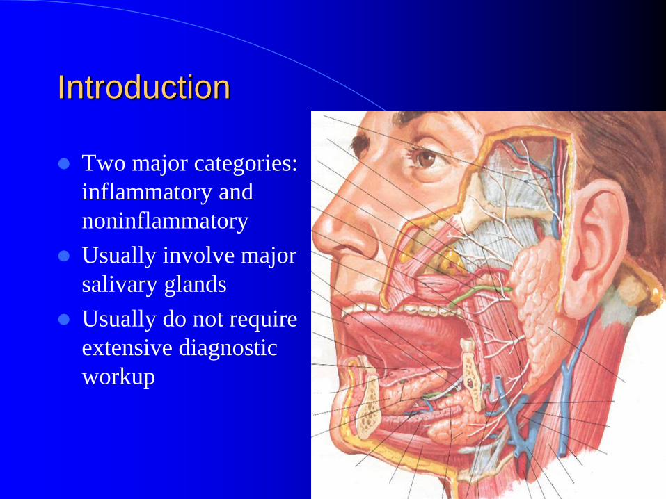
Introduction
Two major categories:
inflammatory and
noninflammatory
Usually involve major
salivary glands
Usually do not require
extensive diagnostic
workup
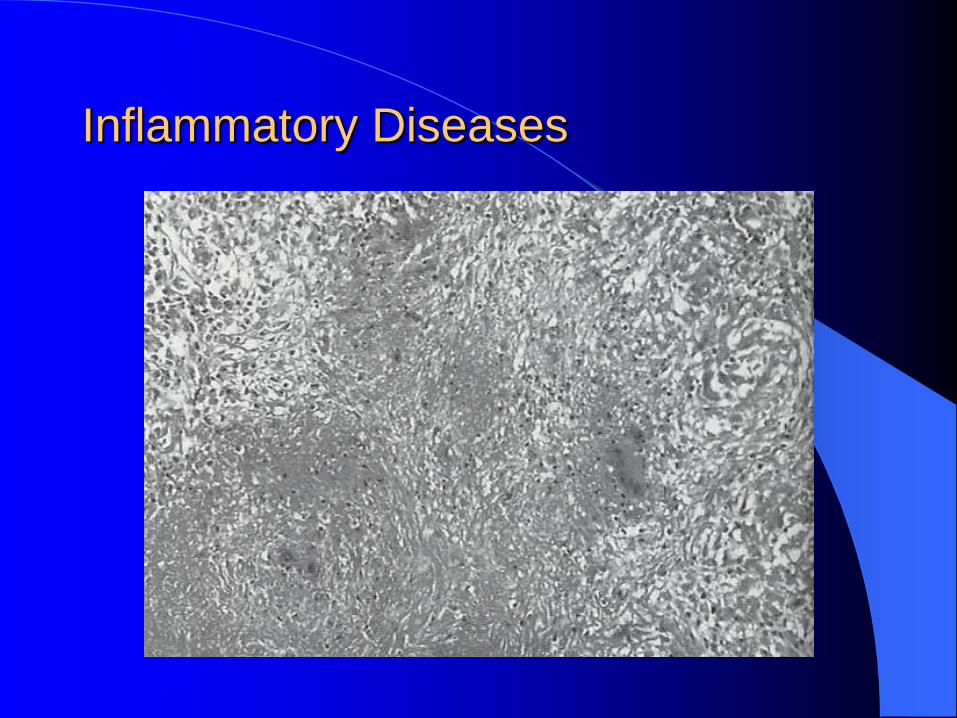
Inflammatory Diseases
Mumps
– Most common viral disorder of salivary glands
– Peak age 4-6
– 1 or both parotids after 2-3 week prodrome
– Diagnosis: serology or urine
– Complications: deafness, pancreatitis,
meningitis, orchitis, Type I DM, chronic
obstructive sialadenitis
Inflammatory Diseases
Other Viruses
– CMV, Coxsackievirus A, Echovirus, Influenza
A, Lymphocytic choriomeningitis Virus
– Treatment: symptomatic for all viral diseases
Acute Suppurative Sialadenitis
– Parotid most common site; peak age 50’s-60’s
– 30-40% in post-op patients; most commonly GI
procedures POD 3-5
Inflammatory Diseases
Acute Suppurative Sialadenitis
– Presentation: sudden, diffuse enlargement with associated induration and tenderness. Massage produces purulent saliva
– 20% of cases bilateral
– Pathogens: Staph aureus most common; Gram negatives, anaerobes also common
– Treatment: hydration, improved oral hygiene, repeated massage of gland, IV abx, warm compresses, sialogogues
Inflammatory Diseases
Acute Suppurative Sialadenitis
– If no significant improvement in 24-48h, then proceed to incision & drainage OR image-guided needle aspiration
Chronic Sialadenitis
– Most commonly parotid
– Usually from permanent damage during acute infection; occasionally from recurrent parotitis of childhood
Inflammatory Diseases
Chronic Sialadenitis
– Histologic changes: sialectasis, progressive
acinar destruction, lymphocytic infiltrates
– Saliva changes; returns to normal between
attacks
– Presentation: mild pain, recurrent parotid
enlargement that worsens with eating; 80%
develop xerostomia
Inflammatory Diseases
Inflammatory Diseases
Chronic Sialadenitis
– Treatment
1) Underlying causes
2) Sialogogues, massage, heat, hydration, abx
during acute attacks
3) Periodic ductal dilatation, duct ligation, total
gland irradiation, tympanic neurectomy
4) Excision
Inflammatory Diseases
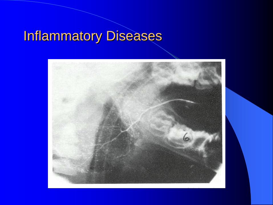
Recurrent Parotitis of Childhood
– More common in males; peak age 5-7
– ¾ give h/o Mumps; heredity plays no role
– Presentation: Usually unilateral; when bilateral,
one side worse
Severe pain, fever, malaise during attacks
Recurs
Inflammatory Diseases
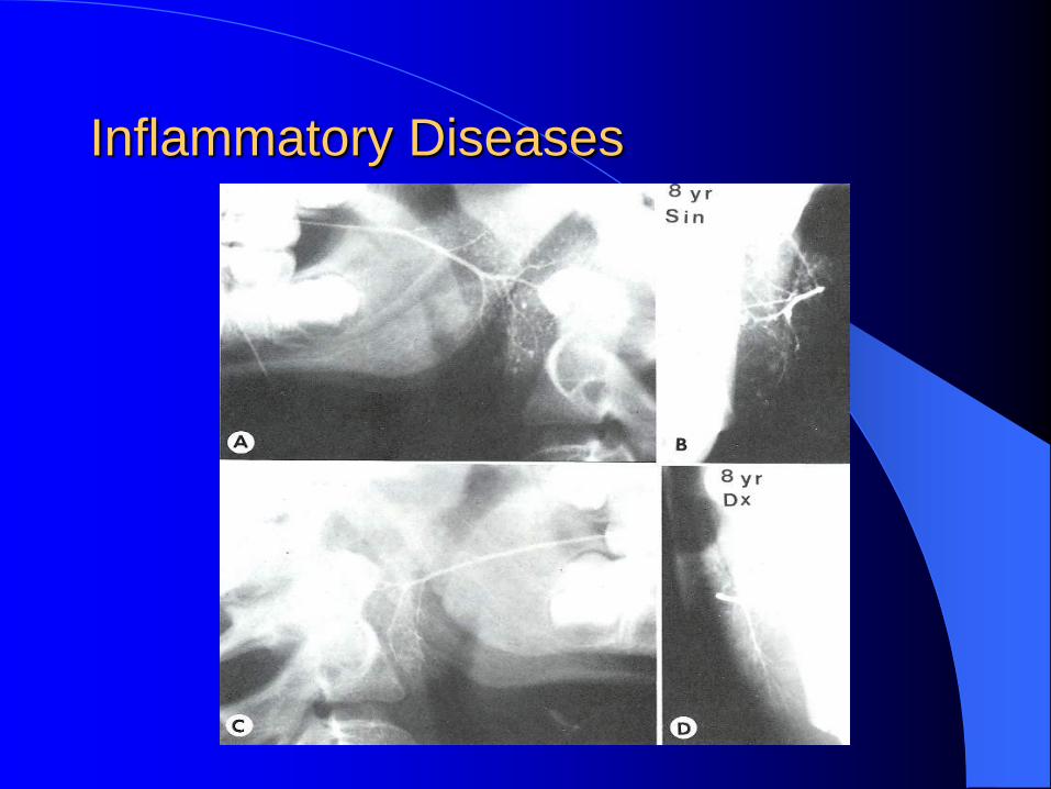
Recurrent Parotitis of Childhood
– Disease course (Ericson): onset age 3 months-
16 years
Exacerbations every 3-4 months
55% of cases resolve with puberty
25% no improvement with puberty
– Histology: massive B-cell infiltration and
dilated intraglandular ducts
Inflammatory Diseases
Inflammatory Diseases
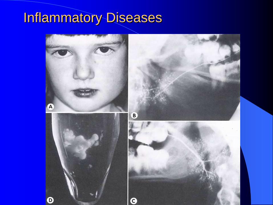
Recurrent Parotitis of Childhood
– Sialogram: multiple peripheral sialectases 1-2 mm in diameter; changes persist w/ resolution of symptoms
– Pathogens: flora ascend from oral cavity
Balls of soft material common, but rarely yields frank pus
– Treatment: Pen VK, massage, warmth, good oral hygiene, sialogogues, chewing gum
Inflammatory Diseases
Inflammatory Diseases
Benign Lymphoepithelial Lesion – Epimyoepithelial islands arise from
1) lymphoreticular infiltrates
2) acinar atrophy
3) ductal metaplasia
– Presentation: Asymptomatic enlargement of 1 gland
– Risk of lymphoma, carcinoma, pseudolymphoma
– No treatment necessary
Inflammatory Diseases
Primary Tuberculosis
– Presentation: Unilateral parotid
May present as acute inflammatory lesion or as chronic tumorous lesion
– Diagnosis: AFB stain of saliva AND PPD test
FNA if tumorous lesion
– Treatment: Anti-TB meds; excision if resistant
– Secondary TB: systemic dz.; submandibular and sublingual glands more often involved
Inflammatory Diseases
Inflammatory Diseases
Animal Scratch Disease
– Typically attacks periparotid lymph nodes
– Pathogens: Bartonella henselae, Afipia felis
– Diagnostic Criteria (3/4):
1) H/o contact w/ a cat and presence of scratch
2) + skin test or + serology for B henselae
3) + Gram stain and Cx
4) Histology: stellate abscesses, pleomorphic intracellular bacilli, Warthin-Starry stain
Inflammatory Diseases
Animal Scratch Disease
– Should place PPD to r/o Tb
– 96% resolve spontaneously within 2-6 months;
close followup needed until adenopathy
subsides
– Treatment: Bactrim X 1 week, or Rifampin X
1-2 weeks
IV Gentamicin in severe cases
Inflammatory Diseases
Actinomycosis
– Infection from tonsil or teeth
– Presentation: 61% visible sinus tracts; 40%
adenopathy; some have purplish skin
discoloration
– Histology: sulfur granules
– Diagnosis: culture
– Treatment: I&D, 2-6 weeks of IV Pen G
Inflammatory Diseases
Inflammatory Diseases
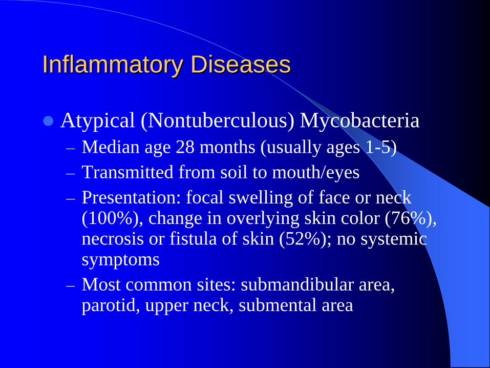
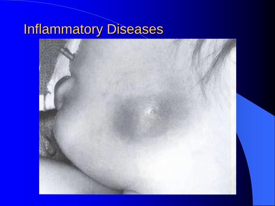
Atypical (Nontuberculous) Mycobacteria
– Median age 28 months (usually ages 1-5)
– Transmitted from soil to mouth/eyes
– Presentation: focal swelling of face or neck (100%), change in overlying skin color (76%), necrosis or fistula of skin (52%); no systemic symptoms
– Most common sites: submandibular area, parotid, upper neck, submental area
Inflammatory Diseases
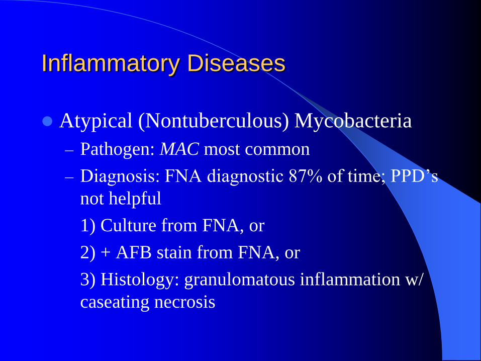
Atypical (Nontuberculous) Mycobacteria
– Pathogen: MAC most common
– Diagnosis: FNA diagnostic 87% of time; PPD’s
not helpful
1) Culture from FNA, or
2) + AFB stain from FNA, or
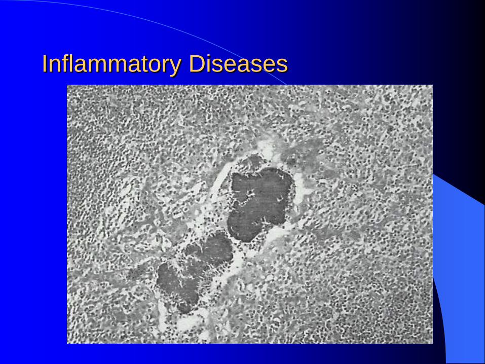
3) Histology: granulomatous inflammation w/
caseating necrosis
Inflammatory Diseases
Inflammatory Diseases
Atypical (Nontuberculous) Mycobacteria
– Treatment: Curettage vs. Excision
Curettage for lesions with extensive skin
necrosis or fluctuant parotid lesions
Surgical excision more effective
Medications controversial; Macrolides may
work for early disease
Inflammatory Diseases
Sarcoidosis
– 6% involve salivary glands clinically, 1/3 histologically
– Heerfordt’s syndrome (Uveoparotid fever):
1) Uveitis
2) Parotid enlargement
3) CN VII paralysis
Self-limited; uveitis can result in glaucoma – requires long term f/u
Inflammatory Diseases
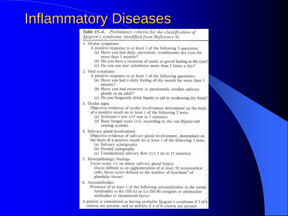
Sjogren’s Syndrome: Background
– Chronic, slowly progressive, benign; 2nd most
common autoimmune disease behind RA
– Lymphocyte-mediated destruction of exocrine
glands producing keratoconjunctivitis sicca and
xerostomia
– 90% middle-aged women
– 44% report PCN allergy
Inflammatory Diseases
Sjogren’s Syndrome: Background
– Primary=exocrine glands only; Secondary=coexisting autoimmune disease
Secondary form more common; salivary gland enlargement more common in primary form
– Serology (similar pattern in SLE):
1) ANA (50-80%)
2) RF (75%)
3) Ro/SS-A antibodies
4) La/SS-B antibodies; 3 or 4 in up to 90%
Inflammatory Diseases
Sjogren’s Syndrome: Presentation
– Xerostomia: most bothersome; difficulty swallowing dry food, difficulty speaking continuously, burning sensation, increased caries, problems wearing dentures; erythematous/sticky oral mucosa, atrophy of filiform papillae
– Keratoconjunctivitis Sicca: gritty feeling under eyelids, blurred vision, burning sensation, thick strands at inner canthi, decreased tearing, redness/itching, photosensitivity; results from destruction of conjunctival epithelium
Inflammatory Diseases
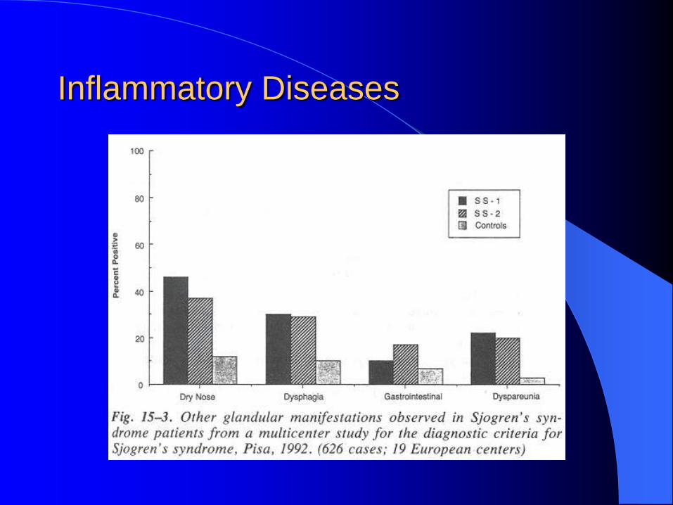
Sjogren’s Syndrome: Presentation
– Other exocrine gland involvement: dry nose, dry throat, xerotrachea, esophageal mucosal atrophy, atrophic gastritis, subclinical pancreatitis, vaginal dryness
– 1/3 = fatigue, low grade fever, myalgias/arthralgias
– Extraglandular involvement in ¼: Lungs, kidneys, vasculitis, nervous system
Inflammatory Diseases
Inflammatory Diseases
Sjogren’s Syndrome: Associated risks
– Increased risk of
1) NonHodgkin’s Lymphoma (RR=44)
2) Multiple Myeloma
Inflammatory Diseases
Inflammatory Diseases
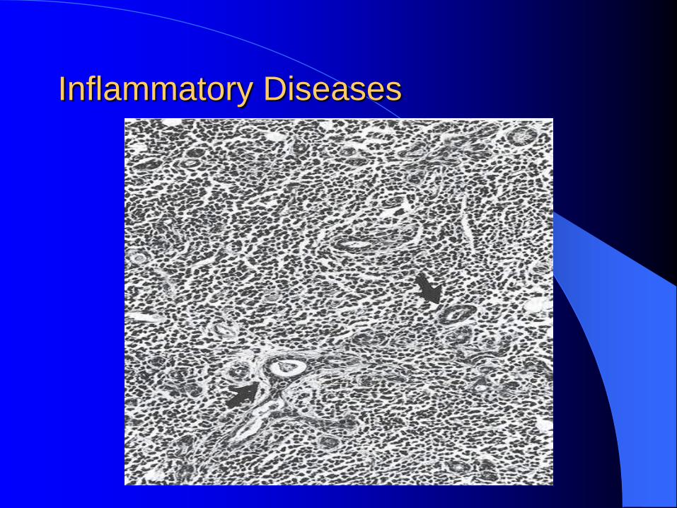
Sjogren’s Syndrome: Histology
– Severe lymphoid (T-cell) infiltrate can mimic lymphoma; heterogenous, lobular architecture preserved
– Enlarged lymph nodes w/ pleomorphic infiltrates and frequent mitotic figures = “pseudolymphoma”
– When biopsying, avoid epinephrine; send specimen in formalin
Inflammatory Diseases
Inflammatory Diseases
Sjogren’s Syndrome: Treatment
– Incurable disease
– Key=fluid replacement
Artificial tears; eye patching, boric acid ointments for corneal ulceration
Avoid diuretics, antihypertensives, antidepressants
– Medications: Pilocarpine 5 mg TID; hydroxychloroquine; glucocorticoids 1 mg/kg/day
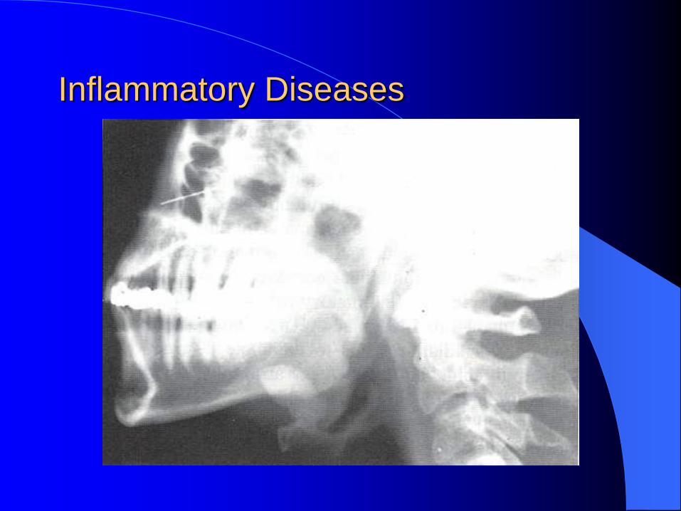
Noninflammatory Diseases Sialolithiasis
– 80% submandibular gland, 20% parotid
– Only 1 stone in ¾ cases
– 90% of submandibular stones radioopaque; 90% of parotid stones radiolucent
– Presentation: recurrent swelling, pain worse with eating
– Complications: sialadenitis, ductal ectasia, and stricture
– Treatment: If near duct orifice, transoral removal of stone with marsupialization
If near hilum, gland excision
Inflammatory Diseases
Noninflammatory Diseases
Cysts
– Mucoceles vs. Mucous cysts: minor salivary
glands
– 2-5% of all parotid lesions
– Congenital: dermoid cysts, ductal cysts, 1st arch
branchial cleft cysts
– Acquired: BLL, trauma, parotitis, calculi,
neoplasms
Noninflammatory Diseases
Trauma
– Identify the duct; can pass probe to ID distal duct; can milk gland to ID proximal duct
– Transected duct: end-to-end anastomosis over polyurethane catheter with 9-0 suture; remove catheter after 2 weeks
– Salivary-cutaneous fistula: repeat aspiration and pressure dressings; sialogram; excision if conservative treatment fails
– Blunt trauma: drain large hematomas early
Noninflammatory Diseases
Sialadenosis
– Nonneoplastic, noninflammatory enlargement
of salivary glands associated with systemic
disorders
– Usually asymptomatic
– Causes=obesity, malnutrition, malabsorption,
and alcoholic cirrhosis; very rarely does
sialadenosis occur in nonalcoholic cirrhosis
Noninflammatory Diseases
Cheilitis Glandularis
– Enlargement of the labial salivary glands; clear, thick, sticky mucus; can result in lower lip eversion
– Treatment: vermilionectomy
Kussmaul’s Disease (Dialodochitis Fibrinosa)
– Mucous plug obstructing duct
– Treatment: rehydration, gentle massage, sialogogues
Noninflammatory Diseases
Necrotizing Sialometaplasia
– Benign, self-healing process of unknown etiology
– Presentation: usually hard palate, usually males; asymptomatic mucosal ulceration
– Histology: easily mistaken for SCCA, mucoepidermoid CA; lobular necrosis + squamous metaplasia + preserved lobular architecture
– Treatment: biopsy for diagnosis, but treatment unnecessary
– Subacute necrotizing sialadenitis=painful, nodular variant