Embed Size (px)
Citation preview
North Carolina Macular Dystrophy Is Causedby Dysregulation of the Retinal TranscriptionFactor PRDM13
Kent W. Small, MD,1 Adam P. DeLuca, PhD,2 S. Scott Whitmore, PhD,2 Thomas Rosenberg, MD,3
Rosemary Silva-Garcia, MD,1 Nitin Udar, PhD,1 Bernard Puech, MD,4 Charles A. Garcia, MD,5
Thomas A. Rice, MD,6 Gerald A. Fishman, MD,7 Elise Héon, MD,8 James C. Folk, MD,2 Luan M. Streb, BA,2
Christine M. Haas, BA,2 Luke A. Wiley, PhD,2 Todd E. Scheetz, PhD,2 John H. Fingert, MD, PhD,2
Robert F. Mullins, PhD,2 Budd A. Tucker, PhD,2 Edwin. M. Stone, MD, PhD2
Purpose: To identify specific mutations causing North Carolina macular dystrophy (NCMD).Design: Whole-genome sequencing coupled with reverse transcription polymerase chain reaction (RT-PCR)
analysis of gene expression in human retinal cells.Participants: A total of 141 members of 12 families with NCMD and 261 unrelated control individuals.Methods: Genome sequencing was performed on 8 affected individuals from 3 families affected with
chromosome 6elinked NCMD (MCDR1) and 2 individuals affected with chromosome 5elinked NCMD (MCDR3).Variants observed in the MCDR1 locus with frequencies <1% in published databases were confirmed usingSanger sequencing. Confirmed variants absent from all published databases were sought in 8 additional MCDR1families and 261 controls. The RT-PCR analysis of selected genes was performed in stem cellederived humanretinal cells.
Main Outcome Measures: Co-segregation of rare genetic variants with disease phenotype.Results: Five sequenced individuals with MCDR1-linked NCMD shared a haplotype of 14 rare variants
spanning 1 Mb of the disease-causing allele. One of these variants (V1) was absent from all published databasesand all 261 controls, but was found in 5 additional NCMD kindreds. This variant lies in a DNase 1 hypersensitivitysite (DHS) upstream of both the PRDM13 and CCNC genes. Sanger sequencing of 1 kb centered on V1 wasperformed in the remaining 4 NCMD probands, and 2 additional novel single nucleotide variants (V2 in 3 familiesand V3 in 1 family) were identified in the DHS within 134 bp of the location of V1. A complete duplication of thePRDM13 gene was also discovered in a single family (V4). The RT-PCR analysis of PRDM13 expression indeveloping retinal cells revealed marked developmental regulation. Next-generation sequencing of 2 individualswith MCDR3-linked NCMD revealed a 900-kb duplication that included the entire IRX1 gene (V5). The 5 mutationsV1 to V5 segregated perfectly in the 102 affected and 39 unaffected members of the 12 NCMD families.
Conclusions: We identified 5 rare mutations, each capable of arresting human macular development. Four ofthese strongly implicate the involvement of PRDM13 in macular development, whereas the pathophysiologicmechanism of the fifth remains unknown but may involve the developmental dysregulation ofIRX1. Ophthalmology 2015;-:1e10 ª 2015 by the American Academy of Ophthalmology. This is an open accessarticle under the CC BY-NC-ND license (http://creativecommons.org/licenses/by-nc-nd/4.0/).
Supplemental material is available at www.aaojournal.org.
Few tissues in the human body are as important to the well-being of a person as the central 3 mm of the human retina.The ability to drive a car, recognize friends in public, andsee words on a computer, cell phone or printed page are justa few of the many activities of daily living that dependheavily on the normal function of the macula.
For all but a few people, the macula functions very wellfor the first 6 or 7 decades of life, but in older individuals,the macula is prone to a genetically and mechanisticallydiverse group of disorders that are known collectively as age-related macular degeneration (AMD). For many years, the
� 2015 by the American Academy of OphthalmologyThis is an open access article under the CC BY-NC-ND license(http://creativecommons.org/licenses/by-nc-nd/4.0/). Published by Elsevier Inc.
neovascular complications of AMD were the most commoncause of irreversible blindness in developed countries.1e4
However, the recent advent of antievascular endothelialgrowth factor drugs5e8 has dramatically reduced vision lossfrom neovascularization, thereby increasing the fraction ofblindness caused by geographic atrophy of the macula.
There are at least 2 approaches that one could envision forreducing the burden of blindness caused by geographic atrophyof the macula. The first would be to understand the patho-physiologic mechanisms of AMD in sufficient detail that onecould detect the disease at a very early stage, perhaps even as an
1http://dx.doi.org/10.1016/j.ophtha.2015.10.006ISSN 0161-6420/15
Ophthalmology Volume -, Number -, Month 2015
asymptomatic genetic predisposition, and deliver a safe andeffective preventive therapy to those at risk, much as statins arenow used to reduce the risk of heart disease. Another strategywould be to rebuild an injured macula with new stemcellederived retinal cells.9,10 Molecular genetics will play animportant role in both of these approaches.
In the 1990s, scientists sought the genetic causes of severalMendelian forms of human macular disease for at least 2 rea-sons. First, it was possible that mild mutations in the genesresponsible for these early-onset conditions might prove to beresponsible for a significant subset of the age-related forms ofthe disease. Second, it was thought that by discovering howrelatively minor alterations of individual genes could causeclinical findings similar to AMD, one would gain valuableinsight into the normal function of the macula. Twenty yearslater, it is clear that none of the genes that cause the classicMendelian macular dystrophies cause a significant fraction ofthe late-onset disease, and none of the genes that have beenshown to predispose people to typical AMD cause any mean-ingful fraction of early-onset Mendelian macular disease.
The first of the classic macular dystrophies to have its genemapped to a chromosome,11 North Carolina macular dystrophy(NCMD), is the last to have its specific disease-causing muta-tions identified. The reason for this delaydthe unusual devel-opmental mechanism of this diseasedmay ultimately makeNCMD themost relevant of theMendelianmacular dystrophiesto the treatment of AMD. North Carolina macular dystrophywas first described in a large kindred from North Carolina byLefler et al12 and later described in more detail by Frank et al.13
The cross-sectional nature of these studies led the investigatorsto believe that the disease was slowly progressive. However,Small14 reexamined the original Lefler kindred approximately20 years later and realized that NCMD is in fact anonprogressive developmental disorder with widely variableexpressivity.
In the decades since the MCDR1 locus was mapped, manyadditional families with NCMD have been described,15e20
including 2 families that link to a separate locus on chromo-some 5 (MCDR3).21,22 The critical region on chromosome 6has been considerably narrowed,23,24 and all of the coding re-gions of genes within this interval have been exhaustivelystudied by us and other investigators.25 The failure of theseexperiments to identify plausible disease-causing mutations inany of these kindreds suggested that the mutations were likelyto exist in nonexomic DNA and to affect the expression of anearby gene or genes rather than the structure of its geneproduct. The purpose of this study was to take advantage ofrecent advances in whole-genome sequencing to comprehen-sively screen the nonexomic sequences within theMCDR1 andMCDR3 loci to identify disease-causing mutations in familiesaffected with these diseases.
Methods
Human Subjects
All subjects provided written informed consent for this researchstudy, which was approved by the institutional review board ofthe University of Iowa and adhered to the tenets set forth inthe Declaration of Helsinki. Blood samples were obtained from
2
all subjects, and DNA was extracted using a nonorganic protocol,as previously described.26
Next-Generation Sequencing of MCDR1 Patients
A targeted genome capture of the linked region was performed on3 members of family A (2 affected and 1 unaffected), 2 membersof family K, and 1 member of family B. Libraries prepared fromthese captures were sequenced on an Illumina HiSeq. In addition,30� whole genomes were obtained from 5 affected individuals:2 from family A, 1 from family K, and 2 from family L. Theselibraries were sequenced on an Illumina HiSeqX. All of theseindividuals are noted in blue in Supplemental Figure 1 (available atwww.aaojournal.org).
Bioinformatic Analysis of Next-GenerationSequencing Data
Sequences were analyzed as described previously.27 Briefly,sequences were aligned to the reference genome using BWA-mem,and single nucleotide variants and small insertions/deletions wereidentified using a GATK-based pipeline.28,29 Variants mappingoutside the MCDR1-linked region and those found at a frequency of1% or greater in public databases30e32 were removed. Variants werethen filtered, requiring that all affected individuals with a givenhaplotype share the heterozygous variant; all other individuals did notshare the variant.
Copy number variants were investigated using Pindel and manualinspection of the aligned sequence data using the Integrative GenomeViewer.33,34 As a control, the identified genes were screened for copynumber variants using Conifer35 in an internal database of 953 wholeexomes of patients with eye disease.
Confirmation of Whole-Genome Sequencing
Variants identified by whole-genome sequencing were confirmedusing automated bidirectional DNA sequencing with dye termi-nation chemistry on an ABI 3730 sequencer (ThermoFisher Sci-entific, Foster City, CA).
Screening of Control Subjects
A total of 261 normal control subjects were screened for the presenceof V1 to V3 (Table 1) using unidirectional automated DNAsequencing. To evaluate these controls for the presence of V4 andV5 (Table 1), oligonucleotide primers were designed to amplifyacross the novel junctions created by these tandem duplications(Supplemental Table 1, available at www.aaojournal.org), and theproducts of these amplifications were evaluated by electrophoresison 6% nondenaturing polyacrylamide gels followed by silverstaining, as previously described.36
Induced Pluripotent Stem Cell Generation and 3-Dimensional Differentiation. Human dermal fibroblasts were iso-lated from skin biopsies obtained from normal individuals afterinformed consent. Cultured fibroblasts were reprogrammed via viraltransduction of the transcription factors OCT4, SOX2, KLF4, andc-MYC, as previously described.27,37,38 Human induced pluripotentstem cells (iPSCs) were maintained in Essential 8 media (Life Tech-nologies, Carlsbad, CA) on Laminin 521ecoated plates (Corning LifeSciences, Tewksbury, MA). To initiate differentiation, iPSCs wereremoved from the culture substrate via incubation with TrypLEExpress Enzyme (Life Technologies) dissociated into a single cellsuspension and subsequently differentiated via the 3-dimensional (3D)differentiation protocol previously published by Eiraku et al.39
RNA Isolation and Reverse Transcription Polymerase ChainReaction. Total RNA was extracted from normal human iPSCsisolated at 0, 30, 60, and 100 days after differentiation using the
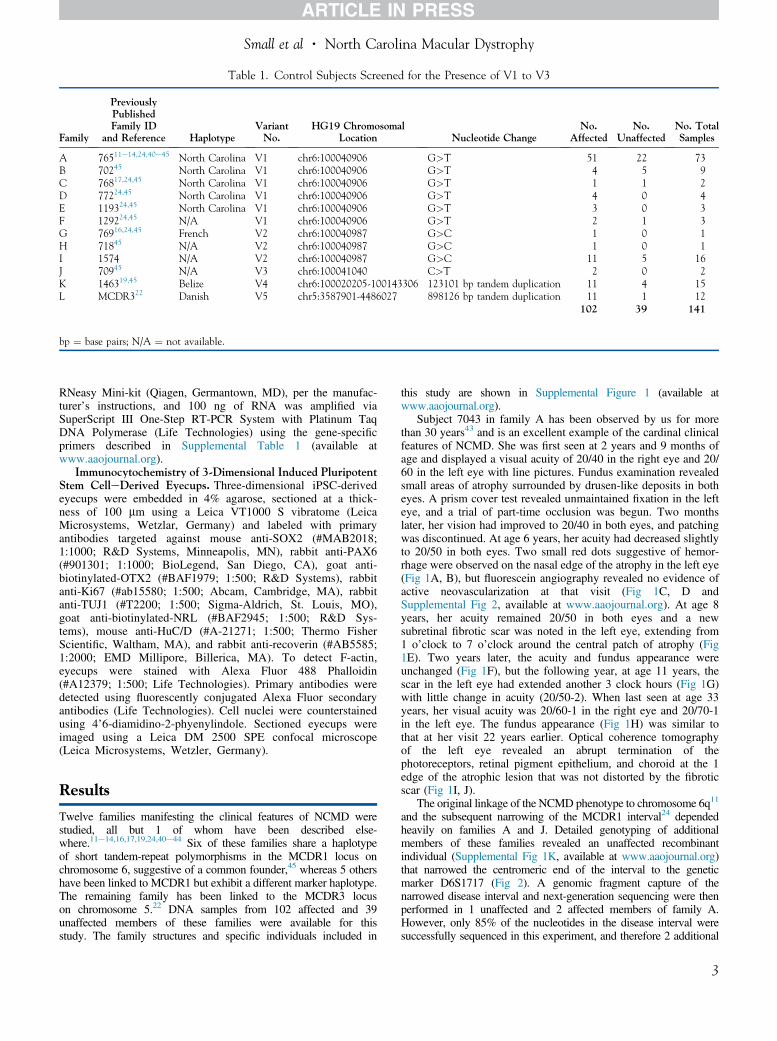
Table 1. Control Subjects Screened for the Presence of V1 to V3
Family
PreviouslyPublishedFamily ID
and Reference HaplotypeVariantNo.
HG19 ChromosomalLocation Nucleotide Change
No.Affected
No.Unaffected
No. TotalSamples
A 76511e14,24,40e45 North Carolina V1 chr6:100040906 G>T 51 22 73B 70245 North Carolina V1 chr6:100040906 G>T 4 5 9C 76817,24,45 North Carolina V1 chr6:100040906 G>T 1 1 2D 77224,45 North Carolina V1 chr6:100040906 G>T 4 0 4E 119324,45 North Carolina V1 chr6:100040906 G>T 3 0 3F 129224,45 N/A V1 chr6:100040906 G>T 2 1 3G 76916,24,45 French V2 chr6:100040987 G>C 1 0 1H 71845 N/A V2 chr6:100040987 G>C 1 0 1I 1574 N/A V2 chr6:100040987 G>C 11 5 16J 70945 N/A V3 chr6:100041040 C>T 2 0 2K 146319,45 Belize V4 chr6:100020205-100143306 123101 bp tandem duplication 11 4 15L MCDR322 Danish V5 chr5:3587901-4486027 898126 bp tandem duplication 11 1 12
102 39 141
bp ¼ base pairs; N/A ¼ not available.
Small et al � North Carolina Macular Dystrophy
RNeasy Mini-kit (Qiagen, Germantown, MD), per the manufac-turer’s instructions, and 100 ng of RNA was amplified viaSuperScript III One-Step RT-PCR System with Platinum TaqDNA Polymerase (Life Technologies) using the gene-specificprimers described in Supplemental Table 1 (available atwww.aaojournal.org).
Immunocytochemistry of 3-Dimensional Induced PluripotentStem CelleDerived Eyecups. Three-dimensional iPSC-derivedeyecups were embedded in 4% agarose, sectioned at a thick-ness of 100 mm using a Leica VT1000 S vibratome (LeicaMicrosystems, Wetzlar, Germany) and labeled with primaryantibodies targeted against mouse anti-SOX2 (#MAB2018;1:1000; R&D Systems, Minneapolis, MN), rabbit anti-PAX6(#901301; 1:1000; BioLegend, San Diego, CA), goat anti-biotinylated-OTX2 (#BAF1979; 1:500; R&D Systems), rabbitanti-Ki67 (#ab15580; 1:500; Abcam, Cambridge, MA), rabbitanti-TUJ1 (#T2200; 1:500; Sigma-Aldrich, St. Louis, MO),goat anti-biotinylated-NRL (#BAF2945; 1:500; R&D Sys-tems), mouse anti-HuC/D (#A-21271; 1:500; Thermo FisherScientific, Waltham, MA), and rabbit anti-recoverin (#AB5585;1:2000; EMD Millipore, Billerica, MA). To detect F-actin,eyecups were stained with Alexa Fluor 488 Phalloidin(#A12379; 1:500; Life Technologies). Primary antibodies weredetected using fluorescently conjugated Alexa Fluor secondaryantibodies (Life Technologies). Cell nuclei were counterstainedusing 4’6-diamidino-2-phyenylindole. Sectioned eyecups wereimaged using a Leica DM 2500 SPE confocal microscope(Leica Microsystems, Wetzler, Germany).
Results
Twelve families manifesting the clinical features of NCMD werestudied, all but 1 of whom have been described else-where.11e14,16,17,19,24,40e44 Six of these families share a haplotypeof short tandem-repeat polymorphisms in the MCDR1 locus onchromosome 6, suggestive of a common founder,45 whereas 5 othershave been linked to MCDR1 but exhibit a different marker haplotype.The remaining family has been linked to the MCDR3 locuson chromosome 5.22 DNA samples from 102 affected and 39unaffected members of these families were available for thisstudy. The family structures and specific individuals included in
this study are shown in Supplemental Figure 1 (available atwww.aaojournal.org).
Subject 7043 in family A has been observed by us for morethan 30 years43 and is an excellent example of the cardinal clinicalfeatures of NCMD. She was first seen at 2 years and 9 months ofage and displayed a visual acuity of 20/40 in the right eye and 20/60 in the left eye with line pictures. Fundus examination revealedsmall areas of atrophy surrounded by drusen-like deposits in botheyes. A prism cover test revealed unmaintained fixation in the lefteye, and a trial of part-time occlusion was begun. Two monthslater, her vision had improved to 20/40 in both eyes, and patchingwas discontinued. At age 6 years, her acuity had decreased slightlyto 20/50 in both eyes. Two small red dots suggestive of hemor-rhage were observed on the nasal edge of the atrophy in the left eye(Fig 1A, B), but fluorescein angiography revealed no evidence ofactive neovascularization at that visit (Fig 1C, D andSupplemental Fig 2, available at www.aaojournal.org). At age 8years, her acuity remained 20/50 in both eyes and a newsubretinal fibrotic scar was noted in the left eye, extending from1 o’clock to 7 o’clock around the central patch of atrophy (Fig1E). Two years later, the acuity and fundus appearance wereunchanged (Fig 1F), but the following year, at age 11 years, thescar in the left eye had extended another 3 clock hours (Fig 1G)with little change in acuity (20/50-2). When last seen at age 33years, her visual acuity was 20/60-1 in the right eye and 20/70-1in the left eye. The fundus appearance (Fig 1H) was similar tothat at her visit 22 years earlier. Optical coherence tomographyof the left eye revealed an abrupt termination of thephotoreceptors, retinal pigment epithelium, and choroid at the 1edge of the atrophic lesion that was not distorted by the fibroticscar (Fig 1I, J).
The original linkage of the NCMD phenotype to chromosome 6q11
and the subsequent narrowing of the MCDR1 interval24 dependedheavily on families A and J. Detailed genotyping of additionalmembers of these families revealed an unaffected recombinantindividual (Supplemental Fig 1K, available at www.aaojournal.org)that narrowed the centromeric end of the interval to the geneticmarker D6S1717 (Fig 2). A genomic fragment capture of thenarrowed disease interval and next-generation sequencing were thenperformed in 1 unaffected and 2 affected members of family A.However, only 85% of the nucleotides in the disease interval weresuccessfully sequenced in this experiment, and therefore 2 additional
3
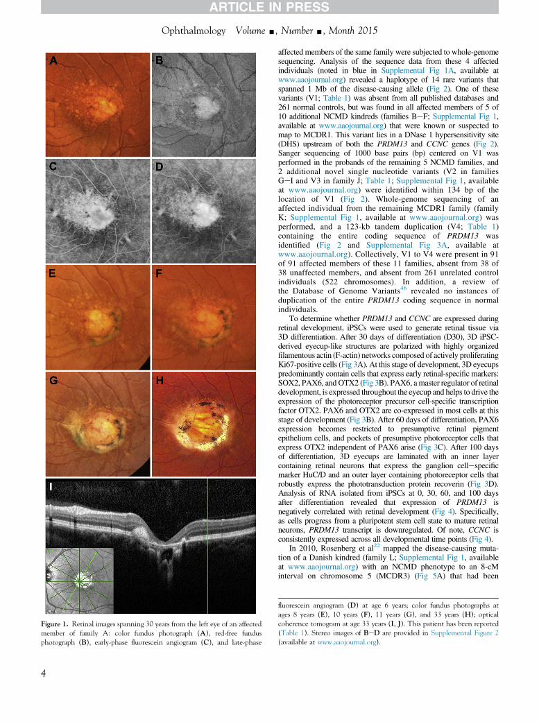
Figure 1. Retinal images spanning 30 years from the left eye of an affectedmember of family A: color fundus photograph (A), red-free fundusphotograph (B), early-phase fluorescein angiogram (C), and late-phase
Ophthalmology Volume -, Number -, Month 2015
4
affected members of the same family were subjected to whole-genomesequencing. Analysis of the sequence data from these 4 affectedindividuals (noted in blue in Supplemental Fig 1A, available atwww.aaojournal.org) revealed a haplotype of 14 rare variants thatspanned 1 Mb of the disease-causing allele (Fig 2). One of thesevariants (V1; Table 1) was absent from all published databases and261 normal controls, but was found in all affected members of 5 of10 additional NCMD kindreds (families BeF; Supplemental Fig 1,available at www.aaojournal.org) that were known or suspected tomap to MCDR1. This variant lies in a DNase 1 hypersensitivity site(DHS) upstream of both the PRDM13 and CCNC genes (Fig 2).Sanger sequencing of 1000 base pairs (bp) centered on V1 wasperformed in the probands of the remaining 5 NCMD families, and2 additional novel single nucleotide variants (V2 in familiesGeI and V3 in family J; Table 1; Supplemental Fig 1, availableat www.aaojournal.org) were identified within 134 bp of thelocation of V1 (Fig 2). Whole-genome sequencing of anaffected individual from the remaining MCDR1 family (familyK; Supplemental Fig 1, available at www.aaojournal.org) wasperformed, and a 123-kb tandem duplication (V4; Table 1)containing the entire coding sequence of PRDM13 wasidentified (Fig 2 and Supplemental Fig 3A, available atwww.aaojournal.org). Collectively, V1 to V4 were present in 91of 91 affected members of these 11 families, absent from 38 of38 unaffected members, and absent from 261 unrelated controlindividuals (522 chromosomes). In addition, a review ofthe Database of Genome Variants46 revealed no instances ofduplication of the entire PRDM13 coding sequence in normalindividuals.
To determine whether PRDM13 and CCNC are expressed duringretinal development, iPSCs were used to generate retinal tissue via3D differentiation. After 30 days of differentiation (D30), 3D iPSC-derived eyecup-like structures are polarized with highly organizedfilamentous actin (F-actin) networks composed of actively proliferatingKi67-positive cells (Fig 3A). At this stage of development, 3D eyecupspredominantly contain cells that express early retinal-specific markers:SOX2, PAX6, andOTX2 (Fig 3B). PAX6, amaster regulator of retinaldevelopment, is expressed throughout the eyecup and helps to drive theexpression of the photoreceptor precursor cell-specific transcriptionfactor OTX2. PAX6 and OTX2 are co-expressed in most cells at thisstage of development (Fig 3B). After 60 days of differentiation, PAX6expression becomes restricted to presumptive retinal pigmentepithelium cells, and pockets of presumptive photoreceptor cells thatexpress OTX2 independent of PAX6 arise (Fig 3C). After 100 daysof differentiation, 3D eyecups are laminated with an inner layercontaining retinal neurons that express the ganglion cellespecificmarker HuC/D and an outer layer containing photoreceptor cells thatrobustly express the phototransduction protein recoverin (Fig 3D).Analysis of RNA isolated from iPSCs at 0, 30, 60, and 100 daysafter differentiation revealed that expression of PRDM13 isnegatively correlated with retinal development (Fig 4). Specifically,as cells progress from a pluripotent stem cell state to mature retinalneurons, PRDM13 transcript is downregulated. Of note, CCNC isconsistently expressed across all developmental time points (Fig 4).
In 2010, Rosenberg et al22 mapped the disease-causing muta-tion of a Danish kindred (family L; Supplemental Fig 1, availableat www.aaojournal.org) with an NCMD phenotype to an 8-cMinterval on chromosome 5 (MCDR3) (Fig 5A) that had been
fluorescein angiogram (D) at age 6 years; color fundus photographs atages 8 years (E), 10 years (F), 11 years (G), and 33 years (H); opticalcoherence tomogram at age 33 years (I, J). This patient has been reported(Table 1). Stereo images of BeD are provided in Supplemental Figure 2(available at www.aaojournal.org).
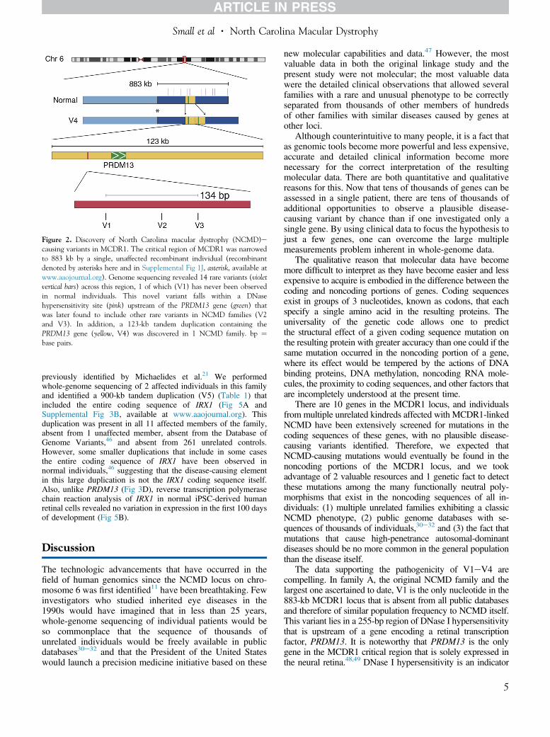
Figure 2. Discovery of North Carolina macular dystrophy (NCMD)ecausing variants in MCDR1. The critical region of MCDR1 was narrowedto 883 kb by a single, unaffected recombinant individual (recombinantdenoted by asterisks here and in Supplemental Fig 1J, asterisk, available atwww.aaojournal.org). Genome sequencing revealed 14 rare variants (violetvertical bars) across this region, 1 of which (V1) has never been observedin normal individuals. This novel variant falls within a DNasehypersensitivity site (pink) upstream of the PRDM13 gene (green) thatwas later found to include other rare variants in NCMD families (V2and V3). In addition, a 123-kb tandem duplication containing thePRDM13 gene (yellow, V4) was discovered in 1 NCMD family. bp ¼base pairs.
Small et al � North Carolina Macular Dystrophy
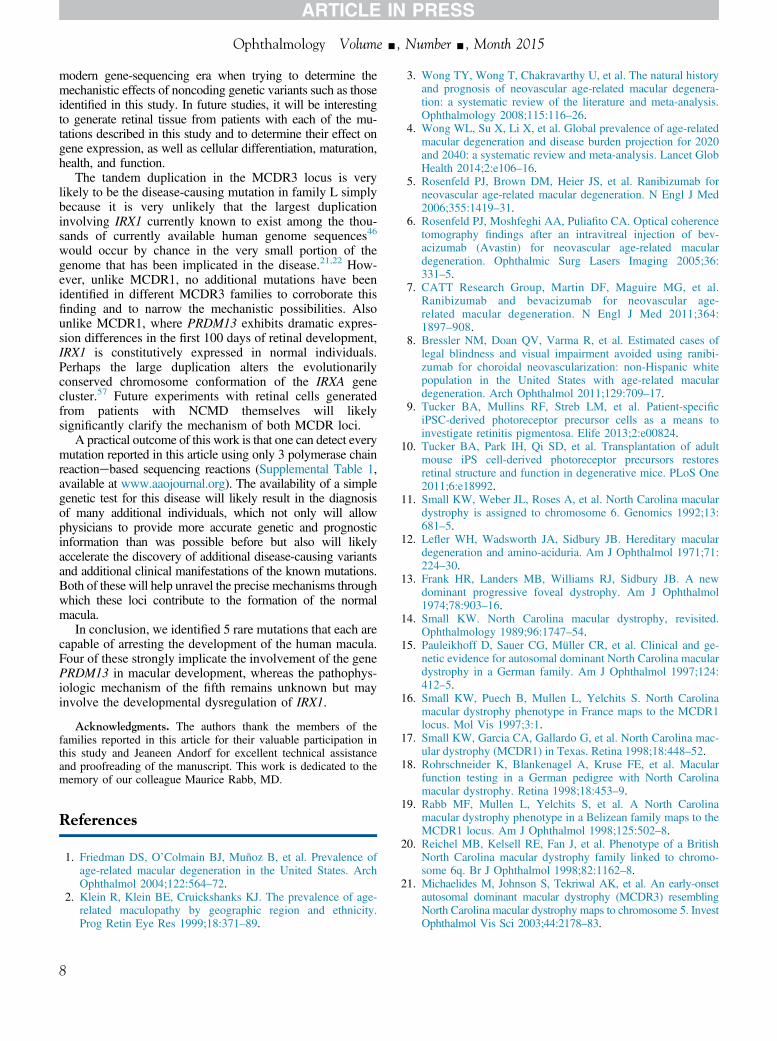
previously identified by Michaelides et al.21 We performedwhole-genome sequencing of 2 affected individuals in this familyand identified a 900-kb tandem duplication (V5) (Table 1) thatincluded the entire coding sequence of IRX1 (Fig 5A andSupplemental Fig 3B, available at www.aaojournal.org). Thisduplication was present in all 11 affected members of the family,absent from 1 unaffected member, absent from the Database ofGenome Variants,46 and absent from 261 unrelated controls.However, some smaller duplications that include in some casesthe entire coding sequence of IRX1 have been observed innormal individuals,46 suggesting that the disease-causing elementin this large duplication is not the IRX1 coding sequence itself.Also, unlike PRDM13 (Fig 3D), reverse transcription polymerasechain reaction analysis of IRX1 in normal iPSC-derived humanretinal cells revealed no variation in expression in the first 100 daysof development (Fig 5B).
Discussion
The technologic advancements that have occurred in thefield of human genomics since the NCMD locus on chro-mosome 6 was first identified11 have been breathtaking. Fewinvestigators who studied inherited eye diseases in the1990s would have imagined that in less than 25 years,whole-genome sequencing of individual patients would beso commonplace that the sequence of thousands ofunrelated individuals would be freely available in publicdatabases30e32 and that the President of the United Stateswould launch a precision medicine initiative based on these
new molecular capabilities and data.47 However, the mostvaluable data in both the original linkage study and thepresent study were not molecular; the most valuable datawere the detailed clinical observations that allowed severalfamilies with a rare and unusual phenotype to be correctlyseparated from thousands of other members of hundredsof other families with similar diseases caused by genes atother loci.
Although counterintuitive to many people, it is a fact thatas genomic tools become more powerful and less expensive,accurate and detailed clinical information become morenecessary for the correct interpretation of the resultingmolecular data. There are both quantitative and qualitativereasons for this. Now that tens of thousands of genes can beassessed in a single patient, there are tens of thousands ofadditional opportunities to observe a plausible disease-causing variant by chance than if one investigated only asingle gene. By using clinical data to focus the hypothesis tojust a few genes, one can overcome the large multiplemeasurements problem inherent in whole-genome data.
The qualitative reason that molecular data have becomemore difficult to interpret as they have become easier and lessexpensive to acquire is embodied in the difference between thecoding and noncoding portions of genes. Coding sequencesexist in groups of 3 nucleotides, known as codons, that eachspecify a single amino acid in the resulting proteins. Theuniversality of the genetic code allows one to predictthe structural effect of a given coding sequence mutation onthe resulting protein with greater accuracy than one could if thesame mutation occurred in the noncoding portion of a gene,where its effect would be tempered by the actions of DNAbinding proteins, DNA methylation, noncoding RNA mole-cules, the proximity to coding sequences, and other factors thatare incompletely understood at the present time.
There are 10 genes in the MCDR1 locus, and individualsfrom multiple unrelated kindreds affected with MCDR1-linkedNCMD have been extensively screened for mutations in thecoding sequences of these genes, with no plausible disease-causing variants identified. Therefore, we expected thatNCMD-causing mutations would eventually be found in thenoncoding portions of the MCDR1 locus, and we tookadvantage of 2 valuable resources and 1 genetic fact to detectthese mutations among the many functionally neutral poly-morphisms that exist in the noncoding sequences of all in-dividuals: (1) multiple unrelated families exhibiting a classicNCMD phenotype, (2) public genome databases with se-quences of thousands of individuals,30e32 and (3) the fact thatmutations that cause high-penetrance autosomal-dominantdiseases should be no more common in the general populationthan the disease itself.
The data supporting the pathogenicity of V1eV4 arecompelling. In family A, the original NCMD family and thelargest one ascertained to date, V1 is the only nucleotide in the883-kb MCDR1 locus that is absent from all public databasesand therefore of similar population frequency to NCMD itself.This variant lies in a 255-bp region of DNase I hypersensitivitythat is upstream of a gene encoding a retinal transcriptionfactor, PRDM13. It is noteworthy that PRDM13 is the onlygene in the MCDR1 critical region that is solely expressed inthe neural retina.48,49 DNase I hypersensitivity is an indicator
5
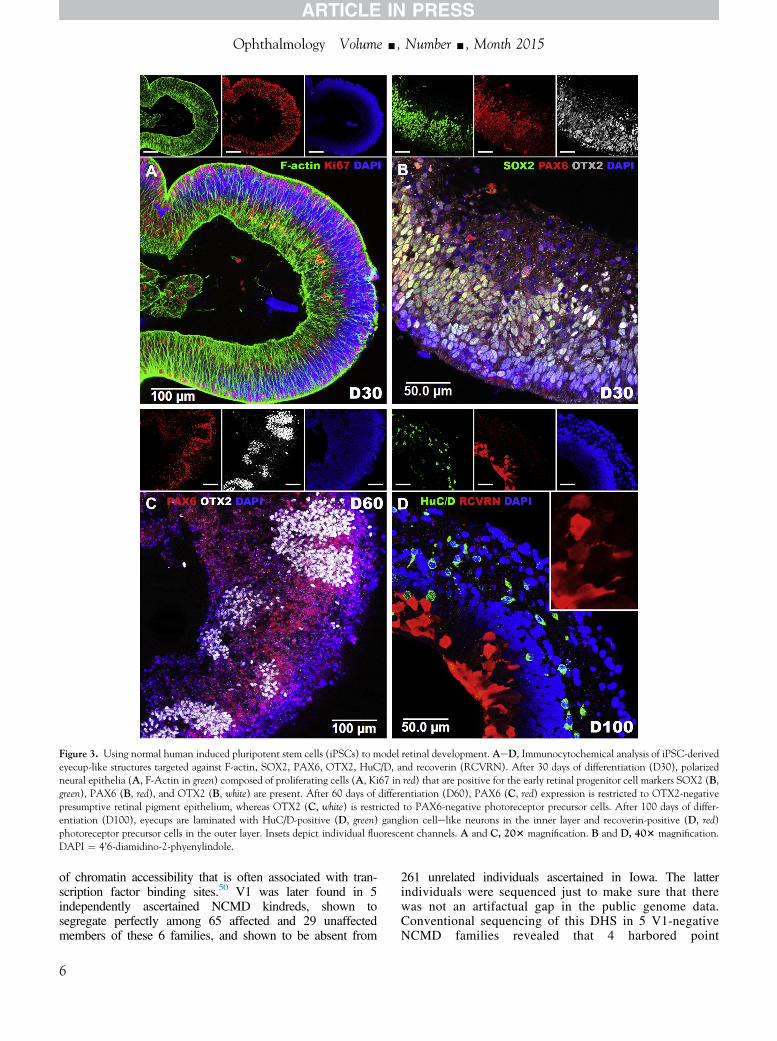
Figure 3. Using normal human induced pluripotent stem cells (iPSCs) to model retinal development. AeD, Immunocytochemical analysis of iPSC-derivedeyecup-like structures targeted against F-actin, SOX2, PAX6, OTX2, HuC/D, and recoverin (RCVRN). After 30 days of differentiation (D30), polarizedneural epithelia (A, F-Actin in green) composed of proliferating cells (A, Ki67 in red) that are positive for the early retinal progenitor cell markers SOX2 (B,green), PAX6 (B, red), and OTX2 (B, white) are present. After 60 days of differentiation (D60), PAX6 (C, red) expression is restricted to OTX2-negativepresumptive retinal pigment epithelium, whereas OTX2 (C, white) is restricted to PAX6-negative photoreceptor precursor cells. After 100 days of differ-entiation (D100), eyecups are laminated with HuC/D-positive (D, green) ganglion cellelike neurons in the inner layer and recoverin-positive (D, red)photoreceptor precursor cells in the outer layer. Insets depict individual fluorescent channels. A and C, 203 magnification. B and D, 403 magnification.DAPI ¼ 4’6-diamidino-2-phyenylindole.
Ophthalmology Volume -, Number -, Month 2015
of chromatin accessibility that is often associated with tran-scription factor binding sites.50 V1 was later found in 5independently ascertained NCMD kindreds, shown tosegregate perfectly among 65 affected and 29 unaffectedmembers of these 6 families, and shown to be absent from
6
261 unrelated individuals ascertained in Iowa. The latterindividuals were sequenced just to make sure that therewas not an artifactual gap in the public genome data.Conventional sequencing of this DHS in 5 V1-negativeNCMD families revealed that 4 harbored point
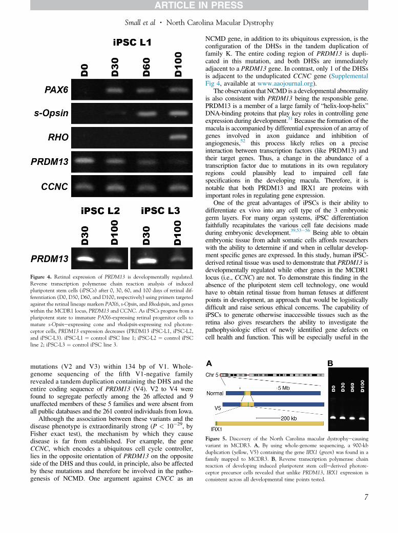
Figure 4. Retinal expression of PRDM13 is developmentally regulated.Reverse transcription polymerase chain reaction analysis of inducedpluripotent stem cells (iPSCs) after 0, 30, 60, and 100 days of retinal dif-ferentiation (D0, D30, D60, and D100, respectively) using primers targetedagainst the retinal lineage markers PAX6, s-Opsin, and Rhodopsin, and geneswithin the MCDR1 locus, PRDM13 and CCNC. As iPSCs progress from apluripotent state to immature PAX6-expressing retinal progenitor cells tomature s-Opsineexpressing cone and rhodopsin-expressing rod photore-ceptor cells, PRDM13 expression decreases (PRDM13 iPSC-L1, iPSC-L2,and iPSC-L3). iPSC-L1 ¼ control iPSC line 1; iPSC-L2 ¼ control iPSCline 2; iPSC-L3 ¼ control iPSC line 3.
Figure 5. Discovery of the North Carolina macular dystrophyecausingvariant in MCDR3. A, By using whole-genome sequencing, a 900-kbduplication (yellow, V5) containing the gene IRX1 (green) was found in afamily mapped to MCDR3. B, Reverse transcription polymerase chainreaction of developing induced pluripotent stem cellederived photore-ceptor precursor cells revealed that unlike PRDM13, IRX1 expression isconsistent across all developmental time points tested.
Small et al � North Carolina Macular Dystrophy
mutations (V2 and V3) within 134 bp of V1. Whole-genome sequencing of the fifth V1-negative familyrevealed a tandem duplication containing the DHS and theentire coding sequence of PRDM13 (V4). V2 to V4 werefound to segregate perfectly among the 26 affected and 9unaffected members of these 5 families and were absent fromall public databases and the 261 control individuals from Iowa.
Although the association between these variants and thedisease phenotype is extraordinarily strong (P < 10�29, byFisher exact test), the mechanism by which they causedisease is far from established. For example, the geneCCNC, which encodes a ubiquitous cell cycle controller,lies in the opposite orientation of PRDM13 on the oppositeside of the DHS and thus could, in principle, also be affectedby these mutations and therefore be involved in the patho-genesis of NCMD. One argument against CNCC as an
NCMD gene, in addition to its ubiquitous expression, is theconfiguration of the DHSs in the tandem duplication offamily K. The entire coding region of PRDM13 is dupli-cated in this mutation, and both DHSs are immediatelyadjacent to a PRDM13 gene. In contrast, only 1 of the DHSsis adjacent to the unduplicated CCNC gene (SupplementalFig 4, available at www.aaojournal.org).
The observation that NCMD is a developmental abnormalityis also consistent with PRDM13 being the responsible gene.PRDM13 is a member of a large family of “helix-loop-helix”DNA-binding proteins that play key roles in controlling geneexpression during development.51 Because the formation of themacula is accompanied by differential expression of an array ofgenes involved in axon guidance and inhibition ofangiogenesis,52 this process likely relies on a preciseinteraction between transcription factors (like PRDM13) andtheir target genes. Thus, a change in the abundance of atranscription factor due to mutations in its own regulatoryregions could plausibly lead to impaired cell fatespecifications in the developing macula. Therefore, it isnotable that both PRDM13 and IRX1 are proteins withimportant roles in regulating gene expression.
One of the great advantages of iPSCs is their ability todifferentiate ex vivo into any cell type of the 3 embryonicgerm layers. For many organ systems, iPSC differentiationfaithfully recapitulates the various cell fate decisions madeduring embryonic development.39,53e56 Being able to obtainembryonic tissue from adult somatic cells affords researcherswith the ability to determine if and when in cellular develop-ment specific genes are expressed. In this study, human iPSC-derived retinal tissue was used to demonstrate that PRDM13 isdevelopmentally regulated while other genes in the MCDR1locus (i.e., CCNC) are not. To demonstrate this finding in theabsence of the pluripotent stem cell technology, one wouldhave to obtain retinal tissue from human fetuses at differentpoints in development, an approach that would be logisticallydifficult and raise serious ethical concerns. The capability ofiPSCs to generate otherwise inaccessible tissues such as theretina also gives researchers the ability to investigate thepathophysiologic effect of newly identified gene defects oncell health and function. This will be especially useful in the
7
Ophthalmology Volume -, Number -, Month 2015
modern gene-sequencing era when trying to determine themechanistic effects of noncoding genetic variants such as thoseidentified in this study. In future studies, it will be interestingto generate retinal tissue from patients with each of the mu-tations described in this study and to determine their effect ongene expression, as well as cellular differentiation, maturation,health, and function.
The tandem duplication in the MCDR3 locus is verylikely to be the disease-causing mutation in family L simplybecause it is very unlikely that the largest duplicationinvolving IRX1 currently known to exist among the thou-sands of currently available human genome sequences46
would occur by chance in the very small portion of thegenome that has been implicated in the disease.21,22 How-ever, unlike MCDR1, no additional mutations have beenidentified in different MCDR3 families to corroborate thisfinding and to narrow the mechanistic possibilities. Alsounlike MCDR1, where PRDM13 exhibits dramatic expres-sion differences in the first 100 days of retinal development,IRX1 is constitutively expressed in normal individuals.Perhaps the large duplication alters the evolutionarilyconserved chromosome conformation of the IRXA genecluster.57 Future experiments with retinal cells generatedfrom patients with NCMD themselves will likelysignificantly clarify the mechanism of both MCDR loci.
A practical outcome of this work is that one can detect everymutation reported in this article using only 3 polymerase chainreactionebased sequencing reactions (Supplemental Table 1,available at www.aaojournal.org). The availability of a simplegenetic test for this disease will likely result in the diagnosisof many additional individuals, which not only will allowphysicians to provide more accurate genetic and prognosticinformation than was possible before but also will likelyaccelerate the discovery of additional disease-causing variantsand additional clinical manifestations of the known mutations.Both of these will help unravel the precise mechanisms throughwhich these loci contribute to the formation of the normalmacula.
In conclusion, we identified 5 rare mutations that each arecapable of arresting the development of the human macula.Four of these strongly implicate the involvement of the genePRDM13 in macular development, whereas the pathophys-iologic mechanism of the fifth remains unknown but mayinvolve the developmental dysregulation of IRX1.
Acknowledgments. The authors thank the members of thefamilies reported in this article for their valuable participation inthis study and Jeaneen Andorf for excellent technical assistanceand proofreading of the manuscript. This work is dedicated to thememory of our colleague Maurice Rabb, MD.
References
1. Friedman DS, O’Colmain BJ, Muñoz B, et al. Prevalence ofage-related macular degeneration in the United States. ArchOphthalmol 2004;122:564–72.
2. Klein R, Klein BE, Cruickshanks KJ. The prevalence of age-related maculopathy by geographic region and ethnicity.Prog Retin Eye Res 1999;18:371–89.
8
3. Wong TY, Wong T, Chakravarthy U, et al. The natural historyand prognosis of neovascular age-related macular degenera-tion: a systematic review of the literature and meta-analysis.Ophthalmology 2008;115:116–26.
4. Wong WL, Su X, Li X, et al. Global prevalence of age-relatedmacular degeneration and disease burden projection for 2020and 2040: a systematic review and meta-analysis. Lancet GlobHealth 2014;2:e106–16.
5. Rosenfeld PJ, Brown DM, Heier JS, et al. Ranibizumab forneovascular age-related macular degeneration. N Engl J Med2006;355:1419–31.
6. Rosenfeld PJ, Moshfeghi AA, Puliafito CA. Optical coherencetomography findings after an intravitreal injection of bev-acizumab (Avastin) for neovascular age-related maculardegeneration. Ophthalmic Surg Lasers Imaging 2005;36:331–5.
7. CATT Research Group, Martin DF, Maguire MG, et al.Ranibizumab and bevacizumab for neovascular age-related macular degeneration. N Engl J Med 2011;364:1897–908.
8. Bressler NM, Doan QV, Varma R, et al. Estimated cases oflegal blindness and visual impairment avoided using ranibi-zumab for choroidal neovascularization: non-Hispanic whitepopulation in the United States with age-related maculardegeneration. Arch Ophthalmol 2011;129:709–17.
9. Tucker BA, Mullins RF, Streb LM, et al. Patient-specificiPSC-derived photoreceptor precursor cells as a means toinvestigate retinitis pigmentosa. Elife 2013;2:e00824.
10. Tucker BA, Park IH, Qi SD, et al. Transplantation of adultmouse iPS cell-derived photoreceptor precursors restoresretinal structure and function in degenerative mice. PLoS One2011;6:e18992.
11. Small KW, Weber JL, Roses A, et al. North Carolina maculardystrophy is assigned to chromosome 6. Genomics 1992;13:681–5.
12. Lefler WH, Wadsworth JA, Sidbury JB. Hereditary maculardegeneration and amino-aciduria. Am J Ophthalmol 1971;71:224–30.
13. Frank HR, Landers MB, Williams RJ, Sidbury JB. A newdominant progressive foveal dystrophy. Am J Ophthalmol1974;78:903–16.
14. Small KW. North Carolina macular dystrophy, revisited.Ophthalmology 1989;96:1747–54.
15. Pauleikhoff D, Sauer CG, Müller CR, et al. Clinical and ge-netic evidence for autosomal dominant North Carolina maculardystrophy in a German family. Am J Ophthalmol 1997;124:412–5.
16. Small KW, Puech B, Mullen L, Yelchits S. North Carolinamacular dystrophy phenotype in France maps to the MCDR1locus. Mol Vis 1997;3:1.
17. Small KW, Garcia CA, Gallardo G, et al. North Carolina mac-ular dystrophy (MCDR1) in Texas. Retina 1998;18:448–52.
18. Rohrschneider K, Blankenagel A, Kruse FE, et al. Macularfunction testing in a German pedigree with North Carolinamacular dystrophy. Retina 1998;18:453–9.
19. Rabb MF, Mullen L, Yelchits S, et al. A North Carolinamacular dystrophy phenotype in a Belizean family maps to theMCDR1 locus. Am J Ophthalmol 1998;125:502–8.
20. Reichel MB, Kelsell RE, Fan J, et al. Phenotype of a BritishNorth Carolina macular dystrophy family linked to chromo-some 6q. Br J Ophthalmol 1998;82:1162–8.
21. Michaelides M, Johnson S, Tekriwal AK, et al. An early-onsetautosomal dominant macular dystrophy (MCDR3) resemblingNorth Carolina macular dystrophy maps to chromosome 5. InvestOphthalmol Vis Sci 2003;44:2178–83.
Small et al � North Carolina Macular Dystrophy
22. Rosenberg T, Roos B, Johnsen T, et al. Clinical and geneticcharacterization of a Danish family with North Carolinamacular dystrophy. Mol Vis 2010;16:2659–68.
23. Sauer CG, Schworm HD, Ulbig M, et al. An ancestral corehaplotype defines the critical region harbouring the NorthCarolina macular dystrophy gene (MCDR1). J Med Genet1997;34:961–6.
24. Small KW, Udar N, Yelchits S, et al. North Carolina maculardystrophy (MCDR1) locus: a fine resolution genetic map andhaplotype analysis. Mol Vis 1999;5:38.
25. Yang Z, Tong Z, Chorich LJ, et al. Clinical characterizationand genetic mapping of North Carolina macular dystrophy.Vision Res 2008;48:470–7.
26. Braun TA, Mullins RF, Wagner AH, et al. Non-exomicand synonymous variants in ABCA4 are an importantcause of Stargardt disease. Hum Mol Genet 2013;22:5136–45.
27. Tucker BA, Scheetz TE, Mullins RF, et al. Exome sequencingand analysis of induced pluripotent stem cells identify thecilia-related gene male germ cell-associated kinase (MAK) as acause of retinitis pigmentosa. Proc Natl Acad Sci U S A2011;108:E569–76.
28. Li H, Durbin R. Fast and accurate long-read alignment withBurrows-Wheeler transform. Bioinformatics 2010;26:589–95.
29. McKenna A, Hanna M, Banks E, et al. The Genome AnalysisToolkit: a MapReduce framework for analyzing next-generation DNA sequencing data. Genome Res 2010;20:1297–303.
30. Exome Aggregation Consortium (ExAC). Available at: http://exac.broadinstitute.org. Accessed December 9, 2014.
31. Exome Variant Server. NHLBI Exome Sequencing Project (ESP)Available at: http://evs.gs.washington.edu/EVS/. Accessed June27, 2012.
32. 1000 Genomes Project Consortium. A map of human genomevariation from population-scale sequencing. Nature 2010;467:1061–73.
33. Ye K, Schulz MH, Long Q, et al. Pindel: a pattern growthapproach to detect break points of large deletions and mediumsized insertions from paired-end short reads. Bioinformatics2009;25:2865–71.
34. Thorvaldsdottir H, Robinson JT, Mesirov JP. IntegrativeGenomics Viewer (IGV): high-performance genomics datavisualization and exploration. Brief Bioinform 2013;14:178–92.
35. Krumm N, Sudmant PH, Ko A, et al. Copy number variationdetection and genotyping from exome sequence data. GenomeRes 2012;22:1525–32.
36. Mykytyn K, Nishimura DY, Searby CC, et al. Identification ofthe gene (BBS1) most commonly involved in Bardet-Biedlsyndrome, a complex human obesity syndrome. Nat Genet2002;31:435–8.
37. Tucker BA, Anfinson KR, Mullins RF, et al. Use of a syntheticxeno-free culture substrate for induced pluripotent stem cellinduction and retinal differentiation. Stem Cells Transl Med2013;2:16–24.
38. Burnight ER, Wiley LA, Drack AV, et al. CEP290 genetransfer rescues Leber congenital amaurosis cellular pheno-type. Gene Ther 2014;21:662–72.
39. Eiraku M, Takata N, Ishibashi H, et al. Self-organizing optic-cup morphogenesis in three-dimensional culture. Nature2011;472:51–6.
40. Fetkenhour CL, Gurney N, Dobbie JG, Choromokos E. Cen-tral areolar pigment epithelial dystrophy. Am J Ophthalmol1976;81:745–53.
41. Hermsen VM, Judisch GF. Central areolar pigment epithelialdystrophy. Ophthalmologica 1984;189:69–72.
42. Small KW, Killian J, McLean WC. North Carolina’s dominantprogressive foveal dystrophy: how progressive is it? Br JOphthalmol 1991;75:401–6.
43. Small KW, Hermsen V, Gurney N, et al. North Carolinamacular dystrophy and central areolar pigment epithelial dys-trophy. One family, one disease. Arch Ophthalmol 1992;110:515–8.
44. Keithahn MA, Huang M, Keltner JL, et al. The variable ex-pressivity of a family with central areolar pigment epithelialdystrophy. Ophthalmology 1996;103:406–15.
45. Small KW. North Carolina macular dystrophy: clinical fea-tures, genealogy, and genetic linkage analysis. Trans AmOphthalmol Soc 1998;96:925–61.
46. MacDonald JR, Ziman R, Yuen RKC, et al. The Database ofGenomic Variants: a curated collection of structural variationin the human genome. Nucleic Acids Res 2014;42:D986–92.
47. Collins FS, Varmus H. A new initiative on precision medicine.N Engl J Med 2015;372:793–5.
48. Whitmore SS, Wagner AH, DeLuca AP, et al. Transcriptomicanalysis across nasal, temporal, and macular regions of humanneural retina and RPE/choroid by RNA-Seq. Exp Eye Res20141–14.
49. Melé M, Ferreira PG, Reverter F, et al. Human genomics. Thehuman transcriptome across tissues and individuals. Science2015;348:660–5.
50. Thurman RE, Rynes E, Humbert R, et al. The accessiblechromatin landscape of the human genome. Nature 2012;489:75–82.
51. Fog CK, Galli GG, Lund AH. PRDM proteins: important playersin differentiation and disease. Bioessays 2012;34:50–60.
52. Kozulin P, Natoli R, O’Brien KMB, et al. Differentialexpression of anti-angiogenic factors and guidance genes inthe developing macula. Mol Vis 2009;15:45–59.
53. Lancaster MA, Renner M, Martin C-A, et al. Cerebral orga-noids model human brain development and microcephaly.Nature 2013;501:373–9.
54. Spence JR, Mayhew CN, Rankin SA, et al. Directed differ-entiation of human pluripotent stem cells into intestinal tissuein vitro. Nature 2011;470:105–9.
55. Xia Y, Sancho-Martinez I, Nivet E, et al. The generation ofkidney organoids by differentiation of human pluripotent cellsto ureteric bud progenitor-like cells. Nat Protoc 2014;9:2693–704.
56. Zhong X, Gutierrez C, Xue T, et al. Generation of three-dimensional retinal tissue with functional photoreceptorsfrom human iPSCs. Nat Commun 2014;5:4047.
57. Tena JJ, Alonso ME, la Calle-Mustienes de E, et al. Anevolutionarily conserved three-dimensional structure in thevertebrate Irx clusters facilitates enhancer sharing and cor-egulation. Nat Commun 2011;2:310.
Footnotes and Financial Disclosures
Originally received: September 15, 2015.Final revision: October 7, 2015.Accepted: October 7, 2015.Available online: ---. Manuscript no. 2015-1616.
1 Molecular Insight Research Foundation, Glendale, California.2 Department of Ophthalmology and Visual Sciences, Stephen A. WynnInstitute for Vision Research, University of Iowa, Iowa City, Iowa.
9
Ophthalmology Volume -, Number -, Month 2015
3 National Eye Clinic, Kennedy Center, Glostrup, Denmark, and Institute ofClinical Medicine, University of Copenhagen, Copenhagen, Denmark.4 Service d’Exploration de la vision et Neuro-ophtalmologie CHRU, Lille,France.5 University of Texas Health Science Center at Houston, Houston, Texas.6 Byers Eye Institute, Stanford University School of Medicine, Palo Alto,California.7 The Pangere Center for Inherited Retinal Diseases, The Chicago Light-house for People Who Are Blind or Visually Impaired, Chicago, Illinois.8 Department of Ophthalmology and Vision Sciences, Programme of Ge-netics and Genomic Medicine, The Hospital for Sick Children, Universityof Toronto, Toronto, Ontario, Canada.
Financial Disclosure(s):The author(s) have made the following disclosure(s): T.R.: Consultant �University Department of Ophthalmology, Glostrup, Denmark.
N.U.: Employee of and owns stock � Illumina.
E.H.: Grants � McLaughlin Foundation, Mira Godard Research Fund, andDepartment of Ophthalmology Research Fund.
J.C.F.: Board member of and has stock options � IDx LLC.
10
J.H.F.: Received institutional grants from Regeneron.
Author Contributions:
Conception and design: Small, Mullins, Tucker, Stone
Data collection: Small, DeLuca, Whitmore, Rosenberg, Silva-Garcia, Udar,Puech, Garcia, Rice, Fishman, Héon, Folk, Streb, Haas, Wiley, Scheetz,Fingert, Mullins, Tucker, Stone
Analysis and interpretation: Small, DeLuca, Whitmore, Rosenberg, Silva-Garcia, Udar, Puech, Garcia, Rice, Fishman, Héon, Folk, Streb, Haas,Wiley, Scheetz, Fingert, Mullins, Tucker, Stone
Obtained funding: Not applicable
Overall responsibility: Small, DeLuca, Whitmore, Mullins, Tucker, Stone
Abbreviations and Acronyms:AMD¼ age-related macular degeneration; bp ¼ base pairs; DHS ¼ DNase1 hypersensitivity site; iPSC ¼ induced pluripotent stem cell;NCMD ¼ North Carolina macular dystrophy; 3D ¼ 3-dimensional.
Correspondence:Edwin M. Stone, MD, PhD, The Stephen A. Wynn Institute for VisionResearch, 375 Newton Road, 4111 MERF, Iowa City, IA 52242. E-mail:[email protected].